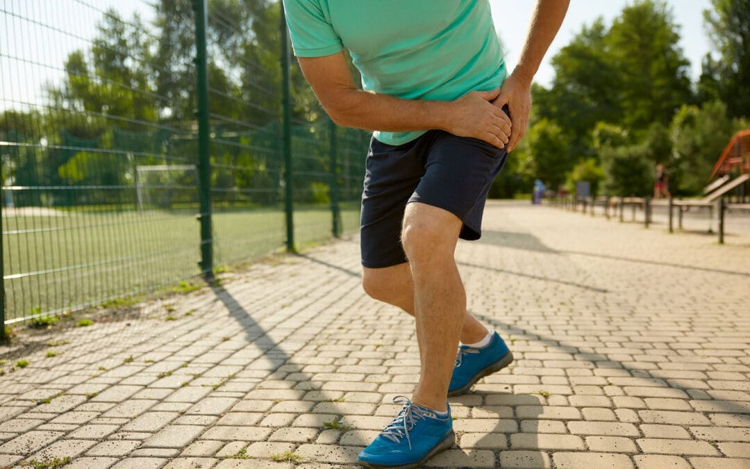
Anterior Hip and Leg Muscles: What They Are, What They Do, and Why They Hurt
A woman holds her aching anterior hip.
Pain in the front of the hip (often felt in the hip crease or groin area) and the front of the thigh is very common. It can show up when you stand up from a chair, climb stairs, run, kick, or even after sitting for a long time. The tricky part is this: front-hip pain is not always “just a tight hip flexor.” Sometimes it’s a muscle or tendon problem, but it can also be related to the hip joint, the pelvis, or the lower back.
This guide is written for everyday people in El Paso who want clear answers, plus a practical explanation of how an integrative chiropractic approach can help reduce pain and prevent flare-ups.
At El Paso Back Clinic, Dr. Alexander Jimenez and the team often observe a pattern: tight, overworked hip flexors, underactive glutes, and poor pelvic control—especially in people who sit a lot, train hard, or are recovering after an accident.
What “anterior hip and leg muscles” means
“Anterior” means the front side. The anterior hip and leg muscles are basically your “go-forward” and “stand-tall” muscles. They help you:
Lift your knee (hip flexion)
Step forward when walking or running
Stabilize your pelvis so your lower back doesn’t overwork
Straighten your knee (knee extension)
Control your leg when you climb stairs or squat
When these muscles get overloaded, they can feel tight, sore, weak, or sharp—depending on the cause.
The main anterior hip muscles (your hip flexors)
Hip flexors are not one muscle. They’re a group that works together.
Key hip flexor muscles
Iliopsoas (iliacus + psoas): the classic “deep hip flexor”
Rectus femoris: part of the quadriceps, crosses the hip and the knee
Sartorius: a long, strap-like muscle across the front of the thigh
Tensor fasciae latae (TFL): supports hip flexion and pelvic control
Pectineus (often grouped with hip flexors in clinical discussions)
Why iliopsoas matters so much
The iliopsoas helps:
Lift the thigh toward the trunk
Support the hip joint and pelvis
Add stability near the lumbar spine/pelvis connection
At El Paso Back Clinic, iliopsoas overuse is commonly discussed among athletes and active individuals who engage in sprinting, jumping, kicking, or repeated hip flexion.
The anterior thigh muscles (front of the thigh)
The main anterior thigh group is the quadriceps. They’re designed to extend the knee and help control motion during walking, stairs, squats, and landing.
Quadriceps muscles
Rectus femoris
Vastus medialis
Vastus lateralis
Vastus intermedius
The anterior thigh compartment is also supplied and controlled by key anatomical structures, such as the femoral nerve (often described as the L2–L4 roots) and the femoral artery system. That’s one reason pain patterns can sometimes feel confusing—muscles, nerves, and joints all influence the sensation you feel.
Why the anterior hip and leg muscles sometimes hurt sometimes
There are a few “big buckets” that explain most front-hip and front-thigh pain.
You’re asking the muscles to do too much, too often (overuse)
Overuse happens when the workload increases faster than your tissues can adapt. Common triggers include:
Sudden jump in running miles
More hills or speed work than usual
Lots of kicking (soccer, martial arts)
Heavy squats/lunges with poor control
Repetitive direction changes (basketball, football)
Overuse can irritate:
The muscle belly (soreness, tightness)
The tendon (tendinopathy-like pain)
The hip flexor attachment area near the front of the hip
Prolonged sitting keeps hip flexors in a “shortened” position
Sitting puts the hips into flexion. Over time, many people notice:
Hip tightness when standing up after sitting
A “pinchy” feeling in the front of the hip
Low back stiffness that shows up with hip tightness
Dr. Jimenez has emphasized in his recent writing that prolonged sitting can contribute to tight hip flexors and poor movement patterns, and that short movement breaks, along with targeted mobility work, can help many people feel better.
The hip flexors can be tight because other muscles are not doing their job
This is one of the most common “root causes” in stubborn cases:
Weak or underactive glutes
Weak deep core stabilizers
Limited hip mobility (the hip joint doesn’t move well)
Pelvic control issues (pelvis tips forward, rotates, or drops during gait)
El Paso Back Clinic explains that when the glutes weaken from inactivity and prolonged sitting, the hips and pelvis can become less stable and shift out of alignment, thereby increasing stress on surrounding tissues.
Sometimes the pain is not in the hip flexor at all
A major clinical point from family medicine guidelines is that hip pain often groups into:
Anterior (front)
Lateral (side)
Posterior (back)
…and the cause changes based on that pattern. Anterior hip pain may result from hip flexor injury, but it can also result from intra-articular hip joint problems (such as femoroacetabular impingement or labral pathology) or from referred pain.
A helpful “body map” concept is presented in educational videos that discuss what different hip pain locations can indicate, but a hands-on evaluation remains important when symptoms persist.
What the pain feels like: common patterns that guide the next step
These are not perfect rules, but they help you decide whether you’re dealing with a likely muscle/tendon issue or something deeper.
More likely muscle/tendon irritation (common hip flexor pattern)
Pain in the front hip crease
Worse with lifting the knee (stairs, marching)
Worse with running sprints, kicking, or hills
Tenderness in the front hip region
Feels tight after sitting
More likely hip joint involvement
Deep groin pain with hip rotation
Catching, clicking, locking, or “pinching”
Pain that persists despite basic stretching/rest
Range of motion feels blocked (especially flexion + rotation)
More likely low back/nerve referral
Front thigh pain plus low back symptoms
Numbness, tingling, and burning sensations
Symptoms that change with spine position
Why “stretching only” often fails
Stretching can feel good short-term, but it may not solve the real driver if the problem is:
Weak glutes and weak core control
A stiff hip joint or pelvic restriction
Poor movement strategy (how you squat, run, or stand)
A training load problem (too much too soon)
In other words, the hip flexors may be tight because they’re protecting you or compensating for something else.
How El Paso Back Clinic approaches anterior hip and leg pain
El Paso Back Clinic describes an integrative model that blends chiropractic care, rehabilitation concepts, and movement-based strategies, with a focus on mobility, flexibility, and the restoration of balanced function.
Here’s how that “integrative” approach commonly helps front-hip and front-thigh problems.
Identify the true driver (not just the sore spot)
A good evaluation typically includes:
History (training, sitting, injury, accident history)
Differentiation between hip joint vs. lumbar referral patterns
Dr. Jimenez has written about the importance of a structured hip evaluation to sort out the likely source of pain and match care to the pattern.
Restore joint motion and reduce protective “guarding”
When the pelvis/hip/lumbar spine isn’t moving well, the body often shifts load to the hip flexors and quads. Chiropractic-style care may focus on restoring smoother motion so the muscles stop overworking.
El Paso Back Clinic also discusses how muscle imbalance and chronic guarding can make it harder for muscles to “relax on their own,” especially after injuries.
Use soft tissue + targeted techniques to normalize muscle function
A common strategy is pairing hands-on care with neuromuscular techniques. El Paso Back Clinic specifically discusses assessing hip flexors with MET therapy (muscle energy technique) as part of reducing tightness and improving hip mobility.
Rebuild strength where it matters (glutes + core + hip control)
To prevent recurrence, the plan usually includes strengthening and control, especially:
Glute bridges and progressions
Hip abduction strength (side-lying or banded work)
Gradual reloading of hip flexors (instead of only stretching)
El Paso Back Clinic’s content repeatedly emphasizes that restoring balanced muscle function around the pelvis and hips supports daily movement and performance.
Practical tips you can start today (safe, simple, and realistic)
If your symptoms are mild and you’re not dealing with red flags, these are common first steps.
For desk workers and drivers (very common in El Paso)
Take 1–2 minute movement breaks every 30–60 minutes
Do a gentle hip flexor stretch (no sharp pinching)
Add a glute activation move (bridges or mini-band walks)
Keep your daily steps consistent (don’t go from 2,000 to 12,000 overnight)
For runners and athletes
Reduce aggravating volume for 1–2 weeks (not “stop forever,” just calm it down)
Avoid sprinting/kicking if it spikes sharp pain
Strengthen glutes and hip stabilizers 2–3x/week
Return to speed and hills gradually, not all at once
Quick self-check idea (mobility clue)
The Thomas Test is commonly used to screen for hip flexor tightness and may help distinguish whether the “tight feeling” is more iliopsoas- or quadriceps-based (rectus femoris). It’s not a diagnosis, but it can be a clue.
When you should get evaluated sooner rather than later
Don’t try to “stretch through it” if you have:
Severe pain after a fall or accident
Inability to bear weight
Fever or feeling unwell with hip pain
Worsening numbness/tingling or leg weakness
Persistent catching/locking and deep groin pain
A structured clinical examination is particularly important when hip pain may involve the hip joint or referral patterns.
The main takeaway
Your anterior hip and leg muscles—especially the hip flexors and quadriceps—are essential for walking, running, stairs, and posture. They often hurt because of:
Too much repeated load (overuse)
Too much sitting (hip flexors stay shortened)
Muscle imbalance (weak glutes/core causing hip flexors to overwork)
Hip joint or low back referral (pain “shows up” in the front)
An integrative chiropractic model—such as the one described in El Paso Back Clinic’s educational resources—focuses on identifying the underlying cause, restoring motion, improving muscle balance, and developing a plan to reduce the likelihood of recurrence.
Is It Safe to Wear a Backpack? Expert Tips on Spinal Health and Back Pain Prevention in the US and El Paso, TX
A woman walking, wearing a backpack with the recommended weight, and maintaining correct posture to prevent back pain and problems.
Back pain is a big issue for many people in the United States
Up to 80% of adults face low back pain at some point in their lives. This is one of the top reasons for doctor visits and missed workdays. The cost is huge too, with over $100 billion spent on spine problems each year. In El Paso, Texas, where people often have active jobs like industrial work or lots of driving, back pain questions focus on things like sciatica, herniated discs, and spinal stenosis. A common concern across the country, including in places like El Paso, is whether wearing a backpack is safe for the spine. The good news is that it can be safe if you follow some simple rules. This article focuses on backpack safety and then addresses other key questions about managing back pain, treatment options, and daily habits to keep your spine healthy.
Understanding Backpack Safety and Spinal Health
Wearing a backpack is common for carrying things, but if it’s too heavy or worn incorrectly, it can hurt your back. Heavy backpacks can strain muscles and joints in your back, neck, and shoulders. This might lead to pain or bad posture over time. However, backpacks do not cause scoliosis, a spinal curvature that affects about 2% to 3% of people. Scoliosis often starts in teens and is more common in girls, but it’s not linked to backpacks.
Is it safe? Yes, as long as you distribute the weight right and follow the tips to avoid strain. Improper use can cause muscle fatigue, poor posture (such as slouching), and even chronic pain if left unaddressed. In El Paso, where people might carry tools or bags for work, this is especially important to prevent issues such as sciatica, where pain radiates down the leg due to nerve pressure.
Here are some key tips for safe backpack use:
Choose the right backpack: Pick one with wide, padded straps and a padded back. It should fit your body size and have a waist strap for heavy loads. Lightweight materials help too.
Limit the weight: Keep the backpack under 10-15% of your body weight. For example, if you weigh 150 pounds, aim for no more than 15-22.5 pounds.
Distribute weight evenly: Put heavier items at the bottom and close to your back. Use compartments to balance things and stop shifting.
Wear it correctly: Always use both straps. Adjust them so the pack sits in the middle of your back, not sagging low. Bend your knees to lift it.
Make smart choices: Remove extra items often. Use lockers or storage if possible. For very heavy loads, try a rolling backpack or crossbody bag.
These steps help distribute the load across your strong back muscles and keep your spine aligned. If you feel pain, stop and adjust. In places like El Paso, with busy lifestyles, following these can help prevent accidents from becoming long-term back issues.
Common Causes of Back Pain in the US
Back pain affects millions. In the US, about 26% of adults have it at any time, and it’s more common after age 45. Among adults aged 50 and older, up to 45.6% experience it. Causes include muscle strains, ligament injuries, herniated discs (where the disc’s soft center protrudes), arthritis, and spinal stenosis (where the spinal canal narrows). Stress can make it worse by causing muscle spasms. Even factors such as obesity or infections can play a role.
Chronic back pain lasts more than 3 months and affects 8% of adults. It often comes from wear and tear on discs or joints. Poor sleep makes it worse because pain disrupts rest, and lack of sleep raises inflammation. In the US, this results in high costs, such as lost work and medical bills.
Symptoms vary. You might feel an ache in your lower back or sharp pain if it’s sciatica. Numbness, tingling, or weakness in the legs are red flags. Scoliosis, which affects 7 million Americans, can cause symptoms such as uneven shoulders or back pain; most cases are mild.
Muscle or ligament strain: From lifting incorrectly or sudden moves.
Disc problems: Bulges or herniations press on nerves.
Arthritis: Joint wear is common in older people.
Stenosis: Narrowing squeezes nerves, causing leg pain.
Stress and lifestyle: Tension builds up, leading to spasms.
Knowing these helps prevent pain. For example, strengthening your core muscles supports your spine and reduces strain from daily activities like wearing a backpack.
Managing Chronic Back Pain
Chronic back pain needs long-term plans. First, see if it’s new or ongoing. Most cases improve with rest and simple fixes, but if it lasts, get checked. Avoid bed rest; gentle movement helps recovery faster.
Daily habits matter. Exercise like walking or swimming builds strength. Maintain a healthy weight to reduce spinal load. Quit smoking, as it negatively affects spinal tissues and raises surgery risk by up to 50%. Good posture and ergonomic setups at work prevent strain.
In El Paso, with industrial jobs and driving, pain from accidents is common. Recovery focuses on building habits to avoid re-injury.
Stay active: Low-impact exercises like yoga or Pilates.
Watch your diet: Healthy foods reduce inflammation.
Manage stress: Deep breathing or mindfulness helps.
Sleep well: Use pillows to maintain spinal alignment.
Stretch daily: Loosen tight muscles, such as the hamstrings.
These steps reduce pain and improve quality of life.
Treatment Options: Surgery vs. Conservative Care
When pain doesn’t go away, choices include conservative care or surgery. Conservative means non-surgical options such as physical therapy, medications, injections, chiropractic care, or massage. These are tried first for 8-12 weeks. Surgery is indicated for severe cases, such as nerve damage or instability.
Ask your doctor: What causes my pain? What tests do I need? What are the risks and benefits? For surgery, ask about the surgeon’s experience, recovery time, and whether you’ll need help at home. Alternatives like spinal decompression stretch the spine to ease disc pressure.
Chiropractic vs. orthopedic: Chiropractors focus on spinal adjustments to realign the spine and relieve pain without medication. Orthopedists may recommend surgery for significant issues. Both can help, but chiropractic care is well-suited to conservative care.
In El Paso, many choose chiropractic for herniated discs or sciatica. It’s safe and effective for back pain, reducing symptoms by fixing alignment and boosting blood flow.
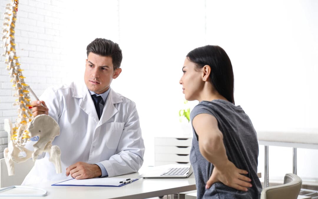
Spinal Health in El Paso, TX
El Paso has unique needs. Active lives, work injuries, and car accidents lead to questions about sciatica, where nerve pain goes down the leg, or spinal stenosis with leg weakness. Herniated discs are common from lifting or falls.
Lumbar stenosis FAQs: It causes leg pain or numbness when walking. Avoid high-impact exercises like running; try swimming instead. Treatments include therapy or decompression.
Local care often combines chiropractic and orthopedic care. Dr. Alexander Jimenez, a chiropractor in El Paso with over 30 years of experience, notes that integrative care is most effective. He uses adjustments, nutrition, and therapy for root causes. For example, a worker’s back pain improved by 50% within weeks with his plan. He stresses non-surgical options for sciatica and injuries, helping people stay active in El Paso’s environment.
Sciatica: From disc pressure; chiropractic eases it.
Chiropractic: Aligns the spine, safe for all ages.
Dr. Jimenez’s work shows personalized plans reduce pain without surgery.
Daily Habits to Prevent Spinal Injury
Preventing pain starts with habits. Lift by bending knees, not back. Stand every 15 minutes if sitting for long. For driving in El Paso, take breaks to stretch.
Core strength is key. Exercises like planks support your spine. Avoid smoking for better healing. Ergonomics: Screen at eye level, chair with back support.
For backpacks, combine with these: Even weight helps posture.
Lift right: Knees bent, close to body.
Posture: Stand tall, no slouch.
Exercise: Core and back focus.
Weight control: Less strain on the spine.
Breaks: Move often.
These reduce the risk of injury and tie into backpack safety.
Conclusion
Wearing a backpack is safe when done properly, with proper weight distribution and habits. This fits into broader questions about spinal health in the US and El Paso. Manage chronic pain with conservative care first, like chiropractic, and build daily routines to prevent issues. Experts like Dr. Jimenez show that integrative approaches work. Stay active, ask questions, and protect your spine for a better life.
Mobility Challenges in Mexican and Mexican American Communities: Insights from El Paso Back Clinic®
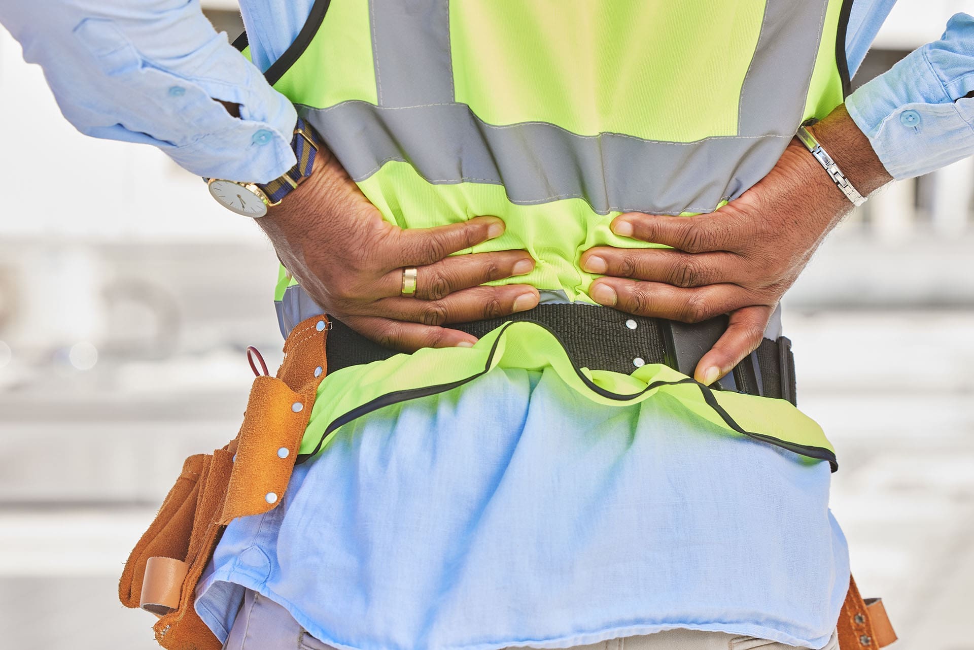
Mexican-American with back pain at a construction site.
At El Paso Back Clinic® in El Paso, TX, we see many patients from Mexican and Mexican American backgrounds facing mobility issues. These problems often stem from tough jobs, health factors like obesity, and aging. Our wellness chiropractic care focuses on pain relief and improved movement. This article discusses common issues such as arthritis and back pain, supported by studies. We’ll explain how our team, including Dr. Alexander Jimenez, DC, APRN, FNP-BC, uses integrative approaches to help. If you’re in El Paso dealing with these, our clinic is here for you.
Common Musculoskeletal Mobility Issues We Treat
Musculoskeletal problems affect your bones, muscles, and joints, making it difficult to move freely. At our clinic, we see these issues often in our community, where many work in demanding fields like farming or construction.
Arthritis, especially in the knees, is a top concern. It causes joint wear-related swelling and pain. In Mexico, about 20-25% of adults aged 40+ have it, with higher rates among women (Villarreal Rizzo et al., 2025). Mexican Americans in the U.S. also face risks, like osteoporosis weakening bones in 16% of women (Wright et al., n.d.). At El Paso Back Clinic®, we help ease this with gentle adjustments and exercises.
Chronic low back pain hits hard, too. It comes from prolonged lifting or standing. In Mexico, it’s the leading cause of disability, with 840.6 cases per 100,000 in 2021 (Clark et al., 2023). Among farmworkers here in Texas, 46.9% report back issues affecting daily life (Weigel et al., 2013). Our chiropractic care targets this to get you moving again.
Work injuries often involve the shoulders, wrists, and legs. Repetitive tasks in jobs cause rotator cuff problems in 19.1% and elbow pain in 20.2% of Latino workers (Mora et al., 2014). Older adults in our area are at risk of frailty due to ongoing pain, leading to reduced mobility (National Institutes of Health, n.d.). Women face more disability in tasks like walking, with arthritis raising risks by 35% over time (Rodriguez et al., 2021).
Here are key facts we see in our patients:
Arthritis rates: 19.6% for knee issues in Mexicans over 40, up to 24.2% in women (Ciampi de Andrade et al., 2022).
Back pain: Affects 16.9% of farmworkers from repetitive strain (Mora et al., 2014).
Craft-related injuries: Neck and knee pain from activities like weaving (Jeanson et al., 2025).
Disability trends: Physical function declines by 0.18 points per year with arthritis (Rodriguez et al., 2021).
Jobs in agriculture and construction drive these, plus obesity adds joint stress. In our Mexican American patients, higher BMI initially slows strength loss but worsens it later (Davis & Al Snih, 2025). About 83% of Hispanic men are overweight, linked to less activity (Valdez et al., 2019). At El Paso Back Clinic®, we address this with personalized plans.
Neuromusculoskeletal Issues Addressed at Our Clinic
These issues combine nerve problems with muscle and bone pain, leading to numbness or weakness. Our wellness approach helps restore nerve function and reduce discomfort.
Chronic low back pain is common, often due to nerve compression. It’s the main cause of disability in Mexico (Alva Staufert et al., 2021). Knee and foot arthritis affects movement, with 25.5% showing joint changes (Ciampi de Andrade et al., 2022). We treat foot pain from standing jobs, seen in 4.8% of workers (Mora et al., 2014).
Shoulder injuries, such as rotator cuff tears, are associated with overhead work and affect 19.1% (Mora et al., 2014). Elbow issues, or epicondylitis, affected 20.2% due to tool use (Mora et al., 2014). MSDs in Mexico rose 57.3% over 30 years (Clark et al., 2023). Obesity plays a role, with 40% of Hispanic men affected (Valdez et al., 2019).
In border areas like El Paso, women report 29.8% low back and 38.3% upper back pain from factory jobs (Harlow et al., 1999). Older patients walk more slowly due to leg pain (Quiben & Hazuda, 2015).
Common issues we handle:
Low back pain: Top disability driver, tied to work and weight (Alva Staufert et al., 2021).
Knee/foot arthritis: More in women, causing stiffness (Ciampi de Andrade et al., 2022).
Rotator cuff: From arm overuse in construction (Mora et al., 2014).
Epicondylitis: Elbow strain, common in 20% (Mora et al., 2014).
How El Paso Back Clinic® Helps with Integrative Care
Our clinic combines nurse practitioners (NPs) and chiropractic methods for culturally sensitive help. We focus on pain management and rehab to fit our community’s needs.
NPs at our clinic offer full check-ups that consider culture and history. They suggest diets rich in veggies and yoga for detox and pain relief (Jimenez, 2026a). We team up for whole-body care (Jimenez, 2026b).
Chiropractic adjustments realign the spine to ease nerve compression. For sitting-related back pain, we restore curves and strengthen the core (El Paso Back Pain Clinic, n.d.). Access to this care is key, though Hispanics use it less (Roseen, 2023).
Dr. Alexander Jimenez shares from his experience: Chronic back pain worsens with poor posture, but adjustments and exercises help (Jimenez, n.d.). For sciatica, decompression relieves pressure on nerves, which is common in laborers. Neuropathy gets therapy for tingling (Jimenez, n.d.). He uses functional medicine to tackle stress, diet, and job factors in our Mexican American patients.
We include mindfulness and natural remedies. Cultural factors, such as family support, help recovery, but delays worsen pain (Arthritis Foundation, n.d.). Our NPs create home plans (Pérez-Stable et al., 2003).
Rehab strengthens areas such as the legs and shoulders (Mora et al., 2014). It cuts frailty risks (National Institutes of Health, n.d.). For farmworkers, it reduces disability (Weigel et al., 2013).
Our care benefits:
Cultural match: Understanding barriers like work migration (Harlow et al., 1999).
Strength building: Targeted exercises (Mora et al., 2014).
Prevention: Nutrition against obesity (Valdez et al., 2019).
Why Choose El Paso Back Clinic® for Your Mobility Needs
In El Paso, with our diverse community, these issues are common but treatable. Our clinic specializes in wellness chiropractic to help you stay active. Contact us for a consultation with Dr. Jimenez and our team.
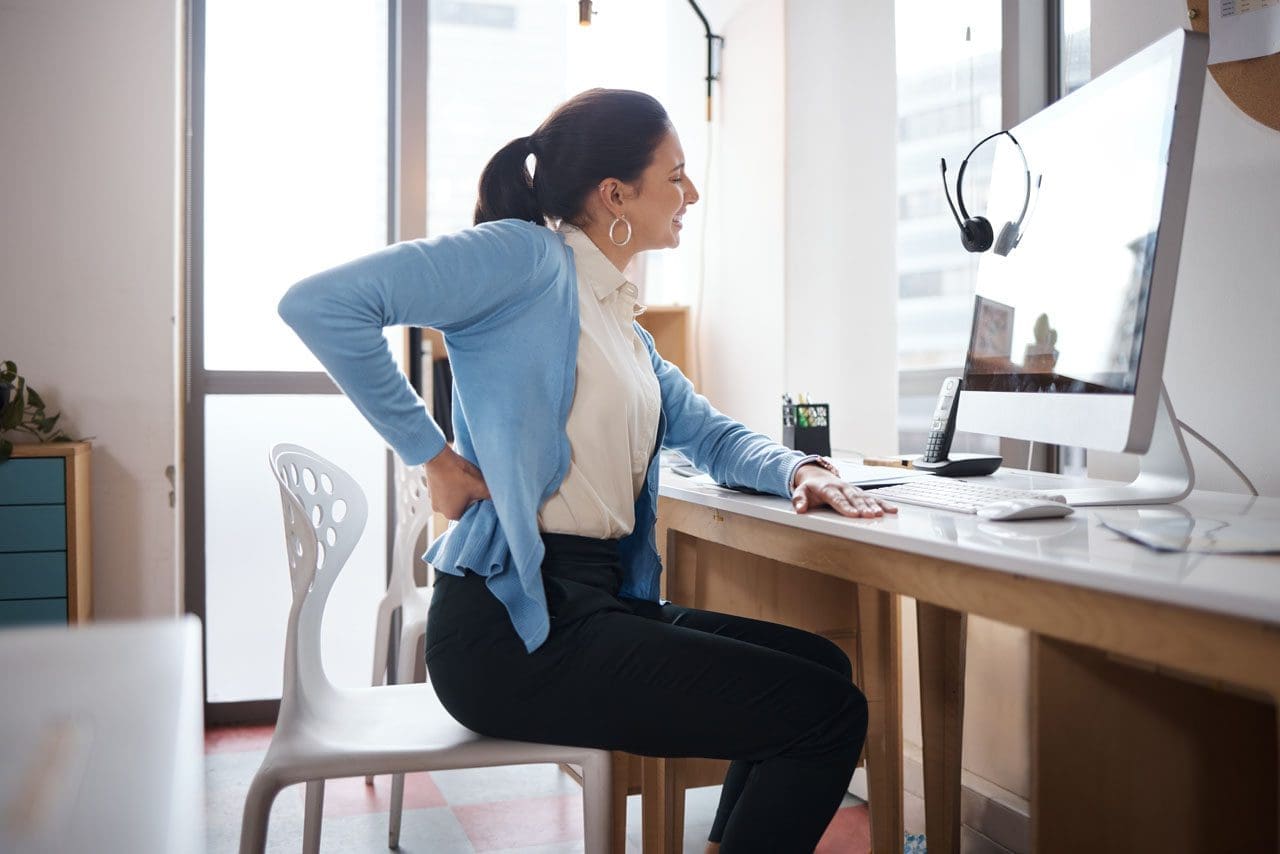
Dr. Alex Jimenez at El Paso Back Clinic®: Beating Back Pain from Long Desk Hours
Businesswoman experiences worsening back pain while sitting at her desk.
If your back pain gets worse the longer you sit at your desk, you are not alone. Many people in El Paso face this issue due to long hours spent in sedentary jobs. Sitting for extended periods can put pressure on the spine, tighten muscles, and reduce blood flow, leading to stiffness, aches, and, in some cases, chronic problems (Colorado Pain Care, n.d.). The positive news is that you can take simple steps to reduce the pain and prevent it from worsening. At El Paso Back Clinic® in El Paso, TX, the wellness chiropractic care team, led by Dr. Alex Jimenez, DC, APRN, FNP-BC, focuses on helping people just like you find natural, long-term relief through personalized plans.
Prolonged sitting stresses the lower back by increasing disc pressure by up to 90% compared to standing. It flattens the spine’s natural curve, strains muscles, and creates imbalances (Colorado Pain Care, n.d.). Slouching or leaning forward adds extra load to the neck and upper back. Over time, this can lead to tight hips, weak core muscles, and ongoing discomfort that affects daily life.
At El Paso Back Clinic®, our experts understand these issues caused by sedentary work. They use a holistic approach that combines chiropractic adjustments, functional medicine, and rehab to address root causes like poor posture and muscle imbalances from desk jobs (Jimenez, n.d.-a).
Here are practical changes to start today:
Move often: Get up every 30 minutes to stand, walk, or shift positions. Short 1-2 minute breaks improve circulation and ease tension (Huntsville Hospital Health System, n.d.; Sydney West Physio, n.d.).
Use regular breaks: Set a timer for quick walks to get water or to stretch. This habit prevents stiffness from building up throughout the day.
Add dynamic movement: While sitting, shift weight, uncross legs periodically, or use a footrest to change angles. These small actions keep the spine mobile (Colorado Pain Care, n.d.).
A proper ergonomic setup supports optimal posture and reduces strain.
Follow these key tips:
Set your chair so that your feet are flat on the floor, your knees are at 90 degrees, and your hips are level with or above your knees.
Add lumbar support (a small pillow or rolled towel works) to maintain the lower back’s curve.
Place your screen at eye level to avoid looking down or up too much.
Keep the keyboard and mouse close so elbows bend at 90 degrees and shoulders stay relaxed.
Avoid crossing legs for long, as it can tilt the pelvis (Senara Chiropractic & Med Spa, n.d.; Huntsville Hospital Health System, n.d.).
Consider alternating between sitting and standing with a standing desk. Even partial standing reduces spinal pressure.
Stretches help loosen tight spots from sitting, such as the hips, shoulders, and neck.
Try these simple ones:
Hip flexor stretch: Kneel on one knee, gently push hips forward, and hold 20-30 seconds per side.
Chest and shoulder opener: Clasp hands behind your back or use a wall to stretch forward.
Neck tilts: Slowly tilt the head side to side or forward/back; hold for 10-15 seconds.
Upper back extension: Hands behind head, gently arch upper back (Sydney West Physio, n.d.).
Do them hourly or during breaks for better flexibility.
Strengthening the core supports the spine and improves posture long-term.
Include these:
Planks: Hold forearm plank 20-30 seconds.
Cat-camel: On hands and knees, arch and round back slowly.
Bridges: Lie back, lift hips while squeezing glutes.
Walking or gentle yoga: Build overall strength (Huntsville Hospital Health System, n.d.; Sydney West Physio, n.d.).
Aim for 20-30 minutes of activity most days.
For lasting relief, professional care targets alignment, mobility, and personalized fixes. At El Paso Back Clinic®, Dr. Alex Jimenez leads a team offering integrated chiropractic care. This includes spinal adjustments to correct misalignments, non-surgical spinal decompression for disc relief, acupuncture, functional medicine for nutrition and stress, and rehab exercises tailored to desk-related issues.
Dr. Jimenez, with dual expertise as a chiropractor and nurse practitioner, emphasizes posture correction, mobility training, and the prevention of sedentary pain through evidence-based methods. The clinic helps restore function without drugs or surgery, focusing on root causes like imbalances from prolonged sitting (Jimenez, n.d.-a; Jimenez, n.d.-b).
Other options in El Paso exist, but El Paso Back Clinic® stands out for its comprehensive wellness approach, advanced diagnostics, and patient-centered plans that go beyond basic adjustments.
If pain includes numbness, tingling, or weakness in the legs, or persists despite changes, seek evaluation to rule out serious conditions (University of Maryland Medical System, n.d.).
Start small: improve movement, setup, and stretches. If needed, contact El Paso Back Clinic® for expert help. Many in El Paso regain comfort and stay active with this care.
Spinal Hygiene Explained: Daily Habits to Keep Your Spine Strong and Pain-Free
A woman performs spinal hygiene exercises on a fitness ball at home, strengthening her back muscles.
Spinal hygiene is the practice of caring for your spine every day to keep it strong, flexible, and healthy. Just like brushing your teeth helps prevent cavities, spinal hygiene helps avoid back problems and keeps your body moving well. The spine supports your whole body, protects the nervous system, and lets you bend, twist, and stand tall. Good habits can make a big difference in how you feel now and as you get older.
What Spinal Hygiene Means
Spinal hygiene includes simple daily actions to protect the spine’s natural shape and movement. The spine has gentle curves that help absorb shock and allow smooth motion. When these curves stay balanced, and the spine moves freely, you feel better overall.
Proper posture: Sit and stand with your shoulders back, head aligned over your spine, and pelvis in a neutral position to avoid extra strain.
Regular movement and exercise: Stay active with walking, swimming, or stretching to keep joints loose and muscles strong.
Proper body mechanics: Lift things by bending your knees, keeping objects close to your body, and avoiding twists to prevent injury.
Core strength: Build muscles around your midsection for better support and stability.
Hydration and nutrition: Drink plenty of water to keep spinal discs cushioned, and eat foods rich in calcium, vitamin D, magnesium, and omega-3 fatty acids to support bone and tissue health.
Stress management: Use deep breathing, meditation, or yoga to reduce tension that tightens back muscles.
These steps help maintain the spine’s integrity and prevent issues like stiffness or pain (Spinenpain.org, n.d.; Lifemovesmt.com, n.d.).
Neglecting spinal hygiene can lead to problems over time. Poor habits can cause muscle imbalances, joint wear, reduced motion, and conditions such as herniated discs or chronic back pain. This can affect your nervous system, since the spine houses nerves that control body functions. When the spine is out of alignment, it may press on nerves, leading to pain, weakness, or other issues (Servinglifedallas.com, n.d.; Drmmalone.com, n.d.).
Benefits of Good Spinal Hygiene
Taking care of your spine brings many advantages. It reduces the chance of back pain, improves how easily you move, and supports better posture. A healthy spine also helps the nervous system function smoothly, boosting energy, coordination, and overall well-being. Regular care can slow age-related changes, lower injury risk, and help you stay active longer.
Prevents muscle tightness and joint problems
Improves blood flow and nutrient delivery to spinal tissues
Enhances balance and reduces fall risk
Supports better sleep and less daily discomfort
Studies and experts note that these habits promote long-term health and vitality (Illinoisspinalcare.com, n.d.; Spinehealth.org, n.d.).
How Chiropractic Care Fits In
Chiropractic care plays a key role in spinal hygiene. Chiropractors use gentle adjustments to fix misalignments, called subluxations, that can stress the spine and nerves. These adjustments restore proper movement, reduce pain, and improve function. Regular chiropractic visits act as preventive maintenance, catching small issues before they grow.
Many people combine chiropractic with other habits for the best results. Adjustments improve alignment, while daily posture and exercise help maintain gains. This approach helps address common complaints such as neck or low back pain and supports recovery from injuries (Illinoisspinalcare.com, n.d.; Eastportlandchiropractor.com, n.d.).
The Power of Integrative Care with Chiropractors and Nurse Practitioners
An integrative approach brings together diverse experts to achieve stronger results. Chiropractors focus on the spine’s structure, alignment, and movement through adjustments and rehab. Nurse practitioners (NPs), especially those with advanced training, address broader health needs such as nutrition, stress management, hormone balance, and lifestyle changes.
This team effort addresses both the physical spine and daily habits that affect it. For example, a chiropractor might correct alignment after an injury, while an NP guides on anti-inflammatory foods or stress reduction to aid healing. Together, they create personalized plans that work better than one alone, especially for complex pain, chronic issues, or recovery from accidents.
Dr. Alexander Jimenez, DC, APRN, FNP-BC, stands out in this field. As a Doctor of Chiropractic and board-certified Family Nurse Practitioner, he combines spinal adjustments, decompression therapy, and functional medicine. His practice emphasizes root-cause care, using nutrition, lifestyle tweaks, and advanced diagnostics for musculoskeletal problems, personal injuries, and overall wellness. Clinical observations from his work show that integrative methods restore mobility, reduce pain, and improve quality of life by treating the whole person—not just symptoms (Dralexjimenez.com, n.d.; A4m.com, n.d.).
Simple Ways to Start Spinal Hygiene Today
You can begin with easy changes that fit into daily life.
Posture checks: Set reminders to sit tall and take breaks from sitting every 30 minutes.
Daily stretches: Try cat-cow pose, child’s pose, or seated twists for 5-10 minutes.
Safe lifting: Always bend at the knees and use your legs.
Stay hydrated: Drink water throughout the day to keep your discs healthy.
Eat spine-friendly foods: Include leafy greens, fish, nuts, and dairy or alternatives for key nutrients.
Move often: Walk or do low-impact activity for at least 30 minutes most days.
Manage stress: Practice deep breathing or short meditation sessions.
Adding regular chiropractic check-ups can help monitor and maintain progress (Lifemovesmt.com, n.d.; Newlifefamilychiropractic.net, n.d.).
Final Thoughts
Spinal hygiene is a smart, everyday way to protect one of your body’s most important parts. By focusing on posture, movement, nutrition, and professional care when needed, you support a strong spine, healthy nerves, and a better quality of life. Small habits add up to big benefits, helping you stay active and pain-free for years.
Innovations in Sciatica Treatment in 2026: A Shift Toward Targeted, Minimally Invasive, and Integrative Care
Sciatica is one of the most common causes of chronic lower back and leg pain. It occurs when the sciatic nerve—the longest nerve in the body—is irritated or compressed, often due to disc herniation, spinal degeneration, inflammation, or biomechanical imbalance. For years, treatment options focused mainly on pain medications, steroid injections, or surgery when symptoms became severe.
In 2026, sciatica care has entered a new phase. Treatment is no longer just about “blocking pain.” Instead, the focus is on precision diagnosis, nerve healing, inflammation reduction, and functional recovery, with fewer complications and faster healing times. These advances also emphasize integrated, interdisciplinary care, combining chiropractic treatment with the diagnostic and clinical oversight of nurse practitioners (NPs).
This article explains the most important innovations shaping sciatica treatment in 2026, using easy-to-understand language while staying grounded in current clinical research and real-world outcomes.
Understanding Sciatica: Why Better Solutions Were Needed
Sciatica is not a single disease. It is a symptom caused by pressure or irritation along the sciatic nerve, usually beginning in the lower spine and traveling into the buttock and leg. Pain can feel sharp, burning, electric, or aching and may include numbness or weakness.
Common contributors include:
Herniated or bulging lumbar discs
Spinal stenosis
Degenerative disc disease
Muscle imbalance or pelvic instability
Inflammation around nerve roots
Traditional treatments often relied on:
Long-term anti-inflammatory or pain medications
Opioids for severe cases
Epidural steroid injections
Surgery as a last resort
While these approaches helped some patients, they did not always address the underlying cause, and many carried risks such as dependency, complications, or prolonged recovery (Stanford Health Care, n.d.).
What Has Changed in 2026?
By 2026, sciatica treatment emphasizes early, targeted, and minimally invasive care. Research and clinical experience now show that addressing nerve irritation early and restoring healthy movement patterns can prevent chronic pain and disability (BioSpace, 2025).
Key changes include:
Improved imaging and diagnostics
Precision-guided nerve procedures
Regenerative medicine options
Advanced neuromodulation technologies
Integrated chiropractic and NP-led care models
Advanced Diagnostic Imaging: Seeing the True Source of Pain
One of the biggest improvements in sciatica care is high-resolution MRI technology. Modern imaging allows clinicians to:
Identify the exact nerve root involvement
Distinguish disc-related pain from muscular or inflammatory causes
Detect subtle nerve inflammation missed in earlier imaging methods
Enhanced MRI protocols now guide treatment decisions more accurately, reducing unnecessary procedures and improving outcomes (Stanford Health Care, n.d.).
Nurse practitioners play a critical role here by:
Ordering and interpreting imaging
Correlating findings with physical symptoms
Coordinating referrals and follow-up care
Minimally Invasive Pain Procedures: Precision Without Surgery
Improved Nerve Blocks
Modern nerve blocks are no longer “blind injections.” In 2026, they are image-guided and highly targeted, delivering medication exactly where inflammation and irritation occur.
Benefits include:
Faster pain relief
Reduced medication dosage
Improved diagnostic clarity
Lower complication risk
Nerve blocks are now often used as diagnostic tools to help clinicians determine whether pain is mechanical, inflammatory, or neuropathic in origin (Apollo Spine & Pain, 2026a).
Radiofrequency Ablation (RFA)
Radiofrequency ablation uses controlled heat to disrupt pain signals traveling through irritated nerves. Newer RFA systems are more precise and selective than earlier versions.
Key advantages:
Long-lasting pain relief
Minimal tissue damage
Short recovery time
Reduced reliance on medications
RFA is especially helpful for chronic sciatica that does not respond to conservative care (Apollo Spine & Pain, 2026b).
Spinal Cord Stimulation: Rewiring Pain Perception
Spinal cord stimulation (SCS) has evolved significantly. New devices are smaller, smarter, and more adaptive. They deliver gentle electrical signals that change how the brain interprets pain signals from the sciatic nerve.
Modern SCS systems offer:
Personalized stimulation patterns
Adjustable settings via external controllers
Reduced side effects compared to older models
SCS is now used earlier in care for select patients, helping many avoid surgery altogether (Pain and Spine Specialists, n.d.).
Regenerative Medicine: Supporting Nerve Healing
Platelet-Rich Plasma (PRP)
PRP therapy uses a patient’s own blood components to promote healing and reduce inflammation. In sciatica care, PRP is increasingly used around:
Inflamed nerve roots
Degenerative disc structures
Surrounding soft tissues
Research suggests PRP may:
Reduce inflammation
Support tissue repair
Improve long-term outcomes
While still evolving, regenerative therapies are driving growth in the global sciatica treatment market (Grand View Research, n.d.; PMC, 2024).
Therapeutic Exercise Innovations: Movement as Medicine
Nerve Flossing and Targeted Rehab
Exercise therapy in 2026 is more specific and personalized. One standout approach is nerve flossing, which involves controlled movements designed to improve nerve mobility and reduce irritation.
Benefits include:
Improved nerve glide
Reduced stiffness
Decreased pain during daily activities
Programs are now customized using imaging findings and functional testing, ensuring exercises match the patient’s specific condition (Goodman Campbell, 2026).
Chiropractic Care: A Drug-Free Foundation
Chiropractic care remains central to non-surgical sciatica treatment. Modern chiropractic approaches focus on restoring spinal alignment, reducing nerve compression, and improving movement patterns.
Common techniques include:
Spinal adjustments
Flexion-distraction therapy
Soft tissue mobilization
Low-level laser therapy
Research continues to associate chiropractic care with reduced opioid use and improved pain outcomes (ReachMD, 2024; UH Hospitals, 2025).
Integrated Care: Chiropractors and Nurse Practitioners Working Together
One of the most important shifts in 2026 is the collaborative care model. Instead of isolated treatment, patients benefit from coordinated care involving chiropractors and nurse practitioners.
This model allows for:
Accurate diagnosis and imaging oversight
Conservative, drug-free pain management
Monitoring of neurological symptoms
Reduced opioid exposure
Clinical observations from Dr. Alexander Jimenez, DC, APRN, FNP-BC, highlight that patients receiving coordinated chiropractic and NP care often experience:
Faster recovery
Better functional improvement
Fewer invasive interventions
Greater long-term pain control
His integrative approach emphasizes treating the cause of nerve irritation, not just the symptoms (Jimenez, n.d.).
Why These Innovations Matter
Together, these advances represent a major shift in sciatica care:
From surgery-first to conservative-first
From symptom masking to root-cause resolution
From isolated care to interdisciplinary teamwork
Patients now have more options, fewer risks, and better long-term outcomes.
Conclusion: The Future of Sciatica Care Is Personalized and Precise
By 2026, sciatica treatment has become more effective, safer, and patient-centered. Innovations in diagnostics, minimally invasive procedures, regenerative medicine, and integrative chiropractic-NP–NP care are reducing the need for surgery and long-term medication use.
The focus is no longer just pain relief—it is nerve health, mobility restoration, and sustainable recovery. As research and technology continue to evolve, patients suffering from sciatica can expect even more targeted, evidence-based solutions in the years ahead.
Relieve Lower Back and Hip Pain with Squats, Core Exercises, and Chiropractic Care at El Paso Back Clinic®
Many people in El Paso suffer from lower back pain and hip discomfort due to daily activities, work demands, injuries, or long-term issues. These problems often stem from muscle strains, poor posture, tight hips or glutes, and weak supporting muscles. At El Paso Back Clinic® in El Paso, TX, we specialize in helping patients overcome these challenges through personalized chiropractic care, rehabilitation, and safe exercises.
Squats and core exercises, performed correctly, strengthen the muscles that support the spine, improve alignment, and enhance hip mobility. This reduces stress on the back during movement. They are effective for chronic low back pain, mild sciatica, and general aches from weak muscles. Proper form is essential—sharp pain, numbness, or weakness means you should seek professional evaluation first.
Strong Core + Chiropractic for Lower Back and Hip Pain Relief
The lower back and hips are closely connected through shared muscles, joints, and nerves. Tight hips or glutes can tug on the back, leading to strain. Weak core muscles cause spinal instability and poor posture, leading to chronic pain.
Muscle imbalances force the back to overcompensate in everyday tasks.
Reduced hip mobility leads to excessive forward leaning, stressing the lower back.
Problems in ankle or upper back mobility contribute further.
These factors can result in lumbar instability or pain radiating from the hips to the back.
How Squats Benefit Lower Back and Hip Conditions
Squats strengthen the legs, glutes, and core. With proper technique, they relieve pressure from the lower back.
Proper squats maintain a neutral spine and engaged core, providing stability and minimizing lumbar strain. Activating core and hip muscles during squats supports the spine, preventing excessive arching or rounding.
Squats also increase hip mobility. Tight hip flexors are a common cause of back pain during deeper squats. Improved flexibility allows the hips to function better, sparing the back from overload.
Builds glutes and legs for stronger spinal support.
Enhances blood flow and reduces inflammation in the area.
Aids mild pain that improves with gentle activity.
Research supports that the correct form reduces risks associated with squats.
Core Exercises: A Key to Back and Hip Relief
Core exercises focus on deep muscles in the abdomen, back, and pelvis, acting as a natural spinal brace.
Strong core muscles enhance posture and balance, easing the load on spinal discs and preventing persistent pain from inadequate support. Studies show core stability exercises effectively reduce non-specific low back pain and improve function.
Core training also supports hip pain by stabilizing the pelvis, which is beneficial for conditions like arthritis or glute tightness.
Planks and bird-dogs develop endurance in stabilizing muscles.
Pelvic tilts and bridges safely activate deep muscles.
Standing core activities help relieve pain from prolonged sitting.
Evidence indicates that core exercises often outperform general workouts in reducing pain.
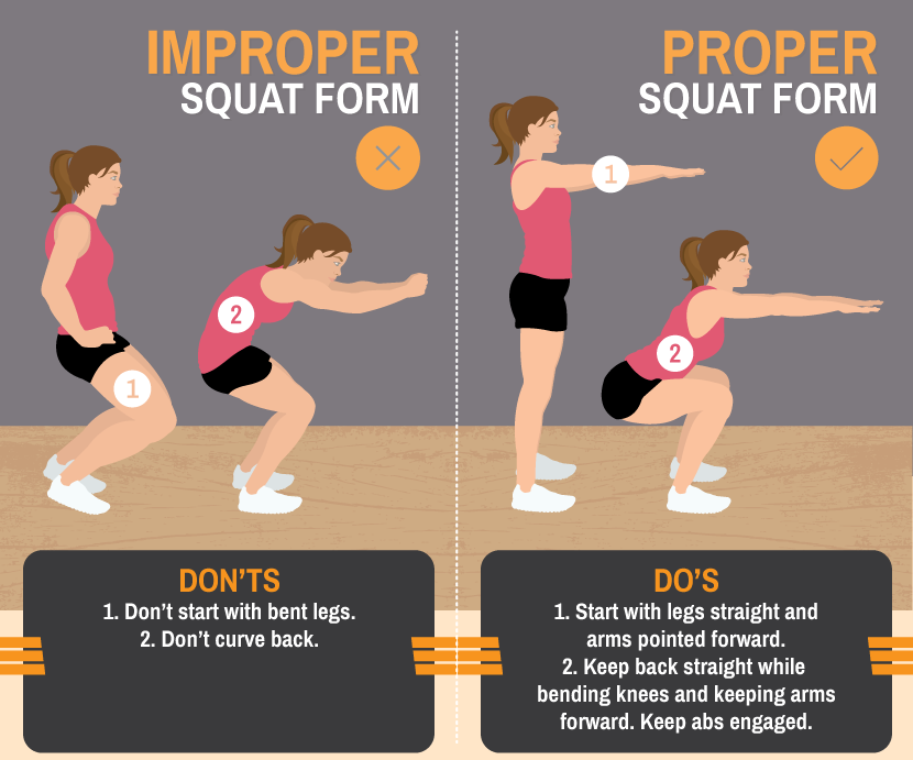
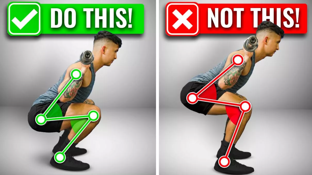
Mastering Proper Form for Safe Squats and Core Work
Incorrect squat form is a leading cause of lower back pain. Frequent mistakes include back rounding, knee collapse, or excessive weight.
Safe squat guidelines:
Position feet shoulder-width apart, toes slightly turned out.
Engage your core as if bracing for impact.
Hinge at the hips, keep the chest high, and descend until the thighs are parallel to the ground.
Drive up through heels, maintaining a neutral spine.
For core exercises, prioritize controlled movement. Hold planks straight with tight abs—avoid dipping or arching.
Begin with bodyweight versions and always warm up to boost circulation and lower injury risk.
Pain during squats typically indicates a weak core, tight hips, or mobility deficits. Address these with targeted stretches and progressive loading.
When Exercises Are Helpful and When to Get Professional Care
Squats and core exercises support:
Chronic low back pain from muscle weakness.
Mild sciatica by decreasing nerve pressure.
Hip tightness referring pain to the back.
Posture-related daily discomfort.
They foster long-term resilience and prevent compensatory back strain. Halt immediately if experiencing severe pain, numbness, weakness, or loss of balance—these may indicate serious conditions such as a disc herniation.
Consult a provider before beginning, especially if you have pre-existing injuries.
Integrative Care at El Paso Back Clinic®
At El Paso Back Clinic®, Dr. Alexander Jimenez, DC, APRN, FNP-BC, leads a team that delivers comprehensive, integrative chiropractic and wellness care for lower back and hip pain. Our approach combines squats and core exercises with chiropractic adjustments, spinal decompression, physical therapy, functional medicine, and rehabilitation programs.
Chiropractic adjustments correct misalignments and joint dysfunctions. A reinforced core helps maintain these corrections by enhancing spinal stability.
Dr. Jimenez creates tailored plans that address root causes through evidence-based protocols, drawing on over 30 years of experience in complex injuries, sciatica, and chronic pain. This multidisciplinary method often yields superior, sustained results compared to isolated treatments.
Visit our main location at 11860 Vista Del Sol, Suite 128, El Paso, TX 79936, or call (915) 850-0900 to schedule your consultation.
Beginner Exercises to Try Under Guidance
Start with these fundamentals, supervised by our team:
Bodyweight Squats: 3 sets of 10-15 repetitions, emphasizing technique.
Glute Bridges: Lie on your back, and elevate your hips by engaging your glutes.
Bird-Dog: On hands and knees, extend opposite arm and leg while bracing core.
Planks: Maintain position for 20-30 seconds, gradually increasing duration.
Pelvic Tilts: On the back, press the lower back into the floor via a pelvic tilt.
Incorporate 2-3 sessions weekly. Include hip mobility work and advance gradually.
Regain Comfort and Mobility Today
At El Paso Back Clinic®, squats and core exercises form integral components of our rehabilitation strategies for lower back and hip pain. They fortify stabilizing muscles, correct alignment, and promote mobility to manage strains, poor posture, instability, and tightness.
Combined with expert chiropractic and integrative care under Dr. Alexander Jimenez, they deliver lasting strength and relief.
Reach out to El Paso Back Clinic® today. Our team will assess your needs and develop a customized plan for optimal recovery.
IFM's Find A Practitioner tool is the largest referral network in Functional Medicine, created to help patients locate Functional Medicine practitioners anywhere in the world. IFM Certified Practitioners are listed first in the search results, given their extensive education in Functional Medicine