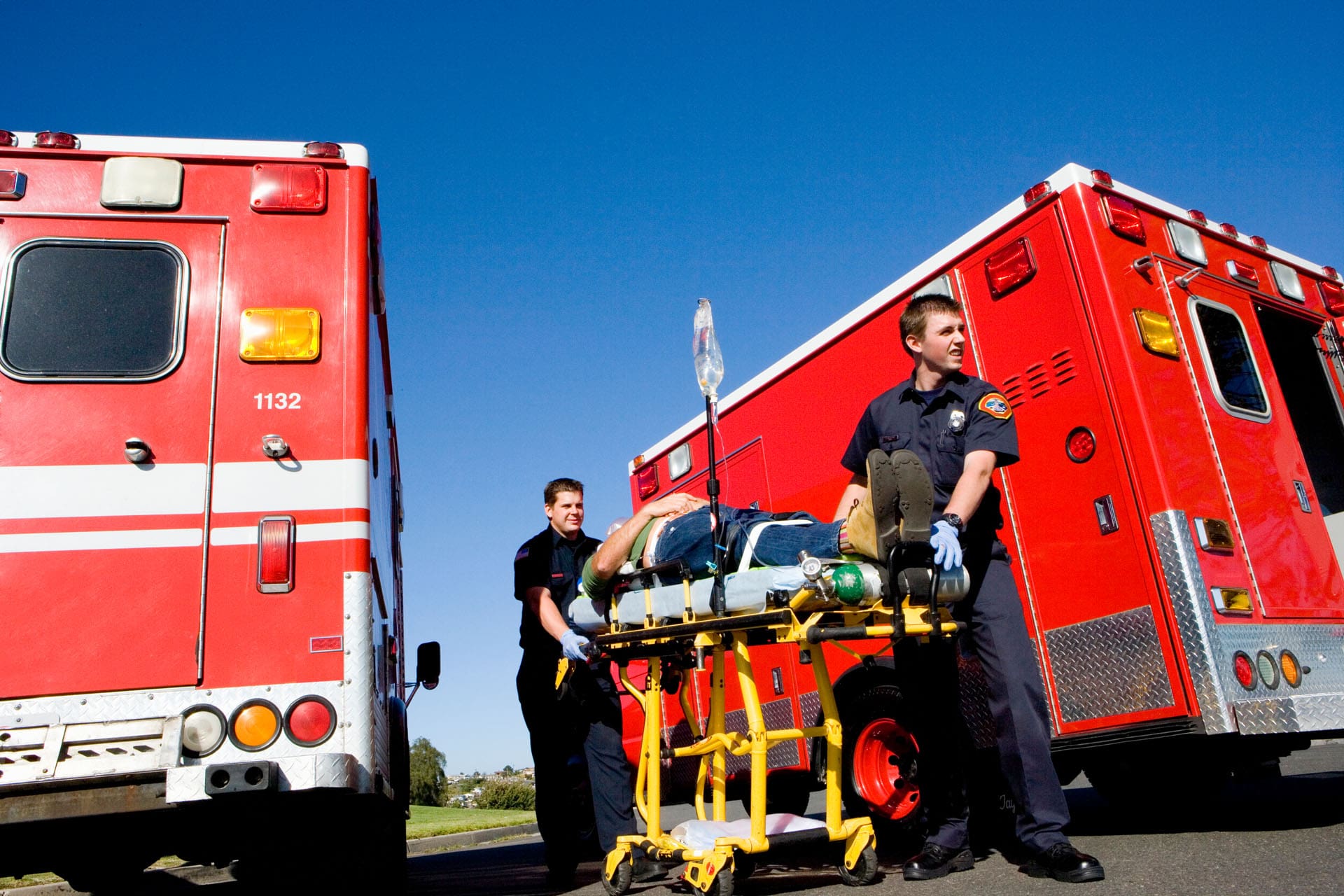
Back Clinic Auto Accident Injuries Chiropractic and Physical Therapy Team. Many automobile accidents occur throughout the world every year, affecting a wide number of individuals, both physically and mentally. From neck and back pain to bone fractures and whiplash, auto accident injuries and their associated symptoms can challenge the daily lives of those who experienced unexpected circumstances.
Dr. Alex Jimenez’s collection of articles discusses auto injuries caused by trauma, including which specific symptoms affect the body and the particular treatment options available for each injury or condition resulting from an auto accident. Being involved in a motor vehicle accident can not only lead to injuries but they can be full of confusion and frustrations.
It is very important to have a qualified provider specializing in these matters completely assess the circumstances surrounding any injury. For more information, please feel free to contact us at (915) 850-0900 or text to call Dr. Jimenez personally at (915) 540-8444.
Annular Tear and Disc Height Loss After Loop 375 Lateral Impact: Mechanism-to-MRI Linkage
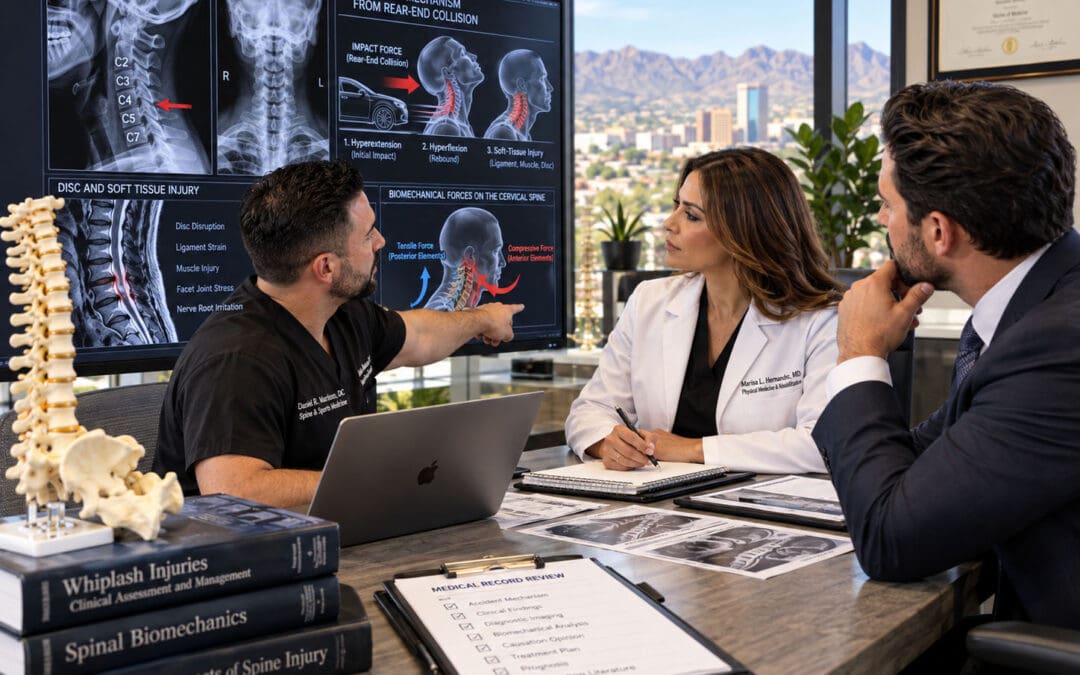
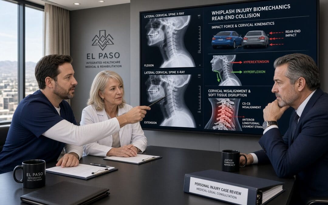
A high-speed side impact on El Paso’s Loop 375 can transmit focused lateral shear, axial compression, and rotational forces through the lumbar or cervical spine in a fraction of a second. When those force vectors exceed the tensile strength of the annulus fibrosus—particularly its relatively weaker posterolateral fibers—an acute annular tear can form, allowing nuclear material to migrate and producing measurable disc height loss visible on MRI. At Injury Medical Clinic PA in El Paso, the multidisciplinary team of Dr. Alex Jimenez, DC, APRN, FNP-BC, CFMP, IFMCP (Texas License #1191402, Prescriptive Authority #59628, NPI 1205907805) and Medical Director Dr. Maria Guadalupe Cardenas, MD (Board Certified Internal Medicine, Texas License #J2933, NPI 1164426748), systematically links these biomechanical mechanisms to objective imaging so that clinical and proximate causation are clear for patients and attorneys.
How a Loop 375 Lateral Impact Generates Disc Injury
Loop 375 (Joe Battle Boulevard and related segments) is a high-volume corridor where lane changes, side-swipes, and T-bone or angle collisions are common. In a pure lateral impact, the striking vehicle imparts a force vector roughly perpendicular to the struck vehicle’s longitudinal axis. Occupants experience rapid lateral torso acceleration while the pelvis and lower extremities lag or rotate, creating shear across the intervertebral discs and facet joints. At the same time, the sudden change in velocity produces axial compression and, often, a twisting moment as the body rebounds against the seatbelt or door.
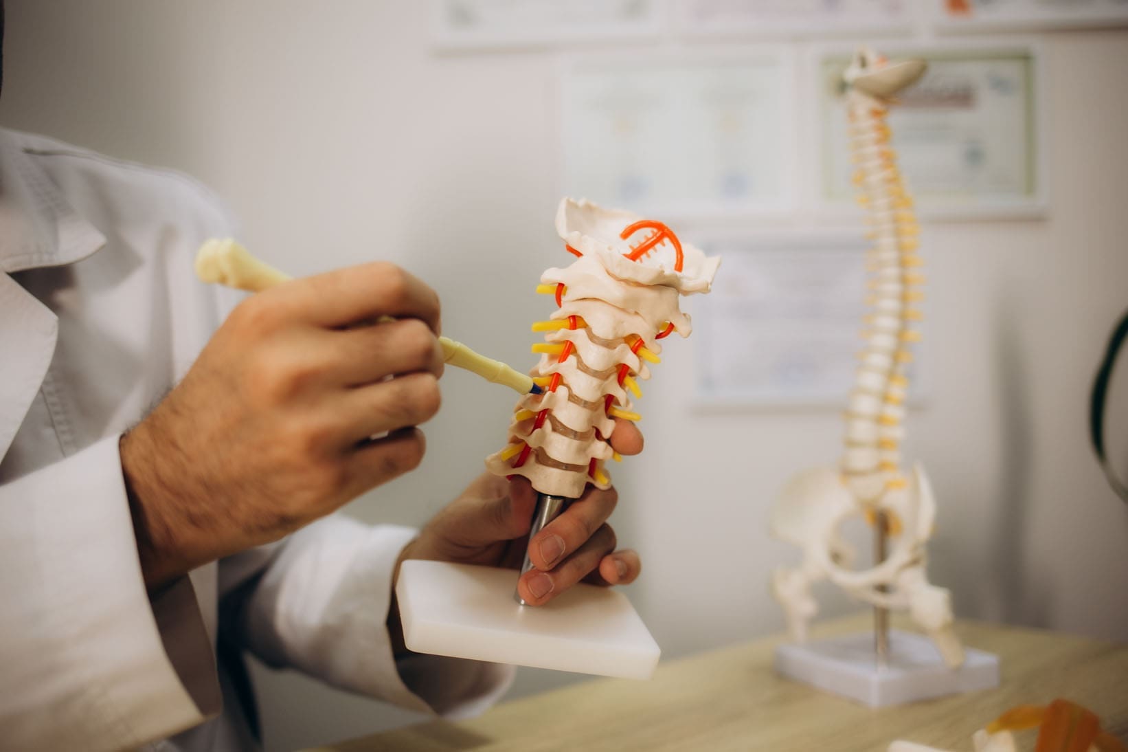
The annulus fibrosus consists of concentric lamellae whose fiber orientation alternates for strength; however, the posterolateral region contains a higher proportion of vertically oriented fibers and is a known site of relative weakness (StatPearls, 2023). When lateral shear and rotation are superimposed on compression, these fibers can separate, producing a radial or transverse annular tear. Nuclear material may then extrude or lose hydration and volume, resulting in a measurable reduction in disc height at the affected level. Because the forces are asymmetric, the tear and height loss are often unilateral or more pronounced on the side of impact—an important morphologic clue when compared with the more uniform, multilevel pattern of ordinary age-related degeneration.
MRI Linkage: Reading the Images Through the Force Vectors
On T2-weighted MRI, an acute annular tear typically appears as a high-intensity zone (HIZ)—a focal bright signal within the normally dark annulus caused by increased water content and inflammatory fluid (StatPearls, 2023; Teraguchi et al., 2022). Supporting signs of acute traumatic change, as opposed to a chronic degenerative baseline, include:
A single or dominant level of abnormality that matches the predicted biomechanics of the impact (for example, L4-L5 or L5-S1 after a low lumbar lateral load, or mid-cervical levels after a higher side-impact).
Asymmetric height loss or annular signal that is greater on the side of the force vector.
Associated soft-tissue findings such as ligamentous edema, facet fluid, or paraspinal muscle signal change at the same or adjacent levels.
Higher T2 hyperintensity and involvement of the outer third of the annulus are more consistent with traumatic disruption than the lower-intensity, inner-two-thirds fissures typical of slow degenerative breakdown (Khurana et al., 2025).
Degenerative disc disease is extremely common and increases with age; imaging studies of asymptomatic volunteers show annular fissures in roughly 19–29% of adults depending on the decade (Brinjikji et al., as cited in clinical radiology reviews). The clinic therefore never equates the mere presence of an HIZ or reduced disc height with trauma. Instead, the team correlates the precise morphology, laterality, and associated acute soft-tissue changes with the documented mechanism, the patient’s immediate post-collision symptoms, and serial clinical examinations. When pre-existing degeneration is present, the records explicitly separate the baseline findings from the new traumatic aggravation—language that prevents defense arguments that “the MRI only shows wear and tear.”
Distinguishing Acute Traumatic Change from Degenerative Baseline
Clear, plain-language documentation is essential. An example of phrasing used in clinic notes and narrative reports reads: “The left posterolateral L4-L5 annular high-intensity zone and 2 mm asymmetric disc-height reduction are morphologically and temporally consistent with the lateral shear and rotational forces of the 14 July Loop 375 side-impact collision. These findings differ from the mild, symmetric multilevel disc desiccation and small inner annular fissures that represent age-related baseline change.” Such statements rest on the mechanism-to-MRI linkage rather than on the radiologist’s impression alone. Digital imaging, functional testing (range-of-motion, neurologic, and strength measures), and Colossus-compliant electronic records further corroborate that the structural changes produce the patient’s objective functional deficits.
Future-Care Modeling with ESWT and Non-Surgical Decompression
Once causation is established, the clinic models evidence-based future care to address both the annular tear and height loss. Non-surgical spinal decompression creates controlled negative intradiscal pressure that can increase disc height and reduce pain; one retrospective cohort demonstrated a statistically significant correlation between restored disc height and decreased low-back pain scores (Apfel et al., 2010). Additional studies confirm improvements in radicular pain and disc morphology after motorized decompression protocols (Choi et al., 2022; Egyptian Rheumatology and Rehabilitation, 2019).
Extracorporeal shock-wave therapy (ESWT) supplies a complementary biologic stimulus. In laboratory models, low-energy ESWT combined with low-tension traction has been shown to restore disc height, improve rehydration, and reduce matrix-degrading enzymes (MMP-3, MMP-13, ADAMTS-4), thereby reshaping the microenvironment for repair (Spine Journal, 2021). Clinical series report pain reduction and facilitation of herniation resorption when ESWT is integrated into staged conservative care (Orthopedic Reviews, 2026; Rehabilitation Medicine, 2024). Because both modalities are non-invasive and drug-free, they align with non-maleficence: they lower the risk of progression to surgery or long-term opioid dependence while still delivering measurable structural and symptomatic benefit.
In the future-care cost spreadsheet these interventions appear as a defined course of decompression sessions (typically 20–30 over 6–8 weeks) plus a series of ESWT treatments, accompanied by supportive chiropractic adjustments, neurofunctional acupuncture or electroacupuncture, and functional-medicine laboratory monitoring directed by Dr. Cardenas. The combination targets root-cause healing—restoring disc height and annular integrity where possible, reducing inflammatory cytokines, and improving segmental stability—rather than merely masking pain (beneficence). Patients receive clear education about expected timelines, self-care strategies, and how the plan coordinates with any outside specialists they already see, preserving autonomy and informed decision-making.
Documentation That Withstands Scrutiny
Every element—mechanism narrative, MRI description distinguishing acute from baseline change, functional deficits, and itemized future-care costs—is recorded in a manner suitable for letters of protection and deposition. The dual-licensed perspective of Dr. Jimenez (structural alignment plus advanced practice medical diagnostics), together with Dr. Cardenas’s internal-medicine oversight, supplies both the biomechanical and medical foundation attorneys require. The result is a transparent, mechanism-linked record that supports the patient’s recovery while meeting El Paso personal-injury litigation evidentiary standards.
Patients and counsel seeking this level of integrated causation analysis and future-care planning may contact Injury Medical Clinic PA for a coordinated evaluation.
References
Apfel, C. C., Cakmakkaya, O. S., Martin, W., Richmond, C., Macario, A., George, E., Schaefer, M., & Pergolizzi, J. V. (2010). Restoration of disk height through non-surgical spinal decompression is associated with decreased discogenic low back pain: A retrospective cohort study. BMC Musculoskeletal Disorders, 11, Article 155. https://doi.org/10.1186/1471-2474-11-155
Choi, E., Gil, H. Y., Ju, J., Han, W. K., Nahm, F. S., & Lee, P. B. (2022). Effect of nonsurgical spinal decompression on intensity of pain and herniated disc volume in subacute lumbar herniated disc. International Journal of Clinical Practice, 2022, Article 6343837. https://doi.org/10.1155/2022/6343837
Rehabilitation Medicine. (2024). Effect of extracorporeal shock wave therapy on different types of lumbar disc herniation. Rehabilitation Medicine, 34(1), 84–89.
Spine Journal. (2021). Low energy extracorporeal shock wave therapy combined with low tension traction can better reshape the microenvironment in degenerated intervertebral disc regeneration and repair. The Spine Journal, 21(1), 160–177. https://doi.org/10.1016/j.spinee.2020.08.012
El-Sayed, A. M., et al. (2019). Spinal decompression therapy as an alternative modality for management of low back pain and radicular pain caused by lumbar disc herniation or protrusion. Egyptian Rheumatology and Rehabilitation, 46, 161–168. https://doi.org/10.4103/err.err_34_18
Brinjikji, W., Luetmer, P. H., Comstock, B., Bresnahan, B. W., Chen, L. E., Deyo, R. A., Halabi, S., Turner, J. A., Avins, A. L., James, K., Wald, J. T., Kallmes, D. F., & Jarvik, J. G. (2015). Systematic literature review of imaging features of spinal degeneration in asymptomatic populations. American Journal of Neuroradiology, 36(4), 811–816. https://doi.org/10.3174/ajnr.A4173
Segmental Disc Compression Patterns Generated by Loop 375 Lateral Impacts
Abstract
Loop 375 lateral impacts in El Paso generate rapid, side-directed force vectors that compress lumbar segments, tear the annulus, and accelerate disc height loss visible on structural MRI. These patterns create clear mechanism-to-injury linkages that support clinical and proximate causation for lumbar claims. At Injury Medical Clinic PA, the dual-licensed team of Dr. Alex Jimenez, DC, APRN, FNP-BC, and collaborating medical director Dr. Maria Guadalupe Cardenas, MD, documents these objective findings with Colossus-compliant notes, functional testing, and imaging timelines. Non-invasive care—chiropractic decompression and ESWT—restores disc hydration and height, reduces surgical risk, and keeps patients in control of their recovery decisions. The resulting impairment ratings are packaged inside the medical-legal causation documentation framework at https://dralexjimenez.com.
Highway-side impacts on El Paso’s Loop 375 do more than crumple doors. They deliver concentrated lateral force vectors that simultaneously load lumbar discs in compression, shear, and side-bending. The result is a recognizable pattern of segmental disc compression: annular fissures, progressive height loss, and altered load-sharing that shows up clearly on structural MRI. For personal-injury attorneys and patients, this pattern clearly links crash mechanics to documented objective injury.
How Lateral Force Vectors Create Segmental Compression
In a typical Loop 375 side-impact or side-swipe, the struck vehicle’s occupant experiences a rapid lateral acceleration. The lumbar spine, already bearing axial load, is forced into lateral bending while the upper body lags. Finite-element and cadaver studies show that axial compression plus lateral bending produces peak stresses along the posterolateral annulus—the same region where clinical annular tears most often appear.
The outer annulus, whose fibers run nearly vertical in the posterolateral quadrant, is mechanically weakest in this direction. Once a tear begins, nuclear material can migrate, and the disc loses height. Sequential MRI demonstrates the progression: an initially bright T2 fissure evolves into a broader annular disruption accompanied by measurable reduction in disc height at the involved segment (commonly L4–L5 or L5–S1).
MRI Correlation That Withstands Scrutiny
Structural MRI is the cornerstone of objective documentation. High-resolution T2 sequences reveal hyperintense annular fissures; sagittal and axial measurements quantify disc-height loss relative to adjacent normal levels. When these imaging findings are correlated with the known crash kinematics—ΔV, principal direction of force, and seat-belt usage—the clinical causation statement becomes precise: the lateral impact produced the observed segmental compression pattern and annular disruption.
Digital diagnostic imaging, functional range-of-motion and strength testing, and detailed electronic notes are packaged so they align with Colossus valuation factors. These records also support Letters of Protection and provide a clear timeline that patients and counsel can share.
Beneficence: Restoring Disc Hydration and Height
The clinical goal is root-cause recovery, not symptom suppression. Non-surgical spinal decompression creates negative intradiscal pressure that draws nuclear material centrally and allows water and nutrients to re-enter the disc. When low-energy extracorporeal shockwave therapy (ESWT) is combined with controlled traction, animal and clinical data show improved disc height, rehydration, and favorable remodeling of the annular matrix.
At the clinic, Dr. Jimenez (DC, APRN, FNP-BC, CFMP, IFMCP; Texas License #1191402, Prescriptive Authority #59628, NPI 1205907805) integrates these mechanical therapies with targeted regenerative options under the medical direction of Dr. Cardenas, MD (Board Certified Internal Medicine, Texas License #J2933, NPI 1164426748). The combination addresses both the structural compression and any concurrent metabolic or inflammatory factors that slow healing.
Non-Maleficence: Delaying or Avoiding Surgery
Invasive procedures carry risks of infection, hardware, and adjacent-segment disease. By restoring disc height and reducing annular stress through decompression and ESWT, many patients experience durable pain relief and improved mobility without immediate surgical intervention. This approach minimizes iatrogenic harm while still producing the objective measurements insurers and defense counsel expect.
Autonomy: Patient-Owned Imaging and Decision Making
Patients retain full access to their imaging timeline and functional data. They review the mechanism-to-injury linkage with both the clinical team and their attorney, then choose the care pathway that fits their goals. Coordination with existing medical providers remains seamless; the clinic’s dual-licensure model keeps chiropractic, acupuncture/electroacupuncture, and advanced medical diagnostics within one coordinated plan.
Future-Care Projections That Drive Case Value
Documented residual disc-height loss and annular disruption support projections for ongoing decompression sessions, periodic ESWT, and monitoring MRI. These future-care items are stated in plain language and tied directly to the original lateral-impact mechanism, so they withstand Colossus review and support appropriate impairment ratings.
The resulting impairment ratings are packaged inside the medical-legal causation documentation framework at https://dralexjimenez.com.
Detailed Biomechanics of Loop 375 Lateral Impacts
Loop 375’s high-speed corridors and frequent lane-change geometry produce a distinctive set of force vectors. When a vehicle is struck broadside or sideswiped, the occupant’s torso is accelerated laterally while the pelvis remains partially restrained by the seat. The resulting moment arm loads the lumbar spine in combined compression, lateral bending, and shear.
Peak annular stresses concentrate at the posterolateral annulus of L4–L5 and L5–S1. In vitro studies using complex loading protocols that include lateral bending demonstrate endplate junction failures and isolated outer-annulus disruptions—exactly the lesions seen clinically after side impacts.
These mechanics explain why patients present with segmental rather than diffuse disc changes. The MRI shows focal height loss and annular high-intensity zones at the levels that experienced the greatest lateral moment. Correlating the police report’s principal direction of force with these segmental findings supplies the proximate-causation statement required for both medical and legal purposes.
Structural MRI Protocols and Quantification
Standard lumbar MRI protocols are augmented with thin-slice T2 sequences and precise mid-sagittal height measurements. Disc height is compared with the adjacent normal disc and with published normative data. We grade annular tears by radial extent and T2 signal intensity. When serial imaging is available, the timeline shows progressive height loss beginning after the documented impact rather than from age-related change alone.
These measurements are entered into the electronic record in a Colossus-ready format: diagnosis codes, objective findings, and functional limitations appear in structured fields. The same images are released to counsel so the patient retains ownership of the visual evidence.
Clinical Examination and Functional Correlates
The physical examination maps MRI findings to clinical signs: restricted lateral flexion, positive straight-leg or femoral-nerve tension, and segmental motion restriction. Instrumented strength and range-of-motion testing quantifies the deficit. These functional data corroborate imaging and show that segmental compression produces real activity limitations—information that directly influences impairment ratings and future care needs.
Multidisciplinary Treatment: Decompression and ESWT
Chiropractic decompression tables apply computer-controlled distraction forces that cycle between hold and rest phases, generating negative intradiscal pressures sufficient to retract nuclear material and promote rehydration. ESWT delivers acoustic energy that, when combined with traction, down-regulates matrix-degrading enzymes and stimulates annular repair.
Sessions are scheduled according to the severity of height loss and annular disruption. We track progress with clinical scores and, when indicated, follow-up MRI. The dual-licensed model allows Dr. Jimenez to integrate these mechanical therapies with medical oversight from Dr. Cardenas, ensuring concurrent management of any metabolic factors.
Beneficence is realized when disc height and hydration improve and patients regain mobility and sleep. Non-maleficence is served by prioritizing non-invasive options that delay or eliminate the need for fusion or discectomy. Autonomy is preserved because every patient receives a clear explanation of the mechanism, imaging findings, and expected outcomes of each treatment choice; the patient retains possession of the imaging timeline and shares it with counsel at the patient’s direction.
Documentation That Survives Defense Scrutiny
Every encounter generates Colossus-compliant electronic notes that link the original crash kinematics to current objective findings. Digital imaging is archived with measurement annotations. Functional testing results are dated and signed. The complete package supports both the Letter of Protection and the impairment rating that is later incorporated into the medical-legal causation report hosted at https://dralexjimenez.com.
Impairment Ratings and Future-Care Projections
Residual disc-height loss and annular disruption are translated into AMA Guides impairment percentages when appropriate. Future-care projections specify the number and frequency of decompression and ESWT sessions needed to maintain restored height and prevent further annular progression. These projections are written in plain language and tied explicitly to the Loop 375 lateral-impact mechanism.
Coordination with Legal Counsel and Existing Providers
The clinic supplies counsel with a concise mechanism-to-injury summary, annotated imaging, and the functional data set. When a patient already has a primary-care or specialty physician, the dual-licensure team coordinates with that relationship rather than replacing it, preserving continuity while adding the structural and regenerative expertise needed for the injury claim.
Multidisciplinary Call to Action and Reference List
Patients injured in Loop 375 lateral impacts and their attorneys can obtain a coordinated evaluation that links crash mechanics to MRI-documented segmental disc compression, restores disc height non-surgically through decompression and ESWT, and packages the entire record for legal use.
Call to Action
Schedule a comprehensive evaluation at Injury Medical Clinic PA in El Paso. Dr. Alex Jimenez, DC, APRN, FNP-BC, and Dr. Maria Guadalupe Cardenas, MD, will map the force vectors of your collision to your current MRI findings, design a drug-free plan to restore disc hydration and height, and prepare the objective documentation your counsel needs. Contact the clinic or visit https://dralexjimenez.com to begin.
References
Che, Y.-J., Guo, J.-B., Wang, Y.-F., Zhu, L.-M., Zhou, C.-X., & Liu, Y.-L. (2021). Low energy extracorporeal shock wave therapy combined with low tension traction can better reshape the microenvironment in degenerated intervertebral disc regeneration and repair. The Spine Journal, 21(1), 91–102. https://doi.org/10.1016/j.spinee.2020.08.004
From Emergency Discharge to Measurable Recovery: How Integrative Care Breaks the Wait-and-See Loop
Abstract
Conventional personal injury treatment often sends accident victims home from the emergency department with muscle relaxers and NSAIDs. Those medicines can hide immediate discomfort. They do little to repair joint, ligament, nerve, or cellular injury. This article explains why that wait-and-see loop can stall healing and weaken a claim. It then follows the path used at Dr. Alex Jimenez’s Injury Medical Clinic PA in El Paso: instant multimodal assessment, chiropractic mechanical alignment, regenerative therapies, tissue-rebuilding peptides, laser therapy, dual FNP-BC and DC care under one roof, SOAP notes, functional impairment mapping, and a three-phase plan from acute pain to Maximum Medical Improvement (MMI).
The Wait-and-See Loop After the Emergency Department
A crash can sprain ligaments, bruise discs, pinch nerves, and knock the spine out of line. The emergency department is built to find life-threatening problems. If no fracture needs surgery, many people go home with rest, ice, a muscle relaxer, and an anti-inflammatory pill. Pain may drop for a short time. Structural and cellular injury can remain.
When discomfort fades, people may think they are fine. Days or weeks later, stiffness, headaches, numbness, or sleep problems appear. Insurers often treat that gap as proof the crash was minor or that something else caused the symptoms. A delay can also let a fresh sprain become a long-term problem. That is how wait-and-see care can leave a person with both a slower recovery and a weaker claim.
Why Delayed Care Hurts Healing and the Record
Personal injury claims rest on records. Strong files show three facts: the crash caused the injury, care started soon, and treatment was reasonable and tracked over time. Late or thin notes give an adjuster room to argue that the injury was small, old, or unrelated.
Notes that list range of motion, nerve findings, daily limits, and progress help turn “I hurt” into measurable facts. Clinics that document function—work, driving, sleep, and lifting—give attorneys a clearer picture of damages. Thorough documentation can support a stronger settlement picture because it shows medical need and real-life impact, not only a pain score.
Harmonized Care Under One Roof
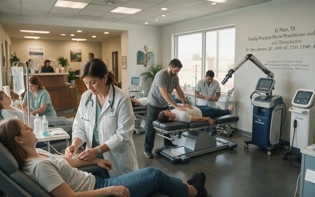
Dr. Alexander Jimenez, DC, APRN, FNP-BC, CCST, CFMP, IFMCP, ATN, is dually credentialed as a Family Nurse Practitioner and a Doctor of Chiropractic. That mix lets patients receive structural care and medical care in one place. They do not have to coordinate with separate clinics for alignment work, medical review, imaging decisions, and injury documentation.
This dual-scope model matters after a crash because injuries are rarely only “a sore neck.” Joints may be restricted. Soft tissue may be inflamed. Nerves may be irritated. Sleep and inflammation can slow repair. A provider who can examine mechanics and medical risk in the same visit can build one plan instead of two disconnected plans.
Medical Direction With Dr. Maria Guadalupe Cardenas
Dr. Maria Guadalupe Cardenas, MD, is board-certified in internal medicine (NPI #1164426749, Texas MD License #J2933). She has more than 40 years of experience as an internist and serves as medical director and collaborative physician at Injury Medical Clinic PA in El Paso, Texas.
This multidisciplinary setup is common in integrative and injury-care clinics. An MD provides medical direction. A chiropractor leads mechanical and functional care. Dr. Jimenez provides chiropractic care, functional medicine, personal injury documentation, and rehabilitation guidance. Dr. Cardenas provides medical oversight, risk review, and coordination for advanced services. The team also integrates rehabilitation and related injury services so care stays aligned under one roof.
Instant Multimodal Assessment
The clinic interrupts the wait-and-see cycle with an early, complete exam rather than a “come back if it still hurts” packet.
A typical first visit includes:
History of the collision and delayed symptoms
Orthopedic and neurologic testing
Range-of-motion and posture checks
Review of emergency-department notes and imaging when needed
Mapping of joints, muscles, ligaments, and nerves involved
Dr. Jimenez has observed that many accident injuries are missed at first because they involve soft tissue rather than a broken bone. Whiplash, ligament sprain, and nerve irritation may not show on a basic X-ray. That is why early motion testing and timely follow-up imaging matter.
From that exam, care can combine chiropractic mechanical alignment with modern medical procedures. Regenerative therapies, tissue-rebuilding peptides, and laser therapy may be added when the findings support them. Functional medicine looks at sleep, nutrition, and inflammation that can stall repair.
How Integrative Chiropractic Care Fits
Integrative chiropractic care is the mechanical core. Gentle adjustments, soft-tissue work, traction, and spinal decompression aim to restore joint motion, ease nerve pressure, and reduce muscle guarding. When the spine and joints move better, blood flow and nerve signals improve. That gives regenerative and rehab work a better chance to succeed.
Integrative chiropractic does not stand alone. The clinic pairs it with:
Regenerative therapies such as PRP, platelet-rich fibrin, or microfragmented adipose tissue when medically appropriate, to support tendon, ligament, and joint repair
Tissue-rebuilding peptides studied for signaling related to inflammation, collagen support, and blood vessel growth
Laser therapy (photobiomodulation / MLS laser) that uses light to support cellular energy and local circulation
Rehabilitation to rebuild strength, balance, and work-ready movement
Medical oversight from Dr. Cardenas for safety, referrals, and internal medicine issues that can affect healing
The point is simple: line up the frame, then support the tissue that must rebuild.
SOAP Notes and Functional Impairment Mapping
Even with proper care, a claim can still be denied if the chart is thin. The clinic uses detailed SOAP notes to track a patient from the initial injury to MMI.
Subjective: pain, sleep, work limits, and daily tasks in the patient’s words
Objective: motion degrees, strength, spasm, and neurologic signs
Assessment: how findings link to the crash
Plan: why the next step is needed
Functional impairment mapping goes further. It tracks how the injury changes driving, sitting, lifting, and household tasks. Those details help show why care continues and when the patient reaches MMI—the point at which further major improvement is not expected. Clear notes also help explain any pause in care so insurers cannot treat every gap as proof the injury was gone.
Attorneys look for this kind of file because it is easier to defend. It shows causation, timely care, and a reasoned path rather than a stack of pain pills.
A Scannable Three-Phase Recovery Plan
Recovery is easier to follow when it has stages. The clinic uses a phase-by-phase approach that patients and attorneys can scan.
Phase 1: Acute pain alleviation (days 1–14)
Calm inflammation and muscle spasm
Restore basic joint motion with gentle chiropractic care
Use laser therapy and supportive medical care as needed
Start a paper trail that links symptoms to the crash
Protect sleep and basic daily function
Care in this window helps prevent the “it must not have been serious” argument. It also reduces the chance that a sprain becomes long-term instability.
Phase 2: Cellular regeneration and stability (weeks 3–8)
Continue alignment work so tissue heals in a better position
Add regenerative therapies or peptides when the exam supports them
Use laser and soft-tissue methods to support repair
Address nutrition, hydration, and inflammation through functional medicine
Recheck motion, strength, and nerve findings
Feeling a little better is not the finish line. The team looks for tissue stability, not only a lower pain score.
Phase 3: Functional rehabilitation to MMI (8+ weeks)
Build strength, endurance, and work or driving tolerance
Correct posture and movement patterns that keep re-irritating the injury
Document remaining limits and future care needs
Reach MMI with a clear summary of what improved and what remains
MMI does not always mean perfect. It means the chart can show the true end of expected medical gain. That honesty protects both the patient and the claim.
Clinical Observations From Dr. Alexander Jimenez
From years of accident and sports-injury work in El Paso, Dr. Jimenez notes that people often look “fine” on a first X-ray and still have ligament, disc, or nerve injury that shows up later. He also notes that care works best when mechanical correction and cellular support run together. Adjusting a joint while nearby tissue stays inflamed or poorly supplied can stall progress. Pairing chiropractic care with regenerative tools, peptides, laser therapy, and medical direction is meant to close that gap.
The same observations apply to documentation. A note that only says “patient improved” is weak. A note that shows degrees of motion gained, tasks the patient can now do, and why the next phase is needed is useful in both the treatment room and a settlement file. Dual-credentialed care, plus medical direction from Dr. Cardenas, lets the clinic tell that story from both a chiropractic and a medical viewpoint.
A Clearer Path From Injury to MMI
Conventional personal injury treatment often keeps accident victims in a prolonged state of uncertainty. Muscle relaxers and NSAIDs can mask immediate discomfort while structural and cellular injury go unaddressed. Dr. Jimenez’s clinic interrupts that cycle with instant multimodal assessment, harmonized structural and medical care under one roof, and objective tracking from first injury to MMI.
The journey is straightforward: ease acute pain in days 1–14, support cellular regeneration and stability in weeks 3–8, then rebuild function from week 8 onward. For people in El Paso facing auto or work injuries, that structure can protect both recovery and the record that supports fair compensation.
Micro-fragmented adipose tissue, or MFAT, is a minimally invasive regenerative treatment. It uses a small amount of a person’s own fat. Mechanical processing cleans the fat and breaks it into tiny pieces, without harsh enzymes or extra chemicals. The finished tissue holds healing cells, growth factors, and a soft natural framework. After a car accident, injuries are often layered. Joints may be misaligned. Ligaments may tear or stretch. Soft tissues may stay swollen and take a long time to heal.
MFAT can be injected into selected joints, tendons, or ligaments to help reduce inflammation and support repair. This article explains what MFAT is, how it is prepared, why auto injuries are complex, and how MFAT works with integrative chiropractic rehabilitation. It also describes the team at Injury Medical Clinic PA in El Paso, Texas, where Dr. Alexander Jimenez provides chiropractic and integrative care and Dr. Maria Guadalupe Cardenas, MD, offers medical direction.
What MFAT Is
MFAT starts with adipose tissue, the medical name for body fat. Fat does more than store energy. It also contains structural tissue, blood-vessel-related cells, signaling cells, and growth factors. In this treatment, a small sample of the patient’s own fat is taken, usually from the abdomen or thigh. The sample is washed and broken into micro-fragments. Those fragments are then placed into an injured joint, tendon, or ligament.
Because the material comes from the same person, the chance of rejection is very low. The goal is to support the local healing environment. MFAT may help calm swelling, add cushion, and send repair signals to nearby tissue.
It does not promise that cartilage will grow back.
It does not replace surgery when a structure is fully torn or unstable.
It is one option for selected injuries that have not settled with basic care.
In plain language, MFAT is the patient’s own tissue, prepared in a limited way, and placed where the body is having trouble healing.
How Mechanical Processing Concentrates Healing Support
The processing step is what sets this treatment apart. The fat is handled in a closed system and reduced in size with mild mechanical force. The process removes oil, excess fluid, and blood residue. The useful tissue stays together. No harsh enzymes are added. No cells are grown in a lab.
A typical visit follows this path:
A clinician reviews the injury, exam findings, and imaging.
A small amount of fat is collected under local anesthesia.
The tissue is washed and micro-fragmented the same day.
The processed tissue is injected, often with ultrasound guidance.
A protection and rehabilitation plan begins after the procedure.
This same-day process is one reason MFAT is called minimally invasive. Most people go home the same day and return to light activity within a few days. Activity then increases under a guided plan.
Why Car Accidents Cause More Than One Problem at a Time
A crash can injure several layers of the body at once. A knee can strike the dashboard. A shoulder can brace against the seat belt. The neck can snap forward and back. Ligaments can stretch. Cartilage can bruise. Muscles can tighten to protect the area. Those changes do not always show up as one simple finding on the first visit.
Common results of auto accidents include:
Joints that no longer move in a smooth, centered way
Ligament tears or sprains that leave the area less stable
Tendon injuries that heal slowly because blood supply is limited
Soft tissues that stay inflamed for weeks or months
Muscle guarding and poor posture that add extra stress
Weakness and balance changes that alter walking, driving, and work tasks
This is why some people still hurt long after the bruise fades. The original tissue injury may remain. The way the body moves may also keep loading that same spot. Older accident injuries can linger as neck pain, low back pain, shoulder or knee pain, stiffness, or reduced motion when those layers are left uncorrected.
Pain medicine may take the edge off symptoms. It does not by itself rebuild tissue or restore motion. That is why an integrative plan looks at both the injured structure and the whole movement pattern.
How MFAT Can Support Auto Injury Recovery
MFAT is usually considered after conservative care has already been tried. That earlier care may include examination, imaging, activity changes, chiropractic care, rehabilitation, and sometimes platelet-rich plasma (PRP). MFAT may be discussed when the injury is more complex, more degenerative, or slower to heal.
When used with an integrative plan, MFAT can address several stages of recovery:
The tissue stage. The injection may reduce local inflammation and support repair inside a joint, tendon, or ligament.
The support stage. The micro-fragments provide a soft scaffold, which can help a worn or injured area feel more cushioned.
The signaling stage. Growth factors and related cells can send repair messages over weeks and months, not only for a few days.
The movement stage. Chiropractic care and rehab then help the body load that tissue more evenly.
The best-studied use is knee osteoarthritis, including cases linked to earlier trauma. Reviews report that some patients have less pain and better function after MFAT. Similar support has been discussed for selected tendon problems, partial soft-tissue tears, and chronic joint irritation. Results vary. When improvement happens, it often builds over weeks to months.
MFAT is not first-line care. It is not a fit for unstable fractures, complete ruptures, active infection, or joints that already need reconstruction. Regenerative treatments used this way are still being studied and are not FDA-approved as a cure for osteoarthritis or tendon injuries. Careful screening matters.
How Integrative Chiropractic Care Fits Into MFAT Treatment
Advanced regenerative medicine and chiropractic rehabilitation work on different parts of the same problem. MFAT supports the biology of the injured tissue. Integrative chiropractic care supports the mechanics of the body around that tissue.
If a joint stays crooked, a hip stays stiff, or the spine keeps moving poorly, the treated area can remain under extra load. Chiropractic adjustments, soft-tissue treatment, posture work, and guided exercise help restore cleaner motion. Rehabilitation then rebuilds strength, balance, and daily function so healing tissue isn’t overloaded.
A clear combined plan often moves through these stages:
Evaluation first. The team identifies the injured tissue and the movement problems that keep stressing it.
Early protection. After MFAT, the injection site needs time. Heavy loading is avoided.
Guided motion. Gentle chiropractic care and light exercise restore range without forcing the area.
Strength and control. Rehab adds stability so work, driving, and home tasks are safer.
Return to function. Care then focuses on lasting mechanics and injury prevention.
This pairing covers several levels of recovery at once: tissue quality, joint position, muscle balance, and whole-body movement. Direct trials of “MFAT plus chiropractic” are still limited, so the combination is based on complementary roles, not on a claim that the pair is proven better than MFAT alone.
A Multidisciplinary Team in El Paso
At Injury Medical Clinic PA in El Paso, Texas, care is built as a coordinated plan rather than a single procedure. Dr. Alexander Jimenez, DC, APRN, FNP-BC, CCST, CFMP, IFMCP, ATN, provides integrative chiropractic care, family nurse practitioner services, functional medicine, personal injury evaluation, and rehabilitation planning. That mix is useful after auto accidents because the same patient may need spinal care, soft-tissue rehab, injury documentation, and a plan that also looks at sleep, nutrition, and inflammation.
Dr. Maria Guadalupe Cardenas, MD, is board-certified in internal medicine and has more than 40 years of experience as an internist. She works with Dr. Alex Jimenez, DC, and serves as the medical director and collaborative physician at Injury Medical Clinic PA. Her NPI is #1164426749, and her Texas MD license is #J2933. In this model, the MD provides medical direction, health screening, and oversight. The chiropractic and rehabilitation team restores motion and function. This multidisciplinary setup is common in integrative or injury care clinics, where an MD provides medical direction alongside a chiropractor.
The team integrates several services into one path:
Chiropractic care from Dr. Jimenez for spinal and joint mechanics
Medical oversight from Dr. Cardenas in internal medicine
Functional medicine support for inflammation, nutrition, sleep, and recovery
Personal injury evaluation and documentation after auto accidents
Rehabilitation and return-to-activity planning
Related services that support whole-person healing
This matters after a crash because the injured joint is rarely the only issue. Body-wide inflammation, poor sleep, blood-sugar stress, or missed medical problems can slow tissue repair. Medical review and chiropractic care can sit side by side instead of sending the patient through disconnected offices.
Clinical Observations From Dr. Alexander Jimenez
Dr. Jimenez has observed that auto-injury patients often present with layered problems. A painful knee may also come with limited hip motion, guarded spinal movement, weak core control, and a walking pattern that keeps twisting the same ligament. Treating only the local tissue can leave those extra forces in place.
His clinical approach asks a simple question: why is this area still hurting? Possible answers include poor alignment, incomplete soft-tissue healing, leftover swelling, nerve irritation, or daily habits that continue to stress the injury. In that view:
MFAT may support the tissue environment.
Chiropractic care may improve motion and reduce compensation.
Rehabilitation may prepare the area for real-life loading.
Functional medicine may address nutrition, inflammation, and recovery capacity.
These observations come from integrated clinical practice in El Paso and from educational posts on his clinical sites and professional profile. Read them as practice-based insight. They are not the same as a controlled trial that proves one exact combination works for every accident injury.
Who May Be Considered and What Recovery Can Look Like
People who may be candidates often have moderate joint damage, a cartilage problem that still causes swelling or catching, a larger partial tendon tear, or a chronic soft-tissue injury that did not settle after therapy or simpler injections. People who are usually not candidates include those with complete tears, unstable fractures, infection, or advanced destruction that already needs surgery.
If MFAT is appropriate, patients should expect a full work-up first. After the procedure, light activity often returns quickly. Structured rehab follows a short protection period. Pain and function, if they improve, usually change over weeks to months. Progress is tracked with motion, strength, swelling, work ability, and daily tasks.
No injection replaces good diagnosis, better mechanics, and follow-through. The strongest plan uses regenerative support when the tissue needs it and chiropractic rehabilitation when the body keeps moving in a way that reloads the injury.
Final Thoughts
MFAT is a minimally invasive regenerative procedure made from a small amount of a patient’s own fat. Mechanical processing concentrates healing cells and growth factors without harsh enzymes or chemicals. After a car accident, this biologic support may help selected joints, tendons, and ligaments calm down and repair more effectively.
Integrative chiropractic care completes the picture by restoring alignment, motion, and strength. At Injury Medical Clinic PA in El Paso, Dr. Jimenez and Dr. Cardenas work within a multidisciplinary model that also includes functional medicine, personal injury care, and rehabilitation. Together, these layers address both the injured tissue and how the whole body moves.
Anyone with lingering pain after an auto accident should get a complete evaluation from a qualified team. That visit can sort out whether continued conservative care, rehabilitation, MFAT, or another option is the safest next step. This article is for education only and is not personal medical advice.
Overlooked Injuries After Work and Auto Accidents: Why X-Rays Often Miss Them
Abstract: Soft-tissue damage, mild concussions, and hairline spinal issues are among the most frequently overlooked injuries after workplace incidents and motor vehicle accidents (MVAs). These problems often lack immediate, visible signs and do not appear on standard X-rays. This article explains why micro-tears in spinal ligaments, subtle annular tears in discs, hidden facet joint capsule trauma, and chronic myofascial trigger points are so often missed. It describes how untreated damage can lead to lasting pain. It then shows how an integrative approach that combines mechanical restoration through chiropractic care with biological tissue repair can support better recovery. Clinical observations from Dr. Alexander Jimenez and the collaborative team at Injury Medical Clinic PA in El Paso, Texas, illustrate how this model works.
A person can walk away from a crash or a workplace incident feeling only somewhat sore. The first exam often looks for broken bones, bleeding, and other obvious emergencies. When the X-rays look normal, many people receive reassurance that they are fine. Days or weeks later, neck stiffness, headaches, back pain, or trouble concentrating can develop and persist. These delayed symptoms are common. They often come from injuries that standard films cannot show.
Right after trauma, adrenaline hides pain. Inflammation then builds slowly. Soft tissues can stretch or tear without breaking bone. That is why so many injuries after MVAs and workplace events stay hidden at first. If they go undiagnosed, they can leave people with persistent discomfort and reduced daily function.
Why Standard X-Rays Miss These Injuries
X-rays are useful for bones. They show fractures and major joint problems. They do not show muscles, ligaments, tendons, fascia, joint capsules, or the outer layers of spinal discs. Mild concussions also leave little clear mark on routine scans. The injury can still be real even when the pictures look clean.
After a collision, the body is thrown forward and then stopped. Ligaments and joint capsules can be stretched past their safe limit. Disc fibers can develop small cracks. Deep muscles can form tight, painful knots. Workplace injuries can follow the same path after a sudden lift, a fall, or months of repetitive strain. Because symptoms often start later, people may assume the pain is only ordinary muscle tightness.
The Injuries Most Often Overlooked After Crashes and Work Events
Several specific problems recur when a more complete evaluation is done.
Micro-tears in spinal ligaments: Ligaments hold the vertebrae in place. Sudden force can create tiny tears that allow slight extra motion. The spine may feel unstable even when X-rays look perfect. People notice stiffness after sitting, pain with certain movements, or a sense that the back “becomes unstable.”
Subtle annular tears in intervertebral discs: The tough outer ring of a disc can crack without a full herniation. These tears can irritate nearby nerves. Pain may travel into an arm or a leg days later. Early tears are easy to miss on routine imaging.
Hidden facet joint capsule trauma: Facet joints guide how the spine bends and twists. Their capsules can stretch or partially tear. This can cause local pain, reduced motion, and headaches. Standard films rarely capture this soft-tissue damage.
Chronic myofascial trigger points: Trauma can create tight bands in deep stabilizing muscles. These knots send pain to other areas. Headaches, shoulder tightness, or low-back discomfort may actually start in these guarded muscles.
Mild concussions are another frequent miss. The brain can move inside the skull even without a direct blow to the head. Symptoms such as foggy thinking, poor concentration, light sensitivity, or mood changes may appear later. A normal scan rules out large bleeding. It does not prove the brain is uninjured.
Workplace injuries often develop in two ways. One is a single event, such as a fall or a sudden lift. The other is cumulative trauma from repeated motions, awkward postures, or vibration. Tendon irritation, shoulder strain, and low-back tightness can build slowly. Because there is no single dramatic moment, these injuries are easy to overlook until function is already limited.
What Happens When These Injuries Stay Untreated
The body tries to protect itself. Muscles tighten around unstable segments. Posture and walking patterns change. Circulation drops in the guarded areas. Scar tissue can form in a less organized way. Over time, the nervous system can become more sensitive to pain. A treatable soft-tissue injury can then settle into chronic pain, reduced movement, and trouble returning to work or daily tasks.
Early, thorough evaluation changes that path. A clear history of how the injury happened, a careful hands-on exam of joint motion and muscle texture, and selected advanced tests can reveal the real sources of symptoms.
An Integrative Approach: Mechanical Restoration Plus Biological Tissue Repair
Recovery works best when two goals are met together. First, the mechanical environment must improve. Joints need proper motion. Muscles need balanced length and strength. Movement patterns need retraining so healing tissue isn’t constantly re-stressed. Second, damaged soft tissues need biological support to rebuild stronger collagen and quiet leftover inflammation.
Integrative chiropractic care addresses the mechanical side. Specific adjustments restore joint motion and reduce extra load on injured segments. Soft-tissue work releases guarded muscles and improves local blood flow. Corrective exercises rebuild strength and coordination. When it is appropriate, spinal decompression can reduce pressure on discs and nerves. These steps create a better setting for repair.
Biological tissue support may include regenerative options such as platelet-rich plasma or related plasma products when clinically appropriate. These approaches use the patient’s own growth factors to help ligaments, tendons, and other soft tissues heal. Mechanical correction and biological support together aim at lasting function, not only short-term relief.
How the El Paso Team Applies This Model
Clinical observations from Dr. Alexander Jimenez, DC, APRN, FNP-BC, CCST, CFMP, IFMCP, ATN, note that patients with overlooked soft-tissue and joint injuries often develop secondary changes in posture, gait, and spinal motion. Treating only the most painful spot can leave those broader patterns in place and slow recovery. A complete assessment that includes biomechanics, nerve function, and movement helps build a more complete plan.
At Injury Medical Clinic PA in El Paso, Texas, care follows a multidisciplinary structure. Dr. Jimenez provides chiropractic care, functional medicine insight, personal injury evaluation, and rehabilitation planning. Dr. Maria Guadalupe Cardenas, MD, board-certified in internal medicine (NPI #1164426749, Texas MD License #J2933), serves as Medical Director and Collaborative Physician. With more than 40 years of experience as an internist, she provides medical oversight and diagnostic direction.
This setup is common in integrative and injury-focused clinics: an MD supplies medical direction while a chiropractor focuses on spinal mechanics, movement, and hands-on restoration. The team integrates chiropractic adjustments and soft-tissue care with medical evaluation, functional medicine strategies, personal injury documentation, and progressive rehabilitation. Patients receive one coordinated plan rather than isolated treatments.
A Clear Next Step After a Crash or Workplace Injury
If pain, stiffness, headaches, or concentration problems linger after a motor vehicle accident or workplace incident—even when early X-rays were normal—further evaluation is reasonable. Soft-tissue damage, mild concussions, and small spinal issues are real. They respond best when they are found early and treated with both mechanical correction and support for biological healing.
An integrative model that pairs chiropractic care for joint and soft-tissue function with medical oversight, functional strategies, and rehabilitation offers a practical path. In the collaborative setting of Injury Medical Clinic PA, chiropractic expertise and internal medicine direction work together to help people move from hidden injury toward better daily function.
A motor vehicle accident (MVA) can place sudden and powerful forces on the body. Knees may strike the dashboard, shoulders may be pulled by a seat belt, and the spine may rapidly bend, twist, or compress. These forces can injure cartilage, ligaments, tendons, muscles, bones, and spinal discs. Common joint trauma injuries include ACL or PCL tears, meniscus injuries, shoulder labrum tears, joint dislocations, sprains, and cartilage damage.
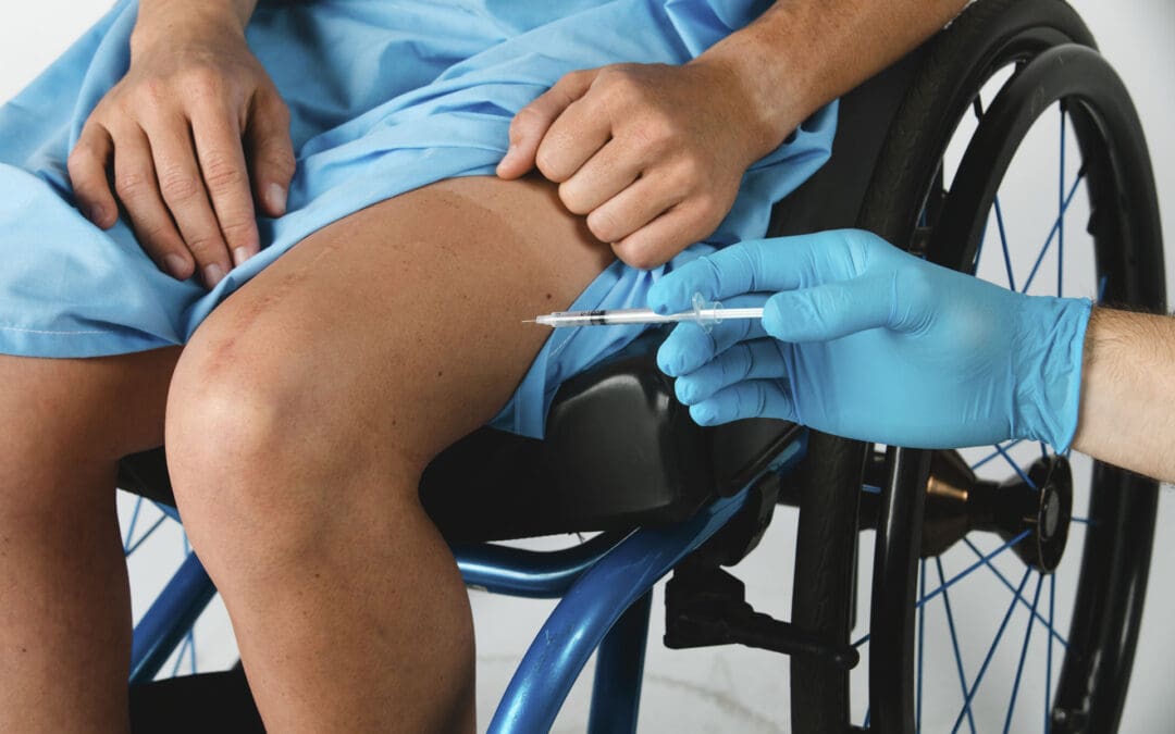
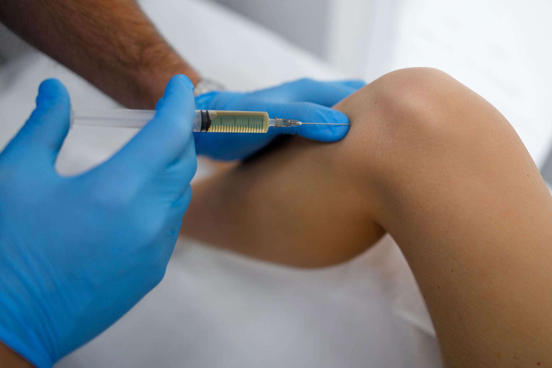
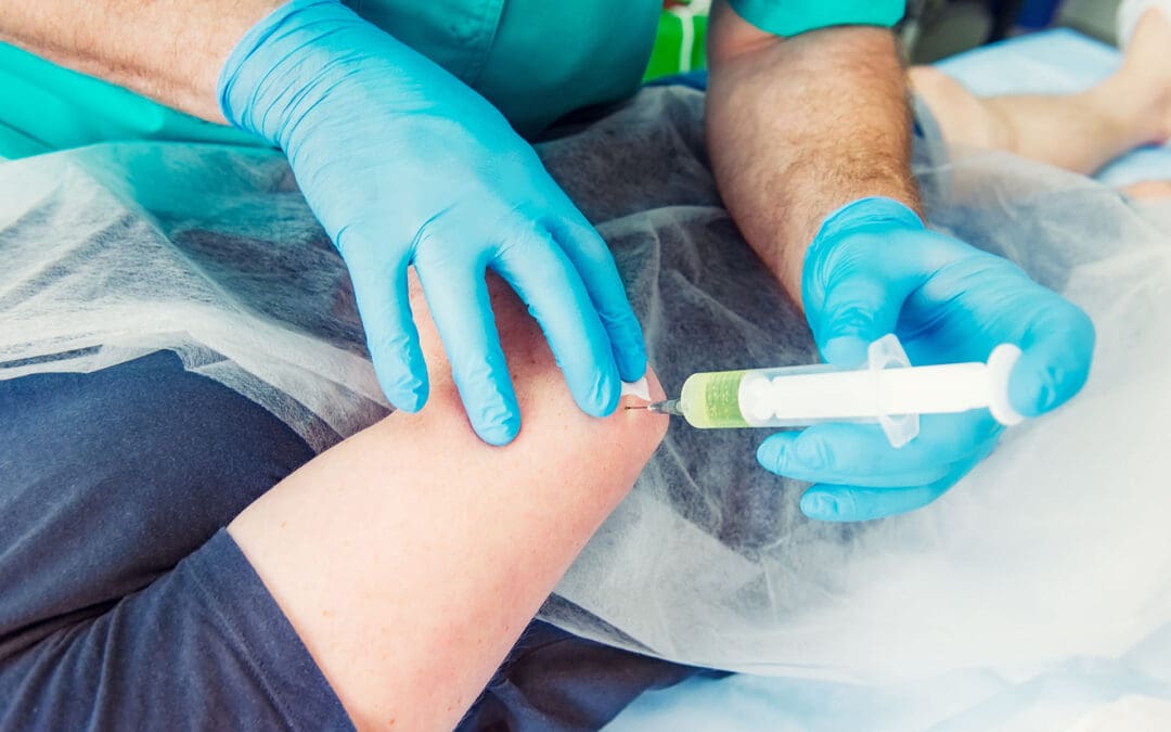
Treatment depends on the type and severity of the injury. Some patients improve with chiropractic care, rehabilitation, exercise, and physical modalities such as shockwave or laser therapy. Others may need targeted pain management or selected orthobiologic procedures, including platelet-rich plasma (PRP), platelet-fibrin products (PFP/PRF), or microfragmented adipose tissue (MFAT). These treatments should be selected after a careful medical and musculoskeletal evaluation because no single therapy is appropriate for every accident injury.
In El Paso, a multidisciplinary approach can bring chiropractic care, medical oversight, functional medicine, personal injury care, rehabilitation, diagnostic testing, and specialty referrals together in one coordinated recovery plan.
What Are Joint Trauma Injuries in a Motor Vehicle Accident?
Joint trauma injuries are sudden injuries caused when crash forces twist, stretch, compress, strike, or dislocate parts of the musculoskeletal system.
A joint is the place where two or more bones meet. Cartilage helps the bones move smoothly. Ligaments connect bone to bone, while tendons connect muscles to bones. When a car crash creates more force than these tissues can safely handle, they may become stretched, torn, bruised, fractured, or displaced.
Common areas affected after an MVA include:
Knees
Shoulders
Hips
Wrists and hands
Ankles
Neck and spinal joints
Lower back
Sacroiliac joints
Spinal discs may also be damaged during an accident. A disc is not technically a joint, but disc injuries often occur together with trauma to the spinal joints, muscles, ligaments, and nerves.
The severity can range from a mild sprain to a complete ligament tear, dislocation, fracture, or complex injury involving several tissues at the same time.
Dashboard Knee Injuries After a Car Accident
The knee is especially vulnerable during a front-end collision.
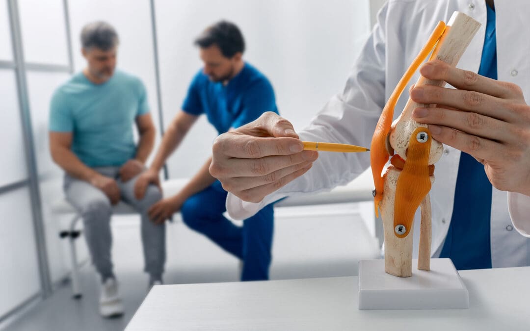
If a person’s bent knee strikes the dashboard, the force may push the shinbone backward. This is a well-known mechanism for injuring the posterior cruciate ligament, or PCL. The American Academy of Orthopaedic Surgeons lists a bent knee striking a dashboard during a motor vehicle collision as a common cause of PCL injury (American Academy of Orthopaedic Surgeons [AAOS], n.d.).
Dashboard trauma can also cause:
PCL tears
ACL or MCL injuries
Meniscus tears
Cartilage damage
Bone bruising
Kneecap fractures
Joint swelling
Reduced range of motion
Knee instability
A direct dashboard impact can even fracture the kneecap. Severe cartilage injury may later contribute to post-traumatic arthritis (AAOS, n.d.).
El Paso educational resources also describe dashboard knee injuries as a combination of ligament, cartilage, meniscus, and bone trauma rather than simply a temporary knee bruise (Jimenez, 2026a).
Shoulder Trauma After an MVA
Shoulders can also absorb major crash forces.
A driver may grip the steering wheel during impact, strike the door, or experience sudden pulling from the seat belt. The arm can be forced backward, upward, or outward.
Possible injuries include:
Rotator cuff strains or tears
Labrum injuries
Shoulder sprains
Tendon injuries
AC joint injuries
Fractures
Partial dislocations
Complete shoulder dislocations
The shoulder labrum is a ring of cartilage that helps stabilize the joint. A traumatic SLAP tear may occur during a motor vehicle collision, and a shoulder dislocation can also result from crash trauma (AAOS, n.d.).
Local El Paso injury resources similarly describe shoulder pain after crashes as potentially involving muscles, tendons, ligaments, nerves, and joint structures.
Hip, Wrist, and Spinal Joint Trauma
An MVA does not affect only the knee and shoulder.
The seat, center console, door, or dashboard may compress the hips. This can lead to strains, joint irritation, cartilage injury, labral damage, or fractures in serious crashes.
When a person braces against the steering wheel or dashboard, the wrists and hands may sustain injuries. Sudden force may cause sprains, tendon injuries, fractures, or joint instability.
The neck and lower back experience a different type of trauma. Rapid acceleration and deceleration can suddenly bend and rotate the spine. This can strain muscles and ligaments, irritate spinal joints, and contribute to disc injury or nerve symptoms.
Because a crash can injure several areas at once, examining only the most painful joint may overlook other injuries.
Symptoms That Should Be Evaluated
Joint trauma does not always cause its worst symptoms immediately. Pain and stiffness can become more noticeable after the initial stress response from the crash fades.
Symptoms may include:
Joint swelling
Bruising
Pain with movement
Reduced range of motion
Clicking, popping, locking, or catching
A feeling that the joint is unstable
Muscle spasms
Weakness
Numbness or tingling
Neck or back stiffness
Pain traveling into an arm or leg
Difficulty walking, climbing stairs, lifting, or working
Obvious deformity, severe swelling, inability to bear weight, new major weakness or numbness, or other severe symptoms require prompt medical evaluation.
Imaging such as X-rays, MRI, CT, or diagnostic ultrasound may be appropriate depending on the suspected injury.
Treating MVA Joint Trauma: More Than Pain Control
The first goal after significant trauma is to determine what was injured.
Treatment may begin with activity changes, bracing when needed, rehabilitation, and methods for controlling pain and swelling. Severe fractures, dislocations, complete ligament tears, or unstable joints may require orthopedic or surgical care.
For other injuries, a coordinated conservative treatment plan may be appropriate.
Integrative Chiropractic Care
Chiropractic care can be one part of MVA rehabilitation when serious fractures, unstable injuries, and other contraindications have been ruled out.
Care may include:
Gentle joint mobilization
Appropriate spinal manipulation
Soft-tissue techniques
Range-of-motion exercises
Posture correction
Corrective exercise
Neuromuscular retraining
Strength and stability work
Clinical guidelines for neck pain and whiplash-associated disorders support multimodal approaches that can include manipulation or mobilization combined with exercise, depending on the patient’s condition and stage of recovery (Bussières et al., 2016).
The purpose is not simply to “put a joint back in place.” The larger goal is to restore safe movement, decrease mechanical stress, improve strength, and help the patient return to normal daily activity.
Regenerative and Orthobiologic Injections
Some patients with persistent joint, ligament, tendon, or cartilage problems may be evaluated for orthobiologic treatment.
Platelet-Rich Plasma
Platelet-rich plasma, or PRP, uses a concentration of platelets prepared from the patient’s own blood. Platelets release signaling proteins involved in tissue repair.
PRP has been studied most strongly for problems such as knee osteoarthritis and several tendon disorders. Research suggests that some patients can experience meaningful improvement in pain and function, although preparation methods, platelet concentrations, and results vary between studies (Berrigan et al., 2024).
PRP should therefore be described as a treatment that may support healing and improve symptoms in selected conditions, rather than a guaranteed way to regenerate every damaged ligament or piece of cartilage.
PFP and Platelet-Fibrin Products
The term PFP may be used in some clinical settings to describe platelet-fibrin products. Related preparations include platelet-rich fibrin, or PRF.
Fibrin creates a matrix that can hold platelets and signaling molecules at the treatment area. Research into these preparations continues, and formulations are not standardized across every clinic or medical study. Reviews of orthobiologic therapies note promising results but also the need for stronger clinical trials and clearer treatment standards (Motejunas et al., 2021).
Microfragmented Adipose Tissue
Microfragmented adipose tissue, or MFAT, is another procedure being studied for musculoskeletal conditions.
Recent research comparing MFAT and PRP for knee osteoarthritis found that both treatments may improve pain and function in selected patients, but researchers also emphasize that the evidence remains limited and further study is needed (Park et al., 2025).
MFAT should not be presented as a proven way to regrow any damaged joint. Patient selection, diagnosis, regulatory requirements, and medical oversight are important. The FDA also warns consumers that many products marketed broadly as “regenerative medicine” are not FDA-approved for treating orthopedic conditions.
Targeted Pain Management
Some MVA injuries involve more than joint damage.
A disc injury may irritate a spinal nerve and cause pain traveling down an arm or leg. In carefully selected patients with radicular pain, an epidural steroid injection may temporarily reduce nerve-root inflammation and pain. Evidence suggests benefits can occur, although the effects and duration vary (Mahmoud et al., 2024).
Trigger point injections may be considered when painful muscle trigger points contribute to myofascial pain. A systematic review found reductions in acute myofascial pain compared with medical management alone, though treatment still needs to be individualized (Hamzoian & Zograbyan, 2023).
These procedures control specific pain generators. They do not replace rehabilitation or correct every damaged structure.
Shockwave and Laser Therapy
Physical modalities can also support selected rehabilitation programs.
Extracorporeal shockwave therapy delivers acoustic energy to targeted tissues and has evidence supporting its use for several chronic tendon problems. It may work best when combined with a progressive exercise program rather than used alone (Elgendy et al., 2024).
Photobiomodulation or therapeutic laser therapy uses light energy. Research suggests it may help reduce pain in some musculoskeletal conditions, although the ideal dose and treatment schedule vary by condition.
Neither therapy replaces proper diagnosis, stabilization of serious injuries, or rehabilitation.
Multidisciplinary MVA Injury Care in El Paso
Joint trauma after a car accident rarely occurs in isolation. A patient may have knee pain, shoulder damage, whiplash, disc irritation, muscle guarding, headaches, weakness, and medical conditions that can affect recovery.
This is where coordinated multidisciplinary care can be useful.
At Injury Medical Clinic PA in El Paso, Texas, practice materials describe Dr. Maria Guadalupe Cardenas, MD, an internal medicine physician, as Medical Director and Collaborative Physician working alongside Dr. Alexander Jimenez, DC, APRN, FNP-BC, CCST, CFMP, IFMCP, ATN. The clinic describes a model that combines medical oversight with chiropractic care, functional medicine, personal injury care, rehabilitation, diagnostic evaluation, pain-management coordination, and related services.
Clinic materials state that Dr. Cardenas is board-certified in internal medicine and has more than 40 years of clinical experience. Independent physician information also identifies her as an internal medicine physician in El Paso, with Texas medical license J2933.
Under this model, Dr. Cardenas provides the medical perspective needed to consider medications, chronic health problems, medical risks, diagnostic findings, and overall patient safety. Dr. Jimenez focuses heavily on the neuromusculoskeletal side of recovery, including spinal and extremity joint function, movement patterns, rehabilitation, and functional health strategies.
Clinical observations published by Dr. Jimenez emphasize examining how an accident affects more than the painful area. His approach looks at joint motion, posture, muscle balance, nerve irritation, gait, strength, flexibility, inflammation, and the patient’s ability to perform everyday activities.
The result is a treatment plan that may move through stages:
Identify the injury and rule out serious damage.
Control pain and inflammation.
Restore joint and spinal movement where appropriate.
Rebuild strength, balance, and stability.
Consider targeted injections or orthobiologic treatment when clinically appropriate.
Track functional improvement and adjust the plan as the patient recovers.
This approach recognizes an important point: reducing pain is valuable, but restoring function is the larger goal.
The Path Forward After an MVA Joint Injury
Motor vehicle accidents can create much more than temporary soreness. Crash forces can tear ligaments, damage cartilage, irritate tendons, dislocate joints, fracture bones, and injure spinal structures.
A successful recovery starts with an accurate diagnosis.
Some patients need orthopedic or surgical care. Others may benefit from chiropractic treatment, rehabilitation, targeted pain management, laser or shockwave therapy, or selected regenerative procedures. PRP, platelet-fibrin preparations, and MFAT may have a role in carefully chosen cases, but they are not universal treatments and should not be marketed as guaranteed tissue regeneration.
For patients in El Paso, multidisciplinary care offers a way to address the medical, mechanical, functional, and rehabilitation sides of an MVA injury within one coordinated plan.
When Micro-Fragmented Adipose Tissue Helps Heal Complex Injuries
Abstract: Injuries from car accidents or workplace events can create lasting problems in joints, tendons, ligaments, and cartilage. This article explains when micro-fragmented adipose tissue (MFAT) is recommended as a regenerative option. It focuses on more complex, severe, or slow-to-heal cases that have not improved enough with conservative care or basic injections such as platelet-rich plasma. Readers will learn how MFAT works, why it is chosen for larger tears and moderate-to-severe joint problems, and how it can be safely combined with integrative chiropractic care when treatments are properly sequenced. The discussion also covers the multidisciplinary team approach used at Injury Medical Clinic PA in El Paso, Texas.
What Is Micro-Fragmented Adipose Tissue?
MFAT starts with a small sample of a person’s own fat, usually taken from the abdomen or side. The tissue is gently cleaned and broken into tiny pieces. These micro-fragments keep natural supportive cells, signaling factors, and a soft framework that can cushion and help repair damaged areas.
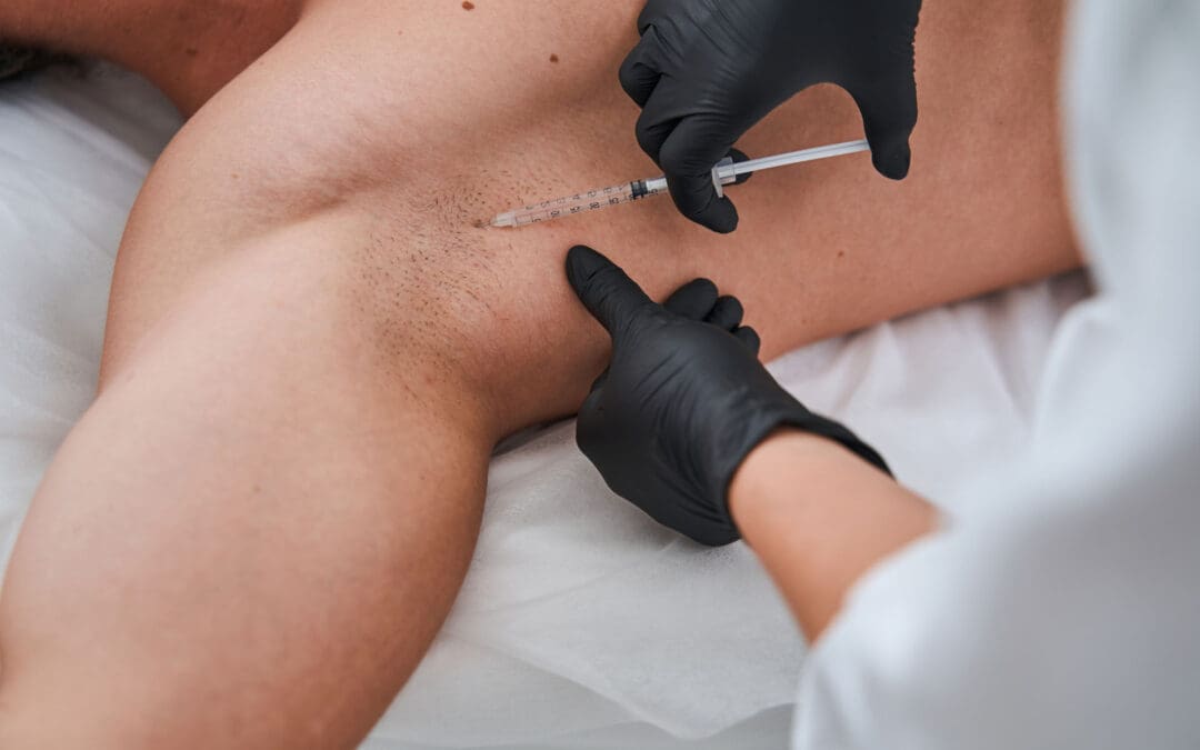
The processed material is then injected into the injured joint, tendon, or soft-tissue site, often with ultrasound guidance for accuracy. Because the material comes from the patient’s own body, the risk of rejection is very low. MFAT supplies both biological signals that calm inflammation and a physical scaffold that supports tissue rebuilding.
When Is MFAT Recommended for Injuries?
When using regenerative therapies for various types of injuries, whether from motor vehicle accidents or work-related events, MFAT is recommended for more complex, severe, or slow-to-heal injuries. These include moderate-to-severe post-traumatic joint degeneration, significant cartilage defects, or larger partial tendon or ligament tears. MFAT could be recommended when conservative care, such as physical therapy or basic injections (such as PRP), fails to restore function.
For moderate-to-severe joint damage, cartilage defects, and larger partial soft-tissue tears, MFAT is the recommended treatment. It can be safely and effectively combined with integrative chiropractic care, provided the treatments are sequenced to facilitate biological healing and restore biomechanical alignment.
A careful evaluation that includes imaging, physical exam findings, and overall health status helps determine whether MFAT is appropriate. The goal is to reduce pain, improve joint support, and aid tissue repair while supporting the body’s natural healing capacity.
How MFAT Compares to Other Regenerative Options
PRP uses concentrated platelets from a blood sample. It delivers growth factors that stimulate healing and is often tried earlier for milder soft-tissue problems or early joint wear. MFAT, by contrast, brings a denser mix of signaling cells plus a natural matrix. This combination is better suited for larger or more advanced problems where simple growth-factor signals alone may not be enough.
In practice:
PRP may be selected first for mild-to-moderate tendon irritation or smaller tears.
MFAT is frequently preferred for moderate-to-severe joint damage, bigger partial tears, or cases that did not respond fully to prior PRP.
Both treatments use the patient’s own tissue and can be performed on an outpatient basis. The choice depends on the size and severity of the injury, previous treatment results, and the specific needs of the joint or soft tissue.
How Integrative Chiropractic Care Fits With MFAT
Regenerative injections address the biological side of healing. Integrative chiropractic care addresses the mechanical side. After an accident or work injury, joints can lose proper alignment, muscles can tighten, and movement patterns can change. These mechanical stresses can slow biological repair or place extra load on recovering tissues.
Chiropractic adjustments, soft-tissue work, and guided movement restore better alignment and joint motion. When the body is mechanically balanced, the regenerative signals from MFAT can work in a more favorable environment. The two approaches complement each other when sequenced carefully:
Early care focuses on reducing acute inflammation and restoring basic motion.
Regenerative injection is timed so the tissue has a stable base for healing.
Ongoing chiropractic and rehabilitation support proper loading while the biological repair continues.
This combined plan helps protect the healing tissue and supports longer-term function.
The Multidisciplinary Team at Injury Medical Clinic PA
At Injury Medical Clinic PA in El Paso, Texas, care is delivered through a coordinated team. Dr. Alexander Jimenez, DC, APRN, FNP-BC, CCST, CFMP, IFMCP, ATN, provides chiropractic care focused on spinal and joint mechanics, along with functional medicine and rehabilitation guidance.
Working alongside him is Dr. Maria Guadalupe Cardenas, MD. She is board-certified in internal medicine, holds NPI #1164426749 and Texas MD License #J2933, and brings more than 40 years of experience as an internist. Dr. Cardenas serves as Medical Director and Collaborative Physician. She provides medical oversight, helps evaluate overall health factors that affect healing, and ensures advanced procedures stay within safe medical guidelines.
This arrangement is common in integrative injury clinics. The medical director supplies diagnostic clarity and medical direction, while the chiropractic and functional-medicine side focuses on biomechanics, nervous-system function, and whole-person recovery. The team integrates chiropractic care from Dr. Jimenez with medical oversight by Dr. Cardenas, as well as functional medicine, personal injury care, rehabilitation, and related services.
Clinical Observations From Practice
Dr. Alexander Jimenez has observed that injury patients often present with mixed tissue and mechanical problems. Larger partial tears or joint surface damage after crashes or workplace incidents frequently respond more completely when biological support is added to careful biomechanical care.
In his clinical experience, sequencing matters. Restoring alignment and reducing abnormal joint stress creates a better setting for regenerative therapies. Patients who receive both tissue-focused injections and ongoing movement correction tend to regain function more steadily than those treated with either approach alone. Functional-medicine elements—nutrition, sleep, and inflammation control—further support the body’s repair capacity. These observations align with the clinic’s emphasis on coordinated, patient-centered plans rather than isolated procedures.
What Patients Can Expect
MFAT is performed in a single outpatient visit under local anesthesia. A small volume of fat is harvested, processed, and injected. Most people resume light activity within days and begin structured rehabilitation after an initial rest period. Improvement often appears over several weeks to a few months as the tissue responds.
Results vary with the severity of the original injury, the patient’s overall health, and adherence to the recovery plan. MFAT is not a guaranteed cure, yet it offers a non-surgical option that can reduce pain, improve joint support, and help many people return to daily activities.
Moving Forward With a Clear Plan
Complex injuries demand more than a single treatment. When larger tears, cartilage problems, or post-traumatic joint changes do not improve with conservative care, MFAT can provide the structural and cellular support needed for better healing. Pairing it with properly timed integrative chiropractic care addresses both the tissue itself and the mechanical forces that act on it.
At Injury Medical Clinic PA, this combination is delivered under the medical direction of Dr. Maria Guadalupe Cardenas and the hands-on musculoskeletal expertise of Dr. Alexander Jimenez and the team. The result is a practical, coordinated path that respects both biology and biomechanics. Anyone recovering from a motor-vehicle or work-related injury can discuss whether this approach fits their specific situation with a qualified care team.
IFM's Find A Practitioner tool is the largest referral network in Functional Medicine, created to help patients locate Functional Medicine practitioners anywhere in the world. IFM Certified Practitioners are listed first in the search results, given their extensive education in Functional Medicine