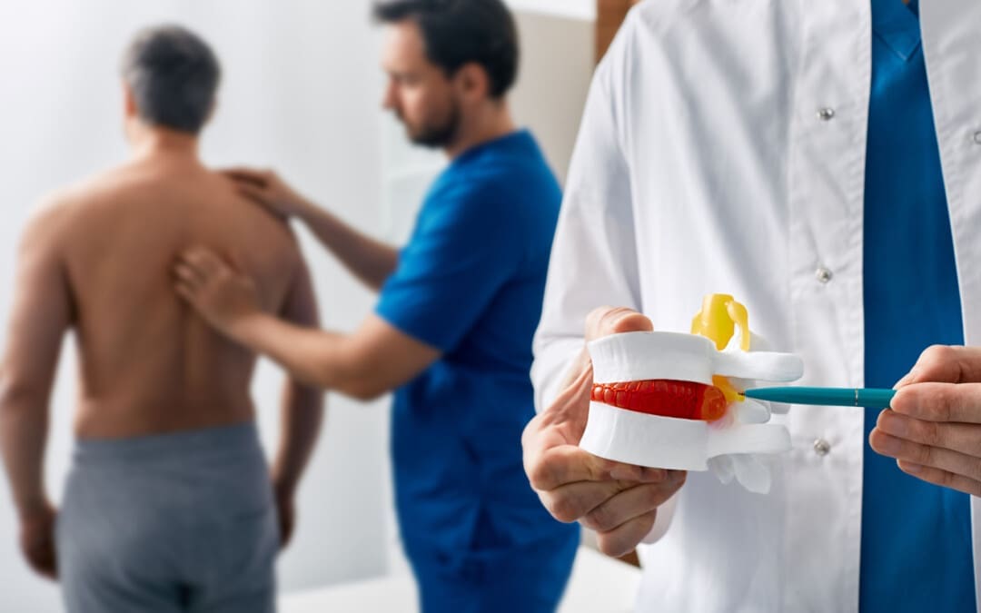
Back Clinic Chiropractic Examination. An initial chiropractic examination for musculoskeletal disorders will typically have four parts: a consultation, case history, and physical examination. Laboratory analysis and X-ray examination may be performed. Our office provides additional Functional and Integrative Wellness Assessments in order to bring greater insight into a patient’s physiological presentations.
Consultation:
The patient will meet the chiropractor which will assess and question a brief synopsis of his or her lower back pain, such as:
Duration and frequency of symptoms
Description of the symptoms (e.g. burning, throbbing)
Areas of pain
What makes the pain feel better (e.g. sitting, stretching)
What makes the pain feel worse (e.g. standing, lifting).
Case history. The chiropractor identifies the area(s) of complaint and the nature of the back pain by asking questions and learning more about different areas of the patient’s history, including:
Family history
Dietary habits
Past history of other treatments (chiropractic, osteopathic, medical and other)
Occupational history
Psychosocial history
Other areas to probe, often based on responses to the above questions.
Physical examination: We will utilize a variety of methods to determine the spinal segments that require chiropractic treatments, including but not limited to static and motion palpation techniques determining spinal segments that are hypo mobile (restricted in their movement) or fixated. Depending on the results of the above examination, a chiropractor may use additional diagnostic tests, such as:
X-ray to locate subluxations (the altered position of the vertebra)
A device that detects the temperature of the skin in the paraspinal region to identify spinal areas with a significant temperature variance that requires manipulation.
Laboratory Diagnostics: If needed we also use a variety of lab diagnostic protocols in order to determine a complete clinical picture of the patient. We have teamed up with the top labs in the city in order to give our patients the optimal clinical picture and appropriate treatments.
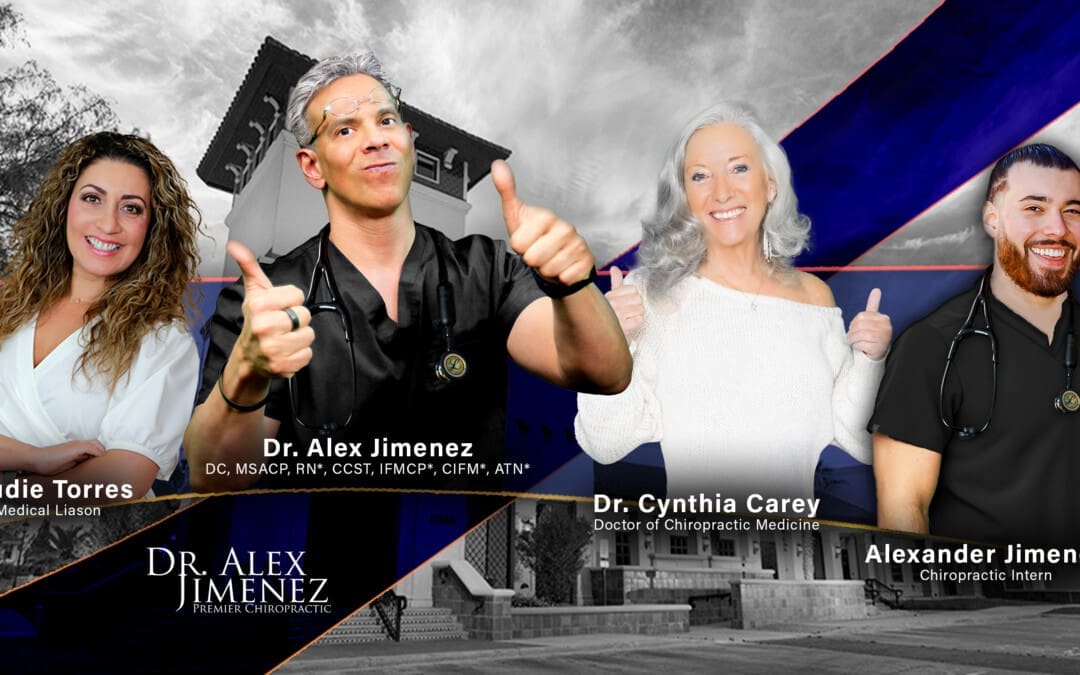
Dr. Alex Jimenez: Pioneering Integrative Care as a Chiropractor and Nurse Practitioner
Dr. Alex Jimenez, a licensed chiropractor and board-certified nurse practitioner based in El Paso, Texas, brings over 25 years of expertise to his Injury Medical & Chiropractic Clinic practice. His dual licensure provides unique insights into the etiologies, pathogenesis, and treatment of complex clinical conditions, enabling him to deliver patient-centered, integrative care that bridges physical medicine, functional medicine, and advanced diagnostics. Combining chiropractic expertise with nurse practitioner-driven medical management, Dr. Jimenez offers comprehensive treatment protocols tailored to acute and chronic conditions, promoting metabolic resilience, longevity, and whole-person wellness.
Dual Expertise: Chiropractic and Nurse Practitioner Roles
Dr. Jimenez’s practice stands out due to his ability to integrate the biomechanical focus of chiropractic care with a nurse practitioner’s diagnostic and therapeutic scope. As a chiropractor, he specializes in restoring musculoskeletal function, particularly after trauma, neck, back, spine, and soft tissue injuries. His chiropractic interventions emphasize non-invasive techniques, such as spinal decompression, manual adjustments, and functional rehabilitation, to alleviate pain and enhance mobility.
As a board-certified nurse practitioner, Dr. Jimenez employs evidence-based medicine to address systemic and metabolic dysfunctions. His expertise extends to managing chronic degenerative disorders, hormonal imbalances, weight loss, sexual health, and pain syndromes. This dual perspective allows him to identify underlying disease causes, from biomechanical misalignments to physiological imbalances, and design treatment regimens that address symptoms and root causes.
The synergy of these roles enables Dr. Jimenez to offer a holistic approach that is particularly effective for complex conditions such as sciatica, fibromyalgia, disc herniation, spondylolisthesis, and chronic neck or back pain. His integrative protocols combine functional medicine assessments, lifestyle interventions, and advanced diagnostics to achieve homeostasis and physiological balance.
Treatment Protocols: Evidence-Based and Patient-Centered
Drawing from integrative protocols outlined on his website, www.chiromed.com, Dr. Jimenez employs a multidisciplinary approach to patient care. His treatment plans are grounded in evidence-based medicine, functional medicine, and lifestyle wellness strategies, tailored to each patient’s unique health profile, lifestyle, and medical history. Below are key components of his integrative care model:
1. Chiropractic Care and Functional Rehabilitation
Spinal Decompression: Non-surgical decompression therapy is used to relieve pressure on spinal discs, addressing conditions like disc herniation, sciatica, and spinal stenosis. This modality promotes healing by improving blood flow and nutrient delivery to affected areas.
Manual Adjustments: Targeted adjustments correct spinal misalignments, reduce nerve compression, and restore joint mobility. These are particularly effective for whiplash, scoliosis, and poor posture.
Functional Strength Training: Dr. Jimenez designs conditioning programs to enhance recovery and prevent re-injury. These programs are tailored for athletes and individuals seeking optimal performance and incorporate mobility, flexibility, and agility training.
2. Functional Medicine Assessments
Functional medicine tools evaluate metabolic, hormonal, and nutritional status. These assessments identify the root causes of conditions such as fatigue, weight gain, or chronic pain, enabling precise interventions.
Advanced diagnostic protocols, including blood panels and imaging, provide data-driven insights into etiologies and pathogenesis. This allows Dr. Jimenez to address underlying dysfunctions rather than merely masking symptoms.
3. Lifestyle and Nutritional Interventions
Weight Loss and Metabolic Resilience: Dr. Jimenez integrates nutritional counseling and metabolic optimization strategies to support sustainable weight loss and prevent chronic diseases like diabetes or cardiovascular disorders.
Hormone and Sexual Health: Personalized regimens address hormonal imbalances and sexual dysfunction, improving quality of life and overall wellness.
Pain Management: Nutritional and lifestyle modifications complement physical therapies to manage chronic pain syndromes, reducing reliance on pharmaceuticals.
4. Specialized Care for Complex Conditions
Sciatica and Disc Injuries: Dr. Jimenez employs targeted decompression and rehabilitation protocols to alleviate nerve compression and restore function.
Chronic Degenerative Disorders: Conditions like fibromyalgia, arthritis, and spondylolisthesis are managed through integrative plans that combine physical therapy, nutritional support, and metabolic optimization.
Sports and Auto Accident Injuries: Tailored rehabilitation programs address soft tissue damage, shoulder injuries, and whiplash, ensuring rapid recovery and long-term resilience.
5. Advanced Wellness Programs
Dr. Jimenez’s clinic offers comprehensive wellness programs on longevity, skin care, and hair loss. These programs integrate nutritional supplementation, lifestyle coaching, and cutting-edge therapies to promote vitality and aesthetic health.
High-level conditioning programs optimize performance for athletes, incorporating functional strength training and recovery-focused interventions.
Integrative Team and Clinic Highlights
Injury Medical & Chiropractic Clinic is El Paso’s largest mobility, flexibility, and agility center, integrating chiropractors, nurse practitioners, registered nurses, nutritionists, and physical performance trainers. The clinic’s multidisciplinary team collaborates to deliver personalized care, ensuring patients achieve measurable health outcomes. Key services include:
Acupuncture: A Complementary therapy to reduce pain and promote relaxation.
Advanced Nutritional Programs: Evidence-based dietary plans to support metabolic health and recovery.
Physical Performance Training: Programs to enhance fitness, prevent injuries, and optimize athletic performance.
The clinic accepts major insurances, including Aetna, Blue Cross Blue Shield, Cigna, and First Health, making care accessible to a broad patient base.
Insights from Dual Licensure
Dr. Jimenez’s licensure as both a chiropractor and nurse practitioner provides him with a comprehensive understanding of disease processes and treatment modalities. His chiropractic training equips him to address biomechanical dysfunctions, while his nurse practitioner expertise allows him to manage systemic conditions with a medical lens. This dual perspective enhances his ability to:
Clarify Etiologies: By combining musculoskeletal assessments with metabolic and hormonal evaluations, Dr. Jimenez identifies multifactorial causes of conditions, ensuring targeted interventions.
Understand Pathogenesis: His knowledge of disease progression informs proactive treatment plans that halt or reverse degenerative processes.
Design Appropriate Regimens: Integrating physical, nutritional, and medical therapies, Dr. Jimenez creates synergistic treatment plans that address both symptoms and underlying dysfunctions.
This integrative approach is particularly valuable for patients with chronic or multifactorial conditions, as it addresses the interplay between physical, metabolic, and lifestyle factors.
Patient-Centered Care: In-Person and Online
Dr. Jimenez’s practice emphasizes personalized attention, whether patients visit in person at 11860 Vista Del Sol, Suite 128, El Paso, TX 79936, or engage through telehealth. His functional medicine series, accessible via www.dralexjimenez.com, educates patients on holistic health principles, covering topics from spinal health to metabolic optimization. The website features:
Informative Content: Blog posts, articles, and videos provide insights into chiropractic care, functional medicine, and injury rehabilitation.
Patient Testimonials: Success stories highlight the transformative impact of Dr. Jimenez’s care, showcasing outcomes for conditions like sciatica, sports injuries, and chronic pain.
Appointment Booking: A user-friendly interface allows patients to schedule in-person or online consultations, with clear guidance on what to expect during their first visit.
Commitment to Education and Community Health
Dr. Jimenez is dedicated to empowering patients through education. His website is comprehensive, offering evidence-based information on health conditions, treatment options, and wellness strategies. By fostering health literacy, Dr. Jimenez enables patients to make informed decisions and take charge of their well-being.
His commitment extends to the El Paso community, where he aims to expand access to integrative care. Dr. Jimenez promotes proactive health management and preventive care through workshops, online content, and community outreach.
Conclusion
Dr. Alex Jimenez’s dual expertise as a chiropractor and nurse practitioner positions him as a leader in integrative medicine. His ability to bridge physical medicine with systemic health management allows him to address complex clinical issues with precision and compassion. Dr. Jimenez delivers personalized care that promotes healing, resilience, and longevity by leveraging evidence-based protocols, advanced diagnostics, and lifestyle interventions.
For more information or to schedule an appointment, visit www.dralexjimenez.com or contact the clinic at +1-915-412-6677.
Should individuals experiencing nerve pain or various sensations get a nerve conduction velocity study to examine nerve health and function?
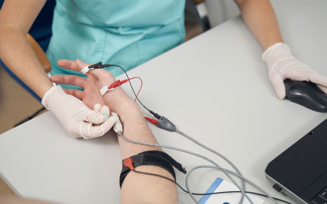
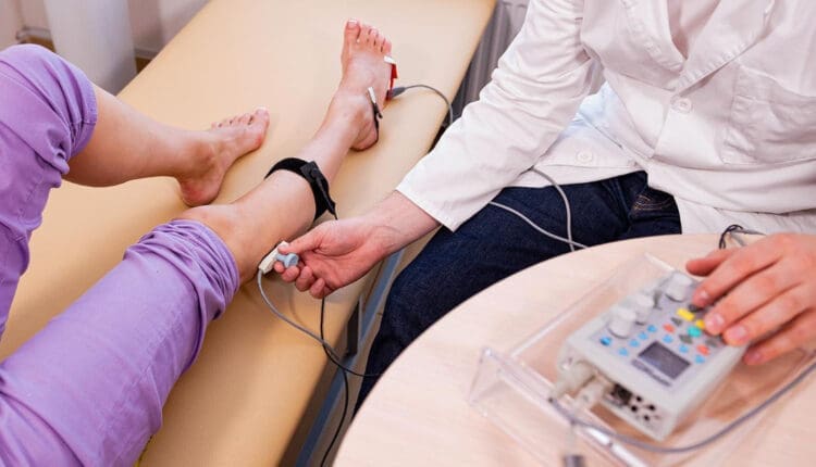
Nerve Conduction Velocity
A nerve conduction velocity (NCV) is a noninvasive test that measures the speed and strength of nerve stimulation using electrical probes placed on the skin. It’s used to diagnose nerve damage or disease, often alongside an EMG (electromyogram) to differentiate between nerve and muscle problems. It can also evaluate sensory issues, pain, and weakness of the extremities.
This test involves safe electrical shocks that can be slightly uncomfortable but not painful.
Nerve conduction velocity (NCV) measures the speed at which electrical impulses travel along a nerve fiber, which measures how quickly electrical signals travel through a nerve.
This information indicates nerve health and function.
Electromyography (EMG) is a nerve test that involves placing tiny needles into the muscles.
A slower NCV can indicate nerve injury or dysfunction.
Test Uses
Generally, the test is ordered to assess peripheral nerve diseases, those that connect from the muscles, organs, and skin to the spinal cord or brain. It can help identify the type and location of nerve damage.
Peripheral nerve conditions typically cause pain, sensory loss, tingling, or burning.
Mild weakness and diminished reflexes can be detected during a neurological examination.
Conditions
Nerve conduction studies are performed to help diagnose conditions.
Nerve damage (neuropathy), such as from diabetes, chemotherapy, or autoimmune disorders
Charcot-Marie-Tooth disease
Nerve compression
Many different conditions, including trauma, inflammation, and tumors, can compress one or more nerves.
Radiculopathy
Often described as a pinched nerve, radiculopathy can affect an arm or a leg, causing pain and weakness.
Peripheral Neuropathy
This nerve damage begins in the most distal nerves, those farthest from the center of the body, such as the toes and fingers. It is often due to chronic alcohol misuse, uncontrolled diabetes, nutritional deficits, and inflammatory diseases. (Ferdousi M. et al., 2020)
Carpal Tunnel Syndrome
Commonly caused by inflammatory diseases or overuse of the wrists, such as from assembly line work, carpal tunnel syndrome causes numbness, pain, and weakness of the fingers and hands. (Tada K. et al., 2022)
Ulnar neuropathy
This common condition causes arm pain and sensory changes, usually due to repetitive movements or a prolonged position that causes pressure on the ulnar nerve.
Guillain-Barré syndrome (GBS)
This inflammatory condition causes demyelination, or loss of the insulating covering around nerves, which results in leg weakness.
It begins in the motor nerves, which send signals to muscles in the legs. (Shibuya K. et al., 2022)
The inflammation travels to nerves of the upper body, often affecting the muscles that control breathing.
Respiratory support is necessary until the condition improves.
Chronic Demyelinating Polyneuropathy (CIDP)
This condition is a chronic, recurrent form of GBS that usually affects the legs and causes episodes of weakness.
ICU neuropathy
Metabolic changes, severe illness, and not moving enough can cause nerves to develop a pattern of weakness and sensory loss.
Myasthenia gravis (MG)
This autoimmune condition affects the junction between the nerves and the muscles.
Myasthenia gravis causes drooping eyelids and weakness of the arms and shoulders.
Amyotrophic lateral sclerosis (ALS)
ALS is a serious, degenerative disease affecting the spinal cord’s motor neurons.
Amyotrophic lateral sclerosis progresses rapidly, resulting in substantial weakness of muscles throughout the body.
How it’s Done
Surface electrodes are placed on the skin over nerves, and a small electrical current is applied to stimulate the nerve.
The time it takes for the electrical signal to travel between the electrodes is measured, and this time is used to calculate the NCV.
Values
Normal NCV values are generally between 50 and 70 meters per second. However, these values can vary depending on the nerve and the individual.
NCV Factors
Various factors can influence NCV.
Age
Sex
Medical conditions like diabetes
Interpretation
A slower NCV can indicate nerve damage or demyelination (loss of the myelin sheath, which insulates nerve fibers), while an EMG can help determine if the problem is with the nerve or the muscle.
Results
The results of NCV testing can be used to determine the type, severity, and location of nerve damage. The results will be ready in report form about a week after the test.
The test measures velocity (how fast a nerve transmits signals) and amplitude (how many nerve fibers were activated). (Tavee J. 2019)
The measurements are transmitted to a computer and shown as waves and numerical values.
The values are compared to a standard measurement based on the tested nerve.
The distance between the electrodes.
The person’s age.
Compared to the standard, the NCV results can identify certain patterns of nerve damage. (Tada K. et al., 2022) Outcomes include: (Tavee J. 2019)
If one or more nerves are affected.
If motor nerves (control movement), sensory nerves (transmit sensory signals), or both are affected.
Whether a nerve is blocked or damaged.
The severity of the damage.
The type of nerve damage
Axonal (damage to the nerve itself)
Demyelination (damage to the protective fatty layer around the nerve)
The results can help point to certain diagnoses.
Preparation Before the Test
Individuals will not need to change their diet before having an NCV. However, patients will be asked to avoid lotions or creams on their skin before the test. Individuals who are also having an EMG at the time of their NCV might be asked to stop taking medications or supplements that increase the risk of bleeding and bruising. If a healthcare provider says not to stop taking the medicines for health reasons, the patient might be warned that they could have some bruising after the EMG test.
NCV may advise against getting the test for those with electrical device implants.
Make sure your healthcare providers are aware of your whole medical history.
Injury Medical Chiropractic & Functional Medicine Clinic
Injury Medical Chiropractic and Functional Medicine Clinic works with primary healthcare providers and specialists to develop an optimal health and wellness solution. We focus on what works for you to relieve pain, restore function, and prevent injury. Regarding musculoskeletal pain, specialists like chiropractors, acupuncturists, and massage therapists can help mitigate the pain through spinal adjustments that help the body realign itself. They can also work with other medical professionals to integrate a treatment plan to resolve musculoskeletal issues.
Peripheral Neuropathy and Chiropractic Care
References
Ferdousi, M., Kalteniece, A., Azmi, S., Petropoulos, I. N., Worthington, A., D’Onofrio, L., Dhage, S., Ponirakis, G., Alam, U., Marshall, A., Faber, C. G., Lauria, G., Soran, H., & Malik, R. A. (2020). Corneal confocal microscopy compared with quantitative sensory testing and nerve conduction for diagnosing and stratifying the severity of diabetic peripheral neuropathy. BMJ open diabetes research & care, 8(2), e001801. doi.org/10.1136/bmjdrc-2020-001801
Tada, K., Murai, A., Nakamura, Y., Nakade, Y., & Tsuchiya, H. (2022). In Carpal Tunnel Syndrome, Sensory Nerve Conduction Velocities Are Worst in the Middle Finger Than in the Index Finger. Frontiers in Neurology, 13, 851108. doi.org/10.3389/fneur.2022.851108
Shibuya, K., Tsuneyama, A., Misawa, S., Suzuki, Y. I., Suichi, T., Kojima, Y., Nakamura, K., Kano, H., Ohtani, R., Aotsuka, Y., Morooka, M., Prado, M., & Kuwabara, S. (2022). Different patterns of sensory nerve involvement in chronic inflammatory demyelinating polyneuropathy subtypes. Muscle & Nerve, 66(2), 131–135. doi.org/10.1002/mus.27530
What is a bone density test, how is it performed, and what do the results mean?
Bone Density Test
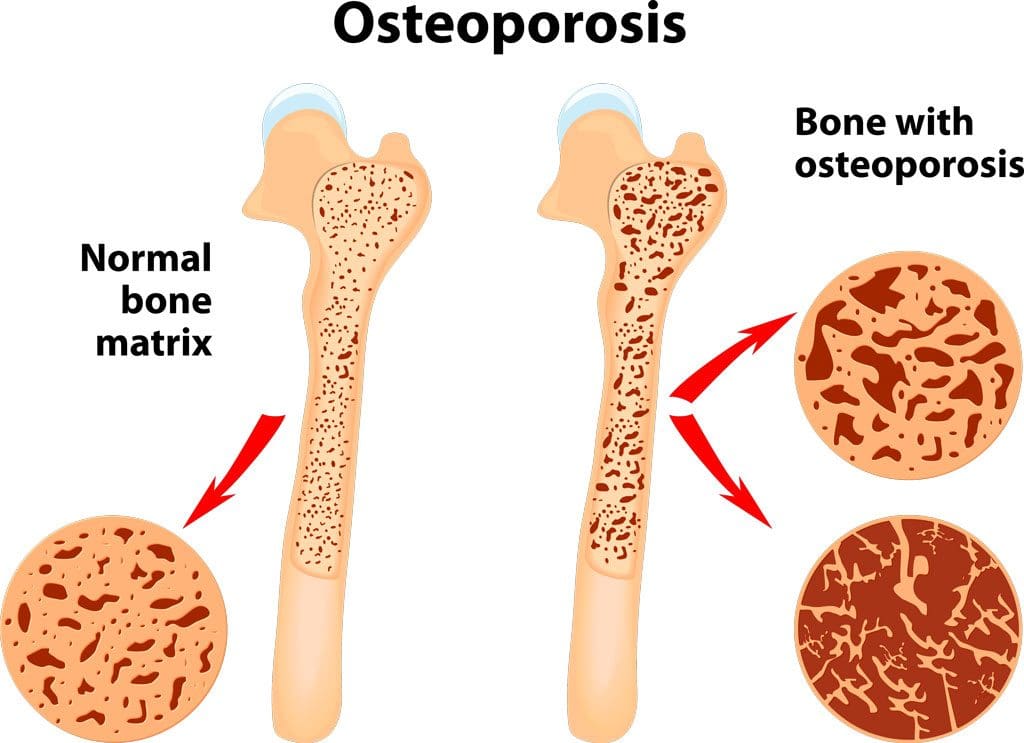
A bone density test examines bone mass, which indicates overall bone strength. Assessing bone density or mass is necessary for diagnosing osteopenia or osteoporosis, conditions that increase the risk of broken bones. The scan is performed through dual-energy X-ray absorptiometry (DEXA), which examines the thickness of the bones. Results from DEXA scans are compared to standardized values to determine whether bone density is lower than normal and whether osteopenia or osteoporosis is present.
Examination
The procedure examines bone density, or bone mass. The bones’ density, or mass, is an overall indicator of bone strength. The greater the bone density, the thicker and stronger the bones are. The test is used to diagnose osteoporosis, a condition characterized by brittle bones at risk of breaking due to significantly low bone density. A bone density test can also diagnose osteopenia, a condition characterized by lower than normal bone mass that can lead to osteoporosis. (National Institute of Arthritis and Musculoskeletal and Skin Diseases, 2025) It is recommended that all women aged 65 and older and all men aged 70 and older have a bone density scan to screen for bone loss to help prevent fractures. (Kling J. M., Clarke B. L., & Sandhu N. P. 2014)
Bone density scans can establish a baseline level of bone density and track changes over time.
For individuals with osteoporosis or osteopenia, a bone density scan can help track how well their bones respond to treatment.
During a DEXA scan, the patient will lie on their back on a table with their legs elevated on a padded platform.
An X-ray scanner will pass over the spine and hips while another scans beneath.
While the scan takes place, the patient will be asked to hold very still to obtain an accurate image.
The scan will obtain bone density readings from the spine and hip, the two most commonly fractured bones, and generally takes less than 30 minutes.
Results
A DEXA scan measures bone density in grams per centimeter squared (g/cm²). This number indicates how densely bone cells are packed together in a specific area of bone. This bone density reading is then compared to a standardized value to determine if bone density is within a normal range or lower than average.
Between minus 1.0 and minus 2.5: Low bone density (osteopenia)
Equal to minus 2.5 or below: Osteoporosis
Bone density values are reported as a Z score for women who have not undergone menopause and men under 50 years old.
Z scores are compared to bone density levels of individuals of the same age and sex.
A Z score of minus 2.0 or lower indicates low bone density, which can be caused by factors other than aging, such as medication side effects, nutritional deficiencies, or thyroid problems.
Arthritis Diagnosis
Because a DEXA scan only measures the thickness of bones, it doesn’t work to diagnose arthritis. An X-ray of the affected joint is currently the most accurate way to diagnose arthritis. The Kellgren-Lawrence classification system categorizes the extent of arthritis based on the severity of joint damage seen on an X-ray. According to this system, arthritis can be classified as: (Kohn M. D., Sassoon A. A., & Fernando N. D. 2016)
Grade 1 (minor)
Minimal or no joint space narrowing, with possible bone spur formation.
Grade 2 (mild)
Possible joint space narrowing, with definite bone spur formation.
Grade 3 (moderate)
Definite joint space narrowing, moderate bone spur formation, mild sclerosis (abnormal thickening of bone), and possible deformation of bone ends.
Grade 4 (severe)
Severe joint space narrowing, large bone spur formation, marked sclerosis, and definite deformation of bone ends.
Injury Medical Chiropractic & Functional Medicine Clinic
Exercise can be incredibly beneficial for improving bone density, joint mobility, and the strength of surrounding muscles, which support and protect joints and bones. Talk to a healthcare provider to learn what interventions and available treatment options would be the most effective. Injury Medical Chiropractic and Functional Medicine Clinic works with primary healthcare providers and specialists to develop an optimal health and wellness solution. We focus on what works for you to relieve pain, restore function, and prevent injury. Regarding musculoskeletal pain, specialists like chiropractors, acupuncturists, and massage therapists can help mitigate the pain through spinal adjustments that help the body realign itself. They can also work with other medical professionals to integrate a treatment plan to resolve musculoskeletal issues.
Kling, J. M., Clarke, B. L., & Sandhu, N. P. (2014). Osteoporosis prevention, screening, and treatment: a review. Journal of women’s health (2002), 23(7), 563–572. doi.org/10.1089/jwh.2013.4611
Kohn, M. D., Sassoon, A. A., & Fernando, N. D. (2016). Classifications in Brief: Kellgren-Lawrence Classification of Osteoarthritis. Clinical orthopaedics and related research, 474(8), 1886–1893. doi.org/10.1007/s11999-016-4732-4
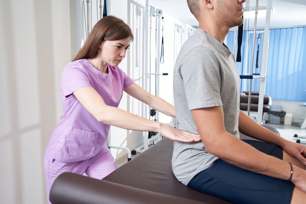
Can postural assessments help identify problems with how a person stands or sits, which can lead to various musculoskeletal issues?
Postural Assessment
Proper posture is an exercise in which the muscles support the skeleton in a comfortable, stable, and efficient alignment. Healthy posture is present when the body is still and when moving. However, numerous factors can affect and hinder posture. These include daily wear and tear, injury, illness, or a condition. A posture assessment is a process that identifies posture issues and their root causes, often using visual and palpation techniques, and can help determine appropriate treatment or exercises. (Science Direct, 2007)
Visual Assessment
Observing the body’s alignment and symmetry from different angles (anterior, posterior, and lateral views).
Consider their daily activities, work environment, and any previous injuries. (Du, S. H. et al., 2023)
Wall Test
The patient stands against a wall with their feet shoulder-width apart and heels about 6 inches from the baseboard.
If they have good posture, their ears will be vertically aligned with their shoulders, and their head will be no more than three finger widths from the wall. (Physiopedia, 2025)
Professionals Who Can Perform a Posture Assessment
Physiotherapists
Professionals trained in assessing and treating musculoskeletal problems.
Chiropractors
Professionals who focus on the spine and nervous system.
Fitness Professionals
Personal trainers or other fitness professionals can use posture assessments to help clients improve their posture and movement.
Ergonomists
Professionals who specialize in designing workspaces and environments to promote good posture and reduce strain.
Injury Medical Chiropractic & Functional Medicine Clinic
Talk to a healthcare provider to learn what interventions would help the most. Injury Medical Chiropractic and Functional Medicine Clinic works with primary healthcare providers and specialists to develop an optimal health and wellness solution. We focus on what works for you to relieve pain, restore function, and prevent injury. Regarding musculoskeletal pain, specialists like chiropractors, acupuncturists, and massage therapists can help mitigate the pain through spinal adjustments that help the body realign itself. They can also work with other medical professionals to integrate a treatment plan to resolve musculoskeletal issues.
Singla, D., & Veqar, Z. (2014). Methods of postural assessment used for sports persons. Journal of clinical and diagnostic research: JCDR, 8(4), LE01–LE4. doi.org/10.7860/JCDR/2014/6836.4266
Du, S. H., Zhang, Y. H., Yang, Q. H., Wang, Y. C., Fang, Y., & Wang, X. Q. (2023). Spinal posture assessment and low back pain. EFORT open reviews, 8(9), 708–718. doi.org/10.1530/EOR-23-0025
Learn about the clinical approach of identity formation and its role in facilitating personal insight and emotional well-being.
Introduction
Many individuals deal with musculoskeletal, autoimmune, gut, or body issues that can affect their daily routine and how they perceive themselves when getting checked out by their doctors. Many healthcare professionals can help assess individuals with these pain-like symptoms causing overlapping risk profiles by creating a safe and positive experience with a clinical approach. By creating a secure environment, many individuals can have good experiences when pain-like issues are affecting their bodies. By delving into today’s article, we are going to look into identity formation, the four identity statuses, and the various areas of identity development.
Theories & Types of Identity Formation
A lot of individuals have an identity, but have they always had one? Everyone’s identity is a conglomeration of the characteristics, values, and attributes they consider significant and use to define themselves. People’s identities are developed as they mature and gain knowledge about the world; they are not forced upon them. Adolescence is the first period when individuals notice a significant change toward identity creation and self-exploration.
A major concern in theories of teenage development is identity building. Identity formation, for instance, was emphasized as the main sign of effective development throughout adolescence in Erikson’s 1968 classic theory of developmental phases. Teenagers who struggle to define their identities may have role confusion, which suggests that they need to do more self-discovery and are unclear of their place in the world.
The Four Identity Statuses
Many healthcare providers will hear their patients describe identity formation during adolescence as it involves decision points and commitments regarding belief systems (e.g., religion, politics) and occupations. The four modes of reacting to late identity crises during the adolescent phase were described, measured, and validated so that individuals could figure out their identity status. (Marcia, 1966) The four described identity statuses are:
Foreclosure: An individual commits to an identity without exploring options.
Identity diffusion: When individuals neither explore nor commit to any identities.
Moratorium: A state in which individuals actively explore options but have not made commitments.
Identity achievement: When individuals have explored different options and made identity commitments.
For example, teens may commit to an identity without exploring if they are content with the values, culture, and religion they were raised in. Teens in foreclosure status may adopt large parts of their parents’ identities or the identity parents have put them on. However, when teens achieve identity, they can explore the world for themselves and decide how they identify due to those experiences. This causes them to relinquish their claims of infantile sources of gratification, thus renouncing lingering fantasies of competence. (Marcia, 1967) At the same time, many people have experienced things that led to a crisis. The thing is that a crisis doesn’t necessarily mean a negative event; it’s simply branching out from what’s comfortable to discover who they are.
Understanding The Effects Of Personal Injury- Video
Various Areas of Identity Development
As a stage in the adolescent life cycle, identity development happens to many people. For many, the search for identity begins in the teenage years. During these years, adolescents are more open to taking on different behaviors and appearances to discover who they are. In an attempt to find their own identity and discover who they are, adolescents tend to cycle through several identities to find one that suits them best. Multiple factors like family life, environment, and social status can make it difficult to develop and maintain an identity. Some studies suggest that this process might be more accurate to identity development rather than formation but confirm this is a typical change process in people’s thoughts about themselves.
Several different areas of identity development are described:
Religious identity: Teens’ religious views are similar to those of their families. Many may question specific customs, practices, or ideas in their parents’ faith, but a few may completely reject their families’ religion. This is due to the dynamic of the parent-adolescent relationship with religion, which exerts positive effects on adolescent adjustments. (Kim-Spoon et al., 2012)
Political identity: Adolescents’ political identity is influenced by their parents’ political beliefs. In the 21st century, a new trend shows decreased political affiliation among young adults. However, many adolescents tend to be more liberal than their elders on social issues. This is because many adolescents encounter events that trigger their civic interest and challenge their beliefs and moralities. (Stattin et al., 2017) Like in other aspects of identity formation, adolescent individuals are predicted by their parents’ involvement and current events when there is an interest in politics.
Vocational identity: Adolescents in earlier generations envisioned themselves working in a particular job and often worked as apprentices or part-time in such occupations. This is a rare case for many people in today’s world. Vocational identity is also related to ego identity by correlating with a successful transition from school to work, allowing individuals to be more confident in their decision-making ability, especially in environmental ambiguities. (Koo & Kim, 2016) Still, vocational identity takes a bit longer to develop since many workplaces require specific skills and knowledge that require additional education or are acquired on the job. Additionally, many job opportunities held by teens are not in occupations many will seek as adults.
Ethnic identity: Ethnic identity refers to how many individuals come to terms with who they are based on ethnicity or racial ancestry. According to the U.S. Census 2012, Americans under 18 are from historically marginalized ethnic groups. Many people who identify under BIPOC (Black, Indigenous, people of color) have discovered their ethnoracial identity as an important part of their identity formation as teens.
Gender identity: Gender identity involves an individual’s sense of gender and can be similar to or different from their biological sex regardless of age group. Gender identity greatly influences many adolescents during these years of self-discovery and can impact other areas of identity, like religion and politics.
Self-Concept
Self-concept and self-esteem are the two primary facets of identity formation. The capacity of an individual to have views and beliefs established with confidence, consistency, and stability throughout the course of their life is the central premise of self-concept. Cognitive growth in early adolescence leads to increased self-consciousness, awareness of others and their opinions, the ability to think about abstract future possibilities, and the ability to weigh many alternatives at once. As a consequence, many teenagers will stop using straightforward, global, and concrete self-descriptions when they are younger. As kids, they use physical characteristics like gender, hair color, or whether they’re quick to identify themselves.
Many teenagers have the ability to imagine many “possible selves” that they may become, and the decisions they make may have long-term effects or possibilities. Exploring these options may cause individuals to make sudden changes in how they show themselves when the teenager selects or rejects traits and actions. In addition, when combining their combinational operation with their degree of identification, both men and females exhibit strong positive connections. (Wagner, 1987) This in turn means directing the real self in the direction of the ideal self. The ideal self differs from person to person; many individuals aspire to be the person they want to be, while many others dread becoming the person they do not want to be. Many may find this unsettling, but it may also serve as motivation by demonstrating consistent conduct that aligns with the ideals and distinguishes the feared potential selves.
Our ideal and frightened selves may be simultaneously explored and discovered. In an effort to create their own identities, many young people may observe characteristics in their family members, friends, or other community members and begin to consider what they like and dislike at the same time. Teenagers learn to identify the factors that impact their conduct and how others see them, which leads to a further distinction in their self-concept known as differentiation. Differentiation seems to be completely established by mid-adolescence and peaks when students enter the seventh or ninth grade. Nowadays, identifying contradictory material in one’s self-concept is a frequent cause of anxiety. Nonetheless, by promoting their exploration and growth, it might help a lot of teenagers.
Self-Esteem
Self-esteem is the other component of identity building. By definition, one’s ideas and emotions about one’s identity and self-concept constitute one’s self-esteem. Many views contend that a strong desire to preserve, defend, and improve oneself is a component of self-esteem. Contrary to common opinion, little evidence supports these views, suggesting that teenage self-esteem has significantly declined. The two sexes have distinct levels of self-esteem; women have higher levels of self-esteem when they have supportive friendships. However, women have poor self-esteem when they are unable to meet someone with similar interests and hobbies or when they are unable to get the acceptance of their friends.
Males have varied levels of self-esteem. Males are more focused on defining authority and establishing and claiming their independence regarding self-esteem. This, in turn, enables men to effectively have high self-esteem via the influence of their peers and friends. However, a male’s poor self-esteem may be further exacerbated by a lack of romantic abilities or the inability to sustain another person’s attachment.
Conclusion
Numerous medical experts may use a clinical approach to identity development to provide a secure environment and a satisfying experience for people when evaluating the pain-like sensations impacting their bodies. Additionally, by offering a variety of alternatives in their individualized treatment plans to improve their health and well-being, a thorough awareness of the significance of identity development helps foster positive relationships with patients.
Injury Medical & Functional Medicine Clinic
We associate with certified medical providers who understand the importance of identity formation when assessing individuals dealing with various pain-like symptoms within their bodies. When asking important questions to our associated medical providers, we advise patients to implement small changes to their daily routine to reduce the pain-like symptoms associated with body pains. Dr. Alex Jimenez, D.C., utilizes this information as an academic service. Disclaimer.
References
Kim-Spoon, J., Longo, G. S., & McCullough, M. E. (2012). Parent-adolescent relationship quality as a moderator for the influences of parents’ religiousness on adolescents’ religiousness and adjustment. J Youth Adolesc, 41(12), 1576-1587. doi.org/10.1007/s10964-012-9796-1
Koo, H.-Y., & Kim, E.-J. (2016). Vocational Identity and Ego Identity Status in Korean Nursing Students. Asian Nursing Research, 10(1), 68-74. doi.org/10.1016/j.anr.2015.11.001
Marcia, J. E. (1966). Development and validation of ego-identity status. J Pers Soc Psychol, 3(5), 551-558. doi.org/10.1037/h0023281
Marcia, J. E. (1967). Ego identity status: relationship to change in self-esteem, “general maladjustment,” and authoritarianism. J Pers, 35(1), 118-133. doi.org/10.1111/j.1467-6494.1967.tb01419.x
Stattin, H., Hussein, O., Ozdemir, M., & Russo, S. (2017). Why do some adolescents encounter everyday events that increase their civic interest whereas others do not? Dev Psychol, 53(2), 306-318. doi.org/10.1037/dev0000192
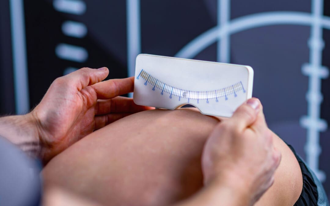
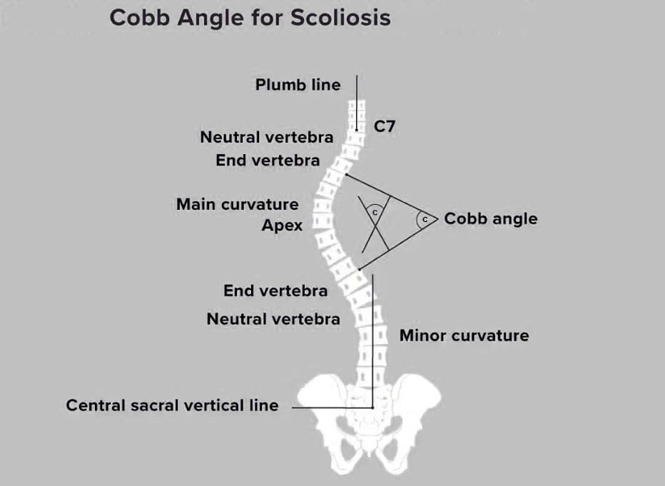
The Cobb angle is a mathematical measurement tool for assessing the curvature of the spine. Along with physical exams and other tests, how is it used to evaluate scoliosis and kyphosis of the spine?
Cobb Angle
The Cobb angle is used to quantify the curvature of the spine, particularly in conditions like scoliosis. It measures the degree of side-to-side spinal curvature, a deformity called scoliosis. The angle’s size helps determine what kind of treatment is needed. Monitoring may be all that’s necessary for mild curvature. With severe scoliosis, treatment may require spinal fusion surgery. Named for orthopedic surgery pioneer John Robert Cobb, it describes the distance a scoliotic curve may deviate from being straight. (Botterbush K. S. et al., 2023) Generally, it takes at least 10 degrees of deviation from straight before scoliosis is confirmed.
X-Ray and Interpretation
An X-ray is taken to measure the Cobb angle. Side and back views are taken. The healthcare provider or examiner then views the X-rays and locates the most affected vertebra in the curve, the apical vertebra. In a scoliotic curve, the apical vertebra is the spinal bone with the greatest degree of rotation that takes the biggest curve away from the center of a normal spine column.
Visualizing the Angle
The apical vertebra is where two lines drawn from the X-rays meet. Two lines are drawn along the edge of the top and bottom bones of the curve. The lines extend out as follows:
On the top bone, the line starts on the high side, continues along the top edge, and then slopes down according to the angle of the vertebra. (Jin, C. et al., 2022)
On the bottom vertebra, the line starts on the low side, continues along the bottom edge, and slopes upward.
The Cobb angle is found by measuring the angle of the two intersecting lines where they meet.
Then, the top and bottom vertebrae of the side-to-side curve are identified to create a number for the Cobb angle. These bones have the most tilt but the least rotation and displacement and are located above and below the apical vertebra. Computer software is commonly used to calculate the Cobb angle. (Jin, C. et al., 2022) Treatment is based on the:
Angle size
Gender: Scoliosis is more common and likely to progress in females
Scoliosis is diagnosed when the Cobb angle reaches 10 degrees or more. However, this is not generally considered a significant curvature (American Association of Neurological Surgeons, 2024). In around 80% of cases, the scoliosis is considered idiopathic or without congenital or other underlying causes.
Less Than 25 Degrees Cobb Angle
If a scoliotic curve is less than 25 degrees, individuals may only need to visit their healthcare provider periodically so long as the scoliosis is monitored. These are mild cases, often without symptoms, but there is a chance that the curvature can progress. This usually means reassessing the Cobb angle every four to six months in a growing child or adolescent. (National Scoliosis Foundation, 2015) A 5-degree or more progression can change the diagnosis and treatment. (Jin, C. et al., 2022)
Between 25 and 40 Degrees Cobb Angle
A Cobb angle of 25 to 40 degrees usually requires wearing a back brace and intensive physical therapy. The goal of these treatments is to help halt the curve’s progression. Braces are generally worn 16 to 23 hours every day. (National Scoliosis Foundation, 2015) The healthcare provider will provide a referral for physical therapy. Many report excellent results with the Schroth or other scoliosis-specific exercise methods. A study found that core stabilization exercise programs can decrease Cobb angles in adolescents with idiopathic scoliosis. (Ko K. J. & Kang S. J. 2017)
Scoliosis in Adults
Scoliosis is diagnosed in adults, usually in those who have had the condition, treated or not, that was identified in their youth. A study that followed various cases for 20 years found disease progression occurred in 40% of adults but was usually less than one degree per year. However, degenerative scoliosis can also occur in individuals aged 65 and older. (American Association of Neurological Surgeons, 2024)
40 Degrees or More Cobb Angle
Surgery may be recommended once the Cobb angle reaches 40 to 50 degrees. A spinal fusion is often used to force the curve to stop developing. In adults, surgery may be needed if the angle reaches 50 degrees and they experience complications, such as nerve damage or bowel/bladder dysfunction. Risk factors in adults include older age, a history of smoking, and a diagnosis of other conditions, including being overweight. (American Association of Neurological Surgeons, 2024)
Variations
Variations occur in measuring scoliosis, and it is important to understand the difference between a change in scoliosis and a change in the tools or measurement. Equipment errors, imaging errors, and the subjective reading of the healthcare provider can change the values. (Jin, C. et al., 2022) Scoliosis measurement software and intelligent medical devices continue to improve how scoliosis is evaluated and treated. Physical exams, symptoms, and careful monitoring of changes in posture or function are still critical to an accurate diagnosis. The healthcare provider will explain the Cobb angle and other test results.
Injury Medical Chiropractic & Functional Medicine Clinic
Injury Medical Chiropractic and Functional Medicine Clinic works with primary healthcare providers and specialists to develop an optimal health and wellness solution. We focus on what works for you to relieve pain, restore function, and prevent injury. Regarding musculoskeletal pain, specialists like chiropractors, acupuncturists, and massage therapists can help mitigate the pain through spinal adjustments that help the body realign itself. They can also work with other medical professionals to integrate a treatment plan to resolve musculoskeletal issues.
Academic Low Back Pain: Impact and Chiropractic Solutions
References
Botterbush, K. S., Zhang, J. K., Chimakurty, P. S., Mercier, P., & Mattei, T. A. (2023). The life and legacy of John Robert Cobb: the man behind the angle. Journal of neurosurgery. Spine, 39(6), 839–846. doi.org/10.3171/2023.7.SPINE23146
Jin, C., Wang, S., Yang, G., Li, E., & Liang, Z. (2022). A Review of the Methods on Cobb Angle Measurements for Spinal Curvature. Sensors (Basel, Switzerland), 22(9), 3258. doi.org/10.3390/s22093258
Ko, K. J., & Kang, S. J. (2017). Effects of 12-week core stabilization exercise on the Cobb angle and lumbar muscle strength of adolescents with idiopathic scoliosis. Journal of Exercise Rehabilitation, 13(2), 244–249. doi.org/10.12965/jer.1734952.476
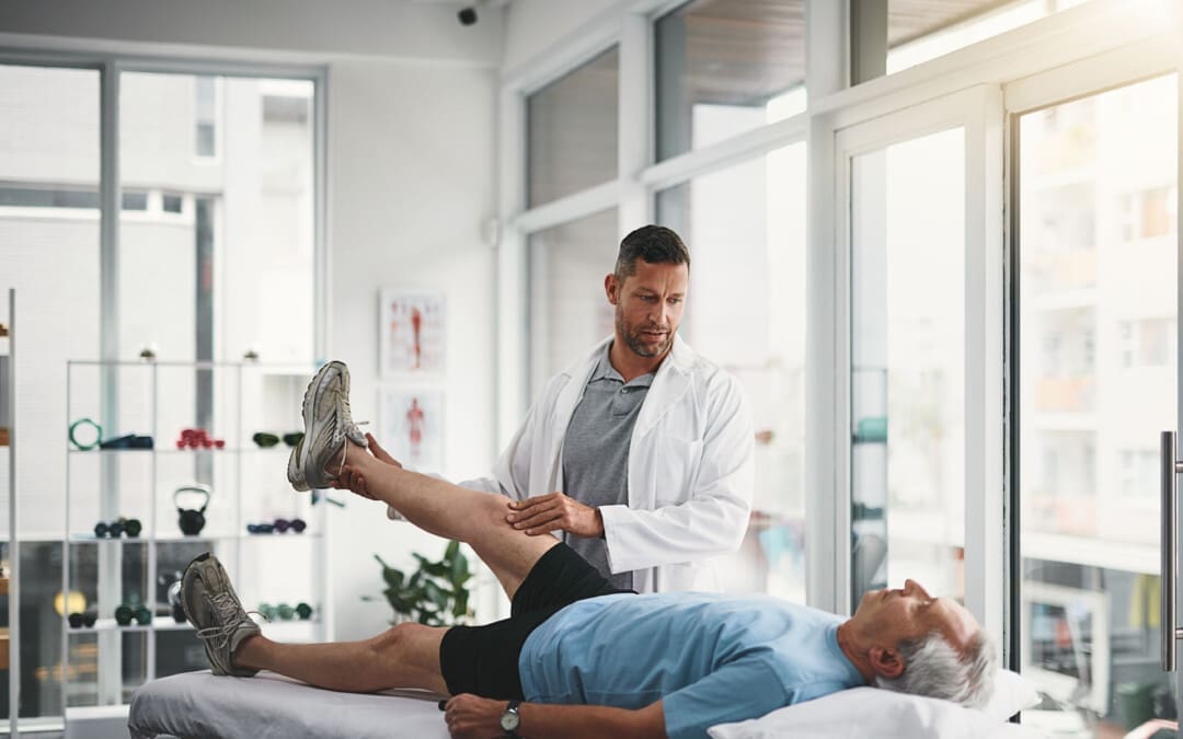
Can the straight leg test help find the cause of back or hamstring pain in individuals experiencing it?
Straight Leg Test
The straight leg raise test is often used to diagnose sciatica/radiculopathy, herniated discs, and other spinal problems. The healthcare provider giving the test performs most of the movement as they assess what’s causing the leg and/or back pain. The patient lies on their back with the legs straight. The provider will have the patient perform specific movements and inform them of how it feels. Then, they’ll raise the leg to see if and at what point symptoms begin to show. Providers often use this test alongside imaging studies.
Some studies suggest the straight leg test helps diagnose sciatica and other causes. (Pesonen J. et al., 2021)
Purpose
The straight leg raise is one of the most common manual tests done during physical exams. The straight leg raise test seeks to reproduce the pain or other symptoms in a controlled fashion to provide clues to what’s happening. It is a manual exam, and the healthcare provider will:
Position the patient
Moves the patient
Create pressure to see how well the patient can resist it
This is often used alongside imaging tests, such as an X-ray or CT scan. (Allegri M. et al., 2016) Its goal is to check for nerve movement and sensitivity of nerve tissue to compression. The straight leg lift test is neurodynamic because it uses movement to diagnose nerve problems. (Baselgia L.T. et al., 2017)
During the Test
Expect to feel some pain during the test, as the whole point is to see what aggravates the symptoms. They may be caused by:
Most of the tests are passive, with the provider doing the lifting. The patient can help achieve the most accurate result by staying as relaxed as possible and being clear about what is felt. (Pande K. 2015) The procedure:
The patient lies on their back with their legs straight.
The provider will ask the patient to turn one of the legs in.
This tells them what hip position affects the lower back symptoms.
They’ll then ask you to bring the leg toward the body’s center.
Then, they’ll lift the straight leg until the patient experiences symptoms.
Pain suggests a herniated disc.
If there is no pain, this also provides valuable information.
The procedure is repeated with the other leg.
Modifications
It’s important to let the examiner know about any limitations. The straight leg raise test has modifications if the patient cannot lift their leg while it’s straight or if they have difficulty lying on their back, which can also help avoid an injury during the test.
Variations
The healthcare provider may repeat the test with the ankle in a dorsiflexed position/raising the foot. Then, they’ll have the patient do it with their chin tucked into their chest. (Young R. et al., 2013) These variations can help check for nerve involvement in specific locations, such as the spinal cord or the dura mater, the membrane covering the brain and spinal cord. (Venne G. et al., 2017) The spinal cord nerves are likely involved and affected if the usual pain is in the back or leg but not the chin, neck, or foot. (Camino Willhuber GO, Piuzzi NS. 2023)
Injury Medical Chiropractic and Functional Medicine Clinic
Injury Medical Chiropractic and Functional Medicine Clinic works with primary healthcare providers and specialists to build optimal health and wellness solutions. We focus on what works for you to relieve pain, restore function, prevent injury, and mitigate issues through adjustments that help the body realign itself. The clinic can also work with other medical professionals to integrate a treatment plan to resolve musculoskeletal problems.
Pesonen, J., Shacklock, M., Suomalainen, J. S., Karttunen, L., Mäki, J., Airaksinen, O., & Rade, M. (2021). Extending the straight leg raise test for improved clinical evaluation of sciatica: validity and diagnostic performance with reference to the magnetic resonance imaging. BMC musculoskeletal disorders, 22(1), 808. doi.org/10.1186/s12891-021-04649-z
Allegri, M., Montella, S., Salici, F., Valente, A., Marchesini, M., Compagnone, C., Baciarello, M., Manferdini, M. E., & Fanelli, G. (2016). Mechanisms of low back pain: a guide for diagnosis and therapy. F1000Research, 5, F1000 Faculty Rev-1530. doi.org/10.12688/f1000research.8105.2
Baselgia, L. T., Bennett, D. L., Silbiger, R. M., & Schmid, A. B. (2017). Negative Neurodynamic Tests Do Not Exclude Neural Dysfunction in Patients With Entrapment Neuropathies. Archives of physical medicine and rehabilitation, 98(3), 480–486. doi.org/10.1016/j.apmr.2016.06.019
Pande K. (2015). The Use of Passive Straight Leg Raising Test: A Survey of Clinicians. Malaysian Orthopaedic Journal, 9(3), 44–48. doi.org/10.5704/MOJ.1511.012
Young, R., Nix, S., Wholohan, A., Bradhurst, R., & Reed, L. (2013). Interventions for increasing ankle joint dorsiflexion: a systematic review and meta-analysis. Journal of foot and ankle research, 6(1), 46. doi.org/10.1186/1757-1146-6-46
Venne, G., Rasquinha, B. J., Kunz, M., & Ellis, R. E. (2017). Rectus Capitis Posterior Minor: Histological and Biomechanical Links to the Spinal Dura Mater. Spine, 42(8), E466–E473. doi.org/10.1097/BRS.0000000000001867
IFM's Find A Practitioner tool is the largest referral network in Functional Medicine, created to help patients locate Functional Medicine practitioners anywhere in the world. IFM Certified Practitioners are listed first in the search results, given their extensive education in Functional Medicine