How PRP Therapy, Chiropractic Adjustments, and Spinal Decompression Can Help Fix Poor Posture Issues in El Paso, TX
Poor posture is a common problem for many adults. Long hours at a desk, looking down at phones, past injuries, or even stress can pull the body out of alignment. Over time, this extra stress does more than cause discomfort. It can weaken muscles, tighten ligaments, and create small tears in the tissues that support the spine.
When these supporting structures break down, it becomes harder to hold good posture without pain or fatigue. Simple stretches or exercises may not be enough if the underlying tissues are damaged. That is where a combined approach using regenerative treatments, chiropractic care, spinal decompression, and supportive therapies can make a real difference. These methods work on both the mechanical alignment of the spine and the biological repair of the tissues that hold everything in place.
How Poor Posture Affects Muscles and Ligaments
Poor posture places uneven pressure on the spine and surrounding tissues. Muscles that should stay balanced often become tight on one side and weak on the other. Ligaments, the strong bands that connect bones and stabilize joints, can stretch beyond their normal range or develop tiny tears from ongoing strain.
This creates a cycle. Weak or damaged tissues make it difficult to maintain proper alignment. The body then compensates with increased tension or guarding, leading to greater pain and stiffness. Many people notice neck tension, low back ache, headaches, or radiating discomfort that makes daily activities harder.
Research on posture and spinal health shows that these changes in muscles and ligaments often contribute to ongoing instability (Darlington Chiropractic Care, n.d.; Square One Health, n.d.). Without addressing both the alignment and the tissue health, progress can stall.
Regenerative Medicine Options Such as PRP Therapy
Regenerative treatments focus on helping the body repair itself at the tissue level. Platelet-Rich Plasma (PRP) therapy is one common option. It uses a small sample of the patient’s own blood, which is processed to concentrate platelets and growth factors. When injected near damaged ligaments or spinal tissues, these concentrated elements send signals that encourage natural healing and new tissue growth.
Similar approaches include Platelet-Free Plasma (PFP) and micro-fragmented adipose tissue (mFAT or MFAT) from the patient’s own fat. These provide growth factors or a natural scaffolding that supports repair in areas worn down by long-term poor posture.
The goal is to strengthen the ligaments so they can better hold the vertebrae in proper position. This biological support is especially helpful when pain or tissue damage has made it difficult to maintain proper alignment through exercise or adjustments alone (Apex Biologix, n.d.; El Paso Chiropractor Blog, 2026).
Chiropractic Adjustments for Better Spinal Alignment
Chiropractic care uses precise, hands-on techniques to gently move vertebrae and joints back toward better alignment. This restores normal motion, reduces pressure on nerves, and helps tight muscles relax. Adjustments also improve the body’s sense of position, called proprioception, making it easier to maintain optimal posture without constant conscious effort.
When tissues are supported by regenerative treatments, chiropractic adjustments often hold their results longer. The mechanical correction works together with the biological repair occurring in the ligaments and muscles (Apex Biologix, n.d.; Darlington Chiropractic Care, n.d.).
Spinal Decompression Therapy
Spinal decompression uses a gentle, controlled pulling force to create more space between the vertebrae. This relieves pressure on bulging discs, pinched nerves, and irritated structures that often result from years of poor posture or compression.
Improved space allows better fluid movement and nutrient flow into the discs. Many patients report that it reduces radiating pain or sciatica-like symptoms, making it easier to participate in rehabilitation and daily movement. Decompression pairs well with other therapies because it relieves pressure on the spine while regenerative treatments promote tissue repair (El Paso Chiropractor Blog, 2026; Square One Health, n.d.).
Supportive Therapies: Shockwave and MLS Laser
Two advanced modalities often enhance results. Shockwave therapy delivers targeted sound waves that increase blood flow, break down scar tissue, and stimulate the body’s repair processes. It is frequently used to “prime” an area before PRP injections or to continue remodeling tissue afterward.
MLS laser therapy uses specific wavelengths of light to reduce inflammation and swelling while boosting cellular energy to support healing. It is particularly beneficial after regenerative injections or adjustments to keep post-treatment soreness low and speed overall recovery. Together, these therapies create a more favorable environment for the main treatments to succeed (CELasers, n.d.; OSpine Medical, n.d.; Carolina Non-Surgical Ortho, n.d.).
How the Therapies Work Together for Better Outcomes
No single treatment fixes posture by itself. The power comes from combining them in a thoughtful sequence.
Regenerative injections such as PRP first deliver growth factors directly to weakened ligaments and damaged tissues. This initiates the biological repair process, allowing the structures that support the spine to become stronger and more stable.
Chiropractic adjustments then provide the mechanical realignment, helping vertebrae sit in better positions while the tissues heal. Spinal decompression creates the necessary space and reduces nerve pressure, allowing the regenerative signals to work without constant compression interfering.
Shockwave therapy improves circulation and tissue responsiveness, helping the PRP or similar treatments reach their full effect. MLS laser therapy calms any temporary inflammation from injections or adjustments, so patients can stay consistent with care and rehabilitation.
Epidural injections may be added in cases of severe nerve inflammation or radiating pain. They calm irritated nerves enough for the patient to safely engage in adjustments, decompression, and exercises.
The result is a supportive environment where the body can heal both structurally and biologically. Patients often report less daily pain first, followed by easier movement and a gradual return to better posture that requires less effort to maintain. This integrated approach is especially useful when underlying tissue damage has made it difficult to progress with conservative care alone (Personal Injury Doctor Group, 2026; El Paso Chiropractor Blog, 2026).
Integrated Care at Injury Medical Clinic in El Paso
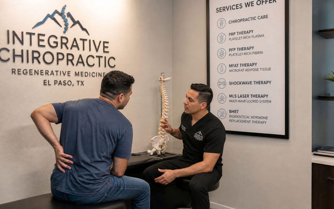
At Injury Medical Clinic PA in El Paso, Texas, patients have access to this type of coordinated care under one roof. Dr. Alexander Jimenez, DC, APRN, FNP-BC, CFMP, IFMCP, ATN, and CCST, brings extensive clinical experience in chiropractic care, regenerative procedures, functional medicine, personal injury support, and rehabilitation. His observations show that many people with posture-related pain from desk work, old injuries, or daily habits benefit when both alignment and tissue health are addressed together.
Working closely with him is Dr. Maria Guadalupe Cardenas, MD, a board-certified internal medicine physician with over 40 years of experience. She serves as Medical Director and Collaborative Physician at the clinic (NPI #1164426749, Texas MD License #J2933). Her role provides medical oversight and direction, ensuring comprehensive evaluation and safe coordination of care.
This collaboration between chiropractic and regenerative expertise (Dr. Jimenez) and internal medicine leadership (Dr. Cardenas) is a common model in integrative and injury-focused clinics. The team also incorporates functional medicine, rehabilitation, soft tissue work, and detailed documentation for personal injury or insurance needs. Patients receive personalized plans that consider the whole picture—structural alignment, tissue repair, inflammation control, and overall function—rather than isolated treatments (El Paso Chiropractor Blog, 2026; LinkedIn pulse on integrated injury care, n.d.; DrAlexJimenez.com, n.d.).
What to Expect from a Combined Treatment Plan
Care usually begins with a thorough evaluation, including a history, an examination, and any necessary imaging or tests. The team then designs a plan that may include regenerative injections, a series of chiropractic adjustments, decompression sessions, shockwave or laser therapy, and guided rehabilitation exercises for posture and core strength.
Progress is monitored closely. Many people notice reduced pain and stiffness within the first few weeks, with continued improvement in mobility and posture comfort over several months. Results vary based on the severity of tissue damage, overall health, and consistency with home exercises and ergonomic changes. The goal is lasting functional improvement, not just temporary relief.
Taking Steps Toward Better Posture and Comfort
Poor posture can create a frustrating cycle of pain and limitation, but addressing both the mechanical alignment of the spine and the biological health of supporting tissues offers a promising path forward. Therapies like PRP and related regenerative options, combined with chiropractic adjustments, spinal decompression, shockwave, and MLS laser therapy, work together to create the conditions the body needs to heal and maintain better alignment.
In El Paso, the integrated team at Injury Medical Clinic PA, led by Dr. Alex Jimenez and under the medical direction of Dr. Maria Guadalupe Cardenas, provides a multidisciplinary approach for patients dealing with posture problems, personal injuries, and related spinal concerns. If ongoing posture discomfort is affecting your daily life, exploring these combined options with experienced providers may help you move toward lasting relief and improved function.
Regenerative Spine Care and Sciatica Relief in El Paso: How Epidural Injections, PRP, mFAT, and Shockwave Therapy Work Together
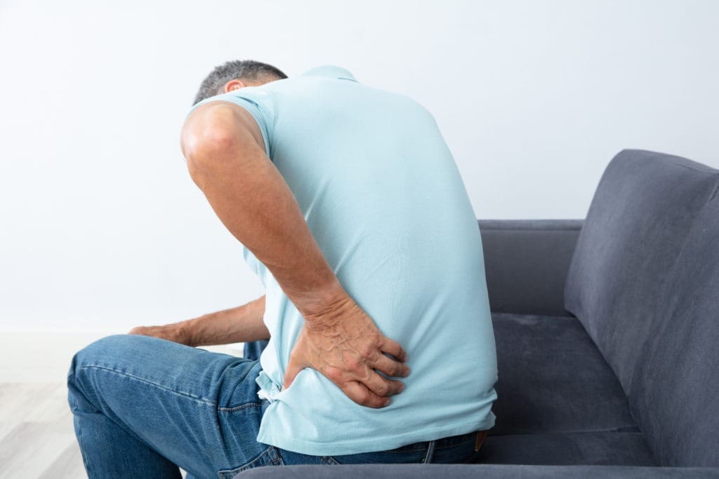
Sciatica and chronic back pain can affect almost every part of daily life. Sitting can hurt. Walking can feel limited. Sleep may be broken. Work, exercise, driving, and family time can become harder than they should be.
At El Paso Back Clinic, the goal is to look deeper than the pain signal. Pain is important, but it is often only the warning light. The real problem may involve an irritated nerve, a damaged disc, a strained ligament, a weak core, poor spinal motion, scar tissue, inflammation, or a past injury that never healed correctly.
This is why a modern spine care plan may combine chiropractic care, rehabilitation, medical oversight, functional medicine, epidural spinal injections, regenerative therapies, and shockwave therapy. Each part has a different job. Together, they may help calm nerve irritation, support tissue repair, improve movement, and help the body return to better function.
What Is Sciatica?
Sciatica is pain that travels along the sciatic nerve. This nerve starts in the lower back and travels through the buttock, hip, leg, and foot. When a spinal nerve root becomes irritated or compressed, pain can travel down the leg.
Common sciatica symptoms may include:
Low back pain
Buttock or hip pain
Burning pain down the leg
Numbness or tingling
Weakness in the leg or foot
Pain that worsens with sitting
Pain that improves when lying down or changing position
Sciatica is not always caused by the same problem. It may come from a herniated disc, disc degeneration, spinal stenosis, facet arthritis, muscle tension, pelvic imbalance, scar tissue, or inflammation. This is why a complete exam matters.
Why Chronic Back Pain Needs More Than Temporary Relief
Chronic back pain is pain that lasts longer than expected. It often continues for more than 12 weeks. By that time, the body may start to change how it moves. Muscles tighten. Joints stiffen. Nerves become more sensitive. The patient may avoid activity, which can lead to weakness and more pain.
Traditional care often focuses on short-term pain relief. That can help during a flare-up, but it may not be enough when the deeper problem is structural or inflammatory.
A more complete plan may look at:
Spinal alignment and joint motion
Disc health
Nerve irritation
Ligament and tendon stress
Muscle weakness
Core control
Inflammation
Nutrition
Sleep
Blood sugar and metabolic health
Prior auto, work, or sports injuries
This whole-person view is important because healing is not only about one painful spot. The spine is part of a larger system.
How Epidural Spinal Injections May Help Sciatica
An epidural spinal injection places medication or biologic material near an irritated spinal nerve. The goal is to reduce inflammation around the nerve root and help calm leg pain.
For a patient with strong nerve pain, this can be helpful. When pain is severe, the patient may not be able to move, stretch, exercise, or sleep well. If an epidural injection reduces the pain enough, the patient may be able to begin rehabilitation and chiropractic care more safely.
Epidural steroid injections are commonly used for spinal stenosis and nerve-related back and leg pain. However, long-term outcomes may vary. In one PCORI-supported report on lumbar spinal stenosis, epidural injections with corticosteroid plus lidocaine did not show long-term benefits over lidocaine alone for pain, function, opioid use, or surgery rates in the studied group (Friedly et al., 2019).
This does not mean epidural injections are useless. It means they should be used carefully and as part of a larger care plan.
Why Some Patients Look Beyond Repeated Steroid Injections
Steroids can reduce inflammation. That is why they are often used during painful flare-ups. But repeated steroid use may carry risks. Cortisone injections can have side effects, including cartilage damage, tendon weakening, blood sugar changes, infection risk, and bone thinning, especially when used too often or in high amounts (Mayo Clinic, 2026).
For some patients, this raises an important question:
Can we reduce pain while also supporting tissue repair?
This is where regenerative therapies may enter the conversation. Regenerative care does not simply try to hide symptoms. It aims to support the body’s natural healing response.
What Are Regenerative Spine Therapies?
Regenerative spine therapies use biologic materials, often from the patient’s own body, to support healing. These treatments may be considered for chronic spine pain, disc-related pain, ligament injury, facet joint pain, and nerve irritation when the patient is a proper candidate.
Common regenerative options include:
PRP: platelet-rich plasma
PFP: platelet-fibrin plasma or platelet-fibrin products
Platelet lysate: a platelet-derived fluid rich in growth factors
mFAT: microfragmented adipose tissue
These therapies are often called orthobiologics. “Ortho” refers to bones, joints, muscles, ligaments, and spine structures. “Biologics” refers to healing materials that come from living tissue.
The University of Iowa Health Care describes regenerative medicine as care that may use a person’s own cells, tissues, or biologic materials to support healing and repair (University of Iowa Health Care, n.d.).
PRP: Platelet-Rich Plasma for Spine and Nerve-Related Pain
PRP is made from a small sample of the patient’s blood. The blood is processed to concentrate platelets. Platelets are best known for helping blood clot, but they also carry growth factors and healing signals.
In spine care, PRP may be used to support damaged or irritated tissues, such as:
Disc-related pain areas
Facet joints
Ligaments
Tendons
Soft tissues around the spine
Research on PRP for low back pain is still growing. A narrative review on regenerative medicine for chronic low back pain described PRP and other biologic therapies as promising options, while also noting that more high-quality research is needed (Wang et al., 2023). A systematic review of PRP for low back pain found PRP was generally effective and safe for degenerative low back pain but also called for stronger studies and better treatment standards (Machado et al., 2023).
In simple terms, PRP is not a magic cure. But for selected patients, it may help support a better healing environment.
Platelet Lysate and Epidural Biologic Injections
Platelet lysate is made from platelets, but it is processed differently than PRP. The platelets are broken open, releasing growth factors into a thinner fluid. Because it is less thick than PRP, platelet lysate may be considered for nerve-related areas, including epidural use in some regenerative medicine settings.
A study of lumbar epidural platelet lysate for radicular pain reported improvements in pain and function through 24 months, with mild adverse events reported in a small percentage of patients (Centeno et al., 2017). More research is still needed, but this area is important because it examines biological support for nerve-related back and leg pain.
A 2025 meta-analysis also compared epidural PRP with steroid injections for lumbar disc disease with radiculopathy. The authors reviewed randomized controlled trials and examined pain and function outcomes over several time points (Muthu et al., 2025). This growing research shows why biologic epidural options are becoming a major topic in modern spine care.
PFP: A Natural Scaffold for Healing
PFP, or platelet-fibrin plasma, is similar to PRP but includes more fibrin activity. Fibrin is a natural protein involved in clotting and wound repair.
You can think of fibrin as a healing web. It may help hold platelets and growth factors in one area longer. This may be useful when the care plan is focused on damaged ligaments, tendons, or joint tissues.
PFP may support:
Local repair signaling
Tissue stability
Collagen remodeling
Longer contact time for healing factors
A more organized repair response
Like other regenerative options, PFP should be used after a detailed exam and proper diagnosis.
mFAT: Microfragmented Adipose Tissue
mFAT stands for microfragmented adipose tissue. Adipose tissue is fat tissue. In this treatment, a small amount of a patient’s own fat is collected, processed, and prepared for injection into a target area.
Fat tissue contains signaling cells and support structures that may help with tissue repair. mFAT is often discussed in regenerative medicine for joint, soft tissue, and orthopedic problems. It does not “regrow” a spine overnight. Instead, it may help support the local repair environment in selected cases.
For chronic spine problems, mFAT may be considered when there is deeper tissue degeneration, joint wear, or long-standing injury patterns. The key is proper patient selection, medical screening, imaging review, and follow-up care.
Shockwave Therapy: The Biological Catalyst
Shockwave therapy, also called extracorporeal shockwave therapy (ESWT), uses sound waves to stimulate tissue. It is non-surgical and does not involve medication.
Shockwave therapy may help painful tissues by creating a controlled healing signal. This process is called mechanotransduction. That means the body turns mechanical energy into a biological response.
ESWT may support healing by helping:
Increase local blood flow
Stimulate new small blood vessel formation
Improve cell activity
Reduce pain signaling
Break down scar-like tissue
Improve collagen remodeling
Support tissue repair pathways
A systematic review and meta-analysis found that ESWT improved pain and lumbar function in patients with chronic low back pain, with no serious adverse effects reported in the included studies (Liu et al., 2023). Another review described shockwave as a tool that may support tissue repair through blood vessel growth, anti-inflammatory effects, and cell signaling (Cheng & Wang, 2015).
Why Shockwave and Regenerative Injections May Work Well Together
Regenerative injections bring healing signals to damaged tissue. Shockwave therapy may help prepare the tissue to respond better.
This is why ESWT can be described as a biological catalyst. A catalyst helps a process move forward. Shockwave does not replace PRP, PFP, platelet lysate, or mFAT. It may help create a better local environment for healing.
A simple way to picture it is this:
PRP, PFP, platelet lysate, and mFAT bring healing signals.
Shockwave therapy helps wake up slow-healing tissue.
Chiropractic care improves joint motion and biomechanics.
Rehabilitation rebuilds strength, balance, and control.
Functional medicine looks for healing barriers inside the body.
When combined correctly, these tools may help the body repair itself more effectively than a single treatment alone.
The Role of Chiropractic Care at El Paso Back Clinic
Chiropractic care is often central to sciatica and back pain recovery because movement matters. If spinal joints, hips, pelvis, and soft tissues are not moving well, stress can build up around the nerves and discs.
At El Paso Back Clinic, chiropractic care may support:
Better spinal motion
Less joint stiffness
Improved posture
Better pelvic and hip mechanics
Reduced muscle guarding
Safer return to activity
Better rehab progress
Dr. Alexander Jimenez, DC, APRN, FNP-BC, CCST, CFMP, IFMCP, ATN, uses a dual-scope clinical view that connects chiropractic evaluation, injury care, functional medicine, and rehabilitation. His clinical observations often focus on how spinal structure, inflammation, metabolic health, and movement patterns work together.
This matters because many patients do not only have “a bad disc.” They may have a body system that is under stress.
Medical Oversight With Dr. Maria Guadalupe Cardenas, MD
At Injury Medical Clinic PA and within the larger integrative care model connected with El Paso Back Clinic, Dr. Maria Guadalupe Cardenas, MD, serves as Medical Director and Collaborative Physician. She is Board Certified in Internal Medicine, has over 40 years of experience as an internist, and is listed with NPI #1164426749 and Texas MD License #J2933.
This medical oversight is valuable because many spine patients have other health issues that can affect treatment safety and healing.
These may include:
Diabetes or blood sugar problems
High blood pressure
Autoimmune conditions
Medication use
Blood thinner use
Hormone changes
Infection risk
Poor sleep
Chronic inflammation
Older injuries or surgeries
A multidisciplinary clinic can help connect the dots between medical history, spine pain, nerve symptoms, and recovery goals.
Functional Medicine: Looking for Healing Barriers
Functional medicine asks a deeper question:
Why is this patient not healing well?
For chronic back pain and sciatica, the answer may lie beyond the spine. The body heals best when it has the right nutrients, blood flow, hormones, oxygen, sleep, and control of inflammation.
Functional medicine support may look at:
Vitamin D status
Blood sugar and insulin
Inflammation markers
Thyroid function
Hormone balance
Gut health
Nutrition
Weight management
Sleep quality
Stress load
This does not replace spine care. It supports spine care. A patient with poor blood sugar control, low protein intake, poor sleep, and high inflammation may heal more slowly. Improving these areas may help the patient respond better to chiropractic care, rehab, injections, and shockwave therapy.
Why Personal Injury Patients May Benefit
After a car crash, work injury, or sports injury, pain may not show up right away. Some symptoms appear hours or days later. Neck pain, back pain, headaches, sciatica, numbness, and stiffness can develop after the body’s stress response calms down.
Personal injury care needs careful documentation and a clear clinical plan. At El Paso Back Clinic, the care model may include:
Injury history
Orthopedic testing
Neurological testing
Range-of-motion findings
Imaging review when needed
Functional limits
Treatment response
Rehab progress
Referrals when needed
This matters because injury recovery is not only about pain relief. It is also about restoring function and documenting how the injury changed it.
A Step-by-Step Spine Recovery Plan
A patient-centered spine plan may include several phases.
Phase 1: Calm the Nerve
When sciatica is active, the first goal is to reduce irritation. This may include careful activity changes, decompression, gentle chiropractic care, targeted injection options, and pain-control strategies.
Phase 2: Improve the Healing Environment
Once pain is more controlled, regenerative therapies and shockwave therapy may be considered. The goal is to support tissue repair, improve circulation, and help chronic tissue move out of a stalled healing state.
Phase 3: Restore Motion
Chiropractic care, soft-tissue therapy, mobility work, and decompression may help the spine and pelvis move more freely.
Phase 4: Rebuild Strength
Rehabilitation helps the patient rebuild core strength, hip control, balance, posture, and endurance. This step helps protect the spine from future flare-ups.
Phase 5: Maintain Long-Term Function
The final goal is not just to feel better for a few days. The goal is to help the patient return to life with improved movement, strength, and awareness of how to prevent future problems.
Who May Be a Candidate?
A patient may be a candidate for this type of care if they have:
Sciatica
Chronic low back pain
Disc herniation
Disc degeneration
Annular tear
Facet arthritis
Ligament injury
Post-accident back pain
Pain that returns after basic care
Difficulty walking, sitting, or sleeping due to nerve pain
Not every patient is a candidate for every treatment. Severe weakness, loss of bowel or bladder control, fever, infection signs, cancer history, major trauma, or rapidly worsening nerve symptoms need urgent medical attention.
Final Thoughts
Sciatica and chronic back pain can be frustrating, but patients now have more options than short-term pain masking. Epidural spinal injections may help calm acute nerve irritation. Regenerative therapies such as PRP, PFP, platelet lysate, and mFAT may support repair in damaged or irritated tissues. Shockwave therapy may act as a biological catalyst by improving blood flow, stimulating cell activity, and helping chronic tissue respond.
At El Paso Back Clinic, this kind of care fits into a larger model that includes chiropractic care, medical oversight, functional medicine, personal injury care, and rehabilitation. With Dr. Alex Jimenez, DC, APRN, FNP-BC, working alongside Dr. Maria Guadalupe Cardenas, MD, Medical Director and Collaborative Physician, patients receive a team-based approach focused on structure, function, safety, and long-term healing.
The goal is simple: reduce pain, restore movement, support healing, and help patients return to the life they want.
Many adults notice extra weight creeping on, especially around the middle, even when they try to eat better and stay active. Hormone changes over time often play a quiet but powerful role in how the body stores fat, burns energy, and controls hunger. Bioidentical hormone replacement therapy (BHRT) offers a way to bring those internal messengers back into better balance. It is not a quick weight-loss fix or a magic pill. Instead, it helps remove some of the metabolic roadblocks that make diet and lifestyle efforts harder to sustain.
When hormone levels are optimized, many people find it easier to manage cravings, keep steady energy, and support lean muscle. This article explains how BHRT, and specifically the EvexiPEL method from Evexias Health Solutions, can work alongside smart eating and daily habits for longer-lasting results.
What Bioidentical Hormones Actually Do in the Body
Hormones act like chemical messengers. They tell the body when to store fat, when to burn it, how hungry to feel, and how well muscles can grow. Key players include estrogen, testosterone, insulin, cortisol, and thyroid hormones. When these get out of balance—often from aging, stress, or other life changes—metabolism can slow, fat can gather more easily around the belly, and cravings for sweets can grow stronger.
Bioidentical hormones are made to match the exact structure of the ones the human body produces naturally. They usually come from plant sources and are customized for each person after lab testing. The goal is to restore balance rather than force rapid change. Because they more closely match the body’s own chemistry, many patients experience smoother effects than with synthetic options.
How Balanced Hormones Help with Weight and Fat Control
Balanced hormones support weight management in several practical ways:
Fewer intense sugar cravings: When estrogen, progesterone, and cortisol signals stabilize, the brain’s hunger cues become easier to manage. People often report a less urgent desire for processed sweets or snacks.
Better insulin sensitivity: Improved insulin function helps the body use blood sugar for energy rather than store it as fat. This makes it easier to maintain a steady weight over time.
More consistent daily energy: Steady hormone levels reduce afternoon slumps. With more energy, it becomes easier to go for a walk, prepare a healthy meal, or stick to an exercise plan.
Support for lean muscle: Testosterone and other hormones help maintain or build muscle. Muscle tissue burns more calories even at rest, which supports a higher everyday metabolism.
Less stubborn abdominal fat: Hormone balance can influence where the body prefers to store fat. Many notice gradual improvement in midsection fat when levels are optimized alongside healthy habits.
These changes do not happen overnight. They create an internal environment where diet and movement efforts can finally show clearer results.
EvexiPEL Pellet Therapy: Steady Delivery Without the Roller Coaster
Evexias Health Solutions developed the EvexiPEL method as a form of BHRT that uses tiny, custom-made pellets. A trained provider places the pellets just under the skin during a short office visit. The pellets then release a steady, consistent dose of bioidentical hormones—such as testosterone or estradiol—over several months, usually three to six.
This steady release mimics the body’s natural rhythm far better than daily creams, gels, pills, or weekly shots. Many patients describe avoiding the ups and downs, or “roller coaster,” that can come with other delivery methods. Consistent levels often translate into more reliable energy, steadier moods, and fewer hormone-driven cravings throughout the day.
Because the delivery stays even, people can focus on building healthy routines instead of managing daily symptom swings. EvexiPEL is always paired with lab testing and a full wellness plan; it is never used alone.
Why Nutrition Matters Even More with BHRT
BHRT works best when paired with a diet built around fresh, whole foods. Think plenty of vegetables, quality proteins, healthy fats from avocados and nuts, and fiber-rich choices. These foods provide the body with the raw materials it needs for hormone production, detoxification, and stable blood sugar.
Cutting back on processed carbohydrates and added sugars helps too. These foods can spike blood sugar and work against the improvements in insulin sensitivity that BHRT supports. Many people find that once hormones stabilize, choosing whole foods feels more natural because energy stays higher and cravings quiet down.
Evexia’s providers often combine pellet therapy with targeted nutraceuticals—high-quality supplements designed to support metabolism, gut health, and mitochondrial energy. This root-cause approach to care addresses multiple systems at once rather than focusing on calories alone.
The Advantage of Multidisciplinary Integrative Care
Hormone balance does not exist in a vacuum. The nervous system, gut health, sleep, stress, and physical structure all influence how well hormones work. That is why care from a coordinated team often produces stronger, longer-lasting outcomes.
A clear example is the collaborative model at Injury Medical Clinic PA in El Paso, Texas. Dr. Alexander Jimenez, DC, APRN, FNP-BC, CFMP, IFMCP, ATN, CCST, brings chiropractic expertise, functional medicine insights, and advanced wellness protocols. He works directly with Medical Director Dr. Maria Guadalupe Cardenas, MD, a board-certified internal medicine physician with more than 40 years of experience (NPI #1164426749, Texas MD License #J2933).
In this setup:
Chiropractic care from Dr. Jimenez helps optimize nervous system function, posture, and mobility, so patients can move more comfortably and handle daily stress more effectively.
Dr. Cardenas provides medical oversight, reviews lab results, manages internal medicine needs, and ensures safe, appropriate hormone monitoring.
Functional medicine and nutrition support address gut health, inflammation, and lifestyle factors that affect metabolism.
Rehabilitation and personal injury services remove physical barriers that might otherwise limit activity and exercise.
Dr. Jimenez’s clinical observations in integrative settings show that patients achieve better metabolic and energy improvements when hormone optimization is combined with whole-person care. The spine and nervous system directly influence hormone signaling and stress responses. When both are supported, the body becomes more efficient at using the benefits of balanced hormones for weight and overall wellness.
This team approach makes BHRT one component of a larger, personalized strategy rather than an isolated treatment.
What Results Typically Look Like
People who combine EvexiPEL BHRT with whole-food nutrition and team-based support often describe:
More stable energy that lasts through the afternoon without relying on caffeine or sugar.
Reduced cravings that once derailed healthy eating plans.
Gradual improvements in body composition—less fat, better muscle tone—as insulin sensitivity and metabolism improve.
Easier adherence to daily movement because joints and energy feel better supported.
These changes build over weeks and months. The steady hormone delivery helps patients stay consistent long enough for new habits to stick. BHRT does not replace the need for healthy food choices and regular activity; it makes those efforts more effective by clearing hormonal interference.
Sample Report
Taking the Next Step Toward Balanced Health
If stubborn weight, low energy, or strong cravings have been ongoing challenges despite sincere efforts, checking hormone levels can be a useful step. A provider trained in EvexiPEL or similar BHRT methods will review full lab results, health history, and lifestyle before recommending a plan. Results vary, and therapy must always occur under proper medical supervision.
Clinics that blend chiropractic care, internal medicine oversight, functional nutrition, and regenerative approaches—like the model with Dr. Jimenez and Dr. Cardenas—can offer the coordinated support many people need. By addressing hormones, nervous system health, nutrition, and daily habits together, patients often move from frustration to steady, inside-out progress.
Balanced hormones alone will not create lasting change. But when they work in harmony with smart daily choices and a supportive care team, weight management becomes less of a constant struggle and more of a natural outcome of a body that is finally working with you instead of against you.
Platelet-Rich Plasma (PRP) Therapy for Better Posture at El Paso Back Clinic: Natural Healing for Spine Strength and Daily Comfort
Many people in El Paso struggle with slouched shoulders or a rounded back that makes everyday tasks feel harder. These posture problems often hide more profound issues like pain, weak ligaments, or worn spinal discs. When it hurts to stand tall, the body chooses easier but unhealthy positions. Over time, this cycle worsens discomfort. At El Paso Back Clinic, platelet-rich plasma (PRP) therapy offers a natural way to break that cycle. PRP therapy can indirectly ease posture issues by calming the pain that forces bad habits, strengthening weak ligaments and tendons, and repairing degenerated spinal discs. When added to a full treatment plan at El Paso Back Clinic, PRP helps address the root musculoskeletal problems that cause poor posture. This leads to smoother movement and better body balance in the neck, back, and shoulders. Patients often turn to this path when exercises or pills stop working.
What Is Platelet-Rich Plasma Therapy at El Paso Back Clinic?
Platelet-rich plasma, or PRP, uses a small sample of your blood. Doctors at El Paso Back Clinic draw the blood, spin it in a centrifuge to concentrate the healing platelets, and inject it into sore areas with ultrasound guidance. These platelets release growth factors that kick-start the body’s repair process. The whole visit takes about 30 minutes, and no foreign drugs are used. This makes PRP a safe, natural choice for many El Paso residents dealing with back or neck pain.
Dr. Alexander Jimenez, DC, APRN, FNP-BC, CFMP, leads the multidisciplinary team at El Paso Back Clinic. His dual training as a chiropractor and family nurse practitioner lets him blend regenerative medicine with chiropractic care. In his clinical work, Dr. Jimenez notes that PRP supports the body’s natural healing processes, especially when combined with functional medicine and rehabilitation (Jimenez, n.d.). The clinic’s locations across El Paso, including the main site at 11860 Vista Del Sol, make this advanced care easy to reach.
PRP first helped athletes recover faster. Today, it is used to treat everyday wear and tear at locations such as El Paso Back Clinic. Johns Hopkins Medicine explains that PRP floods the area with growth factors to speed cell repair and reduce inflammation (Johns Hopkins Medicine, n.d.).
How PRP Injections Repair Damaged Tissues at the Clinic
Once injected, the concentrated platelets go right to work. They release growth factors that handle three key jobs:
Reduce swelling: Chronic inflammation keeps pain going and weakens tissues. PRP calms inflammation, so real healing can start.
Build stronger tissue: Growth factors boost collagen to toughen tendons and ligaments that support the spine.
Speed up repair: Platelets call in cells that fix tears and worn spots.
At El Paso Back Clinic, PRP is used to treat the spine for conditions like degenerative disc disease. Discs act like cushions between bones. When they wear down, pain spreads, and posture slumps. The clinic’s blog on PRP for spinal care reports that patients often experience improved disc health and reduced stiffness without surgery (El Paso Back Clinic, n.d.-a).
For shoulders, PRP helps rotator cuff tendons heal more quickly. Princeton Sports and Family Medicine reports that PRP boosts tendon growth and collagen, so people return to daily tasks faster (Princeton Sports and Family Medicine, n.d.).
Bullet points on the repair steps at El Paso Back Clinic:
Blood draw and spin create PRP with 2 to 8 times the platelet count of normal blood.
Ultrasound guides the needle to the exact spot for the best results.
Growth factors like PDGF, VEGF, and TGF-β promote the formation of new blood vessels and clear waste.
Benefits build over weeks to months, often after two or three sessions with rehab follow-up.
PRP Therapy and Spinal Disc Health in El Paso
Worn discs cause back pain that makes standing straight tough. PRP injections at El Paso Back Clinic go into the disc area or nearby joints. They cut inflammation and help discs hold more water for better cushioning. The Morrison Clinic’s review, used in the clinic’s protocols, notes improved flexibility after PRP for disc problems (The Morrison Clinic, n.d.). This added stability allows the spine to align naturally in daily life.
Dr. Jimenez’s clinical observations highlight that patients with disc wear regain mobility when PRP is combined with chiropractic adjustments. His team checks nutrition and inflammation levels to make results last longer (Jimenez, n.d.).
Strengthening Ligaments and Tendons for Posture Support
Ligaments and tendons hold the spine and shoulders upright like support wires. When they stretch or tear, posture suffers. PRP injections at El Paso Back Clinic strengthen these soft tissues by signaling cells to produce denser collagen. Princeton Medicine shows PRP reduces swelling in rotator cuff injuries and helps shoulders move with less effort (Princeton Sports and Family Medicine, n.d.).
In the neck and low back, stronger ligaments mean less forward head tilt or swayback. Patients at the clinic say they sit taller without constant reminders. Health Coach Clinic, aligned with the clinic’s functional medicine, notes PRP lowers the need for pain pills and keeps people active for natural posture training (Health Coach Clinic, n.d.-a).
How PRP Indirectly Boosts Mobility and Biomechanics
Pain blocks good posture the most. When your back or neck hurts, you hunch to guard it. PRP eases pain at the source at El Paso Back Clinic. With less discomfort, muscles relax and move freely. Better movement creates smoother walking, sitting, and lifting. Over time, the body adopts healthier patterns.
Bullet points on mobility gains from the clinic’s approach:
Less neck and shoulder pain allows the head to balance over the spine.
Stronger back ligaments reduce lower-back sway, which pulls the shoulders forward.
Healthier discs restore the spine’s natural curves.
Faster return to activities builds confidence and encourages movement.
A Journal of Pain Research review backs this, showing PRP gives longer relief for low-back pain by fixing the real damage (Akeda et al., 2019).
Limits of PRP: Not a Magic Fix for Habit-Based Posture
PRP works best for injury or instability. It does not retrain the brain if poor posture comes only from years of desk slouching. All Wells Scoliosis Centre reminds us that posture is a learned habit. Repetition of good movements retrains the brain, but pain must be removed first (All Wells Scoliosis Centre, n.d.).
That is why El Paso Back Clinic uses PRP as part of a bigger plan. Without exercises and habit changes, old ways may return once pain fades. Dr. Jimenez emphasizes that PRP repairs the structure, while chiropractic and rehabilitation address the habit.
The Integrative Chiropractic Approach at El Paso Back Clinic
When regular therapy or medicine falls short, patients choose El Paso Back Clinic’s team. Dr. Jimenez, as DC, APRN, FNP-BC, and CFMP, leads chiropractors, nurse practitioners, physical therapists, and nutritionists. They treat the whole person: spine alignment, nutrition, inflammation, and movement.
The clinic blends PRP with gentle adjustments, spinal decompression, and functional medicine testing. Dr. Jimenez’s writings show patients with sciatica or chronic pain heal faster when PRP repairs tissues and chiropractic keeps the spine moving right (Jimenez, n.d.). Nutrition coaches cut inflammatory foods, while rehab experts teach core strength. This team effort delivers results that single treatments cannot.
Saks Wellness Center ideas, echoed at the clinic, note that chiropractic finds muscle imbalances and fixes them with adjustments and exercises. When paired with PRP, the body receives support from both inside and out (Saks Wellness Center, n.d.).
Functional medicine lowers whole-body inflammation through diet and supplements.
APRNs and FNP-BCs safely oversee injections and track healing.
Regular check-ins catch small issues early.
Patients skip surgery and long-term medication use.
Is PRP Therapy Safe and Effective at the Clinic?
Most people handle PRP well since it uses their own blood. Mild soreness at the injection site fades quickly. Serious side effects are rare. MidJersey Orthopedics and the clinic’s own protocols report PRP eases or ends pain for many without steroid risks (MidJersey Orthopedics, n.d.).
Results vary, but many feel relief in four to six weeks. Riverside Online notes PRP shines with healthy lifestyle changes like better movement (Riverside Online, n.d.). At El Paso Back Clinic, patients see strong outcomes because PRP is integrated into full-body support plans, including recent guides on PRP for sciatica and spinal care (El Paso Back Clinic, n.d.-b).
Real-World Results from El Paso Back Clinic Patients
Picture a local office worker whose neck pain forces them to lean forward. After PRP injections into the cervical ligaments and discs, along with Dr. Jimenez’s chiropractic care, pain decreases and posture improves naturally. A construction worker with low-back disc issues regains lift strength safely. These stories happen often at the clinic because PRP addresses the “why” behind the slump.
Cedars-Sinai describes how platelets release growth factors that rebuild tissue and may avoid surgery (Cedars-Sinai, n.d.). Blue Ridge Ortho adds that PRP helps with back and shoulder problems, making daily life easier (Blue Ridge Ortho, n.d.). Dr. Jimenez’s patient stories on the clinic site echo this success with non-surgical recovery.
Moving Forward with PRP and Posture Care in El Paso
Platelet-rich plasma therapy does not replace good habits, but it clears the path so habits stick. By easing pain, mending discs, and strengthening ligaments and tendons, PRP gives the body a real chance at natural alignment. At El Paso Back Clinic, combining PRP with chiropractic care, functional medicine, and daily practice creates a comprehensive path to better posture and lasting comfort.
If chronic pain or instability keeps you from standing tall, reach out to El Paso Back Clinic. Their non-surgical, team-based approach using the body’s own tools can open the door to a straighter, stronger you. Call 915-850-0900 or visit their El Paso locations to learn more.
Akeda, K., Yamada, T., Takahashi, H., & Sudo, A. (2019). Platelet-rich plasma in the management of chronic low back pain: A critical review. Journal of Pain Research, 12, 753–767. https://pmc.ncbi.nlm.nih.gov/articles/PMC6394242/
Why Poor Posture Habits Develop and How Integrating Chiropractic Care Can Help Restore Alignment
Poor posture is one of the most common physical problems in modern life. It often starts quietly. A person looks down at a phone for hours, leans forward at a desk, drives long distances, or relaxes in a slouched position at home. At first, it may not seem serious. Over time, however, these repeated positions can train the body into unhealthy movement patterns. What feels normal after months or years of slouching may actually be a sign that the muscles, joints, and spine are no longer working in balance.
At El Paso Back Clinic, posture problems are often viewed as more than a simple bad habit. They are usually the result of repeated stress on the body, weak supporting muscles, muscle tension, and changes in how the spine and joints move. Integrative chiropractic care can help address these root causes by improving spinal mobility, reducing soft-tissue tension, and teaching patients how to move, sit, stand, and work in healthier ways. This kind of approach does not just cover up symptoms. It helps restore a more natural, upright, and pain-free posture over time (Harvard Health Publishing, 2025a; OAA Orthopaedic Specialists, 2025).
Poor Posture Usually Develops Slowly
Most people do not suddenly wake up one day with poor posture. It usually develops gradually through daily routines. Modern life encourages a posture pattern that pulls the body forward. Many people spend hours doing the following:
Looking down at smartphones
Leaning toward computer screens
Sitting for long periods without breaks
Driving with rounded shoulders
Carrying tension in the neck and shoulders
Avoiding regular exercise or strength training
These habits can make the body adapt to a slouched position. Muscles in the chest, neck, and hip flexors often become tight, while the core, glutes, and upper back muscles grow weaker. This creates an imbalance. As a result, the head shifts forward, the shoulders round, and the spine loses some of its natural support and alignment (Better Health Channel, n.d.; Brown University Health, 2024).
Technology Has Changed the Way People Hold Their Bodies
One of the primary causes of poor posture today is the constant use of technology. Phones, tablets, and laptops often pull the head and shoulders forward. This forward-leaning pattern is commonly called “text neck” or “tech neck.” The neck must then support the weight of the head in a less efficient position, placing extra strain on the muscles, joints, and ligaments.
Brown University Health explains that looking down at a phone or tablet for long periods is a major contributor to bad posture. Harvard Health also notes that prolonged use of a computer or smartphone can lead to postural changes, muscle fatigue, and pain. These habits do not just affect the neck. They can also influence the shoulders, upper back, mid-back, and even the lower back because the body functions as a single, interconnected system (Brown University Health, 2024; Harvard Health Publishing, 2025a).
Sedentary Living Weakens the Body’s Support System
Poor posture is not only about how someone sits or stands. It is also about whether the body has enough strength and endurance to maintain healthy alignment. Sitting for long periods can weaken the muscles that support posture, especially the deep core muscles, glutes, and upper back stabilizers. When these muscles weaken, the body often relies on passive structures such as ligaments and joint surfaces rather than active muscular support.
This is one reason why slouching can start to feel easier than sitting upright. Slumping reduces the need for muscles to stay active, at least for a short time. However, that temporary comfort can lead to long-term strain. Harvard Health explains that poor posture habits can overstretch some muscles while shortening others, leading to pain and loss of function. Better Health Channel also notes that incorrect posture is often linked with inactivity, muscle fatigue, and poor physical conditioning (Harvard Health Publishing, 2025b; Better Health Channel, n.d.).
Stress and Tension Also Affect Posture
Posture is not only physical. It is also influenced by mental and emotional stress. When people feel stressed, they often tighten their shoulders, clench their jaw, and brace their upper body without realizing it. Over time, that tension pattern can become part of their normal posture. Instead of standing tall with relaxed shoulders and balanced breathing, the body stays guarded and compressed.
Stress-related tension can make it harder to maintain a neutral spine and relaxed shoulder position. It can also reduce normal breathing mechanics, especially when the chest feels tight, and the upper body remains rounded. This may help explain why poor posture is sometimes linked with headaches, neck tension, and fatigue (OrthoCarolina, 2025; Brown University Health, 2024).
The Body Adapts to What It Repeats
A key reason poor posture becomes difficult to fix is that the body adapts to repeated positions. If someone spends enough time in a slouched posture, the body begins to accept that shape as normal. Tight muscles stay tight. Weak muscles stay weak. Joint restrictions may develop. A person may even feel uncomfortable when trying to stand taller because upright posture now feels unfamiliar.
This process helps explain why poor posture is more than a simple choice. It becomes a learned physical pattern. Better Health Channel explains that repeated poor positioning and inactivity can lead to muscle fatigue and strain. Harvard Health also reports that poor posture can contribute to back pain, neck pain, headaches, difficulty breathing, and, in more serious cases, difficulty walking (Better Health Channel, n.d.; Harvard Health Publishing, 2025a).
Common Signs of Poor Posture
Poor posture can show up in many ways. Some signs are easy to see, while others are felt more than seen.
Common visual signs include:
Forward head posture
Rounded shoulders
A slouched upper back
An exaggerated low back arch
Uneven shoulders or hips
A tendency to lean to one side
Common symptoms may include:
Neck pain
Shoulder tightness
Upper back stiffness
Low back discomfort
Headaches
Muscle fatigue
Reduced range of motion
Pain after sitting for long periods
Feeling stiff when standing up after sitting
At El Paso Back Clinic, these patterns would typically be viewed as functional problems that affect more than appearance. They can change the way a person moves, breathes, works, and recovers from daily stress.
Why Integrative Chiropractic Care Can Help
Integrative chiropractic care focuses on the mechanical and functional causes of poor posture. Instead of just telling a patient to “sit up straight,” this approach examines why the posture problem developed in the first place. That may include joint restriction, muscle imbalance, repetitive strain, weak stabilizing muscles, and daily habits that continue to stress the spine.
Chiropractic adjustments can help restore motion in spinal and joint segments that are not moving well. OAA Orthopaedic Specialists explains that adjustments may improve spinal alignment and joint mobility, helping reduce compensatory patterns that contribute to poor posture. When joints move more freely, the body often has an easier time maintaining a more natural posture (OAA Orthopaedic Specialists, 2025).
Soft Tissue Work Helps Reduce Tension
Posture problems often involve more than the spine itself. Tight muscles in the chest, neck, shoulders, and hips can continue to pull the body forward even after a spinal correction. That is why integrative chiropractic care often includes soft tissue work, such as manual therapy, myofascial release, stretching, and mobility work.
This is important because posture is controlled by both joints and muscles. If the muscles remain tight and overactive, it becomes harder to maintain better alignment. Releasing muscle tension can make posture correction feel more natural and less forced. Many chiropractic posture-focused sources describe soft tissue therapy as a helpful component in improving posture and reducing pain associated with muscle imbalances (DE Integrative Healthcare, 2025; Zaker Chiropractic, 2025).
Corrective Exercises Support Long-Term Change
Posture usually does not improve for long unless the body becomes stronger and more aware. Corrective exercises help retrain the muscles that support healthy alignment. This may include exercises for the core, glutes, shoulder blades, upper back, and deep neck stabilizers.
Helpful exercise goals often include:
Strengthening the upper back
Activating the deep core
Improving glute strength
Stretching the chest
Opening tight hip flexors
Training shoulder blade control
Improving balance and body awareness
Harvard Health recommends strengthening the upper back, chest, and core while also reducing the activities that contribute to poor posture. This is one reason why posture care works best when treatment and exercise are combined rather than used alone (Harvard Health Publishing, 2025a).
Ergonomic Education Helps Prevent Recurrence
Even the best treatment plan can lose momentum if a person returns to the same habits that caused the problem. That is why ergonomic education is a major part of posture care. Patients need to understand how they sit, stand, lift, sleep, and use technology during the day.
Simple posture-friendly changes may include:
Raising a screen to eye level
Keeping feet flat while sitting
Taking standing or walking breaks every 20 to 30 minutes
Avoiding long periods of looking down at a phone
Using lumbar support when needed
Keeping shoulders relaxed instead of lifted
Changing positions often instead of holding one posture too long
Brown University Health and Better Health Channel both emphasize that work setup, movement breaks, and body awareness are important in preventing and correcting posture problems (Brown University Health, 2024; Better Health Channel, n.d.).
Clinical Observations from Dr. Alexander Jimenez
The public clinical information shared by Dr. Alexander Jimenez, DC, APRN, FNP-BC, reflects an integrative view of posture-related problems. His materials describe how posture issues are often connected to spinal stress, muscle imbalance, functional movement problems, and broader lifestyle factors. His clinical approach emphasizes looking beyond symptoms alone and considering biomechanics, rehabilitation, and whole-person recovery.
That approach aligns well with posture correction, as poor posture is rarely caused by a single factor. It is usually a combination of sedentary habits, repetitive stress, tight muscles, weak stabilizers, and poor body mechanics. Dr. Jimenez’s public educational content supports a model in which chiropractic care, movement correction, rehabilitation, and lifestyle guidance work together to improve long-term outcomes (DrAlexJimenez.com, 2026a, 2026b).
Better Posture Is About Function, Not Perfection
Proper posture does not mean being rigid or stiff. It means that the body is aligned well enough to move efficiently, breathe more easily, and reduce unnecessary strain. The goal is not to maintain perfect posture every second of the day. The goal is better support, better awareness, and better function.
When posture improves, people may notice benefits such as:
Less neck and back pain
Less tension in the shoulders
Easier breathing
Better movement quality
Less fatigue while sitting or standing
Improved comfort during work and daily life
At El Paso Back Clinic, a posture-centered message would likely focus on helping patients restore natural alignment by addressing the causes of dysfunction rather than only reacting to pain after it appears.
Final Thoughts
People develop poor posture habits mainly because modern life pulls the body into repeated forward, slouched positions. Sitting too much, using phones and computers for long hours, carrying stress, and having weak support muscles all contribute to muscle imbalance and joint strain. Over time, the body adapts to these unhealthy positions until they begin to feel normal.
Integrative chiropractic care can help break that cycle. By improving spinal motion, reducing muscle tension, guiding corrective exercise, and teaching better ergonomic habits, this type of care addresses the root causes of poor posture. That makes it more likely that changes will last. When posture improves, patients often feel better, move better, and place less daily stress on the body.
Understanding Chiropractic Spinal Adjustments: Techniques, Benefits, and Integrated Care
Chiropractic spinal adjustments, also known as spinal manipulations or reductions, offer a natural way to address back pain, improve mobility, and support overall health. These procedures focus on aligning the spine to reduce discomfort and enhance body function without surgery or heavy reliance on medications. Many people seek chiropractic care for issues like chronic back pain, neck strain, or injury recovery. This article explores what happens during an adjustment, its effects on the body, common techniques, and how team-based care can boost results.
What Is a Chiropractic Spinal Adjustment?
A chiropractic spinal adjustment involves a trained practitioner using their hands or a tool to apply a quick, controlled force to misaligned parts of the spine. This helps restore proper alignment and movement to the joints. The goal is to ease pain, improve joint function, and reduce pressure on the nerves and surrounding muscles (Cleveland Clinic, n.d.). It’s a non-surgical method that stretches the joint, often releasing gas bubbles like nitrogen, oxygen, and carbon dioxide, which create that familiar cracking sound—similar to when you crack your knuckles (Chiro One, n.d.).
Adjustments target areas of restriction, called subluxations, where vertebrae are out of place or not moving well. By correcting these, the procedure can improve nervous system function, leading to reduced irritation and better overall health (NCCIH, n.d.). Patients often feel an increase in range of motion right away, along with looser muscles.
Key Aspects of a Chiropractic Adjustment
Here are some main features of this treatment:
Procedure: The chiropractor first checks the spine for problem spots. Then, they use a sudden but precise push to fix the joint (Revive Chiropractic DSM, n.d.).
Sensations: You might hear a pop or crack, but it’s just gas escaping the joint fluid, not bones breaking (Cleveland Clinic, n.d.).
Physical Effects: The thrust stretches tight joints, relaxes tense muscles, and frees trapped gases, reducing built-up pressure (Physicians Group LLC, n.d.).
Benefits: It restores normal joint motion, supports nerve health, and reduces pain from nerve compression (Spine Health, n.d.).
What It Feels Like: Most find it painless, though some notice mild soreness afterward, like after a workout. Many report quick relief and easier movement (Complete Care, n.d.).
These elements make adjustments a popular choice for managing pain without invasive options.
Techniques Used in Chiropractic Adjustments
Chiropractors use different methods based on the patient’s needs. Common ones include:
Manual Adjustment: This is a high-velocity, low-amplitude (HVLA) thrust done by hand. It’s direct and aims to realign the spine quickly (Towson Chiro, n.d.).
Instrument-Assisted: Tools provide gentle taps to the spine, ideal for those who prefer less force (Visit Chiro First, n.d.).
Spinal Decompression: Using a specialized table, the spine is stretched to create space between the vertebrae, helping with issues such as herniated discs (Get Adjusted Columbia, n.d.).
These techniques can be tailored to conditions such as whiplash or back injuries sustained in accidents (Utah Therapeutic Massage, n.d.).
What Happens During a Chiropractic Spinal Adjustment
A typical session starts with an assessment. The chiropractor reviews your health history, performs a physical exam, and may use X-rays to identify subluxations (Dubuque Chiropractic, n.d.). Once identified, the adjustment begins.
The practitioner positions you on a table and applies a fast, targeted thrust to the specific joint. This might cause cavitation—the popping sound from gas release in the joint fluid (Starkwood Chiropractic, n.d.). Right after, muscles relax, nerve irritation drops, and joint motion improves (Personal Injury Doctor Group, 2024).
Sessions often include additional therapies such as soft-tissue work, trigger-point release, or stretches to support the adjustment (Boca Chiropractic SW, n.d.). The whole process is quick and focused on comfort.
Benefits of Chiropractic Spinal Adjustments
Regular adjustments offer several advantages:
Pain Relief: They reduce mechanical stress on the spine and ease nerve compression, helping with back, neck, and headache pain (Chiro One, n.d.).
Improved Function: By fixing alignment, they enhance posture and spinal health, preventing future issues (Boca Chiropractic SW, n.d.).
Nervous System Support: Adjustments promote improved nerve signaling, supporting overall bodily function (Physicians Group LLC, n.d.).
Faster Recovery: For injuries like car accidents, this approach speeds healing by addressing root causes (Dallas Accident and Injury Rehab, n.d.).
Studies show these benefits lead to higher patient satisfaction when combined with other care (My Chiro, n.d.).
Incorporating an Interdisciplinary Team for Better Results
Bringing in a team of experts—like advanced practice registered nurses (APRNs), family nurse practitioners (FNP-BC), certified functional medicine providers (CFMP and IFMCP), advanced translational nutrigenomics specialists (ATN), and certified chiropractic spinal trauma experts (CCST)—makes treatment more effective. This approach combines structural fixes with medical and nutritional support to provide holistic care (Health Coach Clinic, n.d.).
For complex cases, such as auto injuries or chronic pain, this team provides comprehensive plans. It focuses on root causes rather than just symptoms, leading to lasting improvements (LinkedIn, n.d.).
How Each Role Contributes
APRN/FNP-BC: These nurses offer medical checks, diagnose issues, and manage meds if needed. They educate patients and integrate chiropractic with traditional medicine to improve pain control (Nursing World, n.d.; Goodwin University, n.d.).
CFMP/IFMCP: They dig into metabolic and nutritional roots of problems, using functional medicine to heal the musculoskeletal system faster (LinkedIn, n.d.).
ATN: By studying genetics and nutrition, they create custom diets and supplements to cut inflammation and aid repair (Jimenez, n.d.).
CCST: Experts in spinal trauma handle tough injuries like whiplash or disc herniations with advanced techniques (Spine Stop, n.d.).
This teamwork enhances outcomes, especially in recovery from accidents or ongoing conditions (Dallas Accident and Injury Rehab, n.d.).
Clinical Observations from Dr. Alexander Jimenez, DC, APRN, FNP-BC
Dr. Alexander Jimenez, with his dual roles in chiropractic and nursing, observes that adjustments restore function in conditions such as sciatica and herniated discs by reducing nerve compression without surgery (Jimenez, n.d.). He notes that patients often experience rapid pain relief and improved mobility after sessions, especially when combined with functional nutrition.
In trauma cases, such as car accidents, Jimenez highlights how spinal decompression and shockwave therapy speed recovery by addressing inflammation and nerve damage (LinkedIn, n.d.). His integrated approach, blending chiropractic with nutrigenomics, helps address root causes such as gut issues that affect spinal health. Patients report reduced symptoms in fibromyalgia and neuropathies through personalized plans that include team input from therapists and nutritionists.
Jimenez emphasizes holistic care for all ages, using assessments to uncover environmental factors. His observations show that interdisciplinary teams lead to sustained health, with testimonials praising relief from chronic pain and improved vitality (Jimenez, n.d.).
Conclusion
Chiropractic spinal adjustments provide a safe, effective way to manage pain and improve spinal health. By understanding the process, techniques, and benefits, you can see why many choose this path. Adding an interdisciplinary team takes it further by offering comprehensive care for better long-term results. If you’re dealing with back issues or injuries, consider consulting a qualified chiropractor.
Motivation That Lasts: Fun, Low-Impact Workouts and SMART Goal Strategies
Losing weight does not have to feel impossible, even if back pain, low energy, or busy days get in the way. Many people in El Paso start with easy exercises like short walks or gentle stretches, but staying motivated is what brings real results. The good news is that small, smart steps, plus help from a local expert team, can make all the difference. At El Paso Back Clinic, patients discover how chiropractic care and functional medicine remove roadblocks so basic weight-loss exercises feel safe, doable, and even enjoyable. This guide shares straightforward ways to set goals, track progress, choose fun movement, and get professional support right here in El Paso. You will learn practical tips that fit real life and see how the clinic’s team, led by Dr. Alexander Jimenez, helps turn “I can’t” into steady success.
Basic weight-loss exercises like walking, light yoga, or dancing burn calories without stressing your joints. When your body feels better and pain drops, motivation stays strong. El Paso Back Clinic combines chiropractic adjustments, personalized rehab, and health coaching to make these simple moves part of your everyday routine.
Setting Attainable SMART Objectives for Steady Progress
SMART goals keep your weight-loss journey clear and reachable. SMART means Specific, Measurable, Achievable, Relevant, and Time-bound. Instead of saying “I need to lose weight,” try “I will walk for 15 minutes after dinner, five days this week.” This type of goal is easy to follow and gives quick wins. (Hey Life Training, n.d.; El Paso Back Clinic, n.d.-b)
Here are SMART goal examples perfect for basic weight-loss exercises:
Walk briskly for 15 minutes, five days a week, starting this Monday.
Do gentle yoga stretches for 10 minutes each morning for the next two weeks.
Dance to favorite music for 15 minutes, three evenings a week.
Swim or walk in water for 15 minutes twice a week at a local pool.
Take the stairs instead of the elevator at least five times daily this week.
Start small, so you build confidence fast
At El Paso Back Clinic, health coaches help patients turn these goals into custom plans that match their energy and schedule.
Monitoring progress keeps motivation alive. Use a simple notebook or phone app to log your walks, steps, or how your back feels after movement. Seeing checkmarks add up or a line on a graph climb feels rewarding. Patients at the clinic often say watching their own improvements beats staring at the scale. (Zen Habits, n.d.)
To avoid burnout, pick fun, low-impact activities. Yoga, swimming, and walking ease joints and lift mood through natural feel-good chemicals. These basic exercises become something you look forward to instead of dread. (HelpGuide.org, n.d.)
Find accountability with a workout buddy or the clinic’s support network. Many patients walk with family or join gentle group sessions. Reward small wins with non-food treats like new walking shoes or a relaxing evening. Remember your “why”—more energy for family, better sleep, or less back pain. Read it daily on tough days. (Planet Fitness, n.d.-a)
Easy, Efficient Strategies to Stay Motivated Every Day
Consistency beats intensity when building habits. Here are proven strategies that work well with basic weight-loss exercises:
Start small for lasting consistency: Begin with just 10–15 minutes of movement. This avoids burnout and makes exercise a normal part of your day. (Reddit community insights, 2024)
Track your development: Write down workouts, steps, or how clothes fit. Graphs show real progress and keep you excited. (Zen Habits, n.d.)
Make it fun: Choose dancing, swimming, cycling, or active games. Fun turns movement into “me time.” (HelpGuide.org, n.d.)
Reward yourself: After five good days, celebrate with new socks, a movie, or a quiet bath. (Modern Image Aesthetics, n.d.)
Build accountability: Walk with a friend, pet, or join a beginner class. The clinic’s health coaches provide extra check-ins. (Healthline, n.d.)
Recall your “why”: Focus on deeper reasons like steady energy or pride in your posture. (Planet Fitness, n.d.-b)
Prepare for low-energy days: Have a backup like 10 minutes of gentle stretches at home. (Cleveland Clinic, n.d.)
These steps fit real El Paso life—hot days, long work hours, and family needs. Short walks during lunch or evening strolls add up fast.
Walking Your Way to Better Results: Clinic-Approved Tips
Walking is one of the easiest basic weight-loss exercises, and El Paso Back Clinic shares clear ways to burn more fat while protecting your back. Start with 15 minutes daily, five days a week, then add five minutes each week. Walk at a brisk pace faster than normal, swing your arms, and keep a healthy posture. Add short speed bursts or gentle hills for extra calorie burn without hurting knees. Wear supportive shoes and breathe steadily. (El Paso Back Clinic, n.d.-c)
Benefits include stronger bones, less joint pain, better mood, and reduced belly fat linked to heart health. Even short 15-minute walks several times a day work when time is tight. Patients at the clinic combine walking with chiropractic care for faster mobility gains and steady motivation.
Making Fitness Enjoyable and Part of Your Routine
Pick activities you actually like. If running hurts, try dancing at home, water walking, or bike rides on flat paths. Listen to music or podcasts while moving. Many patients discover they enjoy low-impact options once pain eases. (Medical Beauty and Weight Loss, n.d.)
Social support helps too. Walk with neighbors or join light classes. At El Paso Back Clinic, personalized rehab programs make movement feel safe again, so you stay consistent longer.
How El Paso Back Clinic Boosts Motivation Through Integrative Care
Back pain or low energy often stops people from exercising. El Paso Back Clinic, led by Dr. Alexander Jimenez, DC, APRN, FNP-BC, removes these barriers with chiropractic and functional medicine. Their approach helps thousands of El Paso patients move more freely and lose weight sustainably.
Chiropractic adjustments reduce chronic back, hip, and joint pain, so walking or yoga no longer hurts. Better spinal alignment improves nervous system signals that control metabolism and fat burning. When the body works more smoothly, energy rises, and motivation follows naturally. (El Paso Back Clinic, n.d.-a; Adjusted Life Chiropractic, n.d.)
Dr. Alexander Jimenez has observed over 30 years that fixing spinal misalignments breaks the pain-obesity cycle. Pain leads to less movement and comfort eating; extra weight adds more pain. His team uses gentle adjustments, advanced imaging, and lab tests to address root causes such as inflammation, hormonal imbalances, and gut issues. Patients report less pain, better sleep, steadier moods, and fewer cravings. (Jimenez, n.d.; El Paso Back Clinic, n.d.-a)
Custom low-impact exercise plans are a clinic specialty. Instead of heavy gym work, they recommend practical moves: walking programs, water exercises, light resistance bands, and core stretches that fit daily life. These plans build confidence fast because they feel safe. The clinic’s rehabilitation centers offer guided sessions with trainers who understand back issues. (Robinhood Integrative Health, n.d.; El Paso Back Clinic, n.d.-c)
Functional medicine digs deeper. The team checks for slow metabolism, insulin resistance, or stress hormones that block weight loss. Personalized nutrition advice, supplements, and lifestyle tips clear these hurdles. Health coaches then create step-by-step plans with SMART-style process goals—like “walk three to four times this week”—so patients focus on what they can control. (El Paso Back Clinic, n.d.-b, n.d.-d)
Stress management is built in
High stress raises cortisol and belly fat while lowering motivation. Chiropractic care relaxes tight muscles and calms the nervous system. Many patients report feeling more positive and ready to move on after visits. (Dr. P Chiro, n.d.)
Personalized accountability keeps progress on track. Regular check-ins, body scans, and plan updates show results beyond the scale. Improved posture from adjustments makes patients stand taller and feel stronger—boosting confidence to keep going. (Obesity Action Coalition, n.d.; Westport Chiropractic, n.d.)
Dr. Jimenez often reminds patients that big changes start with small, consistent steps. His team at El Paso Back Clinic offers multiple convenient locations across El Paso, including rehab and fitness centers with 24/7 access. Military discounts, virtual coaching options, and meal-prep support make healthy living easier. Patients with past injuries or long-term back pain often return to activities they once avoided, creating a positive cycle of more movement and faster weight-loss results.
By reducing pain, improving mobility, addressing metabolic issues, and providing expert coaching, El Paso Back Clinic turns basic weight-loss exercises into something patients actually enjoy and stick with long-term.
Putting It All Together for Real, Lasting Success
Begin today with one small change. Choose a SMART goal, schedule a 15-minute walk, and note your “why.” Add music or a friend for fun. If back pain or low energy holds you back, contact El Paso Back Clinic for a personalized evaluation. Dr. Alexander Jimenez and his multidisciplinary team combine chiropractic care, functional medicine, and health coaching to support your goals safely.
Motivation comes and goes—some days feel easier than others, and that is normal. The strategies here—SMART goals, tracking, fun movement, rewards, accountability, and professional help—help you bounce back quickly. Over weeks and months, these habits create real momentum.
Basic weight-loss exercises like daily walking or gentle yoga do more than burn calories. They improve heart health, lift mood, strengthen muscles, ease back pain, and raise self-esteem. With support from El Paso Back Clinic, you gain energy for work, family, and life. Celebrate every step, every stretch, and every healthy choice. You have local experts ready to help—one simple, consistent day at a time.
IFM's Find A Practitioner tool is the largest referral network in Functional Medicine, created to help patients locate Functional Medicine practitioners anywhere in the world. IFM Certified Practitioners are listed first in the search results, given their extensive education in Functional Medicine