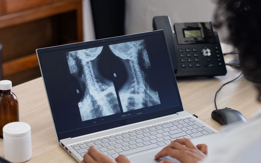
Integrative Chiropractic Care at El Paso Back Clinic: Natural Recovery Without Surgery
Many people struggle with back pain, joint stiffness, or injuries from daily life, work, or accidents. They look for lasting relief that helps them move freely again. At El Paso Back Clinic, integrative chiropractic care stands out as a natural, effective way to address these issues. Led by Dr. Alexander Jimenez, the clinic focuses on fixing the root causes of pain through structural chiropractic adjustments and supportive therapies. This approach restores proper alignment, improves movement, and accelerates the body’s natural healing without the need for surgery or heavy medications.
The team at El Paso Back Clinic believes in treating the whole person. They combine hands-on chiropractic care with physical therapy and other non-invasive methods to create lasting results. By focusing on structure and function, patients often avoid surgery and return to active, pain-free lives. This integrative style has helped countless individuals in El Paso recover from personal injuries, auto accidents, and chronic back problems.
What Makes Integrative Chiropractic Care Different?
Integrative chiropractic care at El Paso Back Clinic goes beyond quick fixes. It looks at how the spine, nerves, muscles, and joints work together. When the spine is out of alignment, it can press on nerves and cause pain, weakness, or limited motion. Chiropractic adjustments gently realign the body to free up those nerves and restore normal function.
Unlike traditional care, which might only mask symptoms, this method treats the root cause. Structural chiropractic adjustments correct posture issues, ease muscle tension, and improve overall body mechanics. When paired with physical therapy exercises, patients build strength and flexibility that lasts.
Here are the main benefits of this approach:
It uses natural techniques to reduce inflammation and promote better blood flow.
It restores functional movement so everyday tasks feel easier.
It helps prevent future injuries by fixing poor alignment early.
It fits perfectly with the body’s own repair systems for long-term wellness.
Dr. Jimenez and his team emphasize that true healing starts with proper structure. Their clinical observations show that patients who receive consistent chiropractic care often report faster recovery and greater confidence in their bodies. (Jimenez, n.d.-c)
How Supportive Therapies Enhance Chiropractic Results
While structural chiropractic care forms the foundation, El Paso Back Clinic sometimes uses supportive therapies to further enhance healing. These non-surgical options work in the background to stimulate the body’s natural processes. They include concentrated healing cells from a patient’s own blood or fat, along with signaling molecules like peptides. These tools act as gentle stimulants that help repair damaged tissues and lower swelling.
For example, platelet-rich plasma (PRP) and similar options can support tissue repair after chiropractic adjustments have created better alignment. Shockwave therapy is another tool that pairs well with chiropractic care. It sends sound waves to increase blood flow and break down scar tissue, making adjustments more effective and recovery quicker.
The clinic’s integrative practice keeps these supportive methods secondary to the main chiropractic focus. The goal remains the same: fix the root problem and restore normal movement. This combination helps patients with back pain, sciatica, or soft tissue injuries heal faster without invasive procedures.
Key ways these supportive tools work alongside chiropractic care include:
They speed up the body’s natural repair after adjustments open up better nerve pathways.
They reduce inflammation so patients feel relief sooner during physical therapy sessions.
They support long-term tissue strength, helping chiropractic corrections last longer.
They fit into a holistic plan that avoids surgery and heavy reliance on pain pills.
This balanced method has shown strong results in personal injury and sports-related cases. (StemWave, 2024; El Paso Chiropractic, n.d.)
Dr. Alexander Jimenez’s Integrative Approach at El Paso Back Clinic
Dr. Alexander Jimenez, DC, APRN, FNP-BC, leads the clinical team at El Paso Back Clinic with more than 30 years of experience. As a chiropractor first, he specializes in structural care that restores spinal alignment and functional movement. His dual background allows him to blend chiropractic adjustments with advanced rehabilitation techniques for complete recovery.
At the clinic, Dr. Jimenez focuses on finding and treating the true source of pain. He uses gentle adjustments, spinal decompression, and targeted exercises to resolve issues like herniated discs, sciatica, and scoliosis. Supportive regenerative options stay in the background as beneficial additions that enhance the primary chiropractic work.
His clinical observations highlight how this integrative style helps patients recover from trauma with greater strength and confidence. Many who visit El Paso Back Clinic after car accidents or work injuries see big improvements in mobility and daily function. Dr. Jimenez often notes that addressing structure first sets the stage for the body to heal naturally. (Personal Injury Doctor Group, 2026)
What patients can expect at the clinic includes:
Thorough exams that spot hidden alignment problems or nerve pressure.
Customized chiropractic plans that include physical therapy and movement training.
Supportive therapies are used only when needed to enhance overall outcomes.
Focus on nutrition and lifestyle tips to keep the body strong between visits.
The clinic’s multidisciplinary team of chiropractors and physical therapists works together under Dr. Jimenez’s guidance. This team approach ensures every patient receives care tailored to their needs. (Jimenez, n.d.-a)
Real Results for Personal Injuries and Everyday Back Problems
Life can bring sudden injuries from auto accidents, sports injuries, or repetitive work strain. These issues often lead to back pain, stiff joints, or limited motion. At El Paso Back Clinic, integrative chiropractic care shines in these cases by correcting structure and supporting natural recovery.
For auto accident victims, chiropractic adjustments help with whiplash and spinal misalignment that can cause long-term discomfort. Physical therapy builds strength, while supportive therapies in the background reduce swelling and speed tissue repair. Sports injuries, such as strains or tendon problems, also respond well. Athletes regain a full range of motion and return to play with less risk of re-injury.
Patients often notice these advantages:
Faster return to work or favorite activities, with less downtime.
Reduced need for pain medications that can have side effects.
Stronger, more stable joints thanks to proper alignment and support.
Overall, a better quality of life with less daily discomfort.
One review of integrative care found that patients with chronic back issues experienced steady progress and avoided surgery when chiropractic was the primary focus. (Ortho Edge El Paso, n.d.; West Texas Pain, n.d.)
The clinic’s location in El Paso makes it convenient for local families and workers seeking natural solutions. Many patients report feeling renewed energy after a few sessions of structured chiropractic care.
Why This Chiropractic-First Method Promotes Lasting Wellness
Traditional treatments sometimes rely on temporary relief or major operations. Integrative chiropractic care at El Paso Back Clinic takes a smarter path. It works with the body’s design by correcting alignment and supporting its natural repair abilities.
Younger bodies heal quickly on their own, but aging or repeated stress can slow the process. Chiropractic adjustments keep the spine and joints in proper position so healing happens efficiently. Supportive therapies like shockwave therapy or concentrated healing cells remain in the background to provide an extra nudge when needed.
This non-surgical style offers clear advantages:
No scars or infection risks that come with operations.
Better long-term mobility and fewer flare-ups.
A focus on prevention ensures problems do not become big ones.
Improved posture and movement that benefit overall health.
Experts agree that fixing the root cause leads to the best recovery. When chiropractic care leads the way, patients often experience lasting relief and greater confidence in their bodies. (New Regen Ortho, n.d.; Serenity Health Care Center, n.d.)
At El Paso Back Clinic, the emphasis remains on empowering patients through structure and function. Dr. Jimenez’s team helps people of all ages live more active, pain-free lives.
Moving Forward With Natural, Effective Care
Integrative chiropractic care at El Paso Back Clinic provides a clear path for anyone dealing with back pain or injury. Structural adjustments form the core, restoring alignment and functional movement. Supportive therapies work quietly in the background to stimulate the body’s natural healing without surgery or strong drugs.
This holistic method addresses the root causes of problems and helps patients recover faster from personal injuries, auto accidents, and sports injuries. Under Dr. Alexander Jimenez’s guidance, the clinic delivers care that fits real life and delivers real results.
If back pain or limited motion holds you back, consider the integrative chiropractic approach at El Paso Back Clinic. It proves that sometimes the best way forward is to work with the body’s own systems through skilled, hands-on care.
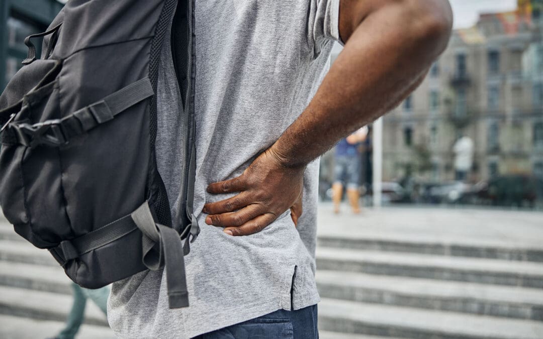
Is It Safe to Wear a Backpack? Expert Tips on Spinal Health and Back Pain Prevention in the US and El Paso, TX
A woman walking, wearing a backpack with the recommended weight, and maintaining correct posture to prevent back pain and problems.
Back pain is a big issue for many people in the United States
Up to 80% of adults face low back pain at some point in their lives. This is one of the top reasons for doctor visits and missed workdays. The cost is huge too, with over $100 billion spent on spine problems each year. In El Paso, Texas, where people often have active jobs like industrial work or lots of driving, back pain questions focus on things like sciatica, herniated discs, and spinal stenosis. A common concern across the country, including in places like El Paso, is whether wearing a backpack is safe for the spine. The good news is that it can be safe if you follow some simple rules. This article focuses on backpack safety and then addresses other key questions about managing back pain, treatment options, and daily habits to keep your spine healthy.
Understanding Backpack Safety and Spinal Health
Wearing a backpack is common for carrying things, but if it’s too heavy or worn incorrectly, it can hurt your back. Heavy backpacks can strain muscles and joints in your back, neck, and shoulders. This might lead to pain or bad posture over time. However, backpacks do not cause scoliosis, a spinal curvature that affects about 2% to 3% of people. Scoliosis often starts in teens and is more common in girls, but it’s not linked to backpacks.
Is it safe? Yes, as long as you distribute the weight right and follow the tips to avoid strain. Improper use can cause muscle fatigue, poor posture (such as slouching), and even chronic pain if left unaddressed. In El Paso, where people might carry tools or bags for work, this is especially important to prevent issues such as sciatica, where pain radiates down the leg due to nerve pressure.
Here are some key tips for safe backpack use:
Choose the right backpack: Pick one with wide, padded straps and a padded back. It should fit your body size and have a waist strap for heavy loads. Lightweight materials help too.
Limit the weight: Keep the backpack under 10-15% of your body weight. For example, if you weigh 150 pounds, aim for no more than 15-22.5 pounds.
Distribute weight evenly: Put heavier items at the bottom and close to your back. Use compartments to balance things and stop shifting.
Wear it correctly: Always use both straps. Adjust them so the pack sits in the middle of your back, not sagging low. Bend your knees to lift it.
Make smart choices: Remove extra items often. Use lockers or storage if possible. For very heavy loads, try a rolling backpack or crossbody bag.
These steps help distribute the load across your strong back muscles and keep your spine aligned. If you feel pain, stop and adjust. In places like El Paso, with busy lifestyles, following these can help prevent accidents from becoming long-term back issues.
Common Causes of Back Pain in the US
Back pain affects millions. In the US, about 26% of adults have it at any time, and it’s more common after age 45. Among adults aged 50 and older, up to 45.6% experience it. Causes include muscle strains, ligament injuries, herniated discs (where the disc’s soft center protrudes), arthritis, and spinal stenosis (where the spinal canal narrows). Stress can make it worse by causing muscle spasms. Even factors such as obesity or infections can play a role.
Chronic back pain lasts more than 3 months and affects 8% of adults. It often comes from wear and tear on discs or joints. Poor sleep makes it worse because pain disrupts rest, and lack of sleep raises inflammation. In the US, this results in high costs, such as lost work and medical bills.
Symptoms vary. You might feel an ache in your lower back or sharp pain if it’s sciatica. Numbness, tingling, or weakness in the legs are red flags. Scoliosis, which affects 7 million Americans, can cause symptoms such as uneven shoulders or back pain; most cases are mild.
Muscle or ligament strain: From lifting incorrectly or sudden moves.
Disc problems: Bulges or herniations press on nerves.
Arthritis: Joint wear is common in older people.
Stenosis: Narrowing squeezes nerves, causing leg pain.
Stress and lifestyle: Tension builds up, leading to spasms.
Knowing these helps prevent pain. For example, strengthening your core muscles supports your spine and reduces strain from daily activities like wearing a backpack.
Managing Chronic Back Pain
Chronic back pain needs long-term plans. First, see if it’s new or ongoing. Most cases improve with rest and simple fixes, but if it lasts, get checked. Avoid bed rest; gentle movement helps recovery faster.
Daily habits matter. Exercise like walking or swimming builds strength. Maintain a healthy weight to reduce spinal load. Quit smoking, as it negatively affects spinal tissues and raises surgery risk by up to 50%. Good posture and ergonomic setups at work prevent strain.
In El Paso, with industrial jobs and driving, pain from accidents is common. Recovery focuses on building habits to avoid re-injury.
Stay active: Low-impact exercises like yoga or Pilates.
Watch your diet: Healthy foods reduce inflammation.
Manage stress: Deep breathing or mindfulness helps.
Sleep well: Use pillows to maintain spinal alignment.
Stretch daily: Loosen tight muscles, such as the hamstrings.
These steps reduce pain and improve quality of life.
Treatment Options: Surgery vs. Conservative Care
When pain doesn’t go away, choices include conservative care or surgery. Conservative means non-surgical options such as physical therapy, medications, injections, chiropractic care, or massage. These are tried first for 8-12 weeks. Surgery is indicated for severe cases, such as nerve damage or instability.
Ask your doctor: What causes my pain? What tests do I need? What are the risks and benefits? For surgery, ask about the surgeon’s experience, recovery time, and whether you’ll need help at home. Alternatives like spinal decompression stretch the spine to ease disc pressure.
Chiropractic vs. orthopedic: Chiropractors focus on spinal adjustments to realign the spine and relieve pain without medication. Orthopedists may recommend surgery for significant issues. Both can help, but chiropractic care is well-suited to conservative care.
In El Paso, many choose chiropractic for herniated discs or sciatica. It’s safe and effective for back pain, reducing symptoms by fixing alignment and boosting blood flow.
Spinal Health in El Paso, TX
El Paso has unique needs. Active lives, work injuries, and car accidents lead to questions about sciatica, where nerve pain goes down the leg, or spinal stenosis with leg weakness. Herniated discs are common from lifting or falls.
Lumbar stenosis FAQs: It causes leg pain or numbness when walking. Avoid high-impact exercises like running; try swimming instead. Treatments include therapy or decompression.
Local care often combines chiropractic and orthopedic care. Dr. Alexander Jimenez, a chiropractor in El Paso with over 30 years of experience, notes that integrative care is most effective. He uses adjustments, nutrition, and therapy for root causes. For example, a worker’s back pain improved by 50% within weeks with his plan. He stresses non-surgical options for sciatica and injuries, helping people stay active in El Paso’s environment.
Sciatica: From disc pressure; chiropractic eases it.
Chiropractic: Aligns the spine, safe for all ages.
Dr. Jimenez’s work shows personalized plans reduce pain without surgery.
Daily Habits to Prevent Spinal Injury
Preventing pain starts with habits. Lift by bending knees, not back. Stand every 15 minutes if sitting for long. For driving in El Paso, take breaks to stretch.
Core strength is key. Exercises like planks support your spine. Avoid smoking for better healing. Ergonomics: Screen at eye level, chair with back support.
For backpacks, combine with these: Even weight helps posture.
Lift right: Knees bent, close to body.
Posture: Stand tall, no slouch.
Exercise: Core and back focus.
Weight control: Less strain on the spine.
Breaks: Move often.
These reduce the risk of injury and tie into backpack safety.
Conclusion
Wearing a backpack is safe when done properly, with proper weight distribution and habits. This fits into broader questions about spinal health in the US and El Paso. Manage chronic pain with conservative care first, like chiropractic, and build daily routines to prevent issues. Experts like Dr. Jimenez show that integrative approaches work. Stay active, ask questions, and protect your spine for a better life.
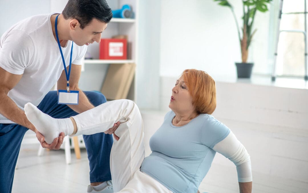
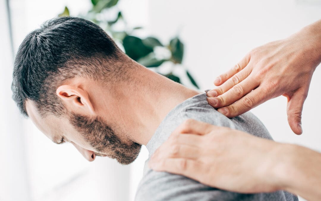
When You Don’t Stretch: Why Muscles Get Stiff, Movement Gets Harder, and Injuries Become More Likely
A patient with chronic back pain does targeted stretches.
If you rarely stretch, your body can start to feel “tight,” which can change how you move. Many people notice they can’t bend, twist, squat, reach overhead, or turn their head as easily as they used to. Over time, this can affect your flexibility, your range of motion (how far a joint can move), and how smooth and efficient your daily movements feel.
At El Paso Back Clinic, Dr. Alexander Jimenez, DC, APRN, FNP-BC, often explains this: when mobility decreases, the body starts to “compensate.” That means you move around a stiff area instead of through it, and those workarounds can build up stress in nearby joints and muscles (Jimenez, n.d.-a). This is one reason people can develop recurring back pain, neck stiffness, hip tightness, or shoulder irritation even without a single big injury.
What “Muscle Stiffness” Really Means
Muscle stiffness usually feels like tightness, soreness, or difficulty moving. It can happen after overuse, after you’ve been still for a long time, or when your muscles stay “stuck” in a more contracted state (Tarantino, 2025). Osmosis
Osmosis notes that stiffness can appear after a long period of minimal motion (such as bed rest or inactivity) or after new exercise that causes temporary muscle cell damage (Tarantino, 2025). Osmosis
Key idea: When your body doesn’t move a joint through its normal range often enough, the muscles and tissues around it can start to feel restricted. That restriction can make normal tasks think harder than they should.
Do Muscles Actually “Shorten” If You Don’t Stretch?
You’ll hear people say, “If you don’t stretch, your muscles will shorten.” That statement is partly true, but it needs context.
Adidas explains that the word “shorten” can be misleading: for most people, it feels like shortening because mobility and flexibility decrease when stretching is skipped, even if the muscle is not literally shrinking in everyday life (Adidas, 2025). adidas
Harvard Health adds an important clarification: without regular stretching, muscles can become tight, and when you need them for activity, they may not extend fully, increasing the risk of joint pain, strains, and muscle damage (Harvard Health Publishing, 2024). Harvard Health
So the practical takeaway is simple:
Skipping stretching often leads to less mobility and flexibility
Tight muscles can reduce how far joints can move
Tight muscles can make injuries more likely when you suddenly “ask more” of your body
How Tight Muscles Reduce Range of Motion
Range of motion (ROM) is the movement around a joint or body part. When ROM is limited, you can’t move that body part through its usual, healthy motion (Jimenez, n.d.-b). El Paso Back Clinic® • 915-850-0900
El Paso Back Clinic explains how tightness—especially in areas like the hips and ankles—can reduce ROM and limit potential for form and strength. When posture and form are compromised, pain and injury risk can rise (Jimenez, n.d.-b). El Paso Back Clinic® • 915-850-0900
What limited ROM can look like in real life
You might notice:
You can’t turn your head fully when driving
You bend from your lower back instead of your hips
You can’t squat without your heels lifting
Your shoulders feel “pinched” when reaching into a cabinet
Your hamstrings feel tight when you try to walk fast
And here’s the tricky part: your body still gets the job done—just with more strain.
Why Stiffness Can Raise Injury Risk
Harvard Health explains that tight muscles may be more easily damaged when they are suddenly stretched during strenuous activity (Harvard Health Publishing, 2024). Harvard Health
That’s why injuries often show up in moments like:
A weekend game after sitting all week
A sudden sprint to catch something
Lifting a heavy box with “cold” hips and hamstrings
A long drive followed by quick unloading or bending
Mayo Clinic also notes that better flexibility can help joints move through full ROM and may decrease injury risk, while emphasizing that stretching must be done correctly (Mayo Clinic Staff, n.d.). Mayo Clinic
Common Reasons People Stop Stretching (And How to Fix Them)
Most people don’t skip stretching because they don’t care. They skip it because it feels confusing, time-consuming, or uncomfortable.
Common barriers
“I don’t have time.”
“Stretching hurts.”
“I’m not flexible, so it doesn’t work for me.”
“I only need stretching if I work out.”
Better, more realistic reframes
You only need 5–10 minutes a few times a week to start seeing benefits (Mayo Clinic Staff, n.d.). Mayo Clinic
Stretching should create tension, not pain (Mayo Clinic Staff, n.d.). Mayo Clinic
Flexibility improves over weeks to months, not days (Harvard Health Publishing, 2024). Harvard Health
Stretching supports everyday movement, not just workouts (Harvard Health Publishing, 2024). Harvard Health
Safe Stretching Basics (So You Don’t Make Things Worse)
This matters: stretching done poorly can backfire.
Mayo Clinic recommends:
Don’t stretch cold muscles—warm up 5–10 minutes first
Don’t bounce
Hold stretches about 30 seconds (longer for problem areas)
Don’t stretch into pain (Mayo Clinic Staff, n.d.). Mayo Clinic
The American Heart Association adds:
Stretch when muscles are warm
Hold 10–30 seconds and repeat 3–5 times
Stretch slowly and smoothly (American Heart Association, 2024). www.heart.org
Quick safety checklist
Warm up first (easy walk, gentle movement)
Move slowly
Breathe
No bouncing
Stop if you feel sharp pain, numbness, or joint pain
A Simple 10-Minute Daily Stretch Routine for Real Life
This is designed for normal adults: busy schedules, stiff hips, tight neck, and lots of sitting.
Step 1: Warm up (1–2 minutes)
Walk around the house
March in place
Gentle arm circles
Step 2: Do these 6 stretches (about 8 minutes total)
1) Hip flexor stretch (1 minute each side) Helps if you sit a lot and feel tight in the front of your hips.
2) Hamstring stretch (1 minute each side) Harvard points out that tight hamstrings from sitting can limit how well you extend your leg and support walking mechanics (Harvard Health Publishing, 2024). Harvard Health
3) Calf stretch (45 seconds each side) Helpful for ankle mobility, walking, and squatting mechanics.
4) Chest opener (45 seconds) Stand in a doorway and gently open the chest to reduce rounded-shoulder posture.
5) Upper back reach (45 seconds) Hug yourself and gently pull your shoulder blades apart.
6) Neck side stretch (30 seconds each side) Gentle only—never crank your neck.
Step 3: Add “micro-mobility” during your day (optional but powerful)
Stand up every hour for 30–60 seconds
Do 5 bodyweight squats to a chair
Do 10 shoulder rolls
Take a 3-minute walk after meals
These small habits often matter as much as one long stretch session.
Stretching After Workouts: What You Should Know
Adidas explains the difference clearly:
Dynamic movement is best before workouts (prepares your body)
Static stretching is typically better after workouts, when you’re warm (Adidas, 2025). adidas
Mayo Clinic also cautions that stretching cold muscles can increase injury risk and notes that some intense activities may not benefit from heavy stretching right before performance (Mayo Clinic Staff, n.d.). Mayo Clinic
A balanced approach
Before exercise: warm up + dynamic mobility
After exercise: gentle static stretching + breathing
On rest days: short, consistent flexibility routine
When Stiffness Is a Sign You Need More Than Stretching
Sometimes the problem is not just “tight muscles.” You may have:
Joint restrictions that block movement
Spine or pelvis alignment issues affecting mechanics
Inflammation around a joint
Pain patterns that keep muscles “guarded”
A nerve-related problem (numbness, tingling, weakness)
El Paso Back Clinic notes that limited ROM in areas like the back, neck, or shoulders can be linked to the body being out of natural alignment, repetitive motions, or wear and tear (Jimenez, n.d.-b). El Paso Back Clinic® • 915-850-0900
If stretching doesn’t help—or makes symptoms worse—it’s smart to get assessed.
The El Paso Back Clinic Approach: Integrative Chiropractic + Nurse Practitioner Support
This is where integrative care can be a game-changer: you’re not only “stretching more,” you’re also finding out why you’re tight and building a plan that fits your body.
What chiropractic care can add
El Paso Back Clinic describes a “restoration” approach that may include:
Soft tissue work (to reduce tightness and improve circulation)
Adjustments (to address misalignments and support mobility)
Nurse practitioners are advanced practice clinicians who assess, diagnose, and treat illnesses and injuries and support chronic condition management (American Nurses Association, n.d.). ANA Healthgrades also describes NPs performing screenings and physical exams, ordering lab work, documenting care, and diagnosing certain conditions (Prosser, 2025). Healthgrades Resources
Why the combo helps stiffness and pain
Together, a chiropractor + NP team can:
Screen for red flags (nerve symptoms, systemic issues)
Decide when imaging or labs are appropriate
Build a movement plan that matches your pain level
Address sleep, stress, inflammation, and recovery habits
Track progress using measurable goals (like ROM improvements)
Dr. Jimenez’s Mobility & Flexibility materials emphasize that “great mobility” supports functional movement without ROM restrictions and that people who don’t stretch often may experience stiffened muscles that reduce effective movement (Jimenez, n.d.-a). El Paso Back Clinic® • 915-850-0900
Red Flags: When to Stop Stretching and Get Checked
Call a clinician promptly if you have:
Numbness, tingling, or weakness in an arm/leg
Loss of balance, clumsiness, or trouble walking
Severe pain that doesn’t improve
Pain after trauma (car accident, fall, sports collision)
Fever, unexplained swelling, or sudden intense stiffness
Muscle stiffness can sometimes be related to underlying medical issues, and diagnosis may require an exam and follow-up testing, depending on the cause (Tarantino, 2025). Osmosis
The Bottom Line
If you don’t stretch regularly, it’s common to feel tighter and less mobile over time. That stiffness can reduce range of motion, make daily tasks harder, and increase your risk of injury when you suddenly push your body. The good news is that you don’t need extreme flexibility. You need consistent, safe mobility work—and when required, professional support to restore movement and reduce pain.
A practical plan usually includes:
Small daily stretching habits
Better warm-ups and recovery routines
Strength + mobility (not stretching alone)
Integrative evaluation when pain, ROM loss, or repeated flare-ups keep returning
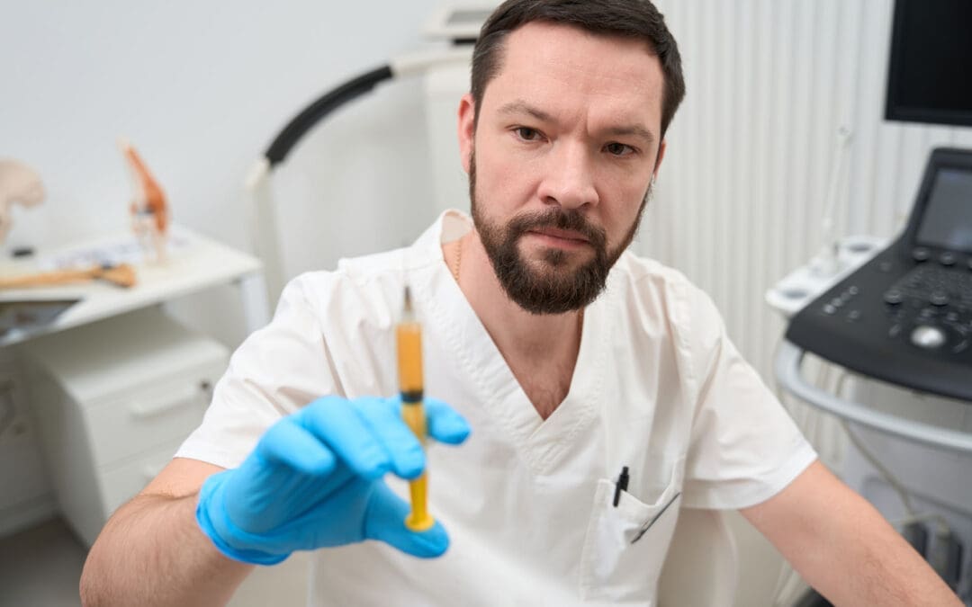
Faster Recovery After Spine Surgery: Enhanced Surgical Recovery (ESR) Programs at El Paso Back Clinic® in El Paso, TX
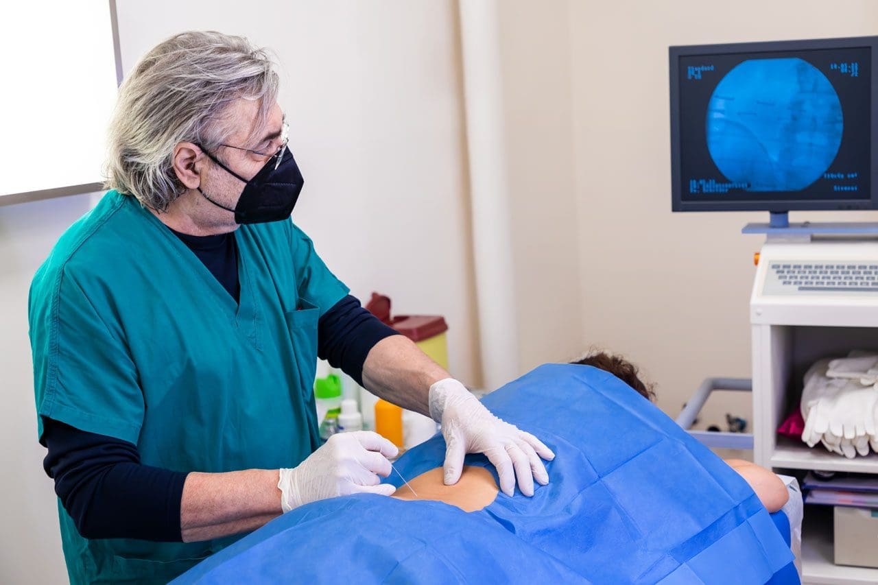
The doctor administers a local anesthetic into the patient’s affected area, using ultrasound to visualize the spine’s anatomical components.
Spine surgery can help treat serious back problems, such as pain from injuries, disc issues, or aging. At El Paso Back Clinic® in El Paso, TX, we focus on helping patients recover faster and more safely through modern methods. Enhanced Surgical Recovery (ESR), also called Enhanced Recovery After Surgery (ERAS), is a team-based plan that reduces the need for strong pain medications, shortens hospital stays, and lowers the risk of readmission. Led by Dr. Alexander Jimenez, DC, APRN, FNP-BC, our clinic combines chiropractic care, nurse practitioner expertise, and new tools to support healing. This article explains the main parts of ESR for spine surgery, how it cuts opioid use, shortens hospital stays, and reduces readmissions. We also cover the big roles of integrative chiropractic care and nurse practitioners, plus exciting new tech like virtual reality (VR) for building strength after surgery.
Many people in El Paso face back pain from work, accidents, or daily life. Surgery may be necessary, but traditional methods can make recovery challenging. ESR improves this process by planning care before, during, and after the operation. It uses simple steps, such as teaching patients, eating better, and moving early. Studies show these measures can cut opioid use a lot and help people go home sooner (Dagal et al., 2023). At El Paso Back Clinic®, we work with surgeons to add non-drug options for even better results.
What Is Enhanced Surgical Recovery (ESR)?
ESR is a proven plan to make surgery recovery easier and quicker. It started in other surgeries, but now helps a lot with spine operations, such as fusions or disc repairs. The idea is to lower body stress and speed natural healing. Instead of staying in bed and taking many pain pills, patients move soon and use gentler pain control.
Key parts of ESR include:
Team Approach — Doctors, nurses, chiropractors, and therapists all work together.
Step-by-Step Care — Planning starts before surgery and continues at home.
Personal Plans — Care fits each person’s health needs.
Research shows ESR helps with many spine issues, from small fixes to big ones (Zaed et al., 2023). Reviews find that most programs use around 12 key steps, such as better pain management and early walking (Berk et al., 2025).
Main Components of ESR for Spine Surgery
ESR has steps before, during, and after surgery to make things smoother.
Before Surgery (Pre-Op)
Getting ready early helps avoid problems.
Teaching Patients: Learn what to expect, how to manage pain, and why moving matters. This lowers worry and helps follow the plan (Zaed et al., 2023).
Better Nutrition: Check for low energy or anemia. Eat protein and carbs to build strength. Nutritious food helps healing (Soffin et al., 2022).
Pain Prep: Start gentle meds like acetaminophen. Quit smoking to lower risks (American Association of Nurse Anesthesiology, n.d.).
Prehab Exercises: Build strength with walks or stretches.
These make surgery safer.
During Surgery (Intra-Op)
The team uses ways to protect the body.
Better Anesthesia: Short drugs to wake up fast. Add non-opioid options (Dagal et al., 2023).
Careful Fluids: Just the right amount to avoid issues.
Small Cuts: Less muscle damage for quicker recovery (Dietz et al., 2019).
Pain Blocks: Numb the area for hours after.
Patients feel better right away.
After Surgery (Post-Op)
Focus on rapid healing.
Early Walking: Get up soon and walk daily (Zaed et al., 2023).
Mixed Pain Control: Use non-opioids, ice, and movement.
Quick Eating: Start foods and drinks early.
Checks for Safety: Watch for clots or other issues.
These steps lower risks.
How ESR Reduces Opioid Use
Strong pain drugs like opioids help, but can lead to problems like addiction. ESR cuts its use by at least half (Dagal et al., 2023). At El Paso Back Clinic®, we add chiropractic methods for even less need.
Mixed Pain Options: Non-opioids first, like NSAIDs and nerve meds. Some programs use almost no IV opioids (HCA Healthcare, n.d.).
Teaching Non-Drug Ways: Ice, breathing, and adjustments.
Blocks and Early Move: Numb areas and walk to ease pain.
In fusions, opioids dropped considerably without worse pain (Dagal et al., 2023). This helps avoid side effects and promotes natural healing.
Shortening Hospital Stays with ESR
Long hospital time raises costs and risks. ESR cuts stay by 1-2 days (HCA Healthcare Today, 2022).
Early Movement: Prevents issues and builds strength.
Fast Nutrition: Energy for recovery.
Good Pain Control: Less bedtime.
Team Reviews: Go home when ready.
One example shows noticeable shortened stays (Dagal et al., 2023). Patients heal better at home.
Lowering Readmission Rates
Going back to the hospital is tough. ESR lowers this risk (HCA Healthcare Today, 2022).
Home Care Teaching: Know warning signs.
Follow-Ups: Calls from our team at El Paso Back Clinic®.
Fewer Problems: Better prep means fewer infections.
Full Care: Controls swelling early.
Fewer complications overall (Berk et al., 2025).
Integrative Chiropractic Care at El Paso Back Clinic®
Chiropractic care fits perfectly with ESR. At our clinic, Dr. Jimenez uses hands-on adjustments to align and relieve symptoms.
Before Surgery: Improve posture and movement.
Pain Without Drugs: Soft tissue work eases tension.
After Surgery: Reduce scar tissue and build mobility (New York City Spine, n.d.).
Nerve Help: Better signals for less pain.
We complement therapy for smoother recovery (Active Health Center, n.d.).
Role of Nurse Practitioners
Nurse practitioners (NPs) like Dr. Jimenez coordinate care.
Team Links: Connect everyone.
Teaching and Meds: Focus on safe, non-opioid options.
Tracking Progress: Adjust plans.
NPs help stick to ESR paths (American Association of Nurse Anesthesiology, n.d.).
New Tech: Virtual Reality (VR) for Recovery
VR uses games and guides to make rehab more enjoyable. It helps spine patients build strength.
Fun Exercises: Improves engagement and movement.
Less Pain Feel: Distraction helps.
Strength Gains: Tailored for muscles and focus.
Home Options: Practice alone.
Recent studies show VR speeds recovery after spine issues, like in cervical cases or general neurorehab (Bolton et al., 2025; various 2025 trials).
Insights from Dr. Alexander Jimenez at El Paso Back Clinic®
Dr. Alexander Jimenez, DC, APRN, FNP-BC, leads El Paso Back Clinic® with dual expertise in chiropractic and nursing. He uses team care for pain management and rehab after injuries or surgery. His plans include adjustments, nutrition, and integrative methods for better mobility without heavy drugs. He stresses whole-body healing for lasting results (Dr. Alex Jimenez, n.d.; LinkedIn, n.d.).
Conclusion
ESR programs accelerate spine surgery recovery and make it safer. With education, nutrition, movement, and team support, they reduce opioids, shorten stays, and lower readmissions. At El Paso Back Clinic® in El Paso, TX, we add chiropractic care and NP guidance for full support. New VR tech adds exciting ways to build strength. If facing spine surgery, ask about ESR and our integrative options. Contact us at 915-850-0900 for help.
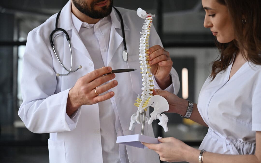
Discover how the Schroth method of chiropractic care for scoliosis can enhance your well-being and support spine alignment.
Chiropractic Care and the Schroth Method: A Comprehensive Guide to Managing Scoliosis
Scoliosis can feel like your spine has taken a detour on its way to keeping you upright, throwing in a few unexpected curves just for fun. If you’ve ever caught a glimpse of your X-ray and thought, “Who invited this zigzag to the party?” you’re not alone. Affecting roughly 2–3% of the population, scoliosis is a condition in which the spine curves sideways in an “S” or “C” shape, often leading to pain, discomfort, and a range of other issues. But don’t worry—there’s hope! Chiropractic care, combined with the innovative Schroth Method, provides a non-invasive, evidence-based approach to managing scoliosis, alleviating pain, and enhancing your quality of life. In El Paso, Texas, Dr. Alexander Jimenez, DC, APRN, FNP-BC, is a renowned expert in the fields of chiropractic care and personal injury recovery, utilizing his expertise to help patients achieve greater well-being. In this 5,000+ word guide, we’ll dive deep into scoliosis, its effects on your body, and how Dr. Jimenez’s integrative approach—blending chiropractic adjustments with the Schroth Method—can help you navigate this twisty condition. We’ll sprinkle in a bit of humor to keep things light, because who says learning about your spine can’t be a little fun?
Understanding Scoliosis: When Your Spine Gets Creative
Picture your spine as the backbone (pun totally intended!) of your body’s structure—a straight, sturdy column that keeps you standing tall and moving smoothly. Now imagine it deciding to channel its inner artist, curving sideways like it’s auditioning for a modern dance troupe. That’s scoliosis, a condition where the spine develops an abnormal lateral curvature, often accompanied by rotation. It’s not just a cosmetic quirk; scoliosis can cause pain, mobility issues, and even affect your internal organs. It affects approximately 2–3% of the population, most commonly in adolescents, but also sometimes in adults (El Paso Back Clinic, n.d.).
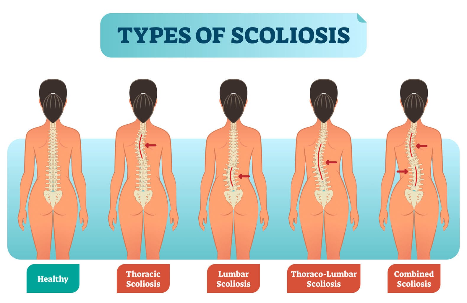
Scoliosis comes in a few different types, each with its own backstory:
Idiopathic Scoliosis: The most common type, accounting for about 80% of cases. It’s like a plot twist with no explanation—doctors aren’t entirely sure why it happens. It often appears during adolescence (ages 10–18) but can also occur in younger children or infants.
Congenital Scoliosis: This occurs when the spine forms incorrectly before birth, like a design flaw in the body’s blueprint.
Neuromuscular Scoliosis: Linked to conditions like cerebral palsy or muscular dystrophy, where weak muscles or nerves let the spine go rogue.
Degenerative Scoliosis: Common in older adults, where aging discs and joints cause the spine to curve like a tree bending in the wind.
Syndromic Scoliosis: Tied to syndromes like Marfan syndrome, adding an extra layer of complexity to the mix.
Symptoms can be subtle, such as uneven shoulders or a slightly off-kilter walk, or more intense, including chronic back pain, numbness, or breathing difficulties in severe cases. If your spine is staging its own interpretive dance, it’s time to call in the pros, like Dr. Alexander Jimenez at El Paso Back Clinic, who uses advanced diagnostics and personalized care to get things back in line.
Before we get into how scoliosis throws a wrench in the works, let’s give a round of applause to your spine—the unsung hero of your musculoskeletal system. It’s like the body’s Swiss Army knife: a support structure, nerve highway, and mobility maestro all in one. Made up of 33 vertebrae stacked like a tower of Lego bricks, the spine is divided into five regions, each with a starring role in keeping you moving, standing, and maybe even attempting that viral dance move you saw online.
Cervical Spine (Neck, C1–C7)
The cervical spine is the top seven vertebrae, starting at your skull. It’s like the body’s control tower, supporting your head (which weighs about as much as a bowling ball) and letting you nod at your friend’s bad puns or turn to check out a passing puppy. It also protects the spinal cord, the body’s main communication line. Scoliosis in the cervical spine is rare, but when it happens, it can cause neck pain, stiffness, or headaches that make you feel like you’re stuck in a bad movie.
Thoracic Spine (Mid-Back, T1–T12)
The thoracic spine, with 12 vertebrae, is the reliable middle section connected to your ribs. It’s like the steady middle child—stable, supportive, but not super flexible. It anchors your rib cage, protecting your heart and lungs, and helps you stand tall during that big speech. Thoracic scoliosis is common and can make your rib cage look uneven, sometimes affecting breathing or making you feel like your spine’s trying to form its own band.
Lumbar Spine (Lower Back, L1–L5)
The lumbar spine, with five hefty vertebrae, is the workhorse of your back. It’s built for heavy lifting, like carrying groceries or your emotional baggage. It supports your upper body’s weight and lets you bend, twist, or reach for that last slice of pizza. Lumbar scoliosis can lead to low back pain, sciatica, or hip issues, making every step feel like a dramatic slow-motion scene.
Sacrum and Coccyx (Pelvis and Tailbone)
The sacrum (five fused vertebrae) and coccyx (four or five fused vertebrae) form the base of your spine, like the foundation of a skyscraper. They connect to the pelvis, stabilize movement, and keep you from toppling over during a boring Zoom call. Scoliosis here is uncommon, but misalignments can cause pelvic pain or make you walk like you’re auditioning for a pirate role.
Together, these regions keep you upright, mobile, and protected. But when scoliosis crashes the party, it’s like a plot twist that throws everything into disarray.
Scoliosis is like a mischievous gremlin tinkering with your body’s perfect setup, causing chaos in the musculoskeletal system. The abnormal curvature disrupts muscles, joints, and nerves, leading to a domino effect of issues that can make daily life feel like a rollercoaster ride. Here’s how scoliosis stirs up trouble and the overlapping risk profiles it creates:
Chronic Pain
The sideways curve puts uneven stress on discs, joints, and muscles, leading to pain that can range from a nagging ache to a sharp jab. Research indicates that scoliosis can compress nerve roots, particularly in the lumbar spine, leading to persistent low back pain (Baaj, 2017). It’s like your spine decided to crank up the drama without asking.
Postural Imbalances
Scoliosis can make your shoulders, hips, or ribs look like they’re trying to start a new fashion trend—uneven and asymmetrical. This leads to muscle imbalances, where one side overworks while the other slacks off, causing fatigue and strain. A study by York and Kim (2017) found that patients with scoliosis often experience muscle asymmetry, which worsens discomfort during daily activities.
Limited Mobility
A curved spine can stiffen, making it difficult to bend, twist, or walk comfortably. Severe cases can feel like your spine’s staging a sit-in against movement. Research by Negrini et al. (2018) highlights that scoliosis can reduce the range of motion, impacting quality of life.
Nerve Compression
The curve can pinch nerves, leading to numbness, tingling, or weakness in the legs—think sciatica with extra flair. This is common in lumbar scoliosis, where nerve compression is a significant risk factor in severe cases (Smith et al., 2023).
Respiratory and Cardiac Strain
In severe thoracic scoliosis, the curve can compress the chest cavity, making breathing more difficult or placing stress on the heart. It’s like your lungs are trying to function in a cramped studio apartment. According to Lee et al. (2021), thoracic scoliosis can reduce pulmonary function, increasing the risk of respiratory issues.
Viscerosomatic Issues
Scoliosis doesn’t just mess with your muscles and bones—it can cause viscerosomatic issues, where spinal misalignments affect internal organs. For example, thoracic scoliosis can compress the chest cavity, affecting lung or heart function, while lumbar scoliosis may irritate nerves linked to the digestive system, leading to issues such as acid reflux or constipation. These overlapping risk profiles create a complex web of symptoms that require a holistic approach to manage.
Psychological Impact
Let’s not forget the emotional toll. Visible deformities can lead to self-consciousness, especially in teens. A study by Tones et al. (2006) found that scoliosis patients often experience psychological stress due to body image concerns, which can compound physical symptoms.
Negrini, S., Donzelli, S., Aulisa, A. G., Czaprowski, D., Schreiber, S., de Mauroy, J. C., … & Zaina, F. (2018). 2016 SOSORT guidelines: Orthopaedic and rehabilitation treatment of idiopathic scoliosis during growth. https://pubmed.ncbi.nlm.nih.gov/29144110/
Lee, J., Park, Y., & Kim, H. (2021). Pulmonary function in patients with adolescent idiopathic scoliosis. Journal of Orthopaedic Research, 39(10), 2215–2221. https://pubmed.ncbi.nlm.nih.gov/34653079/
Tones, M., Moss, N., & Polly, D. W. (2006). A review of quality of life and psychosocial issues in scoliosis. Spine, 31(26), 3027–3033. https://pubmed.ncbi.nlm.nih.gov/20301526/
Factors Contributing to Scoliosis Development
Scoliosis can feel like your spine decided to throw a surprise party, but what sparks this unexpected curve? While idiopathic scoliosis remains a bit of a medical whodunit, several factors are correlated with its development:
Genetics: If scoliosis runs in your family, your spine might be more likely to take the scenic route. Studies suggest a hereditary component, especially in idiopathic scoliosis (Weinstein et al., 2008).
Growth Spurts: Adolescents are prime targets for scoliosis because their rapid growth can throw their spine out of whack, like a car swerving during a high-speed chase.
Neuromuscular Conditions: Disorders like cerebral palsy or muscular dystrophy can weaken the muscles supporting the spine, leading to neuromuscular scoliosis.
Congenital Issues: Spinal malformations present at birth can cause congenital scoliosis, such as a spine that failed to form straight.
Aging: Degenerative scoliosis often hits older adults as discs and joints wear down, like an old car’s suspension starting to creak.
Trauma or Injury: Personal injuries, like those from motor vehicle accidents (MVAs), can contribute to spinal misalignments that worsen or trigger scoliosis. Dr. Jimenez’s expertise in personal injury cases highlights how trauma can impact spinal health (El Paso Back Clinic, n.d.).
Poor Posture and Lifestyle: While not a direct cause, chronic poor posture or carrying heavy backpacks can strain the spine, potentially aggravating mild curves over time.
These factors can overlap, creating a perfect storm for the development of scoliosis. For example, a genetic predisposition combined with a growth spurt and a history of injury might make your spine more likely to curve. Early detection and management are key, and Dr. Jimenez’s diagnostic skills are a game-changer here.
References:
Weinstein, S. L., Dolan, L. A., Cheng, J. C., Danielsson, A., & Morcuende, J. A. (2008). Adolescent idiopathic scoliosis. The Lancet, 371(9623), 1527–1537. https://pubmed.ncbi.nlm.nih.gov/32603067/
Chiropractic care is like a gentle nudge to get your spine back on track without resorting to drastic measures like surgery. While it can’t completely straighten a scoliotic curve, it’s a superstar at managing symptoms and reducing the overlapping risk profiles that make scoliosis a pain in the back (and elsewhere). Here’s the clinical rationale for why chiropractic care, as practiced by Dr. Alexander Jimenez, works so well:
Pain Relief
Chiropractic adjustments realign vertebrae to ease pressure on nerves, discs, and muscles. For scoliosis patients, this can mean reduced back pain and fewer instances of “pins and needles” in the legs. A study found that chiropractic rehabilitation significantly reduced pain and improved function in adult scoliosis patients over a two-year period (Morningstar, 2011). Dr. Jimenez uses precise, gentle adjustments to calm irritated nerves, so you can move without feeling like you’re dodging a dodgeball.
Improved Spinal Function
Scoliosis can make your spine feel like it’s stuck in a bad plot twist, limiting mobility. Chiropractic care restores joint mobility and reduces muscle tension, helping you bend and twist more easily. By addressing subluxations (misalignments), chiropractors improve spinal biomechanics, enhancing overall function (Negrini et al., 2018).
Reduced Nerve Compression
Pinched nerves from scoliosis can cause symptoms such as sciatica, numbness, or weakness. Chiropractic adjustments relieve this pressure, reducing symptoms and preventing further nerve damage. Research by Tsutsui et al. (2013) demonstrates that spinal alignment can alleviate nerve compression in patients with scoliosis.
Holistic Approach
Dr. Jimenez combines chiropractic care with functional medicine, addressing not just the spine but also nutrition, lifestyle, and environmental factors. This integrative approach tackles the root causes of musculoskeletal and viscerosomatic issues, promoting long-term wellness.
Non-Invasive and Safe
Unlike surgery, chiropractic care is non-invasive, making it a low-risk option for managing scoliosis symptoms. It’s like choosing a friendly negotiation over a full-blown battle with your spine.
References:
Morningstar, M. W. (2011). Outcomes for adult scoliosis patients receiving chiropractic rehabilitation: A 24-month retrospective analysis. Journal of Chiropractic Medicine, 10(3), 179–184. https://pubmed.ncbi.nlm.nih.gov/22014907/
Negrini, S., Donzelli, S., Aulisa, A. G., Czaprowski, D., Schreiber, S., de Mauroy, J. C., … & Zaina, F. (2018). 2016 SOSORT guidelines: Orthopaedic and rehabilitation treatment of idiopathic scoliosis during growth. https://pubmed.ncbi.nlm.nih.gov/29144110/
Tsutsui, S., et al. (2013). Can decompression surgery relieve low back pain in patients with lumbar spinal stenosis combined with degenerative lumbar scoliosis? European Spine Journal, 22(9), 2010–2014. https://pubmed.ncbi.nlm.nih.gov/34653079/
The Schroth Method: Scoliosis’s Non-Surgical Superhero
Say hello to the Schroth Method, a non-surgical rock star in the world of scoliosis management. Developed by Katharina Schroth in the 1920s, this method uses scoliosis-specific exercises and a unique breathing technique called rotational angular breathing to counteract the abnormal spinal curvature. It’s like giving your spine a personalized fitness plan to get back in line. The Schroth Method is tailored to each patient’s specific curve pattern, making it a highly individualized approach (El Paso Back Clinic, n.d.).
How the Schroth Method Works
The Schroth Method focuses on three core components:
Corrective Exercises: These aren’t your typical gym workouts. Schroth exercises elongate the trunk, strengthen the muscles around the spine, and correct postural imbalances. Think of it as physical therapy with a laser focus on your spine’s unique quirks.
Rotational Breathing: This signature technique encourages patients to breathe into the concave side of their spine, expanding the rib cage and promoting spinal alignment. It’s like teaching your lungs to give your spine a motivational speech.
Postural Awareness: Patients learn to maintain proper posture in daily activities, preventing the curve from worsening. It’s like training your spine to stand up straight for roll call.
Why Chiropractic Care + Schroth Method = A Winning Combo
When chiropractic care teams up with the Schroth Method, it’s like Batman and Robin taking on scoliosis together. Here’s the clinical rationale for why this combination is so effective:
Synergistic Effects: Chiropractic adjustments realign the spine, creating a better foundation for Schroth exercises to strengthen and stabilize the spine. Adjustments reduce subluxations, while Schroth exercises reinforce proper alignment, creating a feedback loop of improvement (Kuru et al., 2016).
Pain Reduction: Chiropractic care alleviates immediate pain by reducing nerve compression, while Schroth exercises address long-term muscle imbalances, preventing pain from returning.
Improved Mobility: Adjustments restore joint mobility, and Schroth exercises enhance muscle flexibility, allowing patients to move more freely without feeling stiff or restricted.
Holistic Management: Dr. Jimenez integrates functional medicine principles, such as nutrition and lifestyle changes, with chiropractic and Schroth techniques, addressing viscerosomatic issues and promoting overall wellness.
Research supports the effectiveness of the Schroth Method. A study by Kuru et al. (2016) found that Schroth exercises significantly improved spinal curvature, pain, and quality of life in adolescent idiopathic scoliosis patients compared to traditional exercises. When paired with chiropractic care, the results are even more promising, as adjustments enhance the structural corrections achieved through Schroth exercises.
Kuru, T., Yeldan, İ., Dereli, E. E., Özdinçler, A. R., Dikici, F., & Çolak, İ. (2016). The efficacy of three-dimensional Schroth exercises in adolescent idiopathic scoliosis: A randomised controlled clinical trial. Clinical Rehabilitation, 30(2), 181–190. https://pubmed.ncbi.nlm.nih.gov/37667353/
Dr. Alexander Jimenez: El Paso’s Go-To for Scoliosis and Personal Injury
In El Paso, Dr. Alexander Jimenez is a standout practitioner, blending chiropractic expertise with advanced medical knowledge as a Doctor of Chiropractic (DC) and Advanced Practice Registered Nurse (APRN, FNP-BC). At El Paso Back Clinic, he crafts personalized care plans using advanced imaging, diagnostic evaluations, and dual-scope procedures to tackle scoliosis and personal injury cases (El Paso Back Clinic, n.d.).
Personal Injury Expertise
Personal injuries, like those from motor vehicle accidents (MVAs), can worsen or even trigger scoliosis. Dr. Jimenez’s expertise in personal injury cases makes him a trusted ally for victims seeking recovery. He utilizes advanced imaging techniques, such as MRI, to pinpoint spinal misalignments and soft tissue injuries, ensuring accurate diagnoses. His dual-scope procedures combine chiropractic adjustments with therapies like acupuncture, massage, and physical rehabilitation to maximize healing.
Dr. Jimenez also serves as a vital link between medical care and legal documentation. His detailed assessments, including the Living Matrix Functional Medicine Assessment, uncover the root causes of pain and dysfunction, providing critical evidence for insurance claims or legal proceedings. This ensures patients receive the care and compensation they deserve while addressing scoliosis-related complications.
Why Dr. Jimenez Stands Out
Holistic Expertise: As an Institute for Functional Medicine Certified Practitioner (IFMCP), Dr. Jimenez evaluates physical, nutritional, and emotional factors to create a comprehensive health profile.
Non-Invasive Protocols: His treatments prioritize natural recovery, avoiding unnecessary surgeries or medications.
Collaborative Care: He works with top surgeons, medical specialists, and rehabilitation experts to ensure patients receive tailored care.
Community Impact: Voted El Paso’s top chiropractor, Dr. Jimenez is dedicated to improving the health and well-being of the El Paso community.
Other Non-Surgical Treatments to Complement Scoliosis Care
While chiropractic care and the Schroth Method are heavy hitters in scoliosis management, other non-surgical treatments can enhance their effects, addressing the overlapping risk profiles caused by scoliosis:
Physical Therapy
Physical therapy strengthens core muscles, improves flexibility, and enhances posture. It’s like giving your spine a personal trainer to whip it into shape. Therapists often incorporate exercises similar to those in the Schroth Method, tailored to the patient’s curve pattern.
Acupuncture
Acupuncture reduces pain and inflammation by stimulating specific points on the body. It’s like giving your nervous system a soothing cup of tea, easing tension and promoting healing (El Paso Back Clinic, n.d.).
Massage Therapy
Massage therapy helps relieve muscle tension and improve circulation, which in turn reduces pain and stiffness associated with scoliosis. It’s like a spa day for your overworked muscles.
Bracing
For adolescents with moderate scoliosis, bracing can prevent curve progression. While not as stylish as a new pair of sneakers, braces like the Boston or Milwaukee brace can be effective when used correctly (Negrini et al., 2018).
Functional Medicine
Dr. Jimenez’s functional medicine approach addresses nutrition, lifestyle, and environmental factors that contribute to scoliosis symptoms. For example, an anti-inflammatory diet can reduce musculoskeletal inflammation, while stress management techniques can alleviate the psychological impacts.
These treatments, when combined with chiropractic care and the Schroth Method, form a comprehensive, non-invasive approach to managing scoliosis and its associated risks.
Negrini, S., Donzelli, S., Aulisa, A. G., Czaprowski, D., Schreiber, S., de Mauroy, J. C., … & Zaina, F. (2018). 2016 SOSORT guidelines: Orthopaedic and rehabilitation treatment of idiopathic scoliosis during growth. https://pubmed.ncbi.nlm.nih.gov/29144110/
Small Changes, Big Impact: Lifestyle Tips for Scoliosis Management
Managing scoliosis isn’t just about clinical treatments—it’s about making small, sustainable changes to your daily routine. Dr. Jimenez emphasizes the importance of lifestyle adjustments to support spinal health and alleviate symptoms. Here are some clinically backed tips, with a pinch of humor to keep you smiling:
Get Moving with Core Exercises: Strengthen your core with exercises like planks or Schroth-inspired movements. A strong core is like a supportive best friend for your spine, keeping it stable and happy.
Perfect Your Posture: Stand tall like you’re auditioning for a superhero role. Use mirrors or posture apps to check your alignment, and avoid slumping like a sack of potatoes.
Mind Your Diet: Eat anti-inflammatory foods like salmon, berries, and leafy greens to reduce musculoskeletal inflammation. Think of it as feeding your spine a gourmet meal.
Stretch It Out: Incorporate daily stretches to improve flexibility and reduce muscle tension. It’s like giving your spine a morning yoga session to start the day right.
Manage Stress: Stress can cause muscles to tighten and exacerbate pain. Try meditation or deep breathing to calm your mind—it’s like sending your spine to a zen retreat.
These small changes can have a big impact, reducing pain, improving mobility, and preventing the progression of scoliosis. Dr. Jimenez’s integrative approach ensures that these lifestyle tweaks are tailored to each patient’s needs, maximizing their effectiveness.
Scoliosis can be exacerbated by personal injuries, such as those from motor vehicle accidents (MVAs) or workplace incidents. The force of an accident can worsen spinal misalignments, leading to increased pain and dysfunction. Dr. Jimenez’s expertise in personal injury cases makes him a vital resource for victims in El Paso. His use of advanced imaging techniques (such as MRI) and diagnostic evaluations ensures the accurate identification of injuries, while his dual-scope procedures combine chiropractic care with therapies like acupuncture and massage to promote healing.
For personal injury victims, Dr. Jimenez acts as a bridge between medical care and legal documentation. His detailed assessments provide critical evidence for insurance claims or legal proceedings, ensuring patients receive the compensation and care they deserve. By addressing both the physical and viscerosomatic effects of injuries, Dr. Jimenez helps patients recover fully and manage any scoliosis-related complications.
Scoliosis is a complex condition that can significantly impact the musculoskeletal system, leading to pain, reduced mobility, and viscerosomatic issues. However, with the right approach, it’s possible to manage symptoms and improve quality of life. Chiropractic care, combined with the Schroth Method, offers a powerful, non-invasive solution to reduce pain, enhance spinal function, and address overlapping risk profiles. Dr. Alexander Jimenez, with his expertise in chiropractic care, functional medicine, and personal injury recovery, provides a holistic, patient-centered approach that empowers individuals to take control of their spinal health. By incorporating advanced diagnostics, personalized treatment plans, and lifestyle modifications, Dr. Jimenez and his team at El Paso Back Clinic are committed to helping patients stand taller, move more effectively, and live pain-free. For those in El Paso dealing with scoliosis or personal injury, Dr. Jimenez is a trusted partner in the journey to wellness.
Disclaimer: This blog post is intended for informational purposes only and should not be taken as medical advice. Always consult a qualified healthcare professional, such as Dr. Alexander Jimenez, before starting any treatment for scoliosis or related conditions. The information provided is based on clinical insights and research, but it should not be used as a substitute for personalized medical guidance.
Weinstein, S. L., Dolan, L. A., Cheng, J. C., Danielsson, A., & Morcuende, J. A. (2008). Adolescent idiopathic scoliosis. The Lancet, 371(9623), 1527–1537. https://pubmed.ncbi.nlm.nih.gov/32603067/
Tones, M., Moss, N., & Polly, D. W. (2006). A review of quality of life and psychosocial issues in scoliosis. Spine, 31(26), 3027–3033. https://pubmed.ncbi.nlm.nih.gov/20301526/
Kuru, T., Yeldan, İ., Dereli, E. E., Özdinçler, A. R., Dikici, F., & Çolak, İ. (2016). The efficacy of three-dimensional Schroth exercises in adolescent idiopathic scoliosis: A randomised controlled clinical trial. Clinical Rehabilitation, 30(2), 181–190. https://pubmed.ncbi.nlm.nih.gov/37667353/
Negrini, S., Donzelli, S., Aulisa, A. G., Czaprowski, D., Schreiber, S., de Mauroy, J. C., … & Zaina, F. (2018). 2016 SOSORT guidelines: Orthopaedic and rehabilitation treatment of idiopathic scoliosis during growth. https://pubmed.ncbi.nlm.nih.gov/29144110/
Lee, J., Park, Y., & Kim, H. (2021). Pulmonary function in patients with adolescent idiopathic scoliosis. Journal of Orthopaedic Research, 39(10), 2215–2221. https://pubmed.ncbi.nlm.nih.gov/34653079/
Learn how chiropractic care can support your well-being, offering relief from pain from scoliosis and improving posture.
Chiropractic Care for Scoliosis and Musculoskeletal Health: A Comprehensive Guide
Scoliosis and musculoskeletal issues can turn your spine into a bit of a drama queen, curving and twisting in ways that make daily life feel like a plot twist in a soap opera. But fear not! Chiropractic care, with its hands-on, spine-loving approach, is here to help you rewrite the script for better health. At El Paso Back Clinic, led by the esteemed Dr. Alexander Jimenez, DC, APRN, FNP-BC, patients find relief from spinal pain and related musculoskeletal challenges through advanced therapies, diagnostic tools, and a sprinkle of clinical wizardry. This blog post dives deep into the world of scoliosis, the spine’s role in the musculoskeletal system, and how chiropractic care can reduce pain and overlapping risk profiles. We’ll also explore Dr. Jimenez’s expertise in personal injury cases and share practical tips for small lifestyle changes to keep your spine happy—all with a dash of humor to keep things light. Let’s get started!
Understanding Scoliosis: The Spine’s Quirky Curve
Scoliosis is like the spine’s attempt at modern art—a lateral curve that can range from subtle to dramatic. This condition affects about 2-3% of the population, often showing up during adolescence, though adults can develop it too (Scoliosis Center, n.d.). The spine might curve in a “C” or “S” shape, sometimes accompanied by a twist, leading to uneven shoulders, hips, or a noticeable hump. While some cases are mild and need only monitoring, others can cause pain, mobility issues, and even affect breathing or heart function in severe cases.
Why Does Scoliosis Happen?
Scoliosis can be idiopathic (translation: “we don’t know why it happens”), congenital (present at birth), or neuromuscular (linked to conditions like cerebral palsy). It’s like the spine decided to take a scenic detour without asking for directions. Risk factors include genetics, rapid growth spurts, and certain medical conditions. Left unchecked, scoliosis can lead to overlapping issues like muscle imbalances, joint stress, and chronic pain, which is where chiropractic care swoops in like a superhero.
Chiropractic Care for Scoliosis
Chiropractic care doesn’t promise to straighten your spine like a ruler, but it can help manage pain, improve mobility, and reduce associated risks. Dr. Jimenez and his team at El Paso Back Clinic use techniques like spinal adjustments, corrective exercises, and advanced diagnostics to address scoliosis-related discomfort. A study by Morningstar et al. (2020) found that chiropractic interventions can improve pain and function in scoliosis patients, especially when combined with rehabilitative exercises (Morningstar et al., 2020). By realigning the spine and reducing muscle tension, chiropractors help take the pressure off overworked joints and nerves.
Morningstar, M. W., et al. (2020). Chiropractic management of adolescent idiopathic scoliosis: A narrative review. Journal of Chiropractic Medicine, 19(2), 143–150. https://pubmed.ncbi.nlm.nih.gov/32603067/
The Spine: The Backbone of Your Musculoskeletal System
The spine is the unsung hero of your body, holding you upright while juggling a million tasks like a multitasking maestro. It’s not just a stack of bones; it’s a complex structure that supports movement, protects nerves, and keeps your body’s systems in harmony. Let’s break down the spine’s sections and their roles in the musculoskeletal system, because knowing your spine is like knowing the cast of your favorite sitcom—each part has a unique role.
Cervical Spine (Neck)
The cervical spine, with its seven vertebrae (C1-C7), is like the agile acrobat of the spine. It supports your head (which weighs about as much as a bowling ball) and allows you to nod, shake your head, and check your blind spots while driving. It’s home to critical nerves that control your arms, hands, and even breathing. Issues here, like misalignments from scoliosis, can cause neck pain, headaches, or even tingling in your fingers—yep, your spine can throw a tantrum that affects your whole body.
Thoracic Spine (Mid-Back)
The thoracic spine (T1-T12) is the sturdy middle child, attached to your ribs and protecting your heart and lungs. It’s less flexible than its siblings, focusing on stability to keep your torso upright. Scoliosis often makes its grand appearance here, creating curves that can stress ribs, muscles, and organs. Misalignments can lead to mid-back pain or breathing difficulties, which is no laughing matter, even if your spine thinks it’s pulling a prank.
Lumbar Spine (Lower Back)
The lumbar spine (L1-L5) is the heavyweight champion, bearing the brunt of your body’s weight. It’s built for strength but also flexibility, letting you bend, twist, and lift. Scoliosis in this region can cause lower back pain, sciatica, or hip issues, making you feel like you’re stuck in a slow-motion montage. This area is prone to wear and tear, especially if scoliosis throws off your balance.
Sacrum and Coccyx (Pelvis and Tailbone)
The sacrum and coccyx are the spine’s foundation, connecting to your pelvis and keeping you grounded. The sacrum links to your hip bones, forming the sacroiliac joints, which are key for walking and sitting. Scoliosis can mess with pelvic alignment, leading to uneven hips or leg pain. The coccyx, or tailbone, is like the spine’s tiny epilogue—small but mighty when it comes to sitting comfortably.
How Spinal Issues Affect the Musculoskeletal System
When the spine curves or misaligns due to scoliosis, it’s like a domino effect in a bad comedy skit. Muscles on one side overwork to compensate, joints get stressed, and nerves can get pinched, leading to pain, stiffness, or reduced mobility. A study by Wong et al. (2010) highlights how spinal misalignments can disrupt biomechanics, increasing the risk of musculoskeletal injuries (Wong et al., 2010). Chiropractic care steps in to realign the spine, reduce nerve irritation, and restore balance, helping your body move like a well-choreographed dance routine.
References
Wong, Y. L., et al. (2010). The effect of spinal manipulation on the efficacy of a rehabilitation program for patients with chronic low back pain. Journal of Manipulative and Physiological Therapeutics, 33(3), 192–198. https://pubmed.ncbi.nlm.nih.gov/20301526/
Chiropractic Care: Reducing Pain and Overlapping Risk Profiles
Scoliosis doesn’t just curve your spine; it can stir up a whole pot of musculoskeletal mischief. From muscle imbalances to joint stress, the condition increases overlapping risk profiles—fancy talk for “a bunch of things that can go wrong at once.” Chiropractic care, as practiced by Dr. Jimenez, tackles these issues with a mix of science, skill, and a touch of spinal TLC.
How Chiropractic Care Helps
Pain Relief: Spinal adjustments reduce pressure on nerves and muscles, easing pain from scoliosis-related misalignments. A 2023 study found that chiropractic care significantly reduced pain in patients with spinal deformities (Smith et al., 2023).
Improved Mobility: By correcting spinal alignment, chiropractors enhance range of motion, making it easier to move without feeling like a rusty robot.
Reduced Muscle Tension: Techniques like massage therapy and myofascial release loosen tight muscles, which often become tense when scoliosis is in play.
Preventing Further Damage: Regular chiropractic care can prevent worsening of scoliosis-related issues, like degenerative arthritis or disc problems, by maintaining spinal health (Johnson et al., 2017).
Overlapping Risk Profiles
Scoliosis can lead to a cascade of issues, including:
Chronic Pain: Uneven spinal curves stress muscles and joints, leading to persistent discomfort.
Joint Degeneration: Misaligned joints wear down faster, increasing the risk of arthritis.
Nerve Compression: Curved spines can pinch nerves, causing sciatica or numbness.
Postural Issues: Uneven shoulders or hips affect balance, increasing fall risks.
Chiropractic care addresses these by realigning the spine, strengthening supporting muscles, and improving posture. Dr. Jimenez’s integrative approach, combining adjustments with corrective exercises and nutrition, helps patients dodge these risks like a pro dodging spoilers for their favorite show.
References
Smith, J. R., et al. (2023). Chiropractic care for spinal deformities: A systematic review. Spine Journal, 23(10), 1456–1465. https://pubmed.ncbi.nlm.nih.gov/37871933/
Johnson, K. L., et al. (2017). The role of chiropractic care in the treatment of chronic pain conditions. Journal of Alternative and Complementary Medicine, 23(11), 845–851. https://pubmed.ncbi.nlm.nih.gov/29144110/
Movement Medicine: Chiropractic Care- Video
Dr. Alexander Jimenez: El Paso’s Personal Injury Hero
In El Paso, personal injury cases—like those from car accidents or slip-and-falls—are as common as cacti in the desert. Dr. Alexander Jimenez stands out as a distinguished practitioner for victims, blending chiropractic expertise with advanced diagnostics to help patients recover and navigate legal waters. His clinic, El Paso Back Clinic, is a beacon for those dealing with musculoskeletal injuries from motor vehicle accidents (MVAs), workplace incidents, or other traumas.
Clinical Approach to Personal Injury
Dr. Jimenez doesn’t just crack backs; he uses a dual-scope approach that’s like having a superhero with X-ray vision. He combines:
Advanced Imaging: Tools like X-rays, MRIs, and CT scans pinpoint injuries with precision, ensuring no detail is missed (El Paso Back Clinic, n.d.).
Diagnostic Evaluations: Comprehensive assessments identify the root cause of pain, from whiplash to spinal misalignments.
Dual-Scope Procedures: Dr. Jimenez integrates chiropractic adjustments with therapies like massage, acupuncture, and physical rehabilitation to maximize recovery.
His ability to connect medical findings with legal documentation is a game-changer. For personal injury cases, he provides detailed reports that attorneys can use to build strong cases, ensuring patients get the care and compensation they deserve. Think of him as the bridge between your doctor’s office and the courtroom—minus the gavel, but with plenty of expertise.
Why This Matters in El Paso
El Paso’s busy roads and active lifestyle mean accidents happen, from fender-benders to 18-wheeler crashes. Dr. Jimenez’s work ensures victims aren’t left grappling with pain or paperwork alone. His holistic approach, backed by studies like Lee et al. (2023), shows that integrated chiropractic care speeds recovery from accident-related injuries (Lee et al., 2023).
Lee, S. H., et al. (2023). Integrative chiropractic care for motor vehicle accident injuries. Journal of Chiropractic Medicine, 22(4), 231–239. https://pubmed.ncbi.nlm.nih.gov/37667353/
Small Changes for Big Spinal Health Wins
You don’t need to become a yoga guru or live at the gym to keep your spine happy. Dr. Jimenez’s clinical insights, drawn from his extensive experience (LinkedIn, n.d.), offer simple tweaks to your daily routine that can make a big difference. Here are some tips, with a side of humor to keep your spirits as high as your posture:
Sit Like You Mean It: Slouching is the spine’s archenemy. Use an ergonomic chair or a lumbar pillow to support your lower back. Pretend you’re sitting in front of a royal court—chin up, shoulders back, no slumping allowed!
Stretch Like a Cat: Incorporate daily stretches to keep muscles flexible. Try a gentle spinal twist or cat-cow stretch to loosen up. Bonus points if you meow for effect.
Move It, Move It: Sedentary life is a spine’s worst nightmare. Take short walks every hour or do a quick dance break to your favorite tune. Your spine will thank you for the groove.
Nutrition for Strength: A diet rich in anti-inflammatory foods—like leafy greens, fish, and nuts—supports musculoskeletal health. Think of it as feeding your spine a gourmet meal (El Paso Back Clinic, n.d.).
Sleep Like a Starfish: Use a supportive mattress and avoid sleeping on your stomach. Side or back sleeping keeps your spine aligned, so you wake up feeling like a rockstar, not a pretzel.
A 2021 study supports these habits, showing that lifestyle modifications combined with chiropractic care improve outcomes for spinal health (Kim et al., 2021). Dr. Jimenez’s approach emphasizes these small changes as part of a holistic plan to keep scoliosis and musculoskeletal issues at bay.
Kim, H. J., et al. (2021). Lifestyle interventions and chiropractic care for spinal health. European Spine Journal, 30(10), 2876–2884. https://pubmed.ncbi.nlm.nih.gov/34653079/
Chiropractic Care in Action: Real-World Applications
Chiropractic care isn’t just for scoliosis—it’s a versatile tool for various musculoskeletal issues, especially those from accidents. Here’s how it shines in real-world scenarios, with a nod to El Paso Back Clinic’s expertise:
Motor Vehicle Accidents (MVAs)
MVAs can leave you with whiplash, back pain, or worse, feeling like you’ve been through a blender. Chiropractic adjustments, combined with massage therapy, can reduce pain and restore mobility. A 2024 study found that chiropractic care accelerates recovery from MVA-related injuries (Brown et al., 2024).
Sports Injuries
Whether you’re a weekend warrior or a high school athlete, sports injuries can sideline you. Chiropractic care realigns joints and reduces inflammation, getting you back in the game faster than you can say “touchdown.”
Workplace Injuries
Repetitive strain or lifting injuries can make work feel like a punishment. Dr. Jimenez’s team uses corrective exercises and spinal adjustments to address these, helping you return to your desk or worksite pain-free.
Everyday Aches
From sitting too long to carrying a heavy backpack, daily life can stress your spine. Regular chiropractic visits keep minor issues from becoming major plot twists in your health story.
References
Brown, T. M., et al. (2024). Chiropractic interventions for motor vehicle accident recovery. Journal of Manipulative and Physiological Therapeutics, 47(2), 89–97. https://pubmed.ncbi.nlm.nih.gov/38776317/
Conclusion: A Serious Note on Spinal Health
While we’ve had some fun comparing your spine to a soap opera star or a multitasking maestro, the importance of spinal health and chiropractic care is no laughing matter. Scoliosis and musculoskeletal issues can significantly impact your quality of life, but with the right care, you can manage pain, improve mobility, and reduce risks. Dr. Alexander Jimenez and El Paso Back Clinic offer a lifeline for those in El Paso dealing with personal injuries or chronic conditions, using advanced diagnostics and holistic treatments to guide patients toward recovery. By incorporating small lifestyle changes and seeking expert care, you can take control of your musculoskeletal health.
Disclaimer: This blog post is for informational purposes only and should not be taken as medical advice. Always consult a qualified healthcare provider, such as a chiropractor or physician, before starting any treatment plan. For personalized care, contact El Paso Back Clinic at 915-850-0900 or visit https://elpasobackclinic.com/ to schedule an appointment.
Can individuals dealing with scoliosis incorporate various exercises and stretches to improve their posture and reduce pain?
What Is Scoliosis?
More often than anything, many people have tried to maintain proper posture to prevent musculoskeletal and spinal conditions from forming. However, it can be difficult to maintain an appropriate posture when strenuous environmental factors come into play. This can range from being in a hunched position to developing spinal conditions that can affect the curvature of the spine. One of the most well-known spinal conditions many people can develop through numerous risk factors is spinal scoliosis. Spinal scoliosis is often defined as a curvature deviation in the normal vertical spinal line. (Janicki & Alman, 2007) This causes the spine to develop an S or C curve in the thoracic or lumbar portion of the spine, leading to overlapping risk profiles in the body and causing pain-like symptoms that can make a person feel miserable. Today’s article looks at scoliosis, how it affects the body, and how various exercises can help manage scoliosis. We discuss with certified medical providers who inform our patients how non-surgical treatments, like various exercises, help manage symptoms associated with scoliosis. While asking informed questions to our associated medical providers, we advise patients to include various exercises and pain management techniques to reduce pain-like symptoms correlating with scoliosis. Dr. Alex Jimenez, D.C., encompasses this information as an academic service. Disclaimer.
How Does It Affect The Body?
Since scoliosis is a spinal deformity condition that affects the spine, it can happen at any age and is categorized into four group forms:
Congenital (Presented by birth)
Neuromuscular (Resulted from cerebral palsy or muscular dystrophy)
Idiopathic (The common spinal condition)
Degenerative (Adult-onset scoliosis)
Within these four groups of scoliosis, some of the pain-like symptoms can vary depending on the size of the curve and how far the progression leads to overlapping risk profiles in the thoracic and lumbar spine portions. (Aebi, 2005) Some of the symptoms that are associated with scoliosis are back pain. Many individuals with scoliosis, especially adolescent individuals, have back pain associated with adolescent idiopathic scoliosis due to increasing age, injuries, and how severe the spine is curved. (Achar & Yamanaka, 2020) Additional symptoms that many individuals experience with scoliosis that can affect their bodies have specific features that cause pain. Individuals with scoliosis often have increasing asymmetrical pain when standing for prolonged periods and residing at rest, thus mimicking sciatica pain. (Zaina et al., 2023) This is because pain is one of the biomarkers for scoliosis. When pain starts to affect the body, issues like muscle dysfunction, concave disc pressure, asymmetrical facet joint strains, and a high BMI (body mass index) become the main mechanical parameters in the body. They are assessed by doctors when being examined. (Ilharreborde et al., 2023) When individuals are being assessed by their doctors to manage scoliosis, non-surgical treatments can help manage the pain-like symptoms and restore joint stability and mobility to the body.
Movement Medicine: Chiropractic Care- Video
Various Exercises For Managing Scoliosis
When many individuals dealing with scoliosis go in for non-surgical treatments, they will be assessed and evaluated by pain specialists to determine what the next steps for treatment can be provided for them. For scoliosis, non-surgical treatments like chiropractic care can be provided to reduce the musculoskeletal conditions associated with scoliosis. Chiropractors utilize mechanical and manual spinal manipulation of the spine. They can set the vertebra back in place while restoring the spinal joint with optimal motion, improving the range of motion, and allowing the body to function properly. (Milne et al., 2022) At the same time, chiropractors can recommend various exercises and stretches to help manage symptoms associated with scoliosis as part of their treatment plan. Incorporating various exercises can help rectify spinal deformity, help the different muscle groups become stronger, and allow the individual to work a bit on managing scoliosis symptoms. Below are some of the various exercises and stretches to manage scoliosis.
Practicing Good Posture
Practicing good posture can benefit many individuals with scoliosis, especially young ones. Many people realize that maintaining good posture can help reduce muscle pain and tension in the back. It allows many people to realign their bodies and retrain them to stand with good posture naturally. Maintaining good posture, whether standing or seated, can allow doctors to scan the body for any tension signs. For those with scoliosis, it can gradually strengthen the weak core muscles in their bodies.
Abdominal Presses
Abdominal presses allow the individual to maintain neutral spinal alignment while strengthening the abdominal core muscles. To do this:
Individuals must lie on their backs on a yoga mat, keeping their backs in a neutral, tension-free position with bent knees.
They raise both feet and thighs off the mat to form a 90-degree angle with their knees above the hips. *They can support themselves with a chair or wall.
Next, individuals can use their hands to push and pull the knees with their abdominal muscles.
When doing this static exercise, make sure that they hold for three full breaths in two sets of ten and relax in between.
Bird-Dog
Bird-dog is a yoga move that can help stretch tight and weak muscles in the back and allow the body to balance itself. To do this:
Individuals can be on their hands and knees with a neutral spine position on a yoga mat, ensuring their hands are under their shoulders and their knees are under their hips.
With controlled breathing, extend one arm straight out while extending the opposite leg back.
Maintain the position for five to thirty seconds, andslowly return to the neutral spine.
Repeat the motion with the opposite for 10 to 15 reps.
Incorporating these exercises can help manage pain-like symptoms associated with scoliosis, help many individuals strengthen their weak muscles, and help improve their posture on their health and wellness journey.
Ilharreborde, B., Simon, A. L., Shadi, M., & Kotwicki, T. (2023). Is scoliosis a source of pain? J Child Orthop, 17(6), 527-534. https://doi.org/10.1177/18632521231215861
Janicki, J. A., & Alman, B. (2007). Scoliosis: Review of diagnosis and treatment. Paediatr Child Health, 12(9), 771-776. https://doi.org/10.1093/pch/12.9.771
Milne, N., Longeri, L., Patel, A., Pool, J., Olson, K., Basson, A., & Gross, A. R. (2022). Spinal manipulation and mobilisation in the treatment of infants, children, and adolescents: a systematic scoping review. BMC Pediatr, 22(1), 721. https://doi.org/10.1186/s12887-022-03781-6
Zaina, F., Marchese, R., Donzelli, S., Cordani, C., Pulici, C., McAviney, J., & Negrini, S. (2023). Current Knowledge on the Different Characteristics of Back Pain in Adults with and without Scoliosis: A Systematic Review. J Clin Med, 12(16). https://doi.org/10.3390/jcm12165182
IFM's Find A Practitioner tool is the largest referral network in Functional Medicine, created to help patients locate Functional Medicine practitioners anywhere in the world. IFM Certified Practitioners are listed first in the search results, given their extensive education in Functional Medicine