Skateboarding Training Essentials: Strength, Balance, and Injury Prevention with Chiropractic Support at El Paso Back Clinic
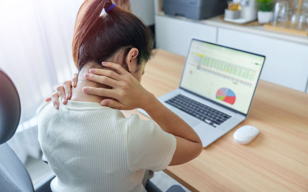
Skateboarding is an exciting sport that mixes skill, speed, and style. It began as a land-based surf practice but has grown into a worldwide hobby for many. To excel in skateboarding, you need targeted training that strengthens your core and legs, improves balance, and teaches safe falling to prevent harm. This training uses repetitive drills, explosive jumps, and endurance workouts to create automatic responses and lasting energy. It also includes mental prep like imagining tricks and steady practice routines.
The sport’s demands, such as repeated one-sided pushes and hard landings, can strain your body. That’s where integrative chiropractic care shines. At El Paso Back Clinic in El Paso, Texas, this approach improves joint mobility, corrects imbalances from skateboarding habits, and accelerates healing after impacts. It improves balance, body sync, and bendiness while offering diet and safety tips to reduce injury risk. Led by Dr. Alex Jimenez, DC, APRN, FNP-BC, the clinic offers tailored care for skateboarders and athletes, blending chiropractic care with rehab and nutrition to support top performance.
This article covers skate training basics and how chiropractic at El Paso Back Clinic supports it. For beginners or pros, these insights can help you advance safely. Visit https://elpasobackclinic.com/ to learn more about their services.
Core Elements of Skateboarding Training
Skateboarding success starts with body and mind prep. Training goes beyond board time—it’s about a solid base for tricks and endurance. Prioritize core and leg power, as these drive your actions (Austin Simply Fit, n.d.). Muscles like abs, lower back, quads, hamstrings, glutes, and calves handle shifts from an upright to a low position in moves like ollies.
Core Workouts: Try planks by holding a straight body pose for 30 seconds. Side versions hit obliques for twist stability.
Leg Boosters: Squats mimic board crouches—lower then rise for three sets of 10 reps.
Importance: Strong cores prevent shakes during jumps, lowering fall risks.
Balance is vital in skating. Poor balance leads to wipeouts on basic maneuvers. Newbies should pick a stance: regular (left-forward) or goofy (right-forward). Place the feet over the truck bolts for maximum stability (Skateboard GB, n.d.).
Balance Practices: Stand on one foot and draw letters with the other toe. Switch sides for ankle strength.
Next Level: Manuals lift the front wheels, balancing on the rear for ramp preparation.
Routine: Dedicate 10 minutes daily to weight shifts on your board for a natural feel.
Safe falling is key to injury avoidance. Falls are part of skating, but proper methods reduce severe damage. Roll instead of bracing with arms to protect wrists (Healthcare.utah.edu, 2024).
Fall Methods: Tuck chin and roll to distribute force. Aim for protected spots like padded knees.
Gear Essentials: Helmets, wrist, knee, and elbow pads absorb shocks.
Safe Start: Use grass or mats for low-risk practice.
Repetitive training builds muscle memory. Repeat actions until they’re instinctive, like pushing and halting (Braille Skateboarding, n.d.). This aids tricks such as frontside kickturns and backwheel pivots (How to Skate, 2018).
Drill Reps: Push 10 times, stop, and redo for fluid flow.
Trick Steps: Divide into parts, like board pop, then foot flick for kickflips.
Side Hops: Mimic skating with 30-second lateral jumps.
Gains: Higher leaps and fast reflexes elevate skills.
Cardio keeps you going strong. Skating provides some, but extras build heart health (Skateboard GB, n.d.).
Rope Skipping: 30 seconds on, rest, three rounds for calf power and breath control.
Crawls: Bear walk forward and back 10 meters.
Cardio Value: Longer sessions with quicker recovery.
Mental training tackles fear. Visualize wins before attempts (Florida Atlantic University, n.d.). Commitment means regular sessions despite setbacks.
Imagery: Eyes shut, see perfect landings.
Fear Busting: Small steps build confidence.
Drive: Love for skating fuels persistence.
Follow principles such as targeted work, gradual increases, and variety to ensure safe progress (The Daily Push, n.d.). Skate-specific drills, slight pushes, and mixes prevent plateaus.
This foundation makes skating enjoyable, but one-sided strains need expert help, like at El Paso Back Clinic.
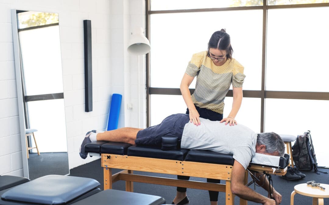
Integrative Chiropractic Care for Skateboarders at El Paso Back Clinic
At El Paso Back Clinic, integrative chiropractic merges adjustments with therapies for whole-body health. For skaters, it enhances joint flow in hips, knees, and ankles, easing restrictions from twists (Push as RX, n.d.). The clinic’s team uses advanced tools for custom plans.
Adjustments: Hands-on fixes realign for better motion.
Skating often causes imbalances—one leg pushes more, enlarging muscles unevenly (Instagram Reel, n.d.). This risks pain or bad posture.
Balance Fixes: Single-side workouts like one-leg squats.
Clinic Approach: Exams spot issues, then adjustments and drills even out.
Prevention: Avoids strains from overuse.
Falls bring impacts, but clinic care hastens recovery by reducing inflammation (Injury 2 Wellness, n.d.). For sprains, they combine rest and rehab.
Healing Tools: Ice, wraps, and elevations cut swelling. Adjustments aid nerves.
Rehab: Planks and stretches rebuild strength.
Quick Return: Less time off the board.
The clinic boosts balance, sync, and flexibility. Core support from deep muscles aids control (Robins, n.d.). Alignment improves awareness.
Balance Enhancers: Fixes heightened position sense.
Sync Training: Patterns restored post-injury.
Flex Moves: Stretches like yoga poses loosen spines.
Nutrition and prevention advice lowers risks. Proteins and veggies aid repair; warm-ups are key (Thompson, n.d.). Clinic experts guide anti-inflammation diets.
Food Advice: Fruits and healthy fats for recovery.
Safety Steps: Check-ups catch problems early; use gear.
Habits: Stay hydrated, foam roll to loosen up.
Dr. Alex Jimenez, a clinic leader with 30+ years, notes that integrative methods prevent injuries by addressing root causes such as imbalances (Jimenez, n.d.). He blends functional medicine, nutrition, and rehab for skateboarders. LinkedIn shares tips on sciatica and balanced routines (Jimenez, n.d.). For skate injuries like ankles or wrists, assessments lead to adjustments and strengthening (Jimenez, n.d.). Teamwork with therapies ensures full recovery.
Chiropractic at the clinic elevates performance, keeping bodies primed (Dallas Thrive, n.d.). Their sports focus includes strength, flexibility, and proprioception for athletes.
Conclusion
Pair skate training with the chiropractic services at El Paso Back Clinic for strength, balance, and safety. Build habits through drills and mental work. Let experts fix strains, speed healing, and advise prevention. Consistency pays off—practice wisely. For personalized care in El Paso, check https://elpasobackclinic.com/.
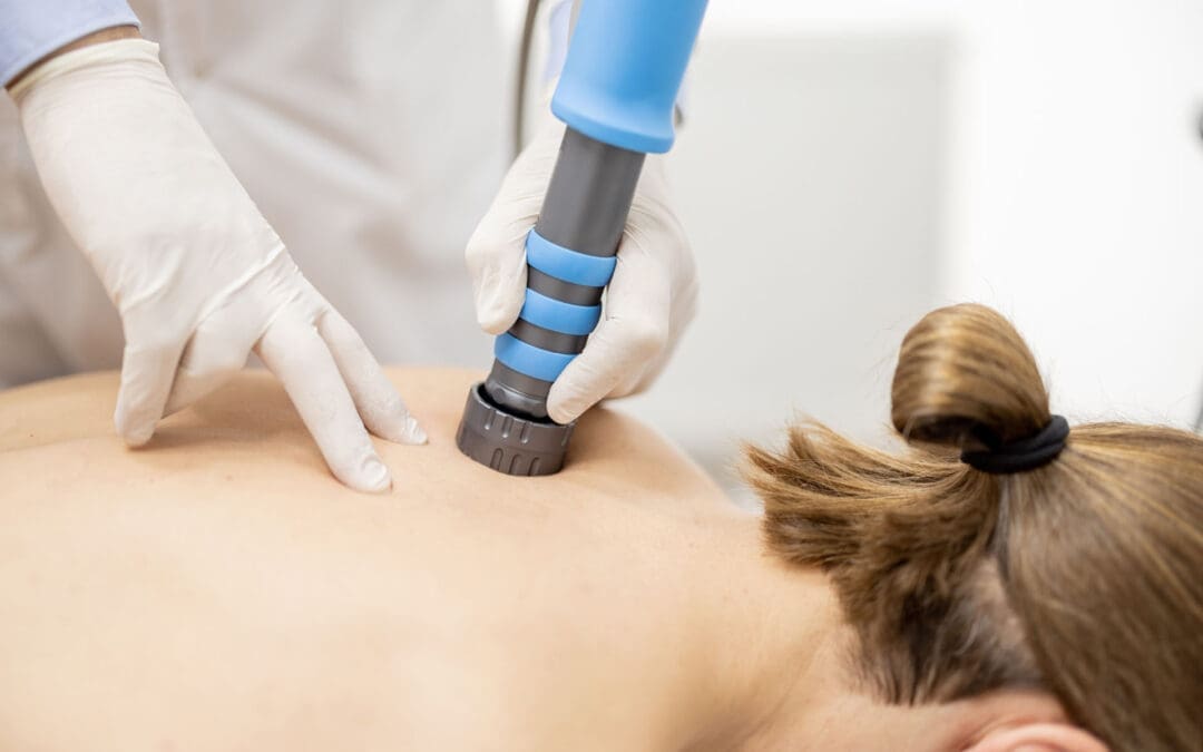
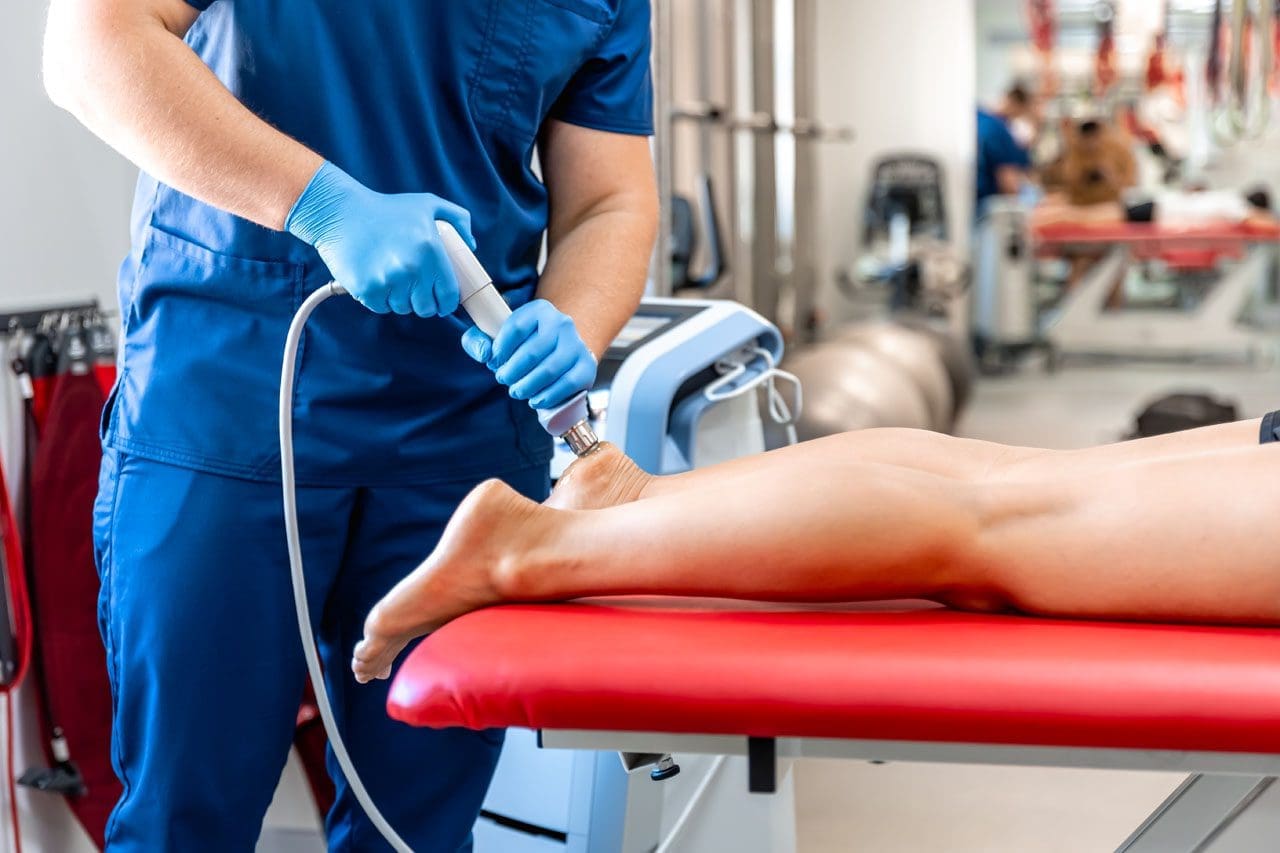
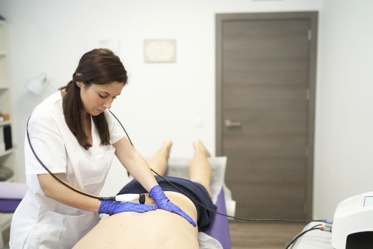
El Paso Back Clinic Shockwave Therapy: A Non-Surgical Option for Chronic Pain
Why Real ESWT Matters for Deep Healing at an Integrative El Paso Back Clinic
When people hear the term shockwave therapy, they often assume every machine is the same. It is not.
Some devices are true medical Extracorporeal Shockwave Therapy (ESWT) systems. Other devices are weaker radial pressure wave tools that are sometimes marketed as shockwave devices, even though they work differently. That difference matters if your goal is real tissue healing, not just short-term soreness relief. Mayo Clinic explains that focused shockwave (FSW) and radial pressure wave (RPW) are distinct waveforms, and only FSW is considered a “true shockwave” in a strict physical sense.
For a clinic like El Paso Back Clinic, where patients often come in with chronic pain, sports injuries, auto injuries, soft-tissue damage, and complex back conditions, the type of device and the treatment plan can make a big difference. The clinic’s site emphasizes multidisciplinary care, non-surgical recovery, and an integrative model that includes chiropractic, rehab, and functional medicine support.
This article explains, in plain language, what “real” shockwave therapy is, why focused shockwave is different from weaker devices, and how it fits into a complete recovery program in an integrative chiropractic setting.
What Is Real Shockwave Therapy?
Extracorporeal Shockwave Therapy (ESWT) is a non-invasive treatment that sends acoustic energy (sound waves) into injured tissue from outside the body. It is used in musculoskeletal care to help reduce pain and support healing in stubborn injuries. UCHealth describes ESWT as a noninvasive option for people who have not responded well to more conventional treatments, noting that it delivers high-energy acoustic waves to injured areas.
Mayo Clinic also describes shockwave therapy as a growing tool in physical medicine and sports medicine, especially for tendon and fascia problems.
In simple terms
Shockwave therapy is used to help the body “restart” healing in tissue that has been painful or stuck for a long time, such as:
tendons
fascia
ligaments
some chronic soft-tissue injuries
certain bone healing problems (in selected cases)
Mayo Clinic lists many musculoskeletal uses, including plantar fasciitis, Achilles tendinopathy, patellar tendinopathy, and lateral epicondylitis (tennis elbow).
Not All “Shockwave” Machines Are the Same
This is the most important part of the topic.
Many clinics use the word shockwave, but there are two main categories of devices used in musculoskeletal care:
Focused Shockwave (FSW / F-ESWT)
Radial Pressure Wave (RPW / radial therapy)
Mayo Clinic clearly explains that these are different technologies and should not be treated as identical. In fact, Mayo states that only focused shockwave generates a true shockwave, while radial devices generate a radial pressure wave.
Why that matters
The difference is not just marketing. It affects:
how deep the energy goes
how precise the treatment is
how much energy reaches the target tissue
what conditions may respond best
If a patient has a deep tendon problem, scar tissue, or a stubborn chronic injury, the provider should know exactly what machine is being used and why.
Focused Shockwave vs. Radial Pressure Wave
Here is the practical difference in plain language.
Focused Shockwave (FSW)
Focused shockwave is designed to deliver energy to a specific target depth. It is more precise and is often the better choice when the provider wants to treat a deeper structure or a smaller, more exact area. Mayo Clinic notes that focused shockwave has different physical properties and can be used alone or in combination with radial treatment, depending on the condition.
Radial Pressure Wave (RPW)
Radial therapy spreads energy more broadly and is often more surface-level. Mayo Clinic explains that radial devices generate pressure waves and notes tissue penetration of about 4 to 5 cm in its 2022 discussion of radial ESWT.
That does not mean radial is “bad.” It means it is different. In many cases, radial therapy remains helpful. But if a clinic claims “shockwave” and the patient expects high-energy focused treatment, the patient should ask which device is being used.
Quick comparison
Focused shockwave
More precise targeting
True shockwave physics
Often used for deeper or more exact lesions
Better fit for some regenerative goals
Radial pressure wave
Broader spread
Pressure-wave technology
Often, more superficial or diffuse treatment
Can still be useful in the right case
Why Energy Dose Matters
Real ESWT is not just “machine on, machine off.” It is dosed.
One of the main ways clinicians describe ESWT dose is Energy Flux Density (EFD), and the standard unit is mJ/mm² (millijoules per square millimeter). A PubMed Central review explains that EFD is the professional parameter used to describe shockwave energy flow through tissue, and specifically notes the unit of measurement as mJ/mm².
This is important because:
stronger energy is not always better
tissue type matters
the diagnosis matters
different injuries need different treatment settings
A quality clinic should be able to explain the treatment plan in a way that matches your condition, rather than using the same approach for every patient.
Does Shockwave Therapy Create “Microtrauma”?
Many people explain shockwave therapy by saying it creates “microtrauma” that triggers healing. That is a common explanation, and Mayo Clinic Sports Medicine uses this language in a patient-friendly way, noting that acoustic waves can create microtrauma to help reinitiate a healing response in tendons.
That said, many experts also describe the process in a more modern way as mechanotransduction—meaning the waves create a mechanical signal that helps cells activate repair pathways. Mayo Clinic’s 2025 article also highlights mechanotransduction and regenerative effects like cellular signaling and neovascular changes.
A simple way to think about it
Shockwave therapy helps by:
stimulating local tissue response
improving healing signaling
reducing pain pathways over time
helping stubborn tissue become more “active” in repair
So the short answer is:
Yes, “microtrauma” is a common way to explain it.
But the bigger idea is that the shockwave creates a healing signal, not uncontrolled tissue damage.
FDA Regulation and Why It Matters
Another reason patients should ask questions is that regulatory status matters.
The FDA has approved/cleared specific extracorporeal shockwave devices for specific uses. For example, the FDA PMA listing for the OrthoSpec Extracorporeal Shock Wave Therapy device states that it is indicated for adults with proximal plantar fasciitis (with or without a heel spur) who have had symptoms for 6 months or more and have failed conservative treatment.
That helps patients understand two important points:
real ESWT is a recognized medical technology
device claims should match actual indications and training
If a clinic says “shockwave,” it is fair to ask:
What exact device is this?
Is it focused or radial?
Is it FDA-cleared/approved for a musculoskeletal indication?
These are smart questions, not rude questions.
Why Real ESWT Is Useful in an Integrative Chiropractic Clinic
Shockwave therapy can be very effective, but it works best when the diagnosis is correct, and the rest of the care plan supports healing.
That is where an integrative clinic model is helpful.
The El Paso Back Clinic describes on its website a multidisciplinary, non-surgical, and functional recovery approach that includes chiropractic care, rehab, and broader wellness support. It also describes care for back, auto, and sports injuries, tendinopathy-related issues, and chronic pain.
Why this pairing makes sense
Shockwave therapy targets soft tissue and the healing response.
Chiropractic and rehab help restore:
joint motion
spinal alignment
posture
movement control
load tolerance
When these are combined, the patient gets a more complete plan.
Example of an integrative recovery setup
A patient with chronic Achilles pain, plantar fasciitis, or post-accident scar tissue restriction may benefit from:
Focused shockwave or radial therapy (depending on the tissue depth and goal)
Chiropractic adjustments to improve joint mechanics
Mobility work to reduce compensation patterns
Strength training/rehab exercise to improve tissue tolerance
Lifestyle support (sleep, inflammation control, nutrition)
This is especially important for back and soft-tissue injuries, as pain often has multiple causes. The tissue may be irritated, but there may also be a movement issue, posture problem, or old compensation pattern keeping it from healing.
Clinical Observations in Dr. Alexander Jimenez’s Integrative Model
Public information on dralexjimenez.com and El Paso Back Clinic describes Dr. Alexander Jimenez as a Doctor of Chiropractic and board-certified Family Nurse Practitioner (DC, APRN, FNP-BC) who uses a multidisciplinary, integrative approach focused on non-surgical recovery, diagnostics, and personalized care.
His El Paso Back Clinic content also emphasizes:
advanced injury rehabilitation
chronic pain care
sports injury care
auto injury care
functional medicine support
team-based recovery planning
These clinic observations support the idea that shockwave therapy should not be used as a stand-alone “gadget” treatment. Instead, it fits best within a broader care plan that includes biomechanics, rehab, and whole-person recovery.
Why dual training matters in this setting
In a clinic model that blends chiropractic and nurse practitioner perspectives, the provider can often look at a case more completely, including:
musculoskeletal pain drivers
nerve irritation patterns
inflammation
healing delays
activity limitations
overall recovery readiness
That type of clinical reasoning is helpful when deciding whether a patient should receive:
focused shockwave
radial therapy
chiropractic and rehab only
imaging first
referral or co-management
What Conditions Often Respond to Shockwave Therapy?
Shockwave therapy is often used for chronic injuries that have not improved enough with standard care.
Mayo Clinic and UCHealth commonly describe these types of cases:
Plantar fasciitis
Tennis elbow (lateral epicondylitis)
Achilles tendinopathy
Patellar tendinopathy
Shoulder tendinopathy
Other chronic tendon or fascia pain problems
Mayo’s clinical articles also note that ESWT has roles in treating tendons, ligaments, fascia, and even in selected bone-healing situations.
It may be especially helpful when:
pain has lasted for months
the patient plateaued in regular therapy
surgery is being considered, but not yet desired
the injury is painful with loading (walking, running, lifting, gripping)
the provider wants a non-invasive option
How to Tell if a Clinic Is Offering “Real” Shockwave Therapy
Because the market uses confusing language, patients should ask direct questions before paying for treatment.
Ask these questions
Is this focused shockwave (FSW) or radial pressure wave (RPW)?
What condition are you treating, and why is this device the right choice?
How do you set the energy dose (EFD/mJ/mm2)?
How many sessions are usually recommended for my condition?
Will I also get rehab or movement treatment?
If my pain is deep, how will you target it?
Is the device FDA-cleared/approved for musculoskeletal use?
A strong clinic should be comfortable answering these questions in simple language.
Why Device Hype Alone Is Not Enough
Some clinics advertise shockwave therapy as a miracle treatment. That is not the best way to present it.
Shockwave therapy can be a powerful tool, but results depend on:
Even the best technology will not work well if the diagnosis is wrong or if the patient returns to the same harmful movement pattern right away.
This is one reason integrated care models, like the one described at El Paso Back Clinic and Dr. Jimenez’s clinical sites, can be so useful for complex injuries: patients receive more than one treatment option and more than one clinical lens.
Bottom Line: Focused ESWT Is the Better Choice for True Regenerative Shockwave Goals
If your goal is real regenerative shockwave therapy, focused shockwave (FSW/F-ESWT) is usually the benchmark because it is the true shockwave form and offers more precise targeting. Mayo Clinic makes this distinction very clearly.
Radial devices can still be helpful in many cases, but they are not the same technology. Patients should not be told they are identical.
For patients in El Paso dealing with:
chronic tendon pain
back-related soft tissue problems
sports injuries
accident-related soft tissue injury
stubborn pain that has not improved
An integrative clinic model like El Paso Back Clinic can be a strong fit because it combines:
non-invasive care
structural assessment
chiropractic and rehab
broader healing support
multidisciplinary planning
That is often what it takes to move from “temporary pain relief” to true recovery.
How Detoxing Can Boost Your Energy Levels: A Simple Guide
Many people feel tired all the time. They drag through the day, relying on coffee or snacks to keep going. But what if there was a way to feel more awake and alert without those quick fixes? Detoxing might be the answer. Detoxing means helping your body get rid of harmful stuff that builds up over time. This can come from the air we breathe, the food we eat, or even stress. When you detox, you lighten your body’s load. This can lead to more energy and better thinking. Let’s explore how this works.
Detoxing can boost your energy levels! Absolutely! By lightening the load on your liver and cutting back on foods that can make you feel tired, you might find yourself with more energy and clearer thinking. Your liver is like a filter for your body. It cleans out bad things. When it’s overloaded, you feel sluggish. Detoxing helps by giving it a break. You do this by eating cleaner foods and drinking more water.
Think about the toxins around us. They come from pollution, processed foods, and chemicals. These can pile up and make you tired. Detoxing clears them out. This lets your liver and kidneys work better. When they do, your body absorbs nutrients more easily. Stable blood sugar means no big crashes after meals. Better oxygen flow helps, too. All this adds up to more energy.
But detoxing isn’t just about feeling less tired. It fights inflammation, the body’s swelling that drains your energy. Toxins cause this swelling, leading to fatigue. Getting rid of them makes your body run more smoothly. It improves how you take in food’s good stuff and boosts energy at the cellular level, in the form of ATP. ATP is like fuel for your cells. More of it means you feel stronger.
Experts like those in chiropractic care, functional medicine, and nursing help with this. They have titles like DC, MSACP, APRN, and IFMCP. They create plans based on science to detox safely. These plans restore balance in your body, reduce swelling, and boost energy. They look for why you’re low on energy, like hidden health issues. Instead of quick fixes, they offer custom solutions backed by research.
Dr. Alexander Jimenez, DC, APRN, FNP-BC, is one such expert. At his clinic in El Paso, Texas, he uses functional medicine to help people detoxify and boost energy. He checks for root causes, such as gut problems or stress. His patients report better sleep, less pain, and more daily energy after following his plans. On LinkedIn, he shares how detox helps with energy production and fights oxidative stress. His approach combines chiropractic adjustments with nutrition to make detoxification more effective.
What Are Toxins and How Do They Affect Energy?
Toxins are harmful things that enter your body. They can be from outside, like car fumes or pesticides on food. Alternatively, toxins can enter your body through internal sources such as stress or unhealthy eating habits. Over time, they build up. This makes your body work harder to stay healthy. The result? You feel worn out.
Environmental toxins, such as heavy metals and pollution, can slow down your cells.
Processed food waste: Sugary treats and junk food create waste that clogs your system.
Daily stress: It adds to the load, making detox harder.
When toxins stay, they cause inflammation. This is your body’s way of fighting back, but it uses up energy. You end up with fatigue, brain fog, and low mood. Detoxing removes these, so your energy comes back.
How Detoxing Works to Boost Energy
Your body has natural ways to detox. The liver, kidneys, skin, and gut all help. But sometimes they need support. Detoxing helps through diet, exercise, and habits.
Detoxing boosts energy by clearing built-up toxins and waste. This eases chronic inflammation and improves nutrient absorption. Stable blood sugar stops energy dips. Better oxygen flow means cells work well.
Here are key ways detox helps:
Clears the liver: Less work for it means more energy for you.
Improves digestion: A better gut means more nutrients for energy.
Functional medicine experts like Dr. Jimenez focus on this. They test for toxins and make plans. This includes foods like garlic and greens to support detox.
Benefits of Detoxing for Energy
People who detox often say they feel renewed. Energy is a big win. But it’s not magic. It’s about better body function.
More daily stamina: No afternoon slump.
Clearer mind: Less fog, better focus.
Better sleep: Detox fixes rhythms for restful nights.
Less fatigue: Your body is efficient, and you feel vital.
One study-like view from experts shows detox can balance hormones, too. This affects energy. But remember, not all detoxes are safe. Some extreme ones tire you more.
Myths and Facts About Detoxing
Not everyone agrees on detox. Some say your body does it on its own. That’s true, but lifestyle helps. Myths say detox diets clean you fast. Facts: They can help when done right, but there are dangers.
Myth: Detox removes all toxins forever. Fact: It’s ongoing.
Myth: You need fancy juices. Fact: Whole foods work best.
Fact: Cutting junk boosts energy from better habits.
Groups like the British Dietetic Association warn against strict detoxes. They can cause low energy due to a lack of food. MD Anderson says switch to healthy eating for real gains, not myths.
Functional Medicine and Personalized Detox
Functional medicine looks at the whole you. Experts find out why energy is low. They use tests for toxins or imbalances. Plans are tailored to each individual’s needs, rather than being universal.
Dr. Jimenez uses this. He combines chiropractic with detox. Patients get more energy from addressing gut or hormone issues. His background in nursing and functional medicine backs this.
Tips from experts:
Eat greens and fiber to help your liver.
Drink water to flush toxins.
Exercise to sweat it out.
Use supplements like milk thistle safely.
Safe Ways to Start Detoxing for Energy
Start slow. Talk to a doctor first. This is especially important if you are dealing with health issues.
Cut back on sugar, alcohol, and processed foods.
Add veggies, fruits, and nuts.
Stay hydrated.
Try sauna or baths for sweat detox.
Get good sleep.
Detox days can reset you. Focus on clean eating one day a week. This builds energy over time.
Potential Side Effects and How to Handle Them
Detox can make you feel worse first. This is due to toxins leaving. Symptoms: Headache, tiredness.
Drink more water.
Eat small meals.
Rest.
If serious, stop and see a pro like Dr. Jimenez.
Long-Term Energy from Detoxing
Detox isn’t a one-time thing. Make it a habit. Eat well, move, manage stress. This keeps energy high.
Patients of functional pros report lasting vitality. It’s about balance, not extremes.
In summary, detoxing boosts energy by clearing toxins, reducing inflammation, and improving body functions. With expert help, it’s safe and effective. Try it for more pep in your step.
Poor posture is more than a back or neck problem. It can also affect how well you breathe and how well your digestive system works. When a person slouches, hunches forward, or carries the head too far in front of the shoulders, the rib cage and abdomen lose space. That change can make it harder for the diaphragm to move well, which may lead to shallow breathing and lower oxygen intake. It can also place extra pressure on the stomach and intestines, which may contribute to reflux, bloating, and constipation (UCLA Health, 2024; Harvard Health Publishing, 2023).
This article is written for the El Paso Back Clinic audience and follows the clinic’s integrative approach: look at posture, spinal alignment, breathing mechanics, mobility, and daily habits together. The clinic and Dr. Alexander Jimenez frequently discuss posture and breathing as a functional pattern, not just a pain issue, on their educational pages. In other words, how you hold your body can shape how your lungs, core, and digestive system work throughout the day (Jimenez, n.d.; El Paso Back Clinic, n.d.).
Why Posture Matters for Breathing
Your diaphragm is the main muscle used for breathing. It sits below the lungs and helps pull air in when it moves downward. For that to happen easily, your rib cage and abdomen need enough room to expand.
When posture collapses (slouching, rounded shoulders, forward head posture), several things can happen:
The chest may cave inward
The upper back may round more
The ribs may not expand as well
The diaphragm may not move as freely
The body may rely more on neck and shoulder muscles to breathe
UCLA Health explains that poor posture can cause the chest to cave in, affecting breathing mechanics (UCLA Health, 2024). Harvard also lists breathing difficulties among the less obvious problems linked to poor posture (Harvard Health Publishing, 2023).
A research article on head-neck posture and respiratory function also found that posture changes can alter normal breathing mechanics, including diaphragm function. This matters because many people spend hours sitting at a desk, driving, or looking down at phones, which can reinforce forward head posture and rounded shoulders (Zafar et al., 2018).
Common signs that posture may be affecting your breathing
You may not always say, “I can’t breathe.” Instead, people often describe it like this:
“I can’t take a full deep breath”
“My chest feels tight when I sit”
“My neck and shoulders always feel tense”
“I sigh a lot”
“I feel winded faster than I should”
Sources on physical therapy and posture education also note a connection between poor posture and reduced diaphragm mobility, poor chest expansion, and shallow breathing (Capital Area PT, 2025; Total Health Chiropractic, 2022).
How Poor Posture Can Affect Digestion
Most people think digestion is only about food choices, enzymes, or stomach acid. Those are important, but body position matters too.
When you slouch, your abdomen compresses. That pressure can affect the stomach and intestines. UCLA Health notes that poor posture can slow digestion and increase abdominal pressure, which may trigger heartburn and acid reflux (UCLA Health, 2024).
BreatheWorks and other posture-focused digestive resources describe similar patterns: slouched alignment can increase abdominal pressure, affect swallowing and breathing coordination, and make reflux or bloating worse for some people (BreatheWorks, 2023a, 2023b).
Digestive symptoms that may be worse with slouching
Some common examples include:
Heartburn after meals
Acid reflux (GERD) symptoms when sitting or bending
Bloating or pressure in the upper abdomen
Feeling overly full
Constipation (especially with long periods of sitting)
Chiropractic and posture education sources (including Nolensville Chiropractic and BreatheWorks) often describe poor posture as a “compression” problem that can interfere with comfortable digestion and gut motility (Nolensville Chiropractic, 2025; BreatheWorks, 2023a).
The Breathing–Digestion Connection
Breathing and digestion are closely linked, and posture affects both simultaneously.
Here’s why:
The diaphragm supports both breathing and abdominal pressure control
The diaphragm is not just a breathing muscle. It also helps regulate pressure in the trunk. If it cannot move well, breathing becomes less efficient, and pressure control in the abdomen may change.
Poor posture can encourage shallow chest breathing
When breathing shifts more into the upper chest and neck, the body often feels more tense. In many people, this goes along with stress and “fight-or-flight” patterns, which can make digestion feel worse.
Slouching compresses the digestive area
A flexed, collapsed posture can reduce the space available to the stomach and intestines. That can be especially noticeable after eating.
BreatheWorks specifically describes how breathing coordination, alignment, and digestive comfort are connected, especially in people with reflux and bloating symptoms (BreatheWorks, 2023a, 2023b). El Paso Back Clinic and Dr. Jimenez’s educational content also emphasize this whole-body view, especially in patients with both musculoskeletal complaints and gut-related symptoms (Jimenez, n.d.; El Paso Back Clinic, n.d.).
Posture Patterns That Commonly Cause Problems
At El Paso Back Clinic, many patients dealing with neck, upper back, or shoulder pain also show posture patterns that can affect breathing and digestion. Dr. Jimenez’s educational content often highlights the same patterns in functional assessments (Jimenez, n.d.).
Forward head posture
This happens when the head moves in front of the shoulders. It increases neck strain and often leads to upper-chest breathing.
Rounded shoulders
Rounded shoulders can limit chest expansion and change rib cage motion.
Excessive upper-back rounding (kyphotic posture)
This can reduce thoracic mobility (mid-back motion), which is important for full breathing.
Slumped sitting posture
A tucked pelvis, a collapsed lower back, and a caved chest can increase abdominal pressure, making both breathing and digestion less efficient.
Why Integrative Chiropractic Care Can Help
A strong posture plan usually needs more than a quick reminder to “sit up straight.” Many people need a combination of mobility work, spinal/rib movement restoration, soft-tissue care, breathing retraining, and strength work to build lasting change.
That is why the El Paso Back Clinic approach is helpful for many people. The clinic’s posture and rehabilitation content describes a broader plan that can include:
Spinal adjustments
Mobility and stretching
Movement retraining
Soft-tissue care
Posture-focused exercises
Health coaching (El Paso Back Clinic, n.d.)
How this may improve breathing
When spinal and rib mobility improve, the chest can move more naturally during breathing. That can support deeper, more efficient breaths and reduce overuse of neck muscles.
How this may improve digestion
When posture improves, abdominal compression may decrease. Better alignment can also make it easier to breathe diaphragmatically, which may support calmer, more comfortable digestion in some patients.
Dr. Jimenez’s educational pages also describe the importance of posture, breathing mechanics, rib mobility, and functional movement in patients with reflux, bloating, and related complaints (Jimenez, n.d.).
Practical Steps to Improve Posture, Breathing, and Digestion
The good news is that small daily changes can make a real difference.
Reset your sitting posture
Try this simple “stacking” setup:
Feet flat on the floor
Hips level (not rolled backward)
The rib cage is stacked over the pelvis
Shoulders relaxed (not rounded forward)
Chin level (not poking forward)
Even a few posture resets per day can help reduce the long stretches of slouching that many people fall into while working or driving (UCLA Health, 2024).
Use posture breaks every 30–60 minutes
Long sitting is a major factor in the worsening of posture over time. A short break helps.
Quick break routine (2 minutes)
Stand up
Roll your shoulders back gently
Take 5 slow breaths
Walk for 1 minute
Reset your sitting position
This kind of movement break can reduce stiffness and help restore better breathing mechanics. General health and posture guidance consistently supports frequent movement to reduce the effects of prolonged sitting (Harvard Health Publishing, 2023; UCLA Health, 2024).
Practice diaphragmatic breathing
Diaphragmatic breathing can help train the body away from shallow chest breathing.
Simple drill (1–2 minutes)
Sit upright or lie on your back
Place one hand on your chest and one on your belly/ribs
Breathe in through your nose
Try to expand the lower ribs and belly gently
Exhale slowly and fully
Keep shoulders relaxed
Posture-focused breathing resources often recommend this type of drill to improve breathing efficiency and reduce tension (Capital Area PT, 2025; Total Health Chiropractic, 2022).
Improve meal posture
How you sit while eating matters, especially if you have reflux.
Better meal posture tips
Sit upright when eating
Avoid eating while slouched on a couch
Chew slowly
Stay upright after meals
Take a light walk after eating if possible
BreatheWorks and UCLA Health both discuss how posture can affect reflux and digestive comfort, especially in people who slouch during or after meals (BreatheWorks, 2023b; UCLA Health, 2024).
When to Get Medical Care Right Away
Posture can affect breathing and digestion, but some symptoms require medical evaluation and should not be blamed solely on posture.
Seek prompt medical care if you have:
Chest pain
Severe shortness of breath
Trouble swallowing
Vomiting blood
Black/tarry stools
Severe abdominal pain
Unexplained weight loss
Ongoing reflux that is not improving
These can be signs of a more serious condition and need a full medical workup (UCLA Health, 2024; Harvard Health Publishing, 2023).
Clinical Perspective from Dr. Alexander Jimenez, DC, APRN, FNP-BC
For the El Paso Back Clinic audience, the key message is simple: posture problems are often functional problems. In Dr. Jimenez’s educational content, posture is not treated as an isolated issue. It is part of a bigger clinical picture that includes spinal mechanics, rib motion, breathing patterns, stress load, and daily movement habits (Jimenez, n.d.).
That is why many patients feel better when care is more comprehensive. Instead of only focusing on pain, an integrative plan may help by:
Improving spinal and rib mobility
Restoring more natural breathing mechanics
Reducing neck and shoulder overuse
Addressing posture during work and meals
Supporting better movement and daily function
The El Paso Back Clinic posture and rehabilitation pages also describe a personalized approach using adjustments, exercise, stretching, and movement retraining, which fits well with this type of whole-body care model (El Paso Back Clinic, n.d.).
Final Takeaway
Poor posture can affect much more than the spine. Slouching and forward head posture can limit diaphragm movement, reduce chest expansion, and lead to shallow breathing. At the same time, abdominal compression can make digestion less comfortable and may worsen reflux, bloating, and constipation in some people.
The good news is that posture can improve. With the right plan—especially one that includes posture correction, breathing retraining, and integrative chiropractic care—many people can breathe better, move better, and feel more comfortable after meals.
For readers of El Paso Back Clinic, this is an important reminder: posture is not just about standing tall. It is about giving your body the space and mechanics it needs to function well.
Understanding Chiropractic Wedges: Their Role in Pain Relief and Spinal Health
Chiropractic care helps people feel better by fixing problems in the spine and body without surgery or strong medicines. One tool that chiropractors often use is called a wedge. These are simple, triangle-shaped blocks made from foam or other firm materials. They are placed on parts of the body, such as the neck, hips, or feet. The idea is to use gravity—the Earth’s natural pull—to gently stretch and align the body. This can help correct spinal curves, ease pain, and improve overall body function (Diamond State Chiropractic, n.d.).
Wedges are not like hard adjustments where the chiropractor pushes on the spine. Instead, they let the body relax and correct itself slowly. Patients lie on them for a few minutes, and gravity does the work. This makes them good for people who want gentle care, such as older adults or pregnant individuals. They can help with back pain, neck strain, and even headaches by improving the body’s alignment (Tiger Lily Chiropractic, n.d.).
In this article, we’ll look at how these wedges work, the different types, and why they fit into a bigger picture of health care. We’ll also discuss how clinics that combine different treatments can improve patient outcomes.
What Are Chiropractic Wedges, and How Do They Work?
Chiropractic wedges are basic tools that look like small ramps. They come in different sizes and shapes, but most are firm enough to support the body’s weight. When a person lies on one side, the wedge lifts a specific area, such as the neck or pelvis. This creates a gentle pull that stretches tight muscles and helps bones return to their proper positions.
The main goal is to restore the spine’s natural curves. The spine isn’t straight; it has gentle bends that help us stand tall and move easily. If these curves become flat or twisted due to poor posture, injuries, or daily stress, it can lead to pain. Wedges use the body’s own weight to fix this over time (Core Chiropractic, n.d.).
Here’s how they typically work:
Placement: The chiropractor places the wedge at the right spot based on the body’s needs.
Time: Patients relax on it for 5 to 10 minutes, sometimes longer, as they get used to it.
Gravity’s Role: No pushing or twisting—just letting gravity pull things into alignment.
Safety: Always start slow to avoid strain, and stop if it hurts (Pure Health, n.d.).
This passive method means no sudden moves, making it comfortable for most people. It’s often part of a plan that includes other care, such as exercises or advice on sitting better.
Types of Chiropractic Wedges
There are a few main kinds of wedges, each for a different part of the body. They target specific issues but can help the whole body feel better.
Neck Wedges (Cervical Wedges)
These are for the upper spine, which includes the neck. Many people lose the natural curve in their neck from looking down at phones or computers all day. This is called forward head posture, and it puts extra pressure on the neck and shoulders.
To use a neck wedge:
Lie on your back on a flat surface.
Place the wedge so the flat side is against your shoulders, and your head rests on the sloped part.
Relax for 5-10 minutes, letting gravity stretch the neck.
Start with short times and build up (YouTube – Cordova & Siegmund, n.d.).
Benefits include less neck pain, fewer headaches, and better posture. It can even help with things like dizziness or tingling in the arms by taking stress off nerves (Pure Health, n.d.). One clinic notes that consistent use, along with adjustments, helps the curve come back and makes changes last longer (Chiropractic First, n.d.).
Pelvic Wedges or SOT Blocks
These are used in the Sacro Occipital Technique (SOT). They go under the hips or pelvis while the person lies face down. The wedges act like a see-saw, using gravity to balance the lower spine and hips.
How they’re placed:
Two wedges under the hips, angled to fix tilts or twists.
The patient lies still, and gravity corrects imbalances.
They are beneficial for conditions such as low back pain, sciatica, or uneven hips (Tiger Lily Chiropractic, n.d.).
They help with conditions like scoliosis or coccydynia (tailbone pain) by aligning the pelvis without hard thrusts. This is ideal for people who can’t tolerate stronger adjustments, such as those with acute pain or older individuals (Walkley Chiropractic Group, n.d.). Dr. Alexander Jimenez, a chiropractor with over 30 years of experience, notes that misaligned hips can cause pain that spreads to the back, legs, and even the knees. He uses non-invasive methods, such as decompression, to fix this, which pairs well with wedge techniques (Jimenez, n.d.a; Jimenez, n.d.b).
Foot Wedges
These smaller wedges go under the feet or in shoes. They fix problems with how the feet roll in or out, called pronation or supination. Bad foot mechanics can affect the knees, hips, and spine.
Uses include:
Placing them to encourage better foot movement.
Helping with pain in the feet, ankles, or higher up the body.
Unlike stiff inserts, they promote natural motion (PhysioFlexx Ayrshire, n.d.).
They can ease nagging aches or prevent injuries by improving the body’s overall movement. For example, if one foot turns in too much, it might tilt the pelvis and cause back issues (Boroondara Osteopathy, n.d.).
Benefits of Using Wedges in Chiropractic Care
Wedges offer many advantages because they’re simple and effective. They don’t require fancy equipment, and patients can often use them at home after learning how to use them.
Key benefits:
Pain Relief: They reduce pressure on nerves and joints, helping with back, neck, and hip pain (Diamond State Chiropractic, n.d.).
Better Alignment: Restore natural spine curves to improve posture and reduce strain (Core Chiropractic, n.d.).
Gentle for Everyone: Safe for pregnant people, older individuals, or those recovering from injuries (Walkley Chiropractic Group, n.d.).
No Side Effects: Unlike pills, they work naturally without risks (National Center for Complementary and Integrative Health [NCCIH], n.d.).
Long-Term Help: When used regularly, they help adjustments last and prevent problems from recurring (Pure Health, n.d.).
Studies show that about 11% of U.S. adults used chiropractic care in 2022, often for pain, and tools like wedges play a big role (NCCIH, n.d.).
Conditions Treated with Wedges
Wedges aren’t a cure-all, but they help with many common issues. Chiropractors check the body first to see if they’re right for you.
Common conditions:
Neck and Shoulder Pain: From poor posture or stress (YouTube – Cordova & Siegmund, n.d.).
Low Back Pain and Sciatica: By balancing the pelvis (Tiger Lily Chiropractic, n.d.).
Scoliosis: Gentle corrections to ease curves (Diamond State Chiropractic, n.d.).
Coccydynia (Tailbone Pain): Using cushions or wedges to reduce pressure while sitting or lying (El Paso Chiropractor Blog, 2019).
Headaches: Less tension in the neck means fewer migraines (Integrated Chiropractic of Boca, n.d.).
Hip Misalignment: Fixes uneven hips that cause limping or leg pain (Jimenez, n.d.a).
Dr. Jimenez notes that hip issues often stem from daily habits, such as carrying heavy bags on one side. He combines alignments with lifestyle changes for better results (Jimenez, n.d.b).
Integrative Clinics and Holistic Approaches
Many chiropractic clinics now take a holistic view, meaning they look at the whole person—not just the spine. This includes mixing wedges with other treatments for better healing.
In an integrative clinic, highly trained experts work together. They might use:
Manual adjustments to move bones.
Physical therapy for strength and flexibility.
Acupuncture to ease pain and inflammation.
Nutritional advice to support the body’s repair (Involve Health, n.d.).
This team approach helps mobility, reduces pain, and boosts quality of life. It’s like what the NCCIH describes: care that combines different methods for overall wellness (NCCIH, n.d.; All Cure Spine and Sports, n.d.).
For example, a patient with back pain might get wedge sessions, then exercises, and tips on eating anti-inflammatory foods. Clinics like Nexus Chiropractic even offer seat wedges for better sitting posture, helping people who work at desks (Nexus Chiropractic, n.d.).
Dr. Jimenez’s practice in El Paso, Texas, shows this well. As a DC, APRN, and FNP-BC, he blends chiropractic with functional medicine. He looks at factors such as diet, stress, and genes to address root causes. For sciatica, he uses adjustments and self-massage tools, including wedge-like supports. His patients report less pain and better movement after integrative plans (Jimenez, n.d.a; Jimenez, n.d.b).
Other benefits of multidisciplinary care:
Faster Healing: Combining therapies speeds up recovery (Dallas Accident and Injury Rehab, n.d.).
Less Medication: Natural methods cut down on pills, including opioids (All Cure Spine and Sports, n.d.).
Personalized Plans: Care fits your life, like adding positive psychology for stress (Involve Health, n.d.).
Prevention: Learn habits to stay healthy in the long term (Poets Corner Medical Centre, n.d.).
Medical doctors often see chiropractors as helpful partners. They value how chiropractic restores movement without surgery (AICA, n.d.).
How to Use Wedges Safely at Home
Some chiropractors teach patients to use wedges at home. Videos show simple steps, like for lumbar or neck stretches (Facebook – West Chiropractic, n.d.; YouTube – Pelvic Wedges, n.d.).
Tips:
Always get checked by a pro first.
Start with 1-2 minutes and add time slowly.
Use on a firm surface, not a soft bed.
Relax fully—don’t tense up.
Stop if you feel pain and talk to your doctor (Pure Health, n.d.).
Consistency matters. Using them daily, along with healthy habits, leads to big changes.
Clinical Observations from Dr. Alexander Jimenez
Dr. Alexander Jimenez has seen thousands of patients over 30 years. He notes that many pains start with small imbalances, such as in the hips or spine. In his clinic, he uses digital X-rays to spot issues, then non-invasive fixes like decompression. While he doesn’t always mention wedges, his focus on gentle alignment aligns with their use. For example, in treating sciatica, he combines adjustments with home tools like foam rollers, which are similar to wedges for pressure relief (Jimenez, n.d.b).
He stresses integrative care: “Addressing the whole person—body, nutrition, and mind—leads to lasting health.” His work with veterans and athletes shows how these methods improve life without drugs (Jimenez, n.d.a).
Conclusion
Chiropractic wedges are a smart, gentle way to support the body’s healing. They fix alignments, ease pain, and fit into bigger health plans. Whether for neck curves, pelvic balance, or foot mechanics, they offer real benefits. In integrative clinics, like Dr. Jimenez’s, they team up with other therapies for the best results. If you’re dealing with pain, talk to a chiropractor—they can show if wedges are right for you.
Why Neuropathy Treatment Costs So Much: Insights from El Paso Back Clinic® in El Paso, TX
Neuropathy is a nerve damage condition that leads to pain, numbness, tingling, or weakness, often in the feet and hands. It can stem from diabetes, injuries, or other health issues. At El Paso Back Clinic® in El Paso, TX, a top wellness chiropractic care center, experts like Dr. Alexander Jimenez help patients manage this through custom, non-invasive treatments. But why does neuropathy therapy cost a lot? It involves long-term care, special tests, complex treatments, and pricey meds. Factors such as regular specialist visits and experimental options add up. Plus, there are hidden costs from missing work. This article breaks down these reasons and offers tips on how El Paso Back Clinic® makes care more affordable and effective for locals in El Paso, TX.
The Need for Long-Term Care in Neuropathy Treatment at El Paso Back Clinic®
Neuropathy is not a quick fix. It is a lasting condition that needs ongoing care to ease symptoms and stop it from getting worse. This long-term nature is a major driver of high costs, as patients return for treatment over time.
Ongoing Check-Ups: Doctors monitor progress and adjust plans, leading to more visits.
Symptom Control: Pain relief might need weekly sessions for months.
Avoiding Worse Problems: Without care, issues like infections or falls can lead to significant hospital bills.
Research shows that neuropathy linked to multiple myeloma can add $16,600 monthly to healthcare costs compared to $15,090 without it (Binder et al., 2019). For diabetic cases, yearly costs can hit $27,931, over four times higher than diabetes alone (Petersen et al., 2023). At El Paso Back Clinic®, Dr. Jimenez uses functional medicine to address root causes, such as inflammation, which can reduce long-term expenses by focusing on natural healing (Jimenez, n.d.a). The clinic offers flexible plans without insurance headaches, making ongoing care easier for El Paso residents (El Paso Back Clinic, n.d.a).
Lifestyle changes are part of the plan as well. Patients receive support with diet and exercise to improve nerve health, but these add costs for experts. Still, at this El Paso, TX clinic, integrated care means better results with fewer future bills.
Specialist Tests and Diagnostic Costs for Neuropathy in El Paso, TX
Identifying the cause of neuropathy requires advanced testing, which is not cheap but is vital to the right treatment.
Nerve Tests: Studies like conduction checks cost $100 to $1,000.
Muscle Tests (EMG): These range from $200 to $500 and assess how muscles respond.
Imaging and Biopsies: MRIs and nerve samples help identify damage, driving costs higher.
Clinics report diagnostic fees ranging from $100 to $1,000, depending on the need (Northstar Joint and Spine, n.d.). Some places repeat tests unnecessarily, adding thousands (Foundation for Peripheral Neuropathy, n.d.). Neuropathy’s complexity, with each nerve different, makes diagnosis tough (London Pain Clinic, n.d.).
At El Paso Back Clinic®, tests such as digital X-rays and nerve checks are included in affordable packages. Dr. Jimenez, with his dual expertise as a chiropractor and nurse practitioner, ensures tests are targeted, saving patients in El Paso, TX, money (Jimenez, n.d.b; El Paso Neuropathy Center, n.d.). This wellness chiropractic approach uses non-invasive methods to diagnose without extra waste.
The High Price of Medications for Neuropathy Relief
Drugs are common for neuropathy, but brand names make them expensive.
Top Brands: Lyrica can cost $200 to $500 per month.
Cheaper Choices: Generics like gabapentin are $10 to $50, but not for all.
Mix of Meds: Multiple pills mean higher totals.
Pregabalin costs more than gabapentin but may reduce overall visits (Sicras-Mainar et al., 2017). For challenging cases, expenses climb with failed trials (Petersen et al., 2023). Side effects require additional monitoring, increasing costs (Cleveland Clinic, 2023).
El Paso Back Clinic® focuses on reducing med reliance through chiropractic and functional medicine. Dr. Jimenez prescribes when needed but prefers natural options like acupuncture to manage pain, cutting drug costs for El Paso, TX patients (Jimenez, n.d.a; Health Coach Clinic, n.d.). Their neuropathy plans include effective prescriptions tailored to minimize symptoms at an affordable cost (Dralexjimenez.com, 2026).
Complex Treatments and Clinic Packages at El Paso Back Clinic®
Treatments can involve technology and multiple sessions, often bundled.
Therapy Rounds: Physical or laser therapy may require 9-12 visits, priced at $600 to $4,200 each.
Devices: TENS units range from $30 to $100, but professional sessions add up.
Advanced Options: Injections or decompression can be $5,000+.
Packages range from $500 to $5,000 yearly (Advantage Health Center, n.d.). Some use laser and bioelectrical for $3,000 to $6,000 over 12 visits (Olympic Spine, n.d.). Nerve healing is slow, so multi-session plans are key (Creekside Chiropractic, n.d.).
In El Paso, TX, El Paso Back Clinic® offers chiropractic care packages for peripheral neuropathy, using adjustments and rehabilitation to ease nerve pain at lower costs (Push as Rx, n.d.). Their affordable plans avoid copays, making complex care accessible (Sciatica Clinic, n.d.). Dr. Jimenez integrates spinal decompression and nutrition for better, faster results.
Regular Meetings with Specialists for Neuropathy Care
Specialist visits are frequent and pricey.
Pain Experts: Higher fees for complex handling.
Foot Specialists: Podiatrists prevent issues with ongoing costs.
Team Approach: Weekly at first, then as needed.
Sessions start at $100, with total costs reaching thousands for complex cases (Northstar Joint and Spine, n.d.). Some setups have brief doctor visits, followed by nurse-led billing (Foundation for Peripheral Neuropathy, n.d.).
At El Paso Back Clinic®, specialists like Dr. Jimenez develop treatment plans with input from chiropractors and nurse practitioners. This El Paso, TX clinic uses team-based care for neuropathy, with routine visits focused on progress and cost control through efficiency (Yelp, n.d.; El Paso Neuropathy Center, n.d.). Military discounts and insurance help too.
Alternative or Experimental Treatments Offered in El Paso, TX
When basics fail, alternatives cost more.
Stem Cells: $5,000 to $50,000, often out-of-pocket.
Acupuncture: $50 to $150 per session; multiple sessions may be needed.
New Tech: TENS or stimulation adds fees.
Trends warn of costly devices without proof (Instagram Reel, 2024). Latest examples include spinal stimulation, which is new and expensive (DVC Stem, n.d.).
El Paso Back Clinic® includes alternatives such as electroacupuncture in its plans, avoiding unproven, high-cost options. Dr. Jimenez’s functional medicine uses evidence-based options for neuropathy, making them affordable for El Paso locals (Jimenez, n.d.a).
Beyond Medical Bills: Lost Productivity and Other Costs
Neuropathy hits more than wallets—work suffers.
Work Absences: Pain causes missed days.
Lower Output: 18% more lost time in diabetic cases.
Family Help: Indirect costs from caregivers.
Monthly extras can be $1,509, including work losses (Binder et al., 2019). Indirect costs, such as leave, account for 48% of the total (Sicras-Mainar et al., 2017). Aids and travel add up (Foundation for Peripheral Neuropathy, n.d.).
In El Paso, TX, El Paso Back Clinic® provides rehabilitation to help patients return to work faster, reducing these losses. Their wellness focus builds strength and reduces downtime (Millennium LC, n.d.).
Specialized Practitioners and Individualized Plans at El Paso Back Clinic®
Experts craft custom plans, which work well but cost.
Full Checks: Look at life, genes, and more.
Mixed Therapies: Functional medicine, manual care, changes.
Whole-Person View: Fix causes, not just pain.
Dr. Alexander Jimenez, DC, APRN, FNP-BC, IFMCP, CFMP, ATN, with 30+ years of experience, leads at El Paso Back Clinic®. He uses “Neuro-Gen” and nutrition for neuropathy, avoiding drugs/surgery. Plans include adjustments, acupuncture, and lifestyle to heal nerves (Jimenez, n.d.a; Jimenez, n.d.b). This clinic in El Paso, TX, is patient-centered, with telemedicine for convenient follow-up.
For diabetic neuropathy, chiropractic care can help improve comfort and overall health (Health Coach Clinic, n.d.). Plans are affordable and prioritize well-being (El Paso Back Clinic, n.d.b).
Wrapping Up: Managing Neuropathy Therapy Costs in El Paso, TX
Neuropathy treatment is pricey due to chronic care, tests, meds like Lyrica, packages, visits, alternatives, and work losses. But at El Paso Back Clinic® in El Paso, TX, Dr. Jimenez and team offer value with custom, natural plans. Call 915-850-0900 for affordable wellness chiropractic care that reduces long-term costs.
Binder, L. M., Chimenti, R. L., Sluka, K. A., & Vardaxis, V. G. (2019). Cost of peripheral neuropathy in patients receiving treatment for multiple myeloma: A US administrative claims analysis. PMC, PMC6444783. https://pmc.ncbi.nlm.nih.gov/articles/PMC6444783/
Common Fastpitch Softball Injuries and How El Paso Back Clinic’s Integrative Chiropractic Care Can Help
Fastpitch softball is a tough sport that asks a lot from players. Pitchers use the underhand windmill throw frequently, and everyone must move quickly and change direction quickly. This leads to pain in muscles and bones. The most common are overuse problems in the shoulder and elbow, like rotator cuff strains and UCL tears from all that pitching. Then there are sudden hurts, such as ACL tears in the knee, ankle sprains, and breaks from sliding, diving, or running into others. Players also deal with finger and hand issues, lower back pain, and concussions. At El Paso Back Clinic in El Paso, TX, they use integrative chiropractic care. This is a gentle, whole-body approach that includes spinal adjustments, muscle therapy, and rehab exercises. It addresses both acute injuries and the root causes of overuse. This care helps softball players heal faster, get stronger, and prevent re-injury. Led by Dr. Alexander Jimenez, DC, APRN, FNP-BC, the clinic focuses on athletes with personalized plans.
Common Injuries in Fastpitch Softball
Fastpitch softball can cause injuries due to its speed and repeated moves. Pitchers throw hard and often, putting stress on their arms. Other players dive, slide, and run, which can twist joints or cause impacts. Research shows shoulder and elbow overuse is the top issue for pitchers because of the windmill pitch (Rothman Orthopaedics, n.d.; Andrews Sports Medicine, n.d.). Lower-body problems result from quick stops and turns (Sports Medicine Clinics, 2025). Head injuries come from hits or crashes (Children’s Health, n.d.).
Here are some main overuse injuries:
Rotator cuff strains: Repeated throwing inflames the shoulder muscles, causing pain. This hits pitchers and throwers hard (Share UPMC, 2020; HDP Chiro, n.d.).
UCL tears: The elbow ligament gets stretched or torn due to the pitching force. Young players who overdo it are at risk (UC Health, n.d.; North Central Surgical, n.d.).
Sudden, acute injuries include:
ACL tears: Knee ligament rips during fast changes in direction. It can keep players out for months (Andrews Sports Medicine, n.d.; PubMed, n.d.).
Ankle sprains: Ankles twist while running or sliding into bases (Rock Valley PT, n.d.; Children’s Hospital, 2022).
Fractures: Breaks in fingers, hands, or wrists from dives or ball hits (Summit Orthopedics, 2022; Therapy Partners Group, n.d.).
Other common problems are:
Finger and hand injuries: From catching or batting (UC Health, n.d.).
Lower back pain: Caused by twisting or bad pitching form (North Central Surgical, n.d.; Share UPMC, 2020).
Concussions: Brain injuries from collisions or head hits (Children’s Health, n.d.; YouTube, n.d.).
These often stem from excessive play without breaks (PubMed, n.d.; PMC, n.d.). Strains and sprains are frequent in arms and legs (PMC, n.d.). To prevent them, use warm-ups, good technique, rest, and pitch limits (Rothman Orthopaedics, n.d.; UC Health, n.d.; NCYS, 2022).
Integrative Chiropractic Care at El Paso Back Clinic
At El Paso Back Clinic, integrative chiropractic care treats the whole body without surgery or meds. It’s holistic, meaning it looks at everything that affects health. The clinic combines chiropractic care with functional medicine and sports rehabilitation to address injuries and their causes (El Paso Back Clinic, n.d.; Integrative Chiro Center, n.d.). Dr. Alexander Jimenez and his team use evidence-based ways to help athletes.
Key parts of their care:
Spinal adjustments: These correct spinal misalignments to reduce pain, improve mobility, and support nerve function (Injury2Wellness, n.d.; SCUHS, n.d.).
Soft tissue therapy: Techniques such as massage reduce swelling and promote muscle healing (SCUHS, n.d.; Peoria Spine and Sport, n.d.).
Functional rehabilitation: Exercises build strength, balance, and flexibility to prevent re-injury (Push as RX, n.d.; Dallas Accident and Injury Rehab, n.d.).
The clinic also offers nutrition, stress management, and lifestyle tips to support full recovery (El Paso Back Clinic, n.d.). This differs from basic care by addressing root causes of softball injuries, such as poor posture or weak muscles (Chiropractic Sports Care, n.d.; El Paso Back Clinic, n.d.).
Benefits for Softball Players at El Paso Back Clinic
El Paso Back Clinic helps softball players recover quickly, play better, and avoid injuries. Their care corrects alignment and reduces inflammation to promote faster healing (SCUHS, n.d.). Players gain more power from balanced bodies, leading to stronger pitches and quicker moves (Dallas Accident and Injury Rehab, n.d.). Prevention is key—they spot problems early (Push as RX, n.d.; El Paso Back Clinic, n.d.).
Dr. Alexander Jimenez shares from his work: Overuse in softball causes inflammation and nerve issues. His methods, such as adjustments and nutrition, can help without surgery (Dr. Alexander Jimenez, n.d.; Dr. Alexander Jimenez LinkedIn, n.d.). He treats shoulders, knees, and backs with movement checks to stop repeats. This fits softball, where arm strain is common.
Benefits include:
Quicker recovery: Adjustments reduce pain and swelling so players return soon (Injury2Wellness, n.d.; SCUHS, n.d.).
Better performance: Stronger muscles and joints mean harder throws and faster runs (Dallas Accident and Injury Rehab, n.d.).
Injury prevention: Regular visits address imbalances, reducing overuse risk (El Paso Back Clinic, n.d.; Push as RX, n.d.).
Studies and videos support this. One shows that therapy for softball injuries is beneficial (YouTube, n.d.). At the clinic, athletes receive custom plans that include rehabilitation and education (El Paso Back Clinic, n.d.).
If you’re in El Paso or nearby, like Horizon City, contact El Paso Back Clinic today. Call +1-915-850-0900 or schedule an appointment. Locations include 11860 Vista Del Sol, Ste 128. Discover how Dr. Jimenez can help your game.
In the end, fastpitch softball risks injuries, but El Paso Back Clinic’s integrative care offers real help. It heals holistically and builds strength. Players stay on the field longer and stronger.
IFM's Find A Practitioner tool is the largest referral network in Functional Medicine, created to help patients locate Functional Medicine practitioners anywhere in the world. IFM Certified Practitioners are listed first in the search results, given their extensive education in Functional Medicine