Back Clinic Imaging & Diagnostics Team. Dr. Alex Jimenez works with top-rated diagnosticians and imaging specialists. In our association, imaging specialists provide fast, courteous, and top-quality results. In collaboration with our offices, we provide the quality of service our patients’ mandate and deserve. Diagnostic Outpatient Imaging (DOI) is a state-of-the-art Radiology center in El Paso, TX. It is the only center of its kind in El Paso, owned and operated by a Radiologist.
This means when you come to DOI for a radiologic exam, every detail, from the design of the rooms, the choice of the equipment, the hand-picked technologists, and the software which runs the office, is carefully chosen or designed by the Radiologist and not by an accountant. Our market niche is one center of excellence. Our values related to patient care are: We believe in treating patients the way we would treat our family and we will do our best to ensure that you have a good experience at our clinic.
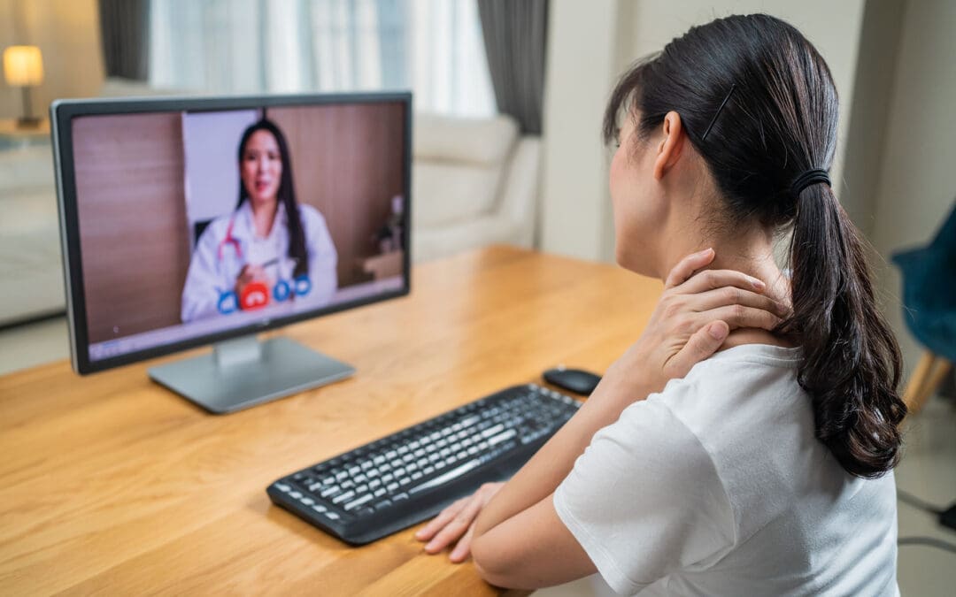
The Role of Telemedicine in Integrative Injury Care at El Paso Back Clinic: Providing Full Support for Car Accident, Work, and Sports Injuries in El Paso, TX
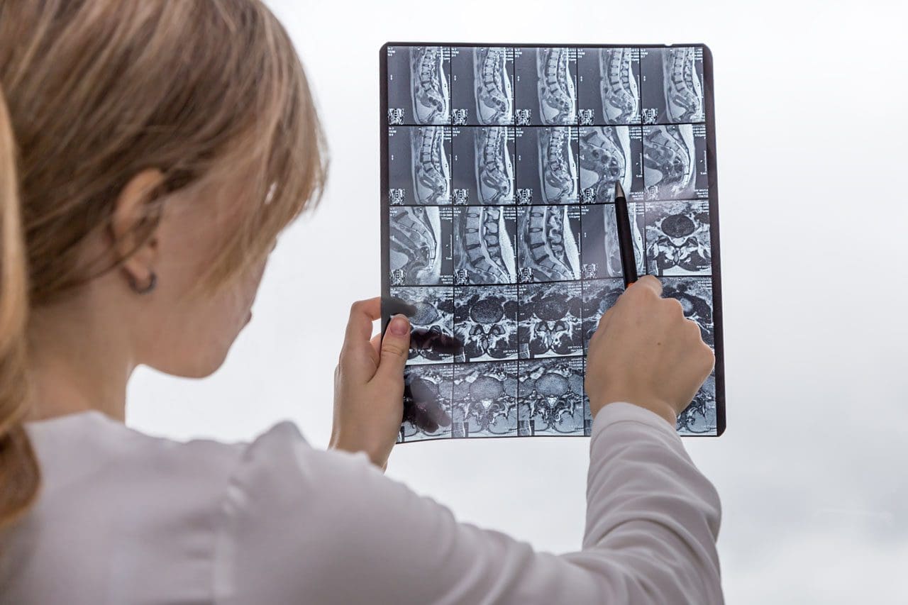
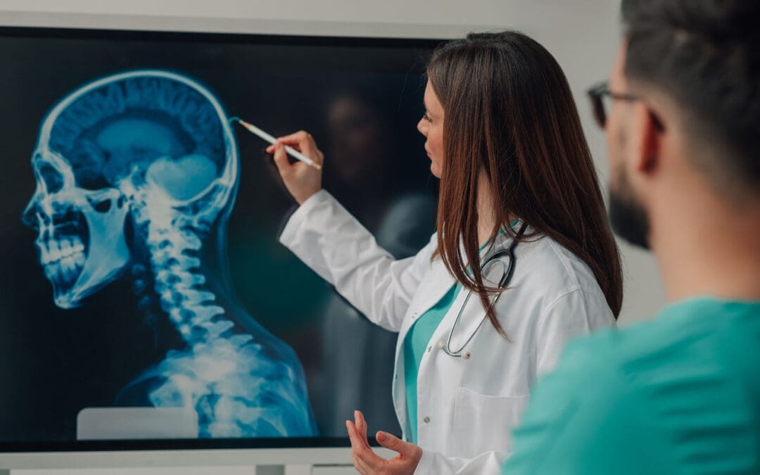
A doctor of chiropractic and a nurse practitioner review the MRI of a patient following a motorcycle collision.
In El Paso, TX, getting injured in a car crash, at work, or during sports can be tough. But at El Paso Back Clinic®, a top wellness chiropractic care spot, new tools like telemedicine make getting help simpler. Telemedicine uses video calls and online apps to let health experts care for you from home. This article explores how the clinic’s integrative nurse practitioner (NP) and chiropractor team up with telemedicine to provide comprehensive injury care. This covers virtual check-ups, treatment planning, and long-term help. It’s super useful for folks who can’t easily move or get to the clinic. The team also shares tips on eating, working out, and daily habits to speed up healing. They keep everything organized and documented for the best outcomes.
El Paso Back Clinic® focuses on functional medicine and holistic healing. Led by Dr. Alexander Jimenez, who is both a chiropractor (DC) and a family nurse practitioner (FNP-BC), the clinic combines conventional medicine with natural approaches to treat injuries. Telemedicine here means you can get exams, diagnoses, and follow-ups without leaving home. This is great for busy El Paso residents or for those who are hurting too much to travel. The clinic’s approach considers your whole body, with the NP and chiropractor working together to create plans that fit your life.
What Is Integrative Care at El Paso Back Clinic?
At El Paso Back Clinic®, integrative care means a team of doctors, therapists, and nutritionists working together to fully heal you. For car accident injuries like whiplash or back strains, the chiropractor adjusts your spine while the NP manages pain and checks for deeper issues. They make custom plans using evidence-based methods.
Common Injuries Treated: Neck pain from crashes, work lifts causing strains, or sports-related twists leading to sprains.
Why Choose Integrative?: It targets the cause, not just pain, blending adjustments with lifestyle changes.
Telemedicine’s Role: Allows remote care, so you start healing right away from home.
This method helps with lasting health. For sports fans in El Paso, tips on better nutrition can speed up recovery (Dallas Accident and Injury Rehab, n.d.).
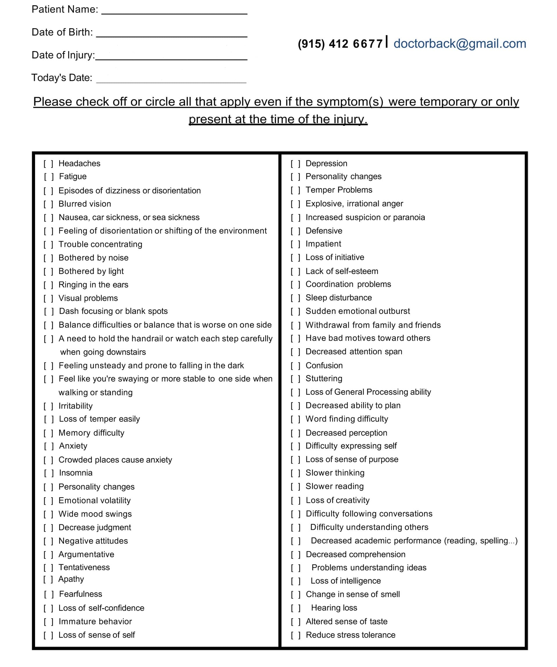
Head Injury/Traumatic Brain Injury Symptom Questionnaire
Virtual Examinations: How El Paso Back Clinic Does It Remotely
Telemedicine at El Paso Back Clinic® starts with virtual exams. You connect via secure video from your phone or computer. Dr. Jimenez or the team talks to you about your injury.
For a car accident, they ask about the crash and pain spots. They watch you move, like bending or walking, to check for swelling or stiffness. Even without hands-on involvement, they spot many problems, such as muscle pulls or nerve issues (Personal Injury Firm, 2025).
Work injuries, like slips, get quick virtual checks to stop things from getting worse. The chiropractor guides home tests, such as balance checks.
Tools in Virtual Exams: Video for movement, apps for sharing photos of injuries, or devices for vital signs.
When It’s Not Enough: Some need in-person touches, so they schedule clinic visits at their El Paso locations.
This remote setup makes getting checked easy, especially in El Paso, where traffic can be a hassle (CK Firm, 2024).
Diagnoses Through Telemedicine at the Clinic
After the exam, the team at El Paso Back Clinic® diagnoses remotely. Common ones from car accidents include whiplash or disc problems. The NP might order X-rays or MRIs, which are performed locally and shared online.
Chiropractors like Dr. Jimenez spot spinal shifts that can cause leg pain, such as sciatica. They explain it clearly on video. The NP assesses whole-body health, including whether swelling worsens.
All sessions are recorded for official documents, insurance keys, or personal injury claims (ChiroMed, n.d.).
Diagnosis Examples: Work-related back pain, sports-related nerve hits, and crash-neck strains.
Team Collaboration: NP handles meds; chiropractor does adjustments.
Tips for Accuracy: Describe pain and show motions well.
This reduces wait times, allowing you to start your El Paso recovery sooner (Complete Care, n.d.).
Managing Treatment Plans Remotely from El Paso Back Clinic
The NP and chiropractor create a treatment plan together, updated via telemedicine. For a sports knee sprain, it might include rest, ice, and shown exercises.
Dr. Jimenez demonstrates stretches on camera. The NP monitors pain and adjusts treatments.
They coordinate to avoid overlaps. For work injuries, plans cover safe job returns. Everything’s online for easy tracking.
Plan Essentials: Pain relief, movement work, and prevention advice.
Integrative Touches: Diet tweaks to cut swelling, like more omega-3 foods.
Telemedicine Updates: Regular video calls to tweak based on progress.
This saves time and money for El Paso patients (Jimenez, n.d.-a).
Ongoing Support and Follow-Up Care at the Clinic
Recovery needs steady help, and El Paso Back Clinic® uses telemedicine for easy follow-ups. Log in to chat about how you’re doing.
For car crash back pain, they check therapy effects and offer encouragement. Support includes mental health tips, as injuries can stress you.
Chiropractors guide home exercises on video. NPs watch for treatment side effects.
Support Types: Mood talks, progress logs, specialist referrals.
How Often: Weekly, early on, then less.
For El Paso Athletes: Safe return-to-play tips, like warm-ups.
This prevents pain from lasting, helping you get back to life fast (Prescient National, n.d.).
Benefits for El Paso Residents with Mobility or Access Issues
Injuries make moving hard, especially in spread-out El Paso. Telemedicine brings care to you.
No travel needed, perfect for remote areas or difficult days. For work injuries, it means less downtime. See pros from home.
Who Gains Most: Those pained by walking, without transport, or packed schedules.
Access Help: Shorter waits than office visits.
Legal Benefits: Docs care for claims without hold-ups.
This makes healing equal for all in El Paso (CK Firm, 2024).
Integrative Advice on Diet, Exercise, and Lifestyle from the Clinic
El Paso Back Clinic® shines with holistic telemedicine tips. They suggest anti-inflammatory foods, such as fruits, to aid healing.
Exercise advice includes easy yoga for pain, demonstrated online. Lifestyle shifts cover better sleep or stress cuts, like apps for calm.
For sports, they teach form to prevent re-injury.
Diet Ideas: Omega-3 for nerves, antioxidants for fixes.
Workout Suggestions: Stretches for range, walks for build-up.
Life Changes: Posture tweaks, drop bad habits.
This addresses root causes for better long-term health (Dallas Accident and Injury Rehab, n.d.).
Coordination and Documentation Between NP and Chiropractor at El Paso Back Clinic
The team shares notes easily on telemedicine platforms. Dr. Jimenez, as both NP and chiropractor, bridges the roles seamlessly.
Records from calls build your file, showing progress for insurance or courts.
Therapies align, like adjustments with rest plans.
Coordination Methods: Shared digital files, joint calls.
Record Value: Shows timely, excellent care.
Your Part: Update honestly for the top plans.
This leads to smooth recoveries in El Paso (Jimenez, n.d.-b).
Insights from Dr. Alexander Jimenez at El Paso Back Clinic
Dr. Alexander Jimenez, DC, APRN, FNP-BC, shares hands-on views from over 30 years at El Paso Back Clinic®. He uses telemedicine for same-day injury exams, like after crashes or sports.
He stresses integrative care for body and mind. For head injuries, he advises sleep, diet, and exercise. His dual license allows him to prescribe medications and adjust spines remotely when possible.
Jimenez highlights tests, such as MRIs, shared online. He combines adjustments in nutrition with other interventions for issues like gut health post-trauma.
Main Observations: Injuries are linked to overall health, like nerves and digestion.
Telemedicine in Practice: Quick virtual help for accidents, with shipped braces.
Tips: Use posture drills and supplements for healing.
His approach shows how the clinic’s NP-chiropractor team excels (Jimenez, n.d.-a; Jimenez, n.d.-b; Jimenez, n.d.-c).
Challenges and Future of Telemedicine at El Paso Back Clinic
Telemedicine has limits, such as the need for touch for some exams. Tech glitches can happen.
But the future is promising. Better apps and AI will improve diagnoses. More insurance covers it.
The clinic trains in remote teamwork.
Fixing Issues: Have in-person backups, help with tech.
Coming Trends: Wearables for live data.
Importance: Makes care more accessible and affordable in El Paso.
Conclusion
At El Paso Back Clinic® in El Paso, TX, telemedicine transforms injury care for car, work, or sports-related injuries. The integrative NP and chiropractor team, led by Dr. Jimenez, offers virtual exams for ongoing support. It includes holistic advice for better living. Ideal for mobility challenges. As Dr. Jimenez proves, this leads to quicker, fuller healing. If injured, reach out to El Paso Back Clinic® for easy, top-notch care at 915-850-0900 or visit their site.
Sports and Activities for TBI Recovery: The Role of Nurse Practitioners and Integrative Chiropractic Care
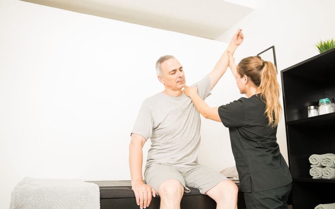
aquatic rehabilitation class for various injuries, including traumatic brain injuries
Traumatic brain injuries, or TBIs, happen when a sudden bump or blow to the head damages the brain. These injuries can come from car crashes, falls, or even sports accidents. Recovering from a TBI takes time and involves many steps to get back strength, balance, and clear thinking. One great way to help is through sports and activities tailored to a person’s needs. These are called adaptive sports. They can boost physical health and also lift moods by making people feel connected and strong. Along with that, healthcare experts like nurse practitioners and chiropractors play big parts in guiding recovery. Nurse practitioners help manage overall health and meds, while chiropractors focus on fixing spine issues and easing pain. This team approach, often called integrative care, mixes different treatments for better results.
In this article, we’ll look at sports that support TBI recovery, such as adaptive basketball and swimming. We’ll also cover calming activities such as tai chi and hiking. Then, we’ll explain how nurse practitioners and chiropractors fit into the picture, drawing on expert perspectives such as Dr. Alexander Jimenez, who combines chiropractic and nursing skills. By the end, you’ll see how these elements work together to create a comprehensive recovery plan.
Understanding TBIs and the Need for Active Recovery
A TBI can mess with how you move, think, and feel. Mild ones, like concussions, might cause headaches or dizziness. Severe ones can lead to long-term problems with balance or memory. The brain has a cool ability called neuroplasticity, which means it can rewire itself to heal. Activities that get you moving help spark this process by building new connections in the brain.
Doctors say rest is key right after a TBI, but then it’s time to add gentle exercise. Starting slow prevents more harm and builds up skills step by step. For example, light walking can improve blood flow to the brain, helping it heal faster. As you get better, more fun activities like games or outdoor adventures can keep things exciting and motivating.
Why activities matter: They improve strength, coordination, and mood.
Start small: Begin with easy tasks at home, like puzzles or stretching.
Build up: Move to group activities for social support.
Research shows that staying active after a TBI lowers the risk of depression and helps people get back to daily life sooner.
Adaptive Sports for Physical and Mental Healing
Adaptive sports are regular sports modified with special tools or rules so everyone can join, regardless of their limitations. For TBI survivors, these sports target balance, hand-eye skills, and thinking on your feet. They also build confidence by letting you achieve goals in a safe way.
Many groups offer adaptive sports programs, making it easy to find local options. Here’s a look at some top ones for TBI recovery:
Adaptive Basketball: Played in wheelchairs or with lower hoops, this sport boosts coordination and teamwork. It helps with quick decisions and arm strength, which TBIs often weaken. Groups like the National Wheelchair Basketball Association run events where players connect and stay motivated.
Cycling: Use adaptive bikes with extra wheels for stability. Cycling improves leg strength and heart health while being low-impact on joints. It’s great for building endurance without straining the brain too much.
Swimming: Water supports your body, making movements easier. Adaptive swimming uses floats or lanes for safety. It enhances balance and breathing control, plus the calm water reduces stress.
Canoeing: In adaptive versions, boats have seats or handles for support. Paddling builds upper body strength and focus. Being on water also calms the mind, helping with anxiety from TBIs.
These sports aren’t just exercise—they create social bonds. Playing with others fights loneliness, a common issue after brain injuries. Studies note that adaptive sports like these keep people active and linked to their communities. One review found that they improve gait and balance in patients with brain injury.
Other Rehabilitative Activities to Enhance Balance and Well-Being
Not all recovery needs to be high-energy. Slower activities like tai chi or hiking can rebuild skills without overwhelming the brain. These focus on mindful movement, which also supports mental health.
Tai Chi: This gentle martial art uses slow, flowing movements to improve balance and focus. For TBI patients, it reduces falls by strengthening core muscles. Classes often adapt poses for sitting if standing is difficult.
Hiking: Adaptive hiking uses trails with smooth paths or walking sticks. It increases heart rate and provides a refreshing change of scenery. Nature-based activities like this restore energy both physically and emotionally.
Adaptive Water Sports: Beyond swimming, try kayaking or water aerobics. These use buoyancy to reduce pressure on the body while improving coordination. Special gear, like life vests, ensures safety.
Home activities can start the process. Activities like balloon tosses or chair yoga build hand-eye coordination and flexibility. Online videos make it easy to try. As skills grow, add group classes for more challenge. Experts say even simple mobilizing, like walking circuits, aids recovery.
Special tools might be needed based on your strengths. For example, use bigger balls in games or stabilizers in cycling. Always check with a doctor to match activities to your healing stage.
The Role of Nurse Practitioners in Coordinating TBI Care
Nurse practitioners (NPs) are advanced nurses who can diagnose, treat, and manage health issues. In TBI recovery, they act as coordinators, making sure all parts of care fit together smoothly.
NPs monitor your overall health during activities. They check for signs like fatigue or headaches that might mean you’re pushing too hard. They also manage meds for pain or mood, adjusting doses as you improve. For instance, if swimming causes dizziness, an NP might suggest changes or add rest days.
In integrative teams, NPs work with other experts to create safe plans. They ensure activities like canoeing don’t clash with your meds or other treatments. Their focus on whole-person care includes emotional support to help with stress during recovery.
Dr. Alexander Jimenez, a chiropractor and family nurse practitioner, notes that NPs play a key role in linking brain health to daily wellness. His observations show they help with sleep and nutrition, which in turn boost activity benefits. This approach ensures activities are effective and safe.
Integrative Chiropractic Care: Supporting Spine and Pain Management
Chiropractors specialize in spine health, which is crucial after a TBI since head injuries often affect the neck. Integrative chiropractic combines adjustments with other therapies, such as exercises, for full recovery.
Chiropractors realign the spine to ease pressure on nerves, reducing headaches and improving balance. For TBI patients, this can help with dizziness from vestibular issues. They also manage pain without heavy meds, using hands-on techniques.
In recovery plans, chiropractors include exercises such as postural training and balance drills. These complement sports by building a strong base. For example, after an adaptive basketball session, a session might address any spine shifts from play.
Dr. Jimenez’s clinical work highlights how chiropractic aids brain healing. He uses gentle adjustments to improve blood flow and nerve function, key for TBIs. His teams integrate this with nutrition and rehab activities, such as light walking, to prevent reinjury. One method he supports is vestibular rehab, which pairs well with sports for better coordination.
Benefits of integrative chiropractic:
Reduces inflammation and pain.
Improves mobility for activities.
Prevents future issues through education.
Combining chiropractic with NP care creates a strong support system. NPs handle meds and monitoring, while chiropractors focus on physical fixes.
Combining Sports, Activities, and Professional Care for Best Results
The best TBI recovery programs combine adaptive sports, calming activities, and expert guidance. Start with a plan from your healthcare team. For example, begin with tai chi for balance, then add cycling as strength grows.
Community outings, like group hikes, apply skills in real life. These build confidence and social ties. Equine therapy, like therapeutic riding, is another option—horses’ movements aid gait and emotional health.
Dr. Jimenez observes that nutrition supports this, like anti-inflammatory foods for brain repair. His work shows that stress management is key, as it affects outcomes.
Track progress with tools like journals or apps. Adjust as needed with your NP or chiropractor. Over time, this leads to independence and joy in activities.
Challenges and Tips for Success
Recovery isn’t always smooth. Fatigue or setbacks can happen. Tips include:
Listen to your body—rest when needed.
Use adaptive gear for safety.
Join support groups for motivation.
With patience, most people see big gains. Studies show stepwise returns to activity, like in sports protocols, work well.
Conclusion
Recovering from a TBI through sports like adaptive basketball or activities like hiking builds both the body and the mind. Nurse practitioners coordinate safe care, while integrative chiropractic handles pain and alignment. Experts like Dr. Jimenez show how this blend speeds healing. Stay active, seek help, and celebrate small wins to pave the way for a brighter path ahead.
Tests Used for Brain Injuries at El Paso Back Clinic® in El Paso, TX
Doctor of Chiropractic and Nurse Practitioner show the imaging result to the patient post-auto-injury rehabilitation with mild brain injury
Brain injuries can strike without warning, from a simple slip at home to a tough hit during sports or a car crash on El Paso’s busy roads. At El Paso Back Clinic® in El Paso, TX, our team of wellness chiropractic care experts knows how vital it is to spot these issues early. We blend chiropractic skills with modern tools to help patients heal and get back to life. Led by Dr. Alexander Jimenez, DC, APRN, FNP-BC, our clinic focuses on whole-body wellness, using safe, non-invasive methods to check for head injuries.
This article dives into the tests we use at El Paso Back Clinic® to find brain injuries. We cover hands-on checks, brain function tests, and high-tech scans. Our goal is to give you clear info so you can seek help fast. Early detection means better recovery and fewer long-term problems.
The Importance of Spotting Brain Injuries Early at Our Clinic
Traumatic brain injuries (TBIs) happen when a bump or jolt shakes the brain. Mild ones, like concussions, might cause short-term headaches or dizziness. Serious cases can lead to lasting memory issues or mood changes. At El Paso Back Clinic®, we see many patients from local accidents or sports-related injuries, and we emphasize prompt action.
Our integrative approach mixes chiropractic care with nurse practitioner expertise. Dr. Jimenez uses his dual training to create custom plans. We check the spine, nerves, and brain together because a head injury often affects the neck as well.
Common signs: Headaches, confusion, nausea, or trouble balancing.
Why act fast: Stops swelling or bleeding from getting worse.
Our edge: Our wellness focus means we look at lifestyle and nutrition, too.
Research backs our methods—early tests lead to stronger outcomes (Pickett et al., 2024). At our El Paso, TX clinic, we guide you through every step.
Starting with Neurological Assessments for Head Injuries
At El Paso Back Clinic®, every brain injury check begins with basic neurological tests. These quick exams help us see how the brain responds right away. No need for big machines; it’s all about skilled observation.
We rely on the Glasgow Coma Scale (GCS) to grade injury severity. The Glasgow Coma Scale (GCS), which scores from 3 to 15, assesses eye opening, verbal response, and motor response. High scores mean mild issues; low ones signal urgency. Our team, including Dr. Jimenez, uses GCS to quickly decide on next steps (Bussières et al., 2022).
We also use the Standardized Assessment of Concussion (SAC). This tests memory and focus with simple questions. For athletes, the Sport Concussion Assessment Tool 5 (SCAT5) adds balance and neck checks. Kids receive the Child SCAT5 version.
GCS breakdown:
Eyes: 1 (none) to 4 (spontaneous).
Verbal: 1 (silent) to 5 (oriented).
Motor: 1 (none) to 6 (obeys commands).
SAC quick tips: Asks things like “What month is it?” or repeats word lists.
SCAT5 extras: Includes symptom checklists and coordination drills.
Dr. Jimenez notes that these tests often reveal neck problems linked to head injuries. At our wellness clinic, we adjust spines to ease related pain.
Hands-On Physical Exams to Uncover Hidden Issues
Physical checks are key at El Paso Back Clinic®. We touch and move areas to find pain, weakness, or limits. This builds on your story about how the injury happened.
Reflex tests tap spots, like the knees, to check nerve pathways. Odd responses might point to brain trouble. The Balance Error Scoring System (BESS) tests stability—stand in poses and count errors. It’s useful for detecting dizziness associated with TBIs (Sillevis et al., 2018).
We measure how far you can move your neck or head without pain. Strength tests have you push against our hands. These help link head injuries to spine misalignments.
BESS poses:
These include the double-leg stance, single-leg stance, and tandem pose.
Perform the exercises on both firm ground and foam to increase the challenge.
The errors to avoid include having hands off the hips, keeping eyes open, and falling.
Reflex checks: Hammer taps for quick reactions.
Motion tests: Gentle turns to spot restrictions.
For El Paso locals in car wrecks, these exams guide our chiropractic adjustments. Dr. Jimenez observes that early physical checks prevent chronic issues.
Cognitive Testing to Measure Brain Function
Head injuries can fog thinking. At El Paso Back Clinic®, we use cognitive tests such as ImPACT to assess memory and reaction time. This computer-based tool is perfect for concussion assessment, as it allows you to compare your scores to norms or baselines.
ImPACT includes modules for word recall, symbol matching, and symptom rating. It’s objective and tracks progress over time. We use it for return-to-work or play decisions (ImPACT Applications, Inc., 2023a).
Our nurse practitioners add deeper checks if needed, like repeating stories or drawing shapes. These rule out other causes.
ImPACT features:
Visual memory: Recall designs.
Reaction time: Click on the colors fast.
Symptom scale: Rate 22 items like fatigue.
Baseline testing: Ideal for athletes before seasons.
Retesting: Every 7-10 days to monitor healing.
Dr. Jimenez integrates ImPACT with chiropractic care, noting better results when spine health supports brain recovery.
Advanced Imaging for Clear Views of Injuries
Imaging lets us see inside. At El Paso Back Clinic®, we start with X-rays for bone alignment and fractures. They’re fast and help plan adjustments.
For deeper looks, CT scans catch bleeds quickly. MRIs show soft-tissue damage, such as bruising or tears—no radiation involved. We order these through our network for full pictures (NYU Langone Health, n.d.).
Digital Motion X-ray (DMX) is a favorite here—it films spine movement to spot instability from whiplash.
X-ray basics: Views bones in still shots.
CT strengths: 3D slices for emergencies.
MRI details: Magnets reveal hidden swelling.
DMX unique: Real-time video of neck motion.
Dr. Jimenez uses imaging to confirm diagnoses, ensuring safe, targeted care at our El Paso wellness clinic.
We love tools that avoid invasives at El Paso Back Clinic®. Surface Electromyography (sEMG) measures muscle activity via skin sensors. It identifies imbalances related to nerve issues post-head injury (Injury 2 Wellness Centers, 2023a).
Our INSiGHT scanners combine scans: Thermal for inflammation, Core for posture, Pulse for stress via heart rate. These insights help us create personalized plan maps (CLA Insights, 2023a).
sEMG benefits:
Detects tense muscles around the neck.
Guides gentle adjustments.
INSiGHT scans:
Heat patterns show hot spots.
Muscle scans check symmetry.
No risks: This procedure is safe for individuals of all ages.
These tools reduce the need for pokes or cuts, aligning with our wellness focus (Injury 2 Wellness Centers, 2023b). Dr. Jimenez says they boost patient involvement.
Nurse Practitioners’ Role in Comprehensive Testing
Our nurse practitioners at El Paso Back Clinic® expand options. They order blood tests for markers like inflammation or clotting risks. This rules out serious issues.
They incorporate a comprehensive approach by integrating chiropractic care into their holistic plans. If scans show problems, they coordinate referrals.
Blood work perks:
Checks for hidden infections.
Monitors healing proteins.
Team integration: NPs and chiros share findings.
Patient plans: Include rest, nutrition, and adjustments.
Dr. Jimenez, with his NP background, ensures seamless care.
Chiropractic Perspectives on Brain Injury Diagnosis
Chiropractors at our clinic see the spine-brain connection. Head hits often shift vertebrae, worsening symptoms. We use tools like Sigma for motion analysis (Kawa, n.d.).
Vestibular tests check eyes and balance. Does the patient experience pain during head movements? The source of the pain could be either the inner ear or the brain.
Spine focus:
Palpate for misalignments.
Grade Whiplash: 0-4.
Red flags: Send to ER for severe signs.
Recovery steps: Adjustments plus exercises.
Dr. Jimenez’s observations show that chiropractic care eases concussion symptoms more quickly.
Collaborative Care for Optimal Recovery
At El Paso Back Clinic®, teamwork rules. NPs order MRIs; chiros use them for adjustments. Shared tests like SCAT5 build complete views.
Plans cover therapy, diet, and follow-ups. Patients return stronger.
Benefits:
Full body healing.
Cost-effective.
Customized to you.
Success stories: Less pain, better function.
Dr. Jimenez’s integrative style shines in El Paso cases.
Insights from Daily Practice at the Clinic
We adapt tests to each patient. A work injury requires X-rays and ImPACT. Follow with BESS for balance gains.
Dr. Jimenez shares how INSiGHT scans catch early nerve stress, preventing long-term woes.
Patients love visual reports—they understand and stick to plans.
Overcoming Challenges in Brain Injury Detection
Access and cost can hinder. But our clinic offers affordable options and education.
Future tools, such as blood biomarkers, promise quicker diagnoses. We stay up to date for the best care.
Hurdles:
Rural limits in TX.
Insurance gaps.
Advances: AI for scan reads, more non-invasives.
Dr. Jimenez pushes for community awareness.
Final Thoughts: Seek Care at El Paso Back Clinic®
Brain injuries need prompt attention. At El Paso Back Clinic® in El Paso, TX, we use GCS, ImPACT, scans, and more for wellness-focused recovery.
If you’ve had a head hit, visit us. Our team, led by Dr. Jimenez, is here for you.
References
Bussières, A., et al. (2022). Concussion knowledge among North American chiropractors. Journal of the Canadian Chiropractic Association, 66(1), 17–26. https://pmc.ncbi.nlm.nih.gov/articles/PMC8791549/
Pickett, W., et al. (2024). Expanding concussion care in Canada: The role of chiropractors and policy implications. Journal of the Canadian Chiropractic Association, 68(2), 145–156. https://pmc.ncbi.nlm.nih.gov/articles/PMC11418793/
Sillevis, R., et al. (2018). Survey of chiropractic clinicians on self-reported knowledge and recognition of concussion injuries. Journal of the Canadian Chiropractic Association, 62(2), 84–95. https://www.ncbi.nlm.nih.gov/pmc/articles/PMC6000952/
Discover the relationship between somatovisceral disorders, head injuries, and effective management strategies.
Understanding Head Injuries and Their Impact on the Brain-Body Connection: A Comprehensive Guide to Somatovisceral Disorders and Non-Surgical Treatment Approaches
Head injuries represent a significant public health concern affecting millions of individuals worldwide each year. When someone experiences trauma to the head, whether from a sports collision, car accident, or fall, the resulting damage extends far beyond the initial impact site. These injuries create a cascade of physiological changes that disrupt the delicate communication system between the brain and body, leading to what researchers now recognize as somatovisceral disorders. Understanding how head injuries affect this vital brain-body connection and exploring effective non-surgical treatment options can make a meaningful difference in recovery outcomes and quality of life.
What Are Somatovisceral Disorders?
Somatovisceral disorders involve complex interactions between the body’s physical structures (somatic system) and its internal organs (visceral system). This intricate process consists of the transmission of nerve signals from bodily structures to visceral organs, creating specific physiological or pathological reactions. The complexity of somatovisceral response lies not only in its dual-system involvement but also in its capacity for bidirectional communication, allowing information to flow from somatic structures to visceral organs and vice versa. foundationhealth
The relationship between head injuries and somatovisceral disorders has gained increasing attention in medical research. Recent studies have demonstrated that mild traumatic brain injury (mTBI) may be a common precipitant of somatic symptom disorder, with research showing that 15-27% of patients who experienced head trauma met criteria for this condition at six months post-injury. This connection highlights how trauma to the brain can disrupt the normal communication pathways that regulate bodily functions, leading to persistent and often debilitating symptoms throughout the body. neurologyopen.bmj
Somatic symptom disorder occurs when individuals experience distressing physical symptoms combined with excessive thoughts, feelings, or behaviors related to those symptoms. Following a head injury, patients frequently report a wide range of somatic complaints, including pain, weakness, difficulty moving, headaches, dizziness, extreme tiredness, changes in vision or hearing, itching, numbness, abnormal movements, stomach aches, and nausea. These symptoms reflect the disrupted communication between the brain and various body systems, demonstrating how neurological damage can manifest as widespread physical dysfunction. chop+1
The Brain-Body Connection and Head Injury
The human nervous system operates through an intricate network that connects the brain to every organ, muscle, and tissue in the body. This communication highway relies on precise signaling between the central nervous system (brain and spinal cord) and the peripheral nervous system (nerves throughout the body). When head trauma occurs, this delicate communication system can become disrupted at multiple levels, affecting both somatic (voluntary) and autonomic (involuntary) nervous system functions.
According to Dr. Alexander Jimenez, a board-certified Family Practice Nurse Practitioner and Doctor of Chiropractic in El Paso, Texas, the spine houses the spinal cord, which acts as the communication superhighway between the brain and body. Any misalignment in the spine can disrupt the nervous system’s signals, and for traumatic brain injury patients, this connection becomes crucial. Dr. Jimenez explains that misalignment caused by the injury itself or associated whiplash can worsen symptoms like headaches, brain fog, and balance issues, emphasizing the importance of addressing both cranial and spinal components in recovery. northwestfloridaphysiciansgroup
The brain-body disconnect following trauma manifests as disrupted somatic sensory processing, encompassing vestibular (balance) and somatosensory (touch, pressure, temperature) processing. These sensory systems are primarily concerned with survival and safety, given the potential consequences of impaired balance or diminished awareness of physical threats. Following a head injury, trauma-related symptoms are conceptualized to be grounded in brainstem-level somatic sensory processing dysfunction and its cascading influences on physiological arousal modulation, affect regulation, and higher-order capacities. pmc.ncbi.nlm.nih
Research has identified that traumatic conditions may manifest as disrupted vertical integration, in which the balance between lower brain regions and higher cortical areas becomes dysregulated, particularly within the midline neural circuitry responsible for generating a primordial sense of a bodily and affective self as a coherent and stable entity in relation to the environment. This alteration has a cascading impact on the horizontal integration of cortical brain structures, meaning that different regions of the brain may be structurally intact yet lack fluid communication. pmc.ncbi.nlm.nih
Autonomic Dysfunction After Head Injury
One of the most significant yet underappreciated consequences of head injury is autonomic nervous system dysfunction. The autonomic nervous system controls involuntary bodily functions, including heart rate, blood pressure, digestion, breathing, and temperature regulation. Following moderate-to-severe traumatic brain injury, patients often experience significant autonomic dysfunction affecting both sympathetic (fight-or-flight) and parasympathetic (rest-and-digest) branches of this critical system. neurologyopen.bmj
Studies have demonstrated that patients with severe traumatic brain injury can experience sympathetic hyperactivity in the acute stages. More importantly, autonomic dysfunction persists in many patients for months or even years after their initial injury, affecting fully ambulant patients whom many might assume to be fully recovered. This persistent dysfunction occurs through various mechanisms, with the hallmark of moderate-to-severe traumatic brain injury being white matter injury caused by axonal shearing due to injury forces, continuing due to inflammation and delayed axonal degeneration in the chronic period, resulting in network disruption.neurologyopen.bmj
Autonomic dysfunction may occur due to injury to regions of the central autonomic network or their connecting white matter tracts. Brainstem nuclei and white matter connections to and from thalamic and basal ganglia regions may be particularly vulnerable to damage, underlying dysfunction that contributes to cognitive impairment post-traumatic brain injury. Given the importance of brainstem, thalamic, and basal ganglia circuits to autonomic function, injury to these white matter tracts may cause centrally mediated autonomic dysfunction. neurologyopen.bmj
The clinical manifestations of autonomic dysfunction after head injury are diverse and often debilitating. Many classic symptoms following concussion are, at least in part, likely a result of injury to the autonomic nervous system. Cognitive difficulties seen after mild traumatic brain injury may be related to autonomic dysregulation, specifically impaired cerebral blood flow. The presence of autonomic dysfunction has been shown to correlate with increased morbidity and mortality in moderate and severe traumatic brain injury, with perturbations of the autonomic nervous system consisting of either increased sympathetic or reduced vagal activity, potentially resulting in serious cardiac complications. health+1
Dr. Jimenez’s clinical practice emphasizes the importance of recognizing autonomic dysfunction in patients recovering from head injuries. His functional medicine approach includes detailed health assessments evaluating lifestyle, environmental exposures, and psychological factors to understand the root causes of chronic disorders and treat patients holistically. This comprehensive evaluation is particularly important for identifying autonomic dysfunction, which may manifest as dizziness, balance problems, temperature dysregulation, digestive issues, and cardiovascular irregularities.
Environmental Factors Affecting Brain Activity and the Body
Environmental factors play a critical role in shaping brain structure and function, as well as the development of mental and physical health conditions. The macroenvironment encompasses immediate factors such as air, noise, and light pollution; proximal factors, including regional socioeconomic characteristics; and distal factors, such as urbanization, natural spaces, and climate. These environmental exposures are mostly modifiable, presenting opportunities for interventions and strategies to promote the structural and functional integrity of the brain and mitigate the burden of illness following head injury. nature
Air pollution has emerged as a significant concern for brain health, particularly following traumatic brain injury, when the brain is already vulnerable. Studies have demonstrated that air pollution may increase vulnerability to mood dysfunction and potentially inhibit an appropriate stress response. Prolonged exposure to fine particulate matter (PM2.5 and PM10) has been associated with negative stress-related brain activation in the prefrontal cortex, frontoinsular cortex, limbic system, inferior parietal cortex, and cingulate regions. Magnetic resonance imaging studies reveal that increased exposure to PM2.5 is associated with changes in brain structure in older adults, including brain atrophy, that occur before the onset of dementia. environmentalhealth.ucdavis+1
Noise pollution, originating from urban traffic, airports, industries, and construction sites, can evoke negative emotions and disrupt recovery following head injury. Prolonged exposure to disruptive noise induces brain alterations through mechanisms such as sleep disturbances, which create a pro-oxidative environment that predisposes to neuroinflammation and heightened hypothalamic-pituitary-adrenal axis reactivity, contributing to mental and physical health problems. For individuals recovering from head trauma, protecting against excessive noise exposure becomes particularly important as the injured brain requires optimal conditions for healing. nature
Light pollution and exposure to artificial light at night have become increasingly prevalent, especially in urban areas, disrupting natural darkness and circadian rhythms. Light is detected by the retina and transmitted through intrinsically photosensitive retinal ganglion cells to the suprachiasmatic nucleus in the hypothalamus and other brain regions involved in regulating circadian rhythms and sleep-wake cycles. Circadian rhythm disruptions have been linked to elevated risk of mood disorders, bipolar disorders, and heightened mood instability, potentially mediated by oscillations in clock gene expression responsive to light-dark transitions. nature
Following traumatic brain injury, circadian rhythm disruptions become even more pronounced. Research has documented that traumatic brain injury can lead to decreased melatonin release, causing circadian rhythm delays. Studies using animal models have revealed that acute subdural hematoma resulted in dysregulation of circadian gene expression and rhythmic changes in body temperature during the first 48 hours post-injury. The regulation of biological rhythms through changes in core body temperature, pineal gland melatonin secretion, and blood cortisol levels becomes disrupted, affecting the body’s ability to anticipate and adapt to environmental changes. practicalneurology+1
Minor traumatic brain injury contributes to the emergence of circadian rhythm sleep disorders, with research identifying two distinct types: delayed sleep phase syndrome and irregular sleep-wake pattern. These disorders differ in subjective questionnaire scores and have distinct profiles of melatonin and temperature circadian rhythms. The alteration in the circadian timing system partially accounts for the presence of post-traumatic brain injury sleep-wake disturbances, which changes in sleep architecture alone cannot fully explain.pubmed.ncbi.nlm.nih+1
How Head Injuries Affect Daily Tasks and Routines
The impact of head injuries extends far beyond the initial trauma, profoundly affecting an individual’s ability to perform everyday activities and maintain normal routines. The disruption to brain-body communication creates challenges across multiple domains of daily functioning, from basic self-care tasks to complex cognitive and social activities. Understanding these impacts helps patients, families, and healthcare providers develop realistic expectations and appropriate support strategies during recovery.
Cognitive fatigue represents one of the most disabling consequences of traumatic brain injury, affecting 21-73% of patients regardless of injury severity or time since injury. Fatigue has been identified as the main cause of disability after traumatic brain injury, negatively affecting social, physical, and cognitive functions as well as participation in daily activities and social life. At the neural level, patients with fatigue following head injury exhibit significant disruption of global resting-state alpha-band functional connectivity between cortical midline structures and the rest of the brain. Furthermore, individuals with fatigue show reduced overall brain activation during cognitive tasks, without time-on-task effects. academic.oup
Adults with a history of even mild traumatic brain injury report significantly greater fatigue and cognitive impairment than those with no history of head trauma, with symptoms becoming more profound with greater injury severity. This persistent fatigue affects the ability to maintain attention, concentrate on tasks, process information efficiently, and sustain mental effort throughout the day. Patients frequently report that activities requiring cognitive engagement become increasingly difficult as the day progresses, leading to a pattern of morning productivity followed by afternoon exhaustion. pubmed.ncbi.nlm.nih+1
Memory difficulties present another significant challenge affecting daily functioning after a head injury. Patients may struggle with both short-term working memory (holding information in mind while using it) and long-term memory formation (creating new lasting memories). These memory challenges affect practical tasks such as remembering appointments, following multi-step instructions, recalling conversations, and learning new information or skills. The impact extends to occupational functioning, with studies finding a correlation between higher levels of mental fatigue and lower employment status following traumatic brain injury. headway+1
Executive function impairments following head injury affect planning, organization, decision-making, problem-solving, and behavioral regulation. These higher-order cognitive processes are essential for managing daily responsibilities, from planning meals and organizing household tasks to managing finances and making important life decisions. Patients may find themselves struggling with tasks that previously seemed automatic, requiring conscious effort and external supports to maintain daily routines. headway
Sensory processing alterations create additional challenges for daily functioning. The vestibular system, which contributes to balance, spatial processing, arousal modulation, first-person perspective, and social cognition, becomes particularly vulnerable following head trauma. Disturbed temporal binding of sensory information creates perceptual chaos and a lack of coherence, which may lead to bodily disconnection and states of hypervigilance. Patients describe feeling disconnected from their bodies, experiencing the world as if through a fog, or feeling constantly on guard against potential threats. pmc.ncbi.nlm.nih
Balance and coordination problems stemming from vestibular dysfunction affect mobility and safety in daily activities. Simple tasks like walking on uneven surfaces, turning the head while moving, or navigating busy environments become challenging and potentially dangerous. Many patients report increased anxiety about falling, leading to activity restriction and social withdrawal. Over one-third of adults over 40 will experience vestibular dysfunction at some point in their lives, and when it occurs, whether by injury, aging, or disease, individuals can experience vertigo, nauseating dizziness, vision, and balance problems affecting every area of life. neuroinjurycare+1
Dr. Jimenez’s practice in El Paso focuses extensively on helping patients restore function and return to daily activities following head injuries. His integrated approach combines chiropractic care, functional medicine, and rehabilitation therapies to address the multiple systems affected by head trauma. By evaluating the connections between physical, nutritional, and emotional factors, Dr. Jimenez develops personalized care plans that recognize the complex ways head injuries disrupt daily functioning and quality of life.
Overlapping Risk Profiles and Symptoms Associated With Head Injuries
Head injuries create overlapping risk profiles affecting multiple body systems simultaneously, leading to complex symptom presentations that can challenge both patients and healthcare providers. Understanding these interconnected risk factors and symptoms is essential for comprehensive assessment and treatment planning. Individuals who sustain head injuries develop an increased risk for somatic symptom disorder, with early illness beliefs playing a significant predictive role. Specifically, believing that mild traumatic brain injury has serious life consequences and causes distress in the weeks following injury is associated with later development of somatic symptom disorder. Patients with somatic symptom disorder after head injury report more pain and post-concussion symptoms. They are significantly more likely to have comorbid major depressive disorder and anxiety disorders compared to those without this condition. neurologyopen.bmj
The systematic review examining the relationship between somatic symptoms and related disorders and mild traumatic brain injury found that the majority of acceptable evidence supported a relationship between these conditions. Nine studies reported associations between functional seizures and a history of mild traumatic brain injury. In comparison, 31 studies assessed relationships between questionnaires measuring somatic symptom disorder burden and mild traumatic brain injury. Three studies investigated healthcare practitioners’ diagnosis of somatic symptoms and related disorders and post-mild traumatic brain injury symptom burden, collectively demonstrating the strong connection between head trauma and subsequent development of somatic complaints. foundationhealth
Cardiovascular complications represent another significant overlapping risk following head injury. Research demonstrates that individuals with moderate-to-severe traumatic brain injury have increased rates of self-reported hypertension and stroke but lower rates of myocardial infarction and congestive heart failure than uninjured adults. The findings highlight the importance of early screening for and management of cardiovascular risk factors in individuals with chronic traumatic brain injury, particularly those of younger age, who are not typically thought to be at risk for these conditions. ahajournals
The relationship between blood pressure and traumatic brain injury follows a complex U-shaped pattern, with both hypotension and hypertension associated with worse outcomes. Early hypotension has been linked with poor outcomes following severe traumatic brain injury, but recent data suggest that arterial hypertension after injury is also associated with poor outcomes. The initial catecholamine response and resulting systemic hypertension may be protective to a point by maintaining cerebral perfusion pressure in the setting of impaired cerebral autoregulation after injury. Yet, catecholamine-induced hypertension may also cause secondary brain damage by aggravation of vasogenic edema and intracranial hypertension. pmc.ncbi.nlm.nih
Post-traumatic headaches affect approximately 40% of individuals who experience concussions, representing one of the most common and persistent symptoms following head injury. Patients can experience tension headaches, migraine headaches, and cervicogenic headaches (radiating from the neck) all at once, making treatment particularly challenging. Ninety-five percent of people with a concussion experience headache associated with that injury, and among those with headache, about two-thirds have migraine features. Individuals with a family history of migraine or preexisting headache disorders face a higher risk of developing post-traumatic headache. wexnermedical.osu+1
Sleep disturbances cluster with other post-traumatic brain injury symptoms, creating compounding difficulties for recovery. Changes in sleep architecture following injury cannot fully explain the extent and intensity of sleep-wake disturbances reported by patients. The current literature supports cognitive-behavioral therapy and sleep hygiene education, light therapy, and certain pharmacologic interventions for treating sleep disturbances in patients with brain injury, with early screening and individualized approaches prioritized to improve sleep and, consequently, speed recovery. pubmed.ncbi.nlm.nih
Exercise intolerance commonly results from a concussion, often limiting return to activities and quality of life. The reviewed studies support clinical suspicion of autonomic dysfunction as an important component of exercise intolerance, though specific mechanisms of impairment and relationships to symptoms and recovery require additional investigation. Post-concussive exercise intolerance has been linked to a reduction in cerebral blood flow, theoretically prolonging the effects of the metabolic energy crisis associated with injury. pmc.ncbi.nlm.nih
Mental health complications, including anxiety, depression, post-traumatic stress disorder, and behavioral changes, frequently develop following head injury. Brain injuries, no matter how severe, commonly cause emotional and behavioral changes, including emotional lability with extreme mood swings, anxiety disorders, depression, impulsive behaviors, flat affect, a lack of emotional expression, and a lack of empathy and social skills. These psychological changes can cause unnecessary suffering and, in cases of severe depression and anxiety, can even halt physical recovery progress. flintrehab
Understanding Long-Lasting Injuries- Video
Non-Surgical Treatments to Improve Somatovisceral Function
Fortunately, numerous non-surgical treatment approaches have demonstrated effectiveness in improving somatovisceral function and promoting recovery following head injuries. These interventions work through various mechanisms to restore proper communication between the brain and the body, balance the autonomic nervous system, and support the brain’s natural healing processes. Dr. Jimenez’s clinical practice emphasizes comprehensive non-invasive protocols, prioritizing natural recovery and avoiding unnecessary surgeries or medications.
Chiropractic Care and Spinal Adjustments
Chiropractic care focuses on the spine and nervous system, recognizing that the spine houses the spinal cord, which acts as the communication superhighway between the brain and body. For traumatic brain injury patients, proper spinal alignment becomes crucial because misalignment caused by the injury itself or associated whiplash can worsen symptoms like headaches, brain fog, and balance issues. Chiropractic care aims to restore proper alignment, thereby improving nervous system function and supporting the brain’s ability to heal. northwestfloridaphysiciansgroup Chiropractic adjustments help alleviate post-traumatic brain injury symptoms by releasing pressure on irritated nerves and improving joint function. For many patients, this results in improved comfort and reduced reliance on pain medication. Proper spinal alignment promotes better blood flow to the brain. Since the brain requires oxygen-rich blood to heal and function, improved circulation directly supports recovery from traumatic brain injury while reducing dizziness and fatigue. northwestfloridaphysiciansgroup
Research demonstrates that chiropractic intervention can modify proprioceptive input from more functional spinal joints, helping restore this input to the brain’s multisensory integration centers. Studies of patients receiving chiropractic care in neurorehabilitation hospitals have shown that spinal manipulation influences pain through complex mechanisms in the central nervous system. A case study documenting concussion treatment using massage and manipulation techniques showed diminished concussion symptoms. It regained ease in cervical range of motion, highlighting the potential importance of manual therapy in reducing headache, dizziness, and nausea during concussion recovery. pmc.ncbi.nlm.nih+2 Dr. Jimenez explains that by realigning the spine through chiropractic adjustments, treatment reduces nerve interference, optimizing mind-body communication, and enhancing overall function. The adjustments improve cerebral blood flow and reduce inflammation, thereby accelerating recovery from head injury. With enhanced nervous system function comes improved mental clarity, including reduced brain fog, sharper focus, and better memory, while also promoting stress relief and alleviating irritability and emotional strain often linked to head injuries. zakerchiropractic
Vestibular Rehabilitation
Vestibular rehabilitation is a specialized form of physical therapy that focuses on strengthening the connections between the brain, eyes, inner ear, muscles, and nerves. This treatment approach proves particularly valuable for post-concussion patients experiencing dizziness, vertigo, balance problems, and spatial impairment. According to a review in the British Journal of Medicine, vestibular therapy reduced symptoms in patients with sports-related concussions faster, with patients three times as likely to return to play within eight weeks of therapy compared to those who didn’t receive treatment. denverphysicalmedicine+1 Vestibular rehabilitation therapy involves exercises designed to improve the functioning between the inner ear, brain, eyes, muscles, and nerves. These exercises help minimize balance issues and treat dizziness, vertigo, and spatial orientation deficits caused by vestibular impairments that some individuals experience after brain injury. The therapy addresses problems in the inner ear through specific exercises designed to improve balance and coordination. biausa
The Epley Maneuver represents a simple yet effective exercise to treat benign paroxysmal positional vertigo, a very specific form of vertigo quite common after traumatic brain injury. During vestibular rehabilitation, benign paroxysmal positional vertigo generally responds well to the Epley Maneuver, and patients learn to perform the movement at home to alleviate symptoms as they arise. Studies have shown that vestibular rehabilitation is an effective modality for managing dizziness, vertigo, and imbalance following concussion. However, careful consideration of the injury’s acuity and effective management of comorbid conditions will optimize results. pubmed.ncbi.nlm.nih+1 Co-morbidities, including cognitive and behavioral issues, visual-perceptual dysfunction, metabolic dysfunction, and autonomic dysfunction, may hamper the effectiveness of traditional vestibular rehabilitation approaches. Working closely with other disciplines well-versed in treating comorbid conditions helps individuals achieve optimal recovery. Dr. Jimenez’s integrated practice model exemplifies this multidisciplinary approach, bringing together chiropractic care, functional medicine, physical therapy, and other specialties to provide comprehensive treatment for patients with vestibular dysfunction following head injuries. pubmed.ncbi.nlm.nih
Physical Therapy and Exercise Rehabilitation
Physical therapy plays a pivotal role in optimizing recovery and enhancing functional independence after brain injury. Therapeutic approaches include gait training to improve walking patterns, balance activities to enhance stability and prevent falls, strength training to rebuild muscle mass and function, coordination exercises to improve fine and gross motor skills, and range-of-motion exercises to maintain flexibility. In some cases, physical therapists recommend body-weight-supported treadmill training to help patients safely relearn walking patterns. Family and caregiver training proves extremely important and helpful, as loved ones can gain an understanding of how the brain works and the specific nature of the injury, supporting the rehabilitation process. biausa
Available evidence demonstrates the potential of exercise in improving cognitive impairment, mood disorders, and post-concussion syndrome following traumatic brain injury. Exercise rehabilitation has been shown to attenuate cognitive deficits in animal models by stimulating cerebral signaling pathways, with treadmill exercise improving memory by modulating neurotransmitter systems and neurotrophic factors. High-intensity interval training helps regulate the autonomic nervous system while boosting brain-derived neurotrophic factor, thereby promoting neuroplasticity, an essential factor for recovery. sciencedirect+1 However, exercise prescription following head injury requires careful consideration, as exercise intolerance commonly results from concussion and autonomic dysfunction. Graded exercise testing while monitoring symptoms and heart rate helps guide a safe return to physical activity. Current clinical practice involves careful assessment to determine appropriate exercise intensity and duration, gradually progressing as autonomic function improves. pmc.ncbi.nlm.nih
Acupuncture and Neuroplasticity Enhancement
Acupuncture has gained widespread recognition as an effective, low-cost treatment for neurological rehabilitation with minimal adverse effects. Clinical and experimental evidence documents the potential of acupuncture to ameliorate injury-induced neurological deficits, particularly sequelae such as dyskinesia, spasticity, cognitive impairment, and dysphagia. These effects relate to acupuncture’s ability to promote spontaneous neuroplasticity after injury. pmc.ncbi.nlm.nih+1 Specifically, acupuncture can stimulate neurogenesis, activate axonal regeneration and sprouting, and improve the structure and function of synapses. These processes modify the neural network and the function of the damaged brain area, leading to improvements in various skills and adaptability. Astrocytes and microglia may be involved in acupuncture-induced regulation of neuroplasticity, for example, by producing and releasing multiple neurotrophic factors, including brain-derived neurotrophic factor and nerve growth factor. pmc.ncbi.nlm.nih
Studies have shown that acupuncture reduces neuroinflammation after brain injury. A study published in The Journal of Neuroinflammation found that acupuncture significantly reduced neuroinflammation and improved cognitive function in animal models of brain injury. By modulating inflammatory pathways, acupuncture helps reduce the production of pro-inflammatory cytokines, promoting brain healing and reducing symptoms such as headaches and dizziness. betsygordonacupuncture Acupuncture enhances neuroplasticity, which is crucial for recovery after brain injury, facilitating improvements in memory, learning, and overall cognitive function. Research in Neural Regeneration highlighted that acupuncture promotes neuroplasticity, which is essential for rehabilitation. Studies demonstrate that acupuncture improves mental performance and reduces anxiety and depression in patients recovering from brain injuries. betsygordonacupuncture+1 Dr. Jimenez’s functional medicine practice incorporates acupuncture and electro-acupuncture as part of comprehensive care plans for patients recovering from head injuries. His team uses these modalities in combination with other therapies to create customized treatment approaches that promote natural healing, mobility, and long-term wellness.
Nutritional Interventions and Functional Medicine
Nutrition plays a positive role during acute traumatic brain injury recovery, with patient needs being unique and requiring individualized approaches. Following mild traumatic brain injury, patients who consumed enough food to meet calorie and macronutrient (particularly protein) needs specific to their injury severity and sex within 96 hours post-injury had reduced length of hospital stay. Patients receiving nutrients and non-nutrient support within 24-96 hours post-injury had positive recovery outcomes, including omega-3 fatty acids, vitamin D, magnesium oxide, N-acetyl cysteine, and hyperosmolar sodium lactate. frontiersin Traumatic brain injury contributes to extensive dysbiosis of the gastrointestinal system, leading to worsened outcomes, making nutritional support essential. Early nutrition supports preservation of muscle mass, decreases infection complications, promotes cerebral homeostasis, and improves recovery outcomes. The human brain consumes 20% of total resting energy, despite accounting for only 2% of total body mass, underscoring the critical role of adequate nutrition for healing. xiahepublishing
A recent clinical trial demonstrated that dietary changes significantly reduce persistent post-traumatic headaches, a common and debilitating consequence of traumatic brain injury. Researchers found that increasing omega-3 fatty acids (commonly found in fatty fish) while reducing omega-6 fatty acids (abundant in seed oils) led to fewer and less severe headaches. Participants assigned to the intervention diet experienced approximately two fewer headache days per month and a 30% reduction in daily headache pain intensity compared to the control diet group. med.unc Supplementing with omega-3 fatty acids can reduce inflammation and oxidative stress, promote brain-cell survival, and help the brain recover from injury. Vitamins D and E, niacin, zinc, and magnesium have neuroprotective benefits, and supplementing with these vitamins and minerals has been shown to improve recovery, especially in deficient patients. An energy-balanced, anti-inflammatory diet with adequate sources of omega-3 fats and appropriate vitamin D supplementation proves especially important for patients with a history of traumatic brain injury. consultant360
Dr. Jimenez’s practice embraces Functional Integrative Medicine, a patient-focused approach that treats the whole person rather than just symptoms. His team offers detailed health assessments that evaluate genetics, lifestyle, environmental exposures, and psychological factors to create comprehensive health profiles. By combining Institute for Functional Medicine programs with personalized nutrition plans, Dr. Jimenez helps patients address chronic conditions and optimize brain health following head injuries.
Massage Therapy and Manual Techniques
Massage therapy provides valuable support in brain injury rehabilitation, enhancing physical, mental, and emotional well-being. Massage significantly improves blood circulation, ensuring that essential nutrients and oxygen are efficiently delivered to brain cells. By increasing circulation, the brain’s healing process is expedited, promoting cellular regeneration and reducing the risk of secondary complications. Improved blood flow also helps reduce swelling and inflammation, common challenges following brain injury. neuropraxisrehab Post-brain injury pain can be debilitating and hinder recovery, but massage therapy helps alleviate pain by targeting tense muscles and releasing built-up tension. Through gentle manipulation, massage therapists can improve muscle flexibility and joint mobility, relieving discomfort and enhancing overall physical comfort. Brain injuries often lead to muscle stiffness and reduced range of motion, but massage therapy techniques such as stretching and kneading help improve flexibility by breaking down scar tissue and adhesions. neuropraxisrehab
Specific massage modalities show promise for traumatic brain injury recovery. Manual Lymphatic Drainage uses light massage to stimulate the flow of lymphatic fluid, potentially increasing the lymphatic system’s ability to clear waste products from the brain. A case study combining Manual Lymphatic Drainage with craniosacral therapy and glymphatic system techniques resulted in an 87% reduction of concussion symptoms after three months of treatment. concussionalliance A case study documenting massage intervention for post-concussion treatment demonstrated complete return to pre-concussion activities and function with no continued symptoms following a short and specific massage series. The therapy focused on restoring ideal alignment of the atlanto-occipital joint, resulting in reduced pain, muscle hypertonicity, headaches, reduced medication use, and improved balance, posture, cervical range of motion, mental focus, and physical activity. pmc.ncbi.nlm.nih
Dr. Jimenez’s comprehensive approach includes specialized massage and manual therapy techniques, integrated with chiropractic care and other modalities. His team focuses particularly on neck and shoulder areas to reduce effects patients experience after traumatic brain injuries, with goals including improved neck mobility, reduction of headaches and nerve pain, and addressing balance, dizziness, and vertigo issues through specific therapeutic techniques. newapproachescenter
Cognitive Behavioral Therapy and Psychological Support
Cognitive Behavioral Therapy has been demonstrated to be effective by over 1,000 studies involving 10,000 patients, making it one of the most scientifically verified psychotherapy treatments available. CBT has been successfully used on a variety of disorders, including traumatic brain injury patients with post-concussional symptoms and secondary effects such as anxiety and fatigue. The therapy focuses on the relationship between thoughts, feelings, and behaviors, built around three core principles: beliefs create feelings, feelings dictate behavior, and behavior reinforces beliefs.flintrehab A new meta-analysis found substantial evidence for the use of cognitive behavioral therapy in managing anxiety and depression in patients with traumatic brain injury. Researchers identified that CBT interventions had immediate effects of reducing depression and anxiety, with sustained impacts for depression at the three-month follow-up. Effects were greater in groups that received individualized CBT than in those that received group-based CBT. headway
CBT proves particularly valuable for addressing recovery expectations and perceived consequences of traumatic brain injury. Behavioral techniques such as relaxation, behavioral activation, and stress management help patients manage the anxiety, depressive symptoms, and insomnia that can be present following injury. In the acute phase of recovery, brief psychoeducational and cognitive behavioral interventions have consistently been shown to result in improvement in managing cognitive and psychological symptoms for brain injury survivors. abct For patients with cognitive impairment, CBT can be adapted with modifications including simplified concepts, concrete behavioral examples, pictorial handouts and cues, considerable repetition, and booster sessions. Studies found that adapted CBT was able to reduce anxiety and depression in patients who suffered moderate to severe traumatic brain injury. CBT helps patients identify and challenge unhelpful or inaccurate thoughts that can arise or intensify after injury, while focusing on behavioral activation and engaging in meaningful, important activities, which can boost mood and decrease isolation. cbtdenver+1
Mind-Body Therapies and Somatic Approaches
Mind-body therapies have gained recognition for their effectiveness in treating trauma-related symptoms and supporting nervous system regulation. More than 80% of specialized programs to treat post-traumatic stress disorder offer some form of mind-body therapy, including yoga, relaxation, tai chi, guided imagery, and mindfulness practices. These approaches prove particularly valuable for individuals experiencing somatic symptoms following head injuries. research.va Somatic therapy helps individuals reconnect with their bodies through awareness of physical sensations and their relationship to emotional experiences. For patients with head injuries who may feel disconnected from their bodies or experience persistent physical symptoms, somatic approaches provide pathways for healing by working through sensations in safe and supportive environments. Techniques such as grounding exercises, deep breathing, mindful observation of physical sensations, and guided movement empower individuals to explore how trauma manifests physically and provide avenues for release. pacmh
Yoga as a whole significantly reduced post-traumatic stress disorder symptoms in research studies, with a positive impact comparable to that of psychotherapeutic and psychopharmacologic approaches. Yoga may improve the functioning of traumatized people by helping them tolerate physical and sensory experiences associated with fear and helplessness, and increasing emotional awareness and affect tolerance. For individuals recovering from head injuries, gentle yoga practices adapted to their current functional abilities can support both physical and psychological healing. research.va The Polyvagal theory provides a powerful framework for understanding how trauma affects the nervous system and pathways for healing. The theory centers on the autonomic nervous system as a key component in trauma recovery, emphasizing the role of the vagus nerve in regulating physiological and emotional states. Basic somatic exercises can bring the nervous system out of dysfunction, beginning to retrain safety and social cues. This proves particularly helpful for individuals with head injuries who experience autonomic dysregulation and hypervigilance. pyramid-healthcare
Breathing Practices and Vagal Tone Restoration
Voluntary regulated breathing practices offer accessible and effective means to support autonomic nervous system regulation and restore vagal tone. These practices draw on both modern scientific studies and ancient concepts, with applications ranging from clinical anxiety treatment to stress reactivity reduction. Effective breathing interventions support greater parasympathetic tone, which can counterbalance the high sympathetic activity intrinsic to stress and dysfunction following head injury. pmc.ncbi.nlm.nih The physiological sigh is a simple yet powerful breathing technique that involves two nose inhales, followed by a long exhale through the mouth. This technique rapidly reduces stress and calms the nervous system by leveraging the interaction between the sympathetic (arousing) and parasympathetic (calming) branches of the autonomic nervous system to control heart rate and promote calm. Studies have shown that this breathing pattern effectively reduces arousal and returns the body to baseline functioning. hubermanlab+1
Deep, slow breathing benefits vagal outflow, with evidence suggesting particular benefits for older adults in restoring vagal tone. One session of deep and slow breathing can produce measurable improvements in heart rate variability metrics associated with parasympathetic activity. Regular practice of paced breathing at approximately six cycles per minute, significantly lower than the standard respiratory rate of 12 to 20 breaths per minute, can enhance vagal tone and improve overall autonomic regulation. pmc.ncbi.nlm.nih+1 Heart rate variability biofeedback is an innovative, non-invasive, evidence-based technique that enhances vagal nerve activity by combining slow-paced breathing with real-time feedback. The practice proves simple to implement, cost-effective, and carries minimal risk, making it an accessible tool for various health interventions. HRV biofeedback likely modulates neuroplasticity in autonomic control centers, enhancing parasympathetic tone and improving cardiac efficiency, reducing sympathetic overactivation, and lowering systemic inflammation. pmc.ncbi.nlm.nih
Improving Central Nervous System Function and Communication
The comprehensive non-surgical treatments described work synergistically to improve central nervous system function and restore proper communication between the brain and body. These approaches target multiple aspects of neurological health, from cellular-level processes to whole-system integration, supporting the brain’s remarkable capacity for adaptation and healing known as neuroplasticity. Neuroplasticity represents the brain’s ability to reorganize and form new neural connections throughout life, enabling recovery from injury by creating alternative pathways when original circuits become damaged. Following a brain injury, neuroplasticity’s ability to adapt becomes crucial, as these injuries frequently result in severe impairments. Rehabilitation strategies exploit neuroplasticity, leveraging the brain’s plasticity to promote healing through approaches ranging from constraint-induced movement therapy to virtual reality and brain-computer interfaces. pmc.ncbi.nlm.nih
The integration of multiple treatment modalities enhances neuroplastic responses and accelerates recovery. Combining chiropractic care with vestibular rehabilitation, for example, addresses both spinal alignment and sensory integration, creating synergistic effects that amplify benefits beyond what either treatment could achieve alone. Similarly, pairing nutritional interventions with physical therapy provides both the structural building blocks and functional stimulation necessary for optimal neural repair and reorganization. frontiersin+4 Dr. Jimenez’s practice exemplifies this integrated approach, combining specialized chiropractic protocols with wellness programs, functional and integrative nutrition, agility and mobility fitness training, and rehabilitation systems for all ages. The team has taken great pride in providing patients with only clinically proven treatment protocols, using an integrated approach to create personalized care plans that often include functional medicine, acupuncture, electro-acupuncture, and sports medicine principles. The goal is to relieve pain naturally by restoring the body’s health and function through holistic wellness as a lifestyle.
Restoring Vagal Tone and Autonomic Balance
The vagus nerve, as the main neural component of the parasympathetic nervous system, plays a crucial role in maintaining physiological homeostasis. The vagus nerve starts in the brain and ends in the abdomen, and it controls the involuntary functions of the heart, lungs, digestive system, liver, and kidneys. Following a head injury, vagal tone frequently becomes diminished, contributing to autonomic dysfunction and associated symptoms. pmc.ncbi.nlm.nih+3 Heart rate variability is a non-invasive biomarker of vagal tone and autonomic flexibility, with reduced HRV associated with cardiovascular diseases, hypertension, inflammation, and mental health disorders. Non-invasive vagal neuromodulation through HRV biofeedback and similar interventions could potentially serve as rehabilitative strategies to restore autonomic balance, mitigate post-injury fatigue, and improve cardiovascular function. pmc.ncbi.nlm.nih
Practices such as breathwork, cold exposure, exercise, meditation, taking probiotics, laughter, singing, massages, and relaxation exercises help improve vagal tone. These accessible interventions provide multiple pathways for patients to actively participate in their recovery, building resilience and enhancing the body’s natural regulatory capacities. High vagal tone is associated with greater resilience to stress, promoting parasympathetic activation and reducing physiological stress responses, such as increased heart rate and muscle tension. neurodivergentinsights+1 The Safe and Sound Protocol represents another non-invasive approach engaging the ventral vagal complex via auditory-motor pathways, facilitating neuroplasticity and enhancing emotional regulation. This protocol may function by modulating the prefrontal cortex’s influence on autonomic outflow, thereby promoting a shift toward parasympathetic dominance. Combined with heart rate variability biofeedback, these approaches offer promising avenues for restoring vagal tone and autonomic balance following head injury. pmc.ncbi.nlm.nih
A Questionnaire Example of TBI Symptoms
Enhancing Communication Between Brain and Body
Effective treatment of head injuries requires addressing the fundamental disruption in communication between the brain and body that occurs following trauma. The somatovisceral response, characterized by intricate interactions between somatic (bodily) and visceral (organ) systems, depends on intact nerve signal transmission for proper function. When head injuries disrupt these communication pathways, comprehensive interventions targeting multiple levels of the nervous system become necessary. foundationhealth
Chiropractic care directly addresses communication disruption by restoring proper spinal alignment, reducing nerve interference, and optimizing signal transmission between the brain and body. Research demonstrates that chiropractic adjustments can improve brain function by supporting proper cerebrospinal fluid flow and blood circulation, which are crucial for healing after traumatic brain injuries. By facilitating a return to the preferred anatomical form through therapy, function is restored, allowing a complete return to pre-injury activities. hmlfunctionalcare+2
Vestibular rehabilitation specifically targets multisensory integration, recognizing that the vestibular system contributes to multisensory binding, giving rise to a unified multisensory experience that underlies self-representation and bodily self-awareness. By addressing vestibular dysfunction through targeted exercises, therapy helps restore temporal binding of sensory information, reducing perceptual chaos and improving coherence of physical experience.pmc.ncbi.nlm.nih
Acupuncture enhances brain-body communication through multiple mechanisms, including stimulation of neuroplasticity, modulation of neurotransmitter systems, and regulation of inflammatory processes. The effect of acupuncture begins with the stimulation of acupoints, which converts physical or chemical information into electrical activity that sends signals along afferent fibers to the spinal cord and brain. This modulation of neural structure and function supports restoration of proper communication throughout the nervous system. pmc.ncbi.nlm.nih
Functional medicine approaches recognize that optimal brain-body communication requires addressing multiple factors, including nutrition, inflammation, gut health, hormone balance, and detoxification. Dr. Jimenez’s practice uses detailed Institute for Functional Medicine Collaborative Assessment Programs focused on Integrative Treatment Protocols, thoroughly evaluating personal history, current nutrition, activity behaviors, environmental exposures to toxic elements, and psychological and emotional factors. This comprehensive approach addresses the root causes of chronic disorders, treating the person holistically rather than just managing symptoms.
Improving Somatic and Autonomic Systems
The ultimate goal of comprehensive treatment for head injuries is to restore balance and proper function to both the somatic (voluntary) and the autonomic (involuntary) nervous systems. The somatic nervous system connects to most senses. The voluntary nervous system controls voluntary muscle movements, while the autonomic nervous system regulates involuntary bodily functions, including heart rate, blood pressure, digestion, and breathing. clevelandclinic Following a head injury, both systems frequently become dysregulated, leading to wide-ranging symptoms affecting physical function, cognitive abilities, and emotional well-being. Addressing this dysregulation requires integrated approaches that simultaneously target physical alignment, sensory processing, autonomic balance, and neuroplasticity. pmc.ncbi.nlm.nih+1
Physical therapy, including vestibular rehabilitation and gait training, directly addresses somatic system function by retraining movement patterns, improving balance and coordination, and rebuilding strength and endurance. These interventions leverage neuroplasticity to establish new motor programs and compensatory strategies, supporting functional recovery even when some neural damage persists. pmc.ncbi.nlm.nih+1
Autonomic system restoration requires approaches specifically targeting vagal tone and parasympathetic activation. Heart rate variability biofeedback, breathing practices, massage therapy, and acupuncture all support enhanced parasympathetic tone, helping shift the nervous system from states of hyperarousal toward balanced regulation. Dr. Jimenez emphasizes that, by focusing on flexibility, agility, and strength through tailored programs, his practice helps patients of all ages thrive despite health challenges.massgeneral+3
Nutritional interventions support both somatic and autonomic function by providing essential building blocks for neural repair, reducing inflammation, supporting mitochondrial function, and optimizing neurotransmitter production. Omega-3 fatty acids, for example, reduce inflammation and oxidative stress while promoting brain cell survival, supporting both structural repair and functional optimization.xiahepublishing+2
Cognitive-behavioral therapy and mind-body approaches address the psychological and emotional factors that influence both somatic and autonomic function. By helping patients reframe unhelpful thoughts, manage anxiety and depression, and develop healthy coping strategies, these interventions support overall nervous system regulation and functional recovery.pacmh+3
The Path Forward: Integrative Care for Head Injury Recovery
Recovery from head injuries represents a complex journey requiring patience, persistence, and comprehensive support. The disruption to brain-body communication and development of somatovisceral disorders following head trauma creates challenges that cannot be addressed through single-modality treatments. Instead, the most effective approach involves integrated care that simultaneously addresses physical alignment, sensory processing, autonomic regulation, nutrition, psychological well-being, and neuroplasticity enhancement. Dr. Jimenez’s practice in El Paso exemplifies this integrative model, bringing together chiropractic care, functional medicine, physical therapy, acupuncture, and other evidence-based approaches to provide comprehensive treatment tailored to each patient’s unique needs. His philosophy recognizes that the body has an innate healing capacity when provided with proper support, emphasizing natural recovery methods over invasive procedures or addictive medications. The evidence reviewed throughout this article demonstrates that non-surgical treatments can effectively improve somatovisceral function, restore vagal tone, enhance brain-body communication, and support recovery of both somatic and autonomic nervous systems. These approaches work synergistically, creating conditions that support the brain’s remarkable capacity for adaptation and healing through neuroplasticity. pubmed.ncbi.nlm.nih+6
For individuals recovering from head injuries, seeking comprehensive evaluation and integrated treatment early in the recovery process offers the best opportunity for optimal outcomes. Dr. Jimenez emphasizes that early identification of at-risk patients appears feasible, with somatic symptom disorder potentially serving as a useful framework for conceptualizing poor outcomes from mild traumatic brain injury in patients with prominent psychological distress and guiding rehabilitation. neurologyopen.bmj The future of head injury treatment lies in continued refinement of these integrated approaches, with ongoing research exploring optimal combinations of interventions, timing of treatment initiation, and personalization based on individual patient characteristics. As understanding of brain-body connections deepens and evidence for non-surgical treatments continues to accumulate, patients have increasing reason for hope that recovery is possible with the right comprehensive support. frontiersin
Conclusion
Head injuries create profound disruptions to the intricate communication systems connecting brain and body, leading to somatovisceral disorders that affect multiple body systems simultaneously. Understanding how environmental factors influence brain activity, how symptoms overlap and cluster, and how daily functioning becomes impaired provides essential context for developing effective treatment approaches. The comprehensive evidence reviewed demonstrates that non-surgical treatments, including chiropractic care, vestibular rehabilitation, physical therapy, acupuncture, nutritional interventions, massage therapy, cognitive behavioral therapy, and mind-body practices, can effectively restore function following head injuries. These interventions work through multiple mechanisms to improve central nervous system function, restore vagal tone and autonomic balance, and enhance communication between the brain and the body, ultimately supporting the recovery of both somatic and autonomic systems.
Dr. Alexander Jimenez’s clinical observations and integrative treatment approach in El Paso, Texas, exemplify how combining these evidence-based modalities into personalized care plans can help patients achieve optimal recovery. By focusing on the body’s natural healing capacity and addressing root causes rather than just symptoms, this comprehensive approach offers hope for individuals recovering from head injuries and experiencing somatovisceral disorders. The journey of recovery requires patience, persistence, and proper support. Still, with integrated care addressing all aspects of health, individuals can work toward restored function, reduced symptoms, and improved quality of life. As research continues to advance understanding of brain-body connections and treatment effectiveness, the future holds promise for even more refined and effective approaches to supporting recovery following head injuries.
References
Jobin, K., Wilson, A. J., King, R., Eliason, P. H., Galarneau, J., Gilmour, G. S., & Debert, C. T. (2025). Somatic symptom and related disorders and mild traumatic brain injury: A systematic review. Biopsychosocial Science and Medicine. https://doi.org/10.1097/PSY.0000000000001427
Silverberg, N. D., Rioux, M., Mikolić, A., Perez, D. L., Burke, M. J., & Howard, A. (2025). Somatic symptom disorder after mild traumatic brain injury. Journal of Head Trauma Rehabilitation. https://doi.org/10.1097/HTR.0000000000001068
Braun, T., & The ENIGMA Consortium. (2024). How does the macroenvironment influence brain and behaviour—a review. Molecular Psychiatry, 29, 3268–3286. https://doi.org/10.1038/s41380-024-02557-x
Harricharan, S., Nicholson, A. A., Densmore, M., Théberge, J., McKinnon, M. C., Neufeld, R. W., & Lanius, R. A. (2022). The brain-body disconnect: A somatic sensory basis for trauma-related disorders. Frontiers in Neuroscience, 16, Article 1015749. https://doi.org/10.3389/fnins.2022.1015749
Katz, D. I., Cohen, S. I., & Alexander, M. P. (2015). Mild traumatic brain injury. Handbook of Clinical Neurology, 127, 131-156.
Hilz, M. J., Wang, R., Xu, X., & Reichmann, H. (2017). Autonomic dysfunction after mild traumatic brain injury. Frontiers in Neurology, 8, Article 511. https://doi.org/10.3389/fneur.2017.00511
Yuan, J., Liu, W., Liang, Q., Wang, Y., Liu, M., & Zhang, J. (2023). Negative environmental influences on the developing brain. Exploration of Medicine, 4(5), 804-820. https://doi.org/10.37349/emed.2023.00169
Baguley, I. J., Heriseanu, R. E., Cameron, I. D., Nott, M. T., & Slewa-Younan, S. (2008). A critical review of the pathophysiology of dysautonomia following traumatic brain injury. Neurocritical Care, 8(2), 293-300.
Calderón-Garcidueñas, L., Hernández-Luna, J., Mukherjee, P. S., Styner, M., Chávez-Franco, D. A., Luévano-Castro, S. C., … & González-Maciel, A. (2024). Air pollution and brain health. Environmental Health Sciences Center. https://environmentalhealth.ucdavis.edu/air-pollution/brain-health
Poldrack, R. A., Shine, J. M., Shine, R., Ferguson, M. A., Pessoa, L., Lindquist, M. A., & Uddin, L. Q. (2024). A longitudinal single-subject neuroimaging study reveals the effects of daily environmental, physiological, and lifestyle factors on functional brain connectivity. PLOS Biology, 22(10), Article e3002797.
Gianaros, P. J., & Jennings, J. R. (2025). Brain–body states as a link between cardiovascular and mental health. Trends in Neurosciences. https://doi.org/10.1016/j.tins.2025.01.017
Russo, M. A., Santarelli, D. M., & O’Rourke, D. (2021). Benefits from one session of deep and slow breathing on vagal tone and anxiety in young and older adults. Scientific Reports, 11, Article 19267. https://doi.org/10.1038/s41598-021-98736-9
Stude, D., & Mick, G. (2018). Patients receiving chiropractic care in a neurorehabilitation hospital: A retrospective case series. Journal of Chiropractic Medicine, 17(2), 127-136. https://doi.org/10.1016/j.jcm.2017.11.008
Haavik, H., Kumari, N., Holt, K., Niazi, I. K., Amjad, I., Pujari, A. N., … & Murphy, B. (2024). Effect of chiropractic intervention on oculomotor and attentional performance. Brain Sciences, 14(5), Article 423.
Choi, W. J., & Kim, M. J. (2023). Nutritional support following traumatic brain injury. Exploratory Research and Hypothesis in Medicine, 8(4), 389-403. https://doi.org/10.14218/ERHM.2022.00086
Schneider, A. L. C., Lakshminarayan, K., Peterlin, B. L., & Gottesman, R. F. (2013). Vestibular rehabilitation following mild traumatic brain injury. NeuroRehabilitation, 32(3), 519-528.
Chavez, L. M., Huang, S. S., MacDonald, I., Lin, J. G., Lee, Y. C., & Chen, Y. H. (2021). Effect of acupuncture on neuroplasticity of stroke patients with motor dysfunction: A meta-analysis of fMRI studies. Evidence-Based Complementary and Alternative Medicine, 2021, Article 8841720.
Wright, A. D., Smirl, J. D., Bryk, K., & van Donkelaar, P. (2023). Autonomic dysfunction and exercise intolerance in concussion: A narrative review. Journal of Athletic Training, 58(7-8), 539-550. https://doi.org/10.4085/1062-6050-0103.22
Morin, M., & Dumoulin, S. O. (2015). Concussion treatment using massage techniques: A case study. Journal of Bodywork and Movement Therapies, 19(2), 252-258. https://doi.org/10.1016/j.jbmt.2014.04.012
Hassett, L. M., Moseley, A. M., Harmer, A. R., & Mackey, M. G. (2021). The benefits of exercise for outcome improvement following traumatic brain injury: A systematic review. Experimental Neurology, 350, Article 113960.
Gisolfi, A., Wagner, A. K., Rasmussen, S., & Borrasso, A. J. (2025). The acceptability of somatic therapy for PTSD among patients in primary care. Families, Systems, & Health. https://doi.org/10.1037/fsh0001007
Kulkarni, M., Brown, R., Stuart, R., Tahir, H., & Granner, M. A. (2025). Altered autonomic cardiovascular function in adults with persisting post-concussion symptoms and exercise intolerance. Physiological Reports, 13(11), Article e70378.
Davis, D. W., Harris, O. A., & Frim, D. M. (2023). Massage therapy may be safe and reduce pain in critically ill patients with acute neurological injury. Brain Injury, 37(10), 998-1005. https://doi.org/10.1080/02699052.2023.2247990
Lim, J. A., Salles, A., & You, J. S. (2025). Exercise effects on cardiovascular, autonomic, cerebrovascular, and cognitive functions in spinal cord injury. Journal of Neurotrauma. https://doi.org/10.1089/neu.2024.0523
Ćwirlej-Sozańska, A., Sozański, B., Wiśniowska-Szurlej, A., & Wilmowska-Pietruszyńska, A. (2023). Adaptive neuroplasticity in brain injury recovery: Mechanisms, challenges and therapeutic implications. International Journal of Molecular Sciences, 24(18), Article 14210. https://doi.org/10.3390/ijms241814210
Clauw, D. J., & Chrousos, G. P. (2015). Chronic pain: Where the body meets the brain. Transactions of the American Clinical and Climatological Association, 126, 167-183.
Chopra, A., Diwan, A. D., & Cooney, M. (2025). Differentiating complex regional pain syndrome from somatic symptom disorder. PLOS Mental Health, 2(1), Article e0000214.
Khadka, S., Zhou, X., Dhiman, S., Hu, M., & Jiang, H. (2025). A comprehensive review of adaptive plasticity and recovery mechanisms following traumatic brain injury. Neurological Practice and Epidemiology, 7(1), Article 70006.
Modarres, M., Kuzma, N. N., Kretzmer, T., Pack, A. I., & Lim, M. M. (2024). Traumatic brain injury-induced disruption of the circadian clock. Brain, Behavior, & Immunity – Health, 36, Article 100724.
Cantor, J. B., Ashman, T., Gordon, W., Ginsberg, A., Engmann, C., Egan, M., … & Dijkers, M. (2024). Sleep and circadian rhythms after traumatic brain injury: A narrative review. Sleep Medicine Reviews, 81, Article 102065.
Willer, B., & Leddy, J. J. (2012). Medical therapies for concussion. Physical Medicine and Rehabilitation Clinics of North America, 23(2), 467-479.
Morrison, E. H., Cooper, D. B., Kennedy, J. E., Peskind, E. R., Cherman, T. S., & Yurgil, K. A. (2012). Desynchrony as a pathological outcome to traumatic brain injury. Translational Research in TBI, 2, Article 100109.
Cattelani, R., Zettin, M., & Zoccolotti, P. (2019). Psychological intervention in traumatic brain injury patients: An overview. Behavioural Neurology, 2019, Article 4182958. https://doi.org/10.1155/2019/4182958
Rasmussen, L. J. H., Caspi, A., Ambler, A., Broadbent, J. M., Cohen, H. J., d’Arbeloff, T., … & Moffitt, T. E. (2025). Neural correlates of fatigue after traumatic brain injury: Resting state and cognitive task performance. Brain Communications, 7(2), Article fcaf082.
Bose, B., Katznelson, L., & Bose, S. (2013). Hypertension after severe traumatic brain injury: Friend or foe? Journal of Neurosurgical Anesthesiology, 25(4), 455-462.
Mollayeva, T., D’Souza, A., Mollayeva, S., & Colantonio, A. (2024). Prevalence of fatigue and cognitive impairment after traumatic brain injury: An online population study. Archives of Physical Medicine and Rehabilitation, 105(6), 1145-1152.
Faden, A. I., Chan, P. H., & Longar, S. (1987). Treatment of hypertension associated with head injury. Annals of Emergency Medicine, 16(5), 545-549.
Jerath, R., Beveridge, C., & Barnes, V. A. (2023). Breathing practices for stress and anxiety reduction: Conceptual framework of implementation based on a systematic review of the published literature. Brain Sciences, 13(12), Article 1612.
Haarbauer-Krupa, J., Pugh, M. J., Prager, E. M., Harmon, N., Wolfe, J., & Yaffe, K. (2024). Prevalence of cardiovascular conditions after traumatic brain injury: A self-report study. Journal of the American Heart Association, 13(9), Article e033673.
Badjatia, N., Carney, N., Crocco, T. J., Fallat, M. E., Hennes, H. M., Jagoda, A. S., … & Vashi, N. (2024). Hypotension and adverse outcomes in moderate to severe traumatic brain injury: A TRACK-TBI study. JAMA Network Open, 7(11), Article e2447751.
Gentle Recovery Strategies After Traumatic Brain Injury: Exercises, Chiropractic Care, and Holistic Support for Lasting Healing
Electromagnetic therapy of the back: a physiotherapist doctor works with a patient with a traumatic brain injury from an occupational accident.
Traumatic brain injury (TBI) affects millions of people every year. A sudden blow or jolt to the head can cause headaches, dizziness, memory problems, neck pain, and poor balance. While the brain needs time and rest to heal, the body also needs gentle movement to recover fully. Early, safe exercises for the neck, core, and balance can speed healing, reduce pain, and lower the risk of falls. Integrative chiropractic care helps restore nerve signals and alignment. Nurse practitioners guide the whole recovery process. When these approaches work together, many people feel stronger and clearer months faster than with rest alone.
This 3,000-word guide uses simple language and proven steps. Every exercise and idea comes from military health guides, rehab centers, and clinical experts. Always get a doctor’s okay before starting. Stop any move that causes sharp pain or new dizziness.
Why Neck Pain Is So Common After TBI
When the head snaps forward and back—like in a car crash or sports hit—the neck takes a huge force. Doctors call this whiplash-associated disorder. Muscles tighten, joints get stiff, and nerves can become irritated. Many people also develop forward head posture, where the head sits inches in front of the shoulders. Each inch forward adds about 10 pounds of stress to the neck muscles (Healthline, 2023a).
Left alone, tight neck muscles pull on the skull base, worsening headaches. They also make balance harder because the brain receives mixed signals from the upper neck. Gentle stretches and posture exercises can effectively address this issue early on.
Common Neck Problems After TBI
Muscle spasms and knots
Stiffness that limits turning the head
Headaches that start at the base of the skull
Forward head posture from pain guarding
Dizziness when moving the head quickly
Safe Neck Stretches to Start in the First Weeks
These four stretches appear on official military and rehab fact sheets. Have them sit in a firm chair with feet flat on the floor. Breathe slowly. Hold each stretch 15–30 seconds and repeat 3–5 times, 2–3 times a day.
Chin Tuck – Slide your chin straight back (like making a double chin) until you feel a stretch behind the neck. Do not tilt down. This is the single best move to fight forward head posture (U.S. Department of Defense, 2020; Healthline, 2023a).
Side Bend – Sit tall. Slowly drop one ear toward the same shoulder until you feel a stretch on the opposite side. Keep your nose pointing forward. Use the hand on top of the head for a gentle extra pull if comfortable (Achieve Brain & Spine, n.d.).
Neck Rotation – Turn your head to look over one shoulder as far as comfortable. Hold, then switch sides. Move only the neck, not the shoulders (U.S. Department of Defense, 2020).
Upper Trapezius Stretch – Sit and place one hand under your thigh to anchor the shoulder. With the other hand, gently pull the head away and slightly forward. You will feel the stretch along the side and back of the neck (Healix Therapy, n.d.).
Tip: Warm the neck first with a warm shower or heating pad for 10 minutes.
Core and Trunk Exercises That Protect the Neck and Brain
A weak core forces the neck muscles to work overtime to keep the head steady. Simple seated core moves wake up the deep stomach and back muscles without jarring the brain.
Do these 3–4 times a week. Start with 8–10 repetitions and build to 15–20.
Sitting Marching – Sit tall with hands on thighs. Lift one knee toward the chest while keeping the back straight, then lower slowly. Alternate legs. This exercise turns on the lower abs and hip flexors (Flint Rehab, 2023a).
Lateral Trunk Flexion (Side Bends) – Sit tall. Slowly slide one hand down the side of the thigh as you bend to that side. Use the opposite core muscles to pull yourself back upright. Works the obliques and reduces side-to-side sway (Illinois Department of Central Management Services, n.d.).
Seated Trunk Extension – Cross arms over chest. Lean forward 10–15 degrees, then slowly sit back tall using the back muscles. Keep the chin tucked to protect the neck (Flint Rehab, 2023a).
Seated Weight Shifts – Scoot forward on the chair so feet are flat and knees are at 90 degrees. Shift weight side to side or front to back while keeping the trunk tall. This exercise is particularly beneficial for promoting early balance (Flint Rehab, 2023a).
Balance Exercises That Are Safe After TBI
Poor balance is one of the biggest fall risks after brain injury. Start every balance exercise seated or holding onto a sturdy surface.
Beginner Level (Weeks 1–4)
Reach in different directions while seated
Heel raises and toe raises while holding a counter
March in place, holding onto a chair
Intermediate Level (Weeks 4–8)
Stand with feet together, eyes open, then eyes closed for 10–20 seconds
Single-leg stance holding a chair (5–10 seconds each leg)
Stand on a firm cushion or folded towel (Neofect, n.d.)
Advanced Level
Tandem stance (heel-to-toe) with arms out
Walk heel-to-toe in a straight line
Step over small objects while watching your feet
Do balance work for 5–10 minutes daily. Progress only when the easier level feels straightforward.
Gentle Yoga and Breathing for Brain and Body Recovery
Modified yoga poses calm the nervous system and safely stretch the entire spine.
Seated Cat-Cow – Hands on knees. Inhale and arch the back while lifting the chest and chin slightly. Exhale and round the back while tucking the chin. Move slowly with the breath (Flint Rehab, 2023b).
Seated Side Stretch – Inhale arms overhead. Exhale and lean to one side, keeping both sit bones on the chair. Hold 3–5 breaths on each side.
Chair Warrior II – Sit sideways on the chair. Extend one leg back and bend the front knee. Reach arms out for a gentle chest and hip opener.
Yoga improves balance by 36% and reduces anxiety in brain-injury patients (Flint Rehab, 2023b).
How Integrative Chiropractic Care Helps TBI Recovery
Chiropractic care is not just about “cracking” the back. Doctors of chiropractic trained in brain-injury care use gentle techniques to:
Remove pressure on nerves, leaving the spine
Restore normal motion to stiff neck joints
Reduce muscle spasms with soft-tissue therapy
Improve blood flow and oxygen to the brain
Correct forward head posture that slows healing
Studies and clinical reports show that spinal adjustments can reduce headache frequency, improve sleep, and speed return to work after concussion (Calibration Mansfield, n.d.; Northwest Florida Physicians Group, n.d.; Pinnacle Health Chiropractic, n.d.).
Dr. Alexander Jimenez, DC, APRN, FNP-BC, a dual-credentialed chiropractor and family nurse practitioner in El Paso, Texas, has treated thousands of patients with TBI, including veterans. He combines precise cervical adjustments, soft-tissue work, and functional neurology exercises. “The upper neck houses sensors that tell the brain where the head is in space. When those joints are stuck, the brain gets fuzzy signals, and balance suffers,” Dr. Jimenez explains in his clinical teaching (Jimenez, 2025). His patients often report clearer thinking and less dizziness within weeks of starting care.
The Important Role of Nurse Practitioners in TBI Care
Nurse practitioners (NPs) are trained to manage complex patients from head to toe. In TBI recovery, they:
Watch for worsening symptoms (increased swelling, seizures, mood changes)
Coordinate physical therapy, chiropractic, counseling, and medications
Teach patients and families what is normal and what needs quick attention
Adjust care plans as healing progresses
Provide follow-up visits to catch problems early (Ackerman, 2012; Mayo Clinic, 2024; Nursing Center, 2023)
Because NPs spend more time with patients than many doctors, they often spot small improvements or setbacks first. Dr. Jimenez, who also holds APRN and FNP-BC credentials, uses this whole-person view in his clinic every day.
Sample 6-Week Gentle Recovery Plan
Week 1–2 (Very Gentle Phase)
5–10 minutes of chin tucks and side bends twice daily
Sitting, marching 2 sets of 10 each leg
Deep breathing for 3 minutes
Short walks with a partner
Week 3–4 (Add Core and Balance)
Add lateral trunk flexion and seated trunk extension
Begin seated weight shifts and reaching
One chiropractic visit for evaluation and gentle adjustment
Week 5–6 (Build Strength and Confidence)
Add standing balance drills with support
Try modified cat-cow and seated yoga stretches
Increase reps to 15–20
Weekly chiropractic care and NP follow-up
Rest for at least one full day between harder sessions. Keep a simple journal: note pain level (0–10), dizziness, and energy. Share it with your team.
Drink water all day (half your body weight in ounces)
Eat protein and colorful vegetables at every meal
Limit screen time in the first weeks—use blue-light glasses if needed
Join an online TBI support group for encouragement
Walk outside in nature when symptoms allow
When to Call the Doctor Right Away
Stop exercising and seek help if you have:
Sudden severe headache
Vomiting or vision changes
Worsening confusion or slurred speech
Seizure or loss of consciousness
Final Thoughts: Healing Is Possible and Often Faster Than You Think
A traumatic brain injury feels overwhelming at first, but the brain and body are built to heal. Gentle neck stretches, core work, balance drills, chiropractic adjustments, and strong nurse practitioner guidance give your recovery the best chance. Start small, stay consistent, and celebrate every tiny win.
Thousands of people—including veterans treated by Dr. Alexander Jimenez—return to work, sports, and family life after TBI by using exactly these safe, evidence-based steps. You can too.
Discover tips and techniques for improving TBI recovery through sleep to support brain health and overall well-being.
The Critical Role of Sleep in Traumatic Brain Injury Recovery: A Comprehensive Guide to Natural Healing
When the brain is injured—by a car crash, a sports accident, or a hard fall—the road to recovery can feel long and uncertain. Sleep, often overlooked, is actually one of the most essential tools for healing the brain and restoring overall health after a traumatic brain injury (TBI). Yet, TBI often disrupts sleep in frustrating ways, making recovery even harder. People recovering from TBI may find themselves struggling with restless nights, constant fatigue, headaches, memory lapses, and physical pain. It’s not just the injury—environmental factors like noise, temperature, and light can make sleep even more difficult. These challenges don’t affect just one part of the body; they ripple through the brain, nerves, muscles, and even how we feel emotionally.
Fortunately, science shows that improving sleep can help the brain and body heal more effectively. There are also many natural, non-surgical treatments—including chiropractic care, acupuncture, physical therapy, massage, and integrative wellness approaches—that can help restore healthy sleep patterns and support recovery. By understanding the vital connection between sleep and brain health, and learning how both our environment and different therapies influence recovery, people with TBI can find hope and real strategies for reclaiming restful nights and stronger days. This article will guide you through the science behind sleep and TBI, explain why sleep is so important for brain and body healing, explore common symptoms and risk profiles after brain injury, and share safe, evidence-based ways to improve sleep and support recovery—so you can move forward on your journey to better health.
Understanding Traumatic Brain Injury and Sleep Disruption
Traumatic brain injury affects millions of people each year, creating a cascade of physical, cognitive, and emotional challenges. The relationship between TBI and sleep is particularly profound, as sleep-wake disturbances are among the most common and debilitating consequences of injury (Sandsmark et al., 2017). Research indicates that approximately 30-85% of individuals who experience a TBI report sleep disturbances, with these problems often persisting for years after the initial injury (Aoun et al., 2019). The brain injury itself triggers multiple mechanisms that disrupt normal sleep architecture. When trauma occurs, the brain undergoes diffuse axonal injury, in which nerve fibers throughout the brain are damaged or torn. This damage particularly affects the arousal and sleep-regulation systems, creating fundamental problems in how the brain controls sleep and wakefulness (Sandsmark et al., 2017). The injury disrupts key brain structures, including the hypothalamus, brainstem, and reticular activating system—all essential components of maintaining healthy sleep-wake cycles.
Beyond the direct structural damage, TBI causes profound hormonal disruptions that further compromise sleep quality. Studies have shown that 95% of patients with acute TBI have low cerebrospinal fluid hypocretin levels, a wake-promoting neurotransmitter (Aoun et al., 2019). When hypocretin levels drop, excessive daytime sleepiness often results. Additionally, traumatic brain injury reduces levels of histamine, another wake-promoting substance, and melatonin, the hormone that regulates sleep-wake cycles. These hormonal imbalances create a perfect storm for sleep dysfunction that can manifest as insomnia, hypersomnia, or disrupted circadian rhythms.
The Glymphatic System: Sleep’s Critical Waste Removal Function
One of the most important discoveries in recent years has been understanding the glymphatic system and its relationship to sleep and brain health. The glymphatic system serves as the brain’s waste-clearance pathway, removing toxic metabolites and proteins that accumulate during waking hours. This system operates primarily during sleep, when it becomes 80-90% more active compared to the waking state (Aoun et al., 2019). During deep sleep, particularly slow-wave sleep, the brain undergoes critical housekeeping functions. Cerebrospinal fluid flows through the brain tissue, washing away cellular debris, proteins such as beta-amyloid and tau, and other potentially harmful substances that accumulate during daily activities (Piantino et al., 2022). When sleep is disrupted after TBI, this waste-clearance process is impaired. The accumulation of these neurotoxic substances can then potentiate cognitive dysfunction, slow recovery, and potentially increase the risk of long-term neurodegenerative conditions.
The bidirectional relationship between sleep disturbances and TBI symptoms creates a vicious cycle. The brain injury disrupts sleep, impairing glymphatic clearance. This impairment leads to increased accumulation of waste products, worsening cognitive symptoms and brain inflammation, and further disrupting sleep (Piantino et al., 2022). Breaking this cycle through targeted sleep interventions becomes essential for optimal recovery.
Common Sleep Disorders Following Traumatic Brain Injury
Understanding the specific types of sleep disorders that develop after TBI helps guide appropriate treatment strategies. The most common sleep disturbances include insomnia, post-traumatic hypersomnia, sleep-disordered breathing, circadian rhythm disorders, and parasomnias (Viola-Saltzman & Watson, 2012).
Insomnia represents the most frequently reported sleep complaint after TBI, affecting 25-29% of patients compared to only 6-10% of the general population (Aoun et al., 2019). People with insomnia following brain injury typically experience difficulty falling asleep, staying asleep throughout the night, or waking too early in the morning. The insomnia often stems from multiple factors, including heightened anxiety about sleep, pain, increased sensitivity to noise and light, and dysfunction in the brain regions that control sleep initiation and maintenance.
Post-traumatic hypersomnia affects approximately 20-25% of individuals after brain injury, manifesting as excessive daytime sleepiness, longer sleep durations, or an increased need for daytime naps (Aoun et al., 2019). This condition can significantly impair daily functioning, making it difficult to maintain work responsibilities, social activities, or rehabilitation programs. The excessive sleepiness often relates to reduced hypocretin levels and disruption of wake-promoting neurochemical systems.
Sleep-disordered breathing, including obstructive sleep apnea, occurs in approximately 23% of TBI patients (Aoun et al., 2019). Brain injury can affect the upper airway muscles, contribute to weight gain due to reduced activity, or damage brainstem regions that control breathing during sleep. When breathing becomes repeatedly interrupted throughout the night, oxygen levels drop, sleep quality plummets, and the brain’s recovery process becomes compromised.
Circadian rhythm disorders develop when the brain’s internal clock becomes disrupted. The suprachiasmatic nucleus in the hypothalamus serves as the master circadian pacemaker, but brain injury can damage this region or the pathways connecting it to other brain areas (Aoun et al., 2019). When circadian rhythms shift, people may find themselves unable to fall asleep until very late at night, waking up at inappropriate times, or experiencing irregular sleep-wake patterns that make maintaining a consistent schedule nearly impossible.
How Environmental Factors Affect Brain Activity and Sleep
The environment plays a powerful role in either supporting or sabotaging sleep quality, particularly for individuals recovering from traumatic brain injury. People with TBI often develop heightened sensitivities to environmental stimuli, making the sleep environment especially critical for recovery.
Light exposure represents one of the most potent environmental influences on sleep and circadian rhythms. Light suppresses melatonin production, the hormone that signals the brain that it’s time to sleep. Artificial light from streetlights, electronic devices, and indoor lighting can delay sleep onset and disrupt circadian phase (Environmental Determinants, 2018). For TBI patients who may already have reduced melatonin production, exposure to light at night can compound sleep difficulties. Even small amounts of light pollution have been shown to significantly affect sleep architecture, reducing sleep efficiency and increasing wakefulness after sleep onset.
Environmental noise creates another major barrier to quality sleep. Traffic sounds, aircraft noise, and urban noise pollution fragment sleep by causing brief arousals throughout the night. Studies have shown that exposure to airplane noise increases the risk of sleeping fewer than 7 hours per night (The Influence of Environmental Factors, 2025). For individuals with TBI, who often experience increased sensitivity to sensory stimuli, noise pollution can be particularly disruptive. The brain’s heightened arousal state makes it more difficult to filter out environmental sounds, leading to more frequent awakenings and lighter, less restorative sleep.
Temperature regulation affects sleep quality by influencing the body’s thermoregulatory system. The ideal sleep environment typically ranges from 60 to 67 degrees Fahrenheit. People living in warmer climates often experience more difficulty sleeping, especially during summer months when higher temperatures can interfere with the natural drop in core body temperature that facilitates sleep onset (Where You Live, 2023). Following TBI, some individuals develop problems with temperature regulation, making environmental temperature control even more important.
Indoor air quality influences sleep by affecting breathing and overall comfort. Poor ventilation, allergens, dust, and chemical pollutants can trigger respiratory issues, allergic reactions, or general discomfort that disrupts sleep. Maintaining clean air through proper ventilation, air filtration, and reducing indoor pollution sources supports better breathing and more restful sleep.
Neurological Disorders and Overlapping Risk Profiles
Traumatic brain injury rarely exists in isolation. The complex neurological changes that follow brain injury often create overlapping symptom profiles that affect multiple body systems simultaneously. Understanding these interconnected symptoms helps explain why TBI recovery requires a comprehensive, whole-person approach.
Headaches represent one of the most common and persistent symptoms following TBI, affecting the majority of individuals during recovery. These headaches can range from tension-type headaches caused by muscle tension and stress to migraine-like headaches with throbbing pain, light sensitivity, and nausea. The relationship between headaches and sleep is bidirectional—poor sleep can trigger or worsen headaches, while severe headaches make falling asleep or staying asleep extremely difficult. Chronic headaches activate pain pathways that increase brain arousal, directly interfering with the relaxation necessary for sleep onset.
Cognitive issues, including problems with memory, attention, concentration, and executive function, create significant challenges after TBI. Sleep plays an essential role in cognitive functioning, as memory consolidation, learning, and cognitive processing all depend on adequate sleep (Sanchez et al., 2022). When sleep becomes disrupted, cognitive symptoms worsen, creating frustration and anxiety that further impair sleep. Research has shown that better sleep during the hospitalization phase after TBI predicts more favorable long-term cognitive outcomes years later (Sanchez et al., 2022).
Fatigue affects 43-73% of people following TBI and differs from normal tiredness (Aoun et al., 2019). This pathological fatigue persists despite rest, creating overwhelming exhaustion that makes even simple daily tasks feel impossible. The fatigue relates to the brain’s increased energy demands during healing, disrupted sleep architecture, and neuroinflammation. When fatigue and sleep disturbances coexist, they create a reinforcing cycle where fatigue makes it harder to maintain normal activity levels, disrupting circadian rhythms and further impairing sleep quality.
Sleep disturbances themselves become both a symptom and a perpetuating factor in TBI recovery. The various forms of sleep disruption—from insomnia to hypersomnia to circadian rhythm shifts—all impair the brain’s ability to heal and regenerate. Poor sleep increases inflammation, impairs immune function, worsens mood and anxiety, and slows cognitive recovery (Zielinski & Gibbons, 2022).
Muscle instability and musculoskeletal pain frequently develop after TBI due to the accident mechanism, reduced activity during recovery, or changes in muscle tone and coordination. The relationship between musculoskeletal pain and sleep is well-established—pain makes finding comfortable sleep positions difficult and triggers frequent awakenings throughout the night. Simultaneously, poor sleep increases pain sensitivity by impairing the body’s natural pain modulation systems (Sleep Disturbance in Musculoskeletal Conditions, 2023).
These overlapping symptoms create what researchers call a “symptom cluster”—a group of interconnected problems that influence and worsen each other. Addressing only one symptom in isolation rarely produces lasting improvement. Instead, comprehensive treatment approaches that target multiple symptoms simultaneously tend to yield better outcomes.
Sleep Disturbances and the Musculoskeletal System
The connection between sleep quality and musculoskeletal health extends beyond simple pain, keeping someone awake. Poor sleep fundamentally changes how the body processes and responds to pain signals, creating physiological changes that perpetuate both sleep problems and musculoskeletal dysfunction. When sleep becomes disrupted, several neurochemical changes occur that affect pain processing. Sleep deprivation increases inflammatory cytokines—proteins that promote inflammation throughout the body. This heightened inflammatory state sensitizes pain receptors, making normally non-painful stimuli feel painful and amplifying existing pain (Sleep Disorders in Chronic Pain, 2023). Additionally, poor sleep impairs the descending pain-inhibitory pathways—the brain’s natural pain-suppression system—making it more difficult for the body to modulate pain signals.
The coexistence of insomnia and chronic musculoskeletal pain results in greater pain intensity and alterations in sleep homeostasis. Among patients with neuropathic pain, those with poor sleep quality experience more severe pain, more severe depressive states, and worse quality of life than patients with good sleep quality (Sleep Disorders in Chronic Pain, 2023). This creates a vicious cycle where pain disrupts sleep, poor sleep increases pain sensitivity, heightened pain further disrupts sleep, and the cycle continues. Sleep disturbances also affect muscle recovery and tissue repair. During deep sleep, the body releases growth hormone, which promotes tissue healing and muscle regeneration. When sleep quality suffers, this repair process becomes impaired, potentially slowing recovery from injuries and contributing to ongoing musculoskeletal dysfunction. The reduced physical activity that often accompanies both TBI and sleep problems can lead to muscle deconditioning, decreased flexibility, and altered movement patterns that increase injury risk and perpetuate pain.
The Autonomic Nervous System: Understanding the Body’s Control Center
To understand how various treatments improve sleep after TBI, it’s essential to grasp the role of the autonomic nervous system (ANS) in sleep regulation. The ANS controls involuntary body functions, including heart rate, breathing, digestion, and the sleep-wake cycle. It consists of two main branches: the sympathetic nervous system (SNS) and the parasympathetic nervous system (PNS). The sympathetic nervous system governs the “fight, flight, or freeze” response. When activated, it increases heart rate, raises blood pressure, heightens alertness, and prepares the body for action. While this system serves important protective functions, chronic activation—common after TBI due to anxiety, pain, and stress—makes falling asleep and staying asleep extremely difficult.
The parasympathetic nervous system promotes “rest and digest” functions. When activated, it slows heart rate, promotes relaxation, aids digestion, and facilitates sleep. The vagus nerve serves as the primary pathway for parasympathetic signals, connecting the brain to organs throughout the body. Strong vagal tone—the measure of vagus nerve activity—indicates good parasympathetic function and associates with better stress resilience, improved sleep quality, and enhanced overall health (The Vagus Nerve, 2024). After traumatic brain injury, the balance between these two systems often becomes disrupted, with excessive sympathetic activation and reduced parasympathetic activity. This imbalance manifests as difficulty relaxing, heightened anxiety, rapid heart rate, and sleep disturbances. Restoring autonomic balance becomes a key goal of many non-surgical treatment approaches.
Neuroinflammation and Sleep Regulation
Neuroinflammation—inflammation within the brain and central nervous system—plays a central role in both TBI pathophysiology and sleep regulation. When a brain injury occurs, the immune system responds by activating inflammatory processes intended to clear damaged tissue and promote healing. However, when this inflammation becomes excessive or prolonged, it can impair recovery and disrupt normal brain function. Inflammatory cytokines, particularly interleukin-1β and tumor necrosis factor-α, directly influence sleep regulation. These molecules can promote sleepiness during acute phases of inflammation, which may explain the excessive sleepiness some people experience immediately after brain injury. However, chronic elevation of these inflammatory markers can disrupt sleep architecture, reduce sleep efficiency, and fragment sleep (Zielinski & Gibbons, 2022).
The relationship between inflammation and sleep is bidirectional. Poor sleep increases inflammatory markers, while elevated inflammation disrupts sleep. This creates another reinforcing cycle that can impede TBI recovery. Inflammation also impairs the glymphatic system’s ability to clear waste products from the brain. The combination of impaired glymphatic function and elevated neuroinflammation creates conditions that slow healing and perpetuate cognitive dysfunction. The vagus nerve plays a crucial role in regulating inflammation through what scientists call the “inflammatory reflex.” When the vagus nerve detects inflammatory signals, it can activate anti-inflammatory pathways that help modulate the immune response (Zielinski & Gibbons, 2022). This connection between the vagus nerve, inflammation, and sleep helps explain why treatments that stimulate vagal activity can improve both inflammation and sleep quality.
Non-Surgical Treatments for Improving Sleep After TBI
While medications can provide short-term relief for sleep problems, they rarely address the underlying causes of sleep dysfunction and can carry risks of dependency and side effects. Non-surgical treatments offer effective alternatives that target the root causes of sleep disturbances while promoting overall healing and recovery.
Chiropractic Care: Restoring Nervous System Function
Chiropractic care focuses on the relationship between the spine and nervous system, recognizing that spinal misalignments can interfere with nervous system function and overall health. For individuals recovering from TBI, chiropractic care offers multiple benefits, including improvements in sleep quality and neurological recovery. Research has demonstrated that chiropractic adjustments can improve brain function, with studies showing up to a 20% boost following a single adjustment (How Chiropractic Neurology Supports, 2025). These improvements include enhanced cerebrospinal fluid flow, reduced pressure on the nervous system, and improved blood circulation to the brain—all factors critical for TBI recovery. Chiropractic care affects sleep through several mechanisms. By addressing misalignments in the spine, particularly in the upper cervical region, chiropractors help improve nervous system function and reduce interference with sleep-regulating pathways (The Relationship Between Chiropractic Care and Sleep, 2023). Spinal adjustments activate the parasympathetic nervous system, promoting the relaxation response necessary for falling asleep. Studies have shown significant improvements in light sleep stages and overall quality of life following chiropractic treatment, along with reductions in anxiety, depression, fatigue, and pain—all factors that commonly disrupt sleep after TBI (Neuroplastic Responses to Chiropractic Care, 2024).
Dr. Alexander Jimenez, DC, FNP-BC, has observed in his clinical practice that chiropractic care combined with functional medicine approaches can significantly improve outcomes for patients with TBI and sleep disturbances. His integrated approach addresses not only structural alignment but also nutritional factors, lifestyle modifications, and the underlying causes of nervous system dysfunction. By restoring proper spinal alignment and nervous system function, chiropractic care helps patients achieve better sleep patterns, reduced pain, and improved overall recovery.
Acupuncture: Modulating Neurotransmitters and Autonomic Function
Acupuncture, a key component of traditional Chinese medicine, involves inserting thin needles at specific points on the body to influence energy flow and promote healing. Modern research has revealed that acupuncture exerts powerful effects on neurotransmitter systems, autonomic nervous system function, and neuroplasticity—all of which are relevant to improving sleep after TBI. Studies have demonstrated that acupuncture therapy can effectively treat sleep disorders by modulating several key neurotransmitter systems. Acupuncture increases gamma-aminobutyric acid (GABA), an inhibitory neurotransmitter that promotes calmness and sleep, while decreasing glutamate, an excitatory neurotransmitter that promotes wakefulness (The Effects of Acupuncture on Sleep Disorders, 2023). This shift in the excitatory-inhibitory balance creates conditions more conducive to falling asleep and maintaining sleep throughout the night.
Acupuncture also affects the autonomic nervous system by modulating vagus nerve activity. Research shows that acupuncture can directly influence peripheral nerves and muscles, which in turn modulate autonomic tone and central nervous system activation (Autonomic Activation in Insomnia, 2011). By activating parasympathetic pathways, acupuncture promotes the relaxation response, reduces stress hormone levels, and improves sleep quality. For stroke patients with sleep disorders—conditions that share similarities with TBI—acupuncture combined with conventional treatments produced significant improvements in sleep quality and neurological function (Effect of Acupuncture on Sleep Quality, 2021). The treatment reduced the time needed to fall asleep, increased total sleep duration, improved sleep efficiency, and decreased the frequency and duration of breathing interruptions during sleep. In Dr. Jimenez’s integrative practice, acupuncture serves as a valuable tool for addressing sleep disturbances in TBI patients. The treatment’s ability to reduce pain, decrease anxiety, improve autonomic balance, and directly influence sleep-regulating neurotransmitters makes it particularly effective when combined with other therapeutic modalities.
Physical Therapy: Exercise and Movement for Better Sleep
Physical therapy uses targeted exercises, manual techniques, and movement strategies to restore function, reduce pain, and improve overall physical health. For individuals recovering from TBI, physical therapy offers benefits that extend well beyond musculoskeletal improvements, enhancing sleep quality and neurological recovery. Exercise represents one of the most effective non-pharmacological interventions for improving sleep. A meta-analysis demonstrated that exercise interventions resulted in significant improvements in overall sleep quality, subjective sleep perception, and sleep latency—the time needed to fall asleep (Sleep Disturbance in Musculoskeletal Conditions, 2023). Exercise promotes better sleep through multiple mechanisms, including reducing anxiety and depression, regulating circadian rhythms, increasing sleep drive, and promoting deeper, more restorative sleep stages.
Physical therapy also addresses the musculoskeletal pain that commonly disrupts sleep after TBI. Through manual therapy techniques, therapeutic exercises, and posture education, physical therapists help reduce pain, improve mobility, and restore normal movement patterns. When pain decreases, sleep quality typically improves as individuals can find comfortable positions and experience fewer pain-related awakenings (How Physical Therapy Supports Better Sleep, 2025). The timing and type of exercise matter for sleep quality. Regular aerobic exercise improves sleep, but exercising too close to bedtime can be stimulating and delay sleep onset. Physical therapists help patients develop appropriate exercise programs that promote sleep without interfering with the ability to fall asleep. Moderate-intensity exercise training has been shown to have significant beneficial effects on both sleep quality and cardio-autonomic function (Sleep Disturbance in Musculoskeletal Conditions, 2023). For TBI patients specifically, research has shown that physical therapy exercises represent a safe and useful strategy for managing sleep disorders in neurorehabilitation (Physical Therapy Exercises for Sleep Disorders, 2021). The combination of improved physical function, reduced pain, better mood, and normalized circadian rhythms creates optimal conditions for restorative sleep.
Massage Therapy: Activating the Parasympathetic Response
Massage therapy involves manipulating soft tissues to promote relaxation, reduce muscle tension, and improve circulation. This hands-on approach offers powerful benefits for sleep quality by directly influencing the nervous system and supporting the body’s natural healing processes. The scientific foundation for massage therapy’s sleep benefits lies in its effects on the autonomic nervous system. Massage activates the parasympathetic nervous system, signaling the body to shift from the stress response to the relaxation response (How Massage Therapy Improves Sleep Quality, 2024). This activation reduces heart rate, lowers blood pressure, decreases cortisol (the primary stress hormone), and increases production of serotonin and dopamine—neurotransmitters associated with mood regulation and relaxation.
Massage therapy supports better sleep by increasing serotonin levels, which serve as a precursor to melatonin. By promoting the production of these sleep-regulating hormones, massage helps the body naturally fall into a healthy sleep cycle (How Massage Therapy Can Improve Sleep Quality, 2024). This natural approach to improving melatonin production can be particularly valuable for TBI patients who may have reduced melatonin levels due to brain injury. Research has demonstrated that massage therapy reduces muscle pain and tension, improves circulation and oxygen flow, and creates overall physical relaxation that facilitates sleep (Massage Positively Influences Daytime Brain Activity, 2025). For individuals with musculoskeletal pain following TBI, massage addresses both the pain itself and the muscle guarding and tension that develop in response to pain.
Studies examining massage therapy in postmenopausal women with insomnia found significant improvements in sleep architecture, including decreased REM latency, reduced time in stage 1 sleep, and increased time in the deeper stages 3 and 4 sleep (The Beneficial Effects of Massage Therapy, 2014). These changes represent meaningful improvements in sleep quality, as deeper sleep stages provide more restorative benefits. In clinical practice, massage therapy is often integrated with other treatment modalities to provide comprehensive care for TBI patients. The combination of massage with chiropractic care, physical therapy, and other approaches creates synergistic effects that enhance overall outcomes.
Restoring Communication Between Brain and Body
All of these non-surgical treatments share a common goal: restoring proper communication between the brain and body. Traumatic brain injury disrupts this communication on multiple levels—from direct damage to neural pathways to hormonal imbalances to autonomic dysfunction. By addressing these disruptions through various therapeutic approaches, practitioners help reestablish the connections necessary for healing. The central nervous system coordinates all body functions through intricate networks of neurons that transmit signals between the brain, spinal cord, and peripheral nerves. When TBI occurs, this communication system becomes compromised. Chiropractic care addresses structural barriers to nerve transmission; acupuncture modulates neurotransmitter activity; physical therapy restores movement patterns that influence neural feedback; and massage therapy activates sensory pathways that signal safety and relaxation to the brain.
Vagal tone—the activity level of the vagus nerve—serves as a key indicator of how well the brain and body communicate. Higher vagal tone associates with better stress resilience, improved mood, better cognitive function, and enhanced sleep quality (The Vagus Nerve, 2024). Many of the non-surgical treatments discussed here work, in part, by improving vagal tone. Chiropractic adjustments, acupuncture, massage, and certain breathing exercises can all activate the vagus nerve, strengthening the parasympathetic response and improving autonomic balance. The somatic nervous system, which controls voluntary movements and processes sensory information, also plays a role in sleep quality. When musculoskeletal pain or movement dysfunction affects the somatic system, it can create ongoing sensory signals that keep the nervous system in a heightened state of alertness. Treatments that address these somatic issues—through physical therapy, massage, and manual techniques—help quiet these alerting signals and allow the nervous system to transition into sleep states more easily.
The Science of Motion- Video
Developing an Effective Sleep Routine After TBI
Creating and maintaining a consistent sleep routine represents one of the most important steps for improving sleep quality after traumatic brain injury. A well-designed sleep routine helps regulate circadian rhythms, signals the brain that it’s time for sleep, and creates optimal conditions for restorative rest.
Establish Consistent Sleep and Wake Times
The foundation of good sleep hygiene involves going to bed and waking up at approximately the same time every day, including weekends. This consistency helps program the brain’s internal clock, making it easier to fall asleep at bedtime and wake up feeling more refreshed (Enhancing Sleep Quality After TBI, 2024). After TBI, when circadian rhythms may be disrupted, this consistency becomes even more critical for reestablishing normal sleep-wake patterns.
Choose a bedtime that allows for 7-9 hours of sleep before your desired wake time. While individual sleep needs vary, most adults require at least seven hours of sleep per night for optimal health and recovery. Avoid the temptation to “sleep in” to make up for poor sleep, as this can further disrupt circadian rhythms and make it more difficult to fall asleep the following night.
Create a Relaxing Pre-Sleep Routine
Dedicate the 60-90 minutes before bedtime to calming activities that help transition from wakefulness to sleep. This wind-down period signals to the brain and body that sleep is approaching, allowing physiological systems to prepare for rest (Sleep After Traumatic Brain Injury, 2025).
Consider incorporating the following elements into your pre-sleep routine:
Dim the lights throughout your living space in the evening. Bright light suppresses melatonin production, making it harder to feel sleepy. Use soft, warm-toned lighting and avoid bright overhead lights as bedtime approaches.
Avoid screens from phones, tablets, computers, and televisions for at least 30-60 minutes before bed. The blue light emitted by electronic devices particularly suppresses melatonin and can delay sleep onset by up to two hours (Assessment and Management of Sleep Disturbances, 2024). If you must use devices, enable night mode or a blue light filter, and keep the screen brightness low.
Practice relaxation techniques such as deep breathing exercises, progressive muscle relaxation, gentle stretching, or meditation. These activities activate the parasympathetic nervous system, reduce stress hormone levels, and prepare the body for sleep. Even 10-15 minutes of focused relaxation can significantly improve your ability to fall asleep.
Take a warm bath or shower 60-90 minutes before bed. The subsequent cooling of body temperature after getting out of the bath mimics the natural temperature drop that occurs at sleep onset, helping to trigger sleepiness.
Engage in quiet, non-stimulating activities like reading a book (preferably a physical book rather than an e-reader), listening to calming music, or journaling. Avoid activities that are mentally or emotionally stimulating, such as work-related tasks, intense discussions, or watching exciting or disturbing content.
Optimize Your Sleep Environment
The bedroom environment significantly influences sleep quality, particularly for individuals with TBI who may have heightened sensory sensitivities.
Keep the bedroom cool, ideally between 60 and 67 degrees Fahrenheit. A cooler room temperature supports the natural drop in core body temperature that facilitates sleep onset and helps maintain sleep throughout the night (Where You Live, 2023).
Make the room as dark as possible. Use blackout curtains or shades to block outside light, cover or remove electronic devices with glowing lights, and consider using a sleep mask if complete darkness isn’t achievable. Even small amounts of light can disrupt sleep architecture and reduce sleep quality.
Minimize noise by using earplugs, white noise machines, or fans to create a consistent background sound that masks disruptive environmental noises. For some individuals, complete silence works best, while others find gentle, consistent sounds more soothing.
Ensure your bed is comfortable with a supportive mattress and pillows appropriate for your preferred sleep position. If musculoskeletal pain disrupts your sleep, consider using additional pillows for support or trying different sleep positions to reduce pressure on painful areas.
Use the bedroom only for sleep and intimacy. Avoid working, watching television, or engaging in other wakeful activities in bed. This helps strengthen the mental association between the bedroom and sleep, making it easier to fall asleep when you get into bed.
Manage Daytime Behaviors That Affect Nighttime Sleep
Actions taken during the day significantly impact nighttime sleep quality.
Get exposure to natural light early in the morning and throughout the day. Sunlight exposure helps regulate circadian rhythms, suppresses daytime melatonin production, and strengthens the contrast between day and night signals to the brain (Assessment and Management of Sleep Disturbances, 2024). Aim for at least 30 minutes of natural light exposure in the morning.
Exercise regularly, but not within 2-3 hours of bedtime. Regular physical activity improves sleep quality, but exercising too close to bedtime can be stimulating and delay sleep onset (Warding Off Sleep Issues, 2024). Morning or early afternoon exercise provides the best sleep benefits.
Limit naps to 20-30 minutes and avoid napping after 3:00 PM. While short naps can be refreshing, long or late-day naps can interfere with nighttime sleep. If you’re experiencing excessive daytime sleepiness after TBI, discuss appropriate napping strategies with your healthcare provider, as this may indicate an underlying sleep disorder requiring specific treatment.
Avoid caffeine for at least 5-6 hours before bedtime. Caffeine has a half-life of 5-6 hours, meaning half of the caffeine consumed remains in your system that long after consumption. For sensitive individuals or those with sleep difficulties, avoiding caffeine after noon may be necessary (Warding Off Sleep Issues, 2024).
Limit alcohol consumption and avoid alcohol close to bedtime. While alcohol may initially make you feel sleepy, it disrupts sleep architecture, reduces REM sleep, and causes more frequent awakenings during the night. Alcohol also affects breathing during sleep and can worsen sleep-disordered breathing.
Avoid large meals within 2-3 hours of bedtime. Eating too close to bedtime can cause digestive discomfort that interferes with sleep. If you’re hungry before bed, choose a light snack that combines complex carbohydrates with a small amount of protein.
Address Specific Sleep Problems
Different sleep problems require targeted strategies.
For difficulty falling asleep, try the “cognitive shuffle” technique or counting backwards by threes from a random number. These activities occupy the mind with neutral content, preventing anxious or racing thoughts that can delay sleep onset. If you don’t fall asleep within 20-30 minutes, get out of bed and engage in a quiet, non-stimulating activity until you feel sleepy.
For frequent nighttime awakenings, practice staying calm and avoiding clock-watching, which can increase anxiety about sleep. Use the same relaxation techniques you employ before bed to help return to sleep. If awakening relates to pain, work with your healthcare providers to address pain management strategies.
For early morning awakening, ensure you’re getting adequate light exposure during the day and avoiding light exposure in the evening. This helps shift your circadian rhythm to a more appropriate schedule.
When to Seek Professional Help
While good sleep hygiene provides the foundation for better sleep, it’s not sufficient as a standalone treatment for specific sleep disorders. If you’re implementing these strategies consistently for 2-3 weeks without significant improvement, consult with healthcare providers who specialize in sleep medicine or TBI rehabilitation (Assessment and Management of Sleep Disturbances, 2024).
A professional evaluation can identify specific sleep disorders like sleep apnea, narcolepsy, or circadian rhythm disorders that require targeted treatments. Sleep studies, including polysomnography and multiple sleep latency testing, provide objective measurements of sleep architecture and can reveal problems not apparent from self-report alone.
A Questionnaire Example of TBI Symptoms
The Role of Functional Medicine in TBI and Sleep Recovery
Functional medicine takes a comprehensive, patient-centered approach to health, seeking to identify and address the root causes of illness rather than simply managing symptoms. For individuals recovering from TBI with sleep disturbances, functional medicine offers valuable insights and treatment strategies that complement other therapeutic interventions. Dr. Alexander Jimenez’s clinical approach exemplifies the principles of functional medicine applied to TBI and sleep disorders. As both a chiropractor and board-certified Family Practice Nurse Practitioner with training in functional and integrative medicine, Dr. Jimenez conducts detailed assessments that evaluate personal history, current nutrition, activity behaviors, environmental exposures, genetic factors, and psychological and emotional elements that may contribute to sleep problems.
This comprehensive evaluation often reveals multiple contributing factors that conventional approaches might miss. For example, nutrient deficiencies in magnesium, vitamin D, or B vitamins can significantly impact sleep quality and neurological recovery. Chronic inflammation driven by dietary factors, environmental toxins, or gut health problems can impair both sleep and healing. Hormonal imbalances, blood sugar dysregulation, and mitochondrial dysfunction can all contribute to the fatigue, cognitive problems, and sleep disturbances that follow TBI. By identifying these underlying issues, functional medicine practitioners can create personalized treatment plans that address multiple factors simultaneously. This might include nutritional interventions to correct deficiencies and reduce inflammation, dietary modifications to support stable blood sugar and gut health, targeted supplementation to support mitochondrial function and neurological healing, stress management strategies to balance the autonomic nervous system, and environmental modifications to reduce toxic exposures and optimize the sleep environment. The integration of functional medicine with chiropractic care, physical therapy, acupuncture, and other modalities creates a truly comprehensive approach to TBI recovery. Rather than viewing sleep problems as an isolated issue, this integrated perspective recognizes sleep as one component of overall health that both affects and is affected by multiple body systems.
The Science of Recovery: Why Comprehensive Care Matters
The evidence supporting non-surgical, integrative approaches to TBI and sleep disorders continues to grow. Research consistently demonstrates that addressing sleep problems after TBI can improve multiple outcomes, including cognitive function, pain levels, mood and anxiety, quality of life, and overall recovery trajectories (Wickwire, 2020). Studies examining sleep quality during the acute hospitalization phase after TBI have found that better sleep during this critical period predicts more favorable long-term cognitive outcomes years later (Sanchez et al., 2022). Specifically, less fragmented sleep, more slow-wave sleep, and higher spindle density during hospitalization are associated with better memory and executive function at long-term follow-up. Importantly, these sleep measures were better predictors of cognitive outcomes than traditional injury severity markers, highlighting sleep’s critical role in recovery.
Cognitive behavioral therapy for insomnia (CBT-I) has emerged as a highly effective treatment for TBI-related sleep problems, with 70-80% of patients experiencing lasting benefit and approximately 50% achieving complete resolution of insomnia (Perspective: Cognitive Behavioral Therapy, 2023). CBT-I teaches skills and strategies that address the perpetuating factors maintaining insomnia, including dysfunctional beliefs about sleep, behaviors that interfere with sleep, and cognitive processes that increase arousal at bedtime. The combination of non-surgical treatments—chiropractic care, acupuncture, physical therapy, and massage therapy—with behavioral interventions like CBT-I and functional medicine approaches creates optimal conditions for recovery. Each modality addresses different aspects of the complex pathophysiology underlying TBI and sleep disturbances. Together, they work synergistically to restore nervous system function, reduce inflammation, improve autonomic balance, address pain and musculoskeletal dysfunction, optimize nutritional status, and reestablish healthy sleep-wake cycles.
Conclusion: Hope for Recovery Through Holistic Healing
Traumatic brain injury and the sleep disturbances that follow can feel overwhelming, but effective treatments exist that can significantly improve quality of life and support the brain’s remarkable capacity for healing. By understanding the complex relationships among brain injury, sleep, inflammation, autonomic function, and overall health, individuals can make informed decisions about their care and take an active role in their recovery. The non-surgical approaches discussed in this article—chiropractic care, acupuncture, physical therapy, and massage therapy—offer safe, effective options for improving sleep quality while supporting overall neurological recovery. These treatments work by restoring proper nervous system function, reducing inflammation, improving autonomic balance, addressing pain and musculoskeletal dysfunction, and reestablishing healthy communication between the brain and body.
Implementing consistent sleep hygiene practices and developing personalized sleep routines provides the foundation for better rest. When combined with professional guidance from healthcare providers trained in functional and integrative medicine, such as Dr. Alexander Jimenez, individuals can address the root causes of their sleep problems rather than simply managing symptoms. Recovery from TBI is rarely linear, and sleep problems may persist for months or years. However, with patience, persistence, and comprehensive care that addresses the whole person rather than isolated symptoms, meaningful improvement is possible. The brain possesses remarkable neuroplasticity—the ability to form new neural connections and pathways—that continues throughout life. By creating optimal conditions for healing through quality sleep, proper nutrition, appropriate therapies, and supportive environments, individuals can harness this neuroplasticity to support recovery and reclaim their lives after traumatic brain injury.
References
Aoun, R., Rawal, H., Attarian, H., & Sahni, A. (2019). Impact of traumatic brain injury on sleep: An overview. Nature and Science of Sleep, 11, 131-140. https://doi.org/10.2147/NSS.S182158
Landvater, J., Kim, S., Caswell, K., Kwon, C., Odafe, E., Roe, G., Tripathi, A., Vukovics, C., Wang, J., Ryan, K., Cocozza, V., Brock, M., Tchopev, Z., Tonkin, B., Capaldi, V., Collen, J., Creamer, J., Irfan, M., Wickwire, E. M., Williams, S., & Werner, J. K. (2024). Traumatic brain injury and sleep in military and veteran populations: A literature review. NeuroRehabilitation, 55(3), 245-270. https://doi.org/10.3233/NRE-230380
Physical therapy exercises for sleep disorders in a rehabilitation setting: A systematic review and meta-analysis. (2021, September 4). PubMed Central. https://pmc.ncbi.nlm.nih.gov/articles/PMC8416054/
Piantino, J. A., Iliff, J. J., & Lim, M. M. (2022). The bidirectional link between sleep disturbances and traumatic brain injury symptoms: A role for glymphatic dysfunction? Biological Psychiatry, 91(5), 478-487. https://doi.org/10.1016/j.biopsych.2021.06.025
Poulsen, I., Langhorn, L., Egerod, I., & Aadal, L. (2021). Sleep and agitation during subacute traumatic brain injury rehabilitation: A scoping review. Australian Critical Care, 34(1), 76-82. https://doi.org/10.1016/j.aucc.2020.05.006
Sanchez, E., Blais, H., Duclos, C., Arbour, C., Van Der Maren, S., El-Khatib, H., Baril, A. A., Bernard, F., Carrier, J., & Gosselin, N. (2022). Sleep from acute to chronic traumatic brain injury and cognitive outcomes. Sleep, 45(8), zsac123. https://doi.org/10.1093/sleep/zsac123
Sandsmark, D. K., Elliott, J. E., & Lim, M. M. (2017). Sleep-wake disturbances after traumatic brain injury: Synthesis of human and animal studies. Sleep, 40(5), zsx044. https://doi.org/10.1093/sleep/zsx044
The beneficial effects of massage therapy for insomnia in postmenopausal women. (2014, September 15). Sleep Medicine Research, 5(2), 51-54. https://doi.org/10.17241/smr.2014.5.2.51
The effects of acupuncture on sleep disorders and its underlying mechanism: A literature review of rodent studies. (2023, August 7). Frontiers in Neuroscience, 17. https://doi.org/10.3389/fnins.2023.1243029
Viola-Saltzman, M., & Watson, N. F. (2012). Traumatic brain injury and sleep disorders. Neurologic Clinics, 30(4), 1299-1312. https://doi.org/10.1016/j.ncl.2012.08.008
Wickwire, E. M. (2020). Why sleep matters after traumatic brain injury. Journal of Clinical Sleep Medicine, 16(Suppl 1), 5S-6S. https://doi.org/10.5664/jcsm.8872
Zielinski, M. R., & Gibbons, A. J. (2022). Neuroinflammation, sleep, and circadian rhythms. Frontiers in Cellular and Infection Microbiology, 12, 853096. https://doi.org/10.3389/fcimb.2022.853096
Nourishing Your Brain: Diet and Integrative Care for Recovery After Brain Injury
A couple prepares a healthy meal after the husband sustained a major head trauma in a construction accident
Brain injuries can happen from accidents, sports, or falls. They affect how the brain works, leading to problems such as memory loss, headaches, or difficulty moving. Recovery takes time, but what you eat and how you care for your body can make a big difference. A good diet provides your brain with the building blocks it needs to heal. Supplements might add extra support, but always check with a doctor first. Integrative care, such as chiropractic methods, can address body issues related to the injury. This article looks at simple ways to eat better, use supplements wisely, and get expert help for better recovery.
Many people recover from brain injuries with the right support. Nutrition plays a big role because the brain uses a lot of energy and nutrients. After an injury, the body loses some key vitamins and minerals. Eating foods rich in protein, healthy fats, and antioxidants can rebuild cells and reduce swelling. Diets like the ketogenic or Mediterranean style are often suggested because they focus on whole foods that boost brain health (UCLA Health, 2023). Adding care from chiropractors and nurse practitioners can address pain and overall health.
Why Nutrition Matters in Brain Injury Recovery
The brain needs fuel to repair itself after an injury. Trauma can cause inflammation, cell damage, and energy shortages. A nutrient-rich diet helps fight these issues. For example, proteins help fix tissues, while good fats like omega-3s protect brain cells. Antioxidants from fruits and veggies help reduce the harm caused by free radicals, which are like harmful particles that damage cells.
Proteins are key because they provide amino acids for healing. The brain uses more protein after injury to rebuild.
Healthy fats, especially omega-3s, make up much of the brain’s structure. They help with thinking and memory.
Antioxidants fight swelling and protect against further damage.
Lean proteins keep energy steady without extra calories that could lead to weight gain.
Studies show that starting healthy nutrition early can improve outcomes. People who eat well have better cognition and less fatigue (Flint Rehab, 2023). Without proper nutrition, recovery might slow down because the body lacks essential nutrients.
Brain injuries often lead to changes in metabolism. The brain might crave sugar, but too much can cause crashes. Instead, focus on balanced meals. Hydration is also important—drink plenty of water to avoid dehydration, which worsens symptoms like tiredness.
Recommended Diets for Brain Injury Recovery
Two diets stand out for brain injury recovery: the ketogenic diet and the Mediterranean diet. Both emphasize whole foods and limit junk. The ketogenic diet is low in carbs and high in fats, which helps the brain use ketones for energy when glucose is hard to process after injury. The Mediterranean diet includes lots of plants, fish, and olive oil, which support long-term brain health.
The Ketogenic Diet
This diet shifts the body to burn fat for fuel. It’s helpful after a brain injury because the brain can struggle with sugar metabolism. Ketones provide a steady energy source.
Eat high-fat foods like avocados, nuts, and olive oil.
Include proteins such as eggs, cheese, and fatty fish.
Limit carbs from bread, pasta, and sweets.
Benefits include better cognition and reduced inflammation.
Animal studies show this diet boosts recovery, and it’s promising for humans (Flint Rehab, 2023). Start slowly and track how you feel.
The Mediterranean Diet
This diet is based on eating like people in Mediterranean countries. It’s rich in fruits, veggies, grains, and fish.
Focus on vegetables like spinach, kale, and broccoli for their vitamin content.
Add fruits such as berries for antioxidants.
Use whole grains like brown rice for steady energy.
Include fish twice a week for omega-3 fatty acids.
Use olive oil instead of butter.
This diet helps with memory and reduces cognitive decline. It’s easy to follow and tasty (Headway, n.d.). People recovering from TBI often see better brain function with this approach.
Both diets stress quality over quantity. Aim for colorful plates to get a mix of nutrients. For example, add berries to yogurt or salmon to salads.
Key Foods to Include in Your Diet
After a brain injury, pick foods that rebuild the brain. Focus on proteins, fats, and antioxidants. These help with healing and energy.
Proteins for Tissue Repair
Protein is like the building material for cells. After an injury, the body needs more to fix the damage.
Lean meats like chicken or turkey provide zinc, which is low after TBI.
Fish such as salmon offer protein plus omega-3s.
Plant options like beans and lentils are good for vegetarians.
Eggs provide choline for memory.
Eat protein at every meal to keep levels steady (Gaylord, n.d.).
Good Fats, Especially Omega-3s
Fats are essential for the structure of brain cell walls. Omega-3s reduce swelling and improve thinking.
Fatty fish: Salmon, mackerel, sardines.
Nuts and seeds: Walnuts, flaxseeds, pumpkin seeds.
Oils: Olive oil, flaxseed oil.
These fats protect against further damage (Lone Star Neurology, 2023).
Antioxidant-Rich Fruits and Vegetables
Antioxidants fight free radicals that harm cells after injury.
Berries: Blueberries, strawberries for flavonoids.
Citrus: Oranges, lemons for vitamin C.
Veggies: Broccoli, spinach, bell peppers.
Others: Dark chocolate, turmeric.
These foods boost brain growth factors like BDNF (Brain Injury Hope Foundation, n.d.).
Lean Proteins and Other Essentials
Choose lean sources to avoid extra fat.
Poultry and fish over red meat.
Legumes provide both fiber and protein.
Dairy, like Greek yogurt, for probiotics.
Combine these for balanced meals, like a salad with chicken, veggies, and nuts.
Supplements to Consider for Brain Injury Recovery
Supplements can fill gaps in your diet, but they’re not a replacement for food. Always talk to your doctor before starting, as they might interact with meds.
Omega-3 Fatty Acids
These help with inflammation and brain function.
Benefits: Improve memory, reduce swelling.
Sources: Fish oil supplements.
Caution: May thin blood.
Studies show they aid recovery (DeNeuro Rehab, 2023).
B Vitamins
These support energy and cell repair.
B2 (Riboflavin): Reduces recovery time in concussions.
B3 and B6: Heal damage, reduce stress.
B12: Helps with nerve protection.
A trial found B2 shortens recovery (PMC, 2024).
Creatine
This boosts energy in brain cells.
Benefits: Protects during energy crises.
Caution: More research needed.
It may delay symptoms (Rezilir Health, n.d.).
Magnesium
Helps with nerve function and reduces excitotoxicity.
Benefits: Improves cognition.
Sources: Supplements or foods like chocolate.
Low levels worsen damage (PMC, 2017).
Other supplements like vitamin D or antioxidants can help, but get tested for deficiencies first.
Foods to Reduce or Avoid
Some foods can slow recovery by causing more inflammation or energy dips.
Processed foods: High in unhealthy fats and additives.
Sugary foods: Cause crashes and weight gain.
Salty foods: Raise blood pressure.
Limit these to focus on healing foods (Headway, n.d.). Choose fresh over packaged.
Spinal manipulation: Adjusts the spine to improve function.
Non-surgical decompression: Relieves pressure on nerves.
This aids musculoskeletal problems and nervous system health. It can reduce headaches and improve memory (Chiro-Med, n.d.).
Dr. Alexander Jimenez, a chiropractor and nurse practitioner, notes that TBIs can cause hidden nerve damage and symptoms such as tinnitus or sciatica. His integrative approach uses chiropractic to rebuild mobility and relieve pain without surgery (DrAlexJimenez.com, n.d.). He combines this with nutrition for better outcomes.
Role of Nurse Practitioners in Recovery
Nurse practitioners oversee overall care. They order lab tests to find deficiencies and suggest changes.
Test for low vitamins or minerals.
Prescribe supplements or diet plans.
Monitor progress.
This ensures personalized care (LinkedIn, n.d.).
Dr. Jimenez, as an APRN and FNP-BC, uses functional medicine to address root causes. He notes that nutrition supports immune and gut health, which are key to brain recovery.
Combining Diet, Supplements, and Care
Put it all together for the best results. Eat a Mediterranean or keto diet, add supplements if needed, and get chiropractic help. Track weight and energy. Small changes add up.
Meal ideas: Salmon with veggies, berry smoothies.
Daily tips: Walk gently, sleep well.
Seek help from doctors like Dr. Jimenez for integrated plans.
Recovery is possible with these steps (Cognitive FX, n.d.).
Conclusion
Healing from brain injury involves smart eating, careful supplements, and expert care. Focus on proteins, omega-3s, and antioxidants while avoiding junk. Chiropractic and nurse practitioner support make a difference. Dr. Jimenez’s work shows that integrative methods work well. Talk to your doctor and start small for better brain health.
Lucke-Wold, B., Sandsmark, D. K., & Menon, D. K. (2017). Supplements, nutrition, and alternative therapies for the treatment of traumatic brain injury. Nutritional Neuroscience, 21(2), 79-91. https://pmc.ncbi.nlm.nih.gov/articles/PMC5491366/
Vonder Haar, C., & Hall, K. D. (2024). Mitigating traumatic brain injury: A narrative review of supplementation and dietary protocols. Nutrients, 16(16), 2665. https://pmc.ncbi.nlm.nih.gov/articles/PMC11314487/
IFM's Find A Practitioner tool is the largest referral network in Functional Medicine, created to help patients locate Functional Medicine practitioners anywhere in the world. IFM Certified Practitioners are listed first in the search results, given their extensive education in Functional Medicine