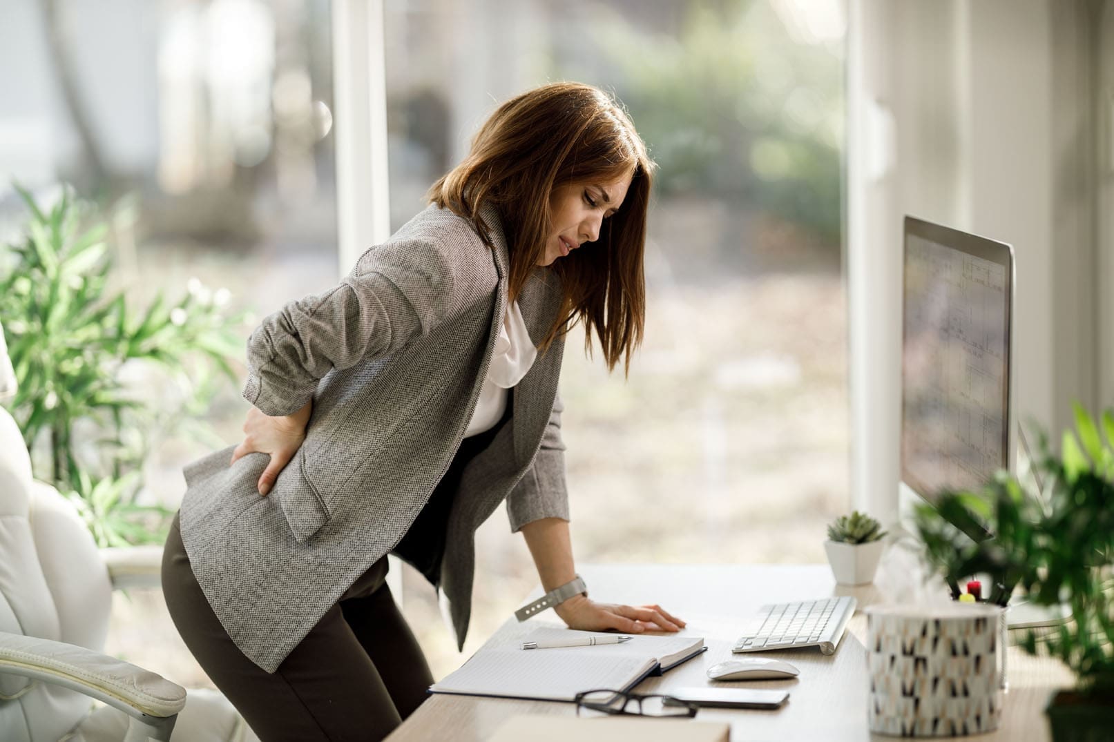
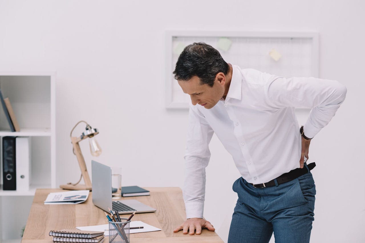
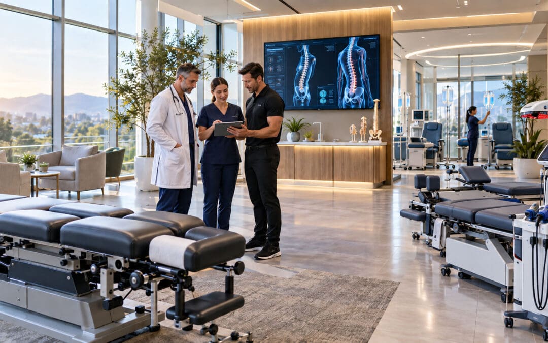
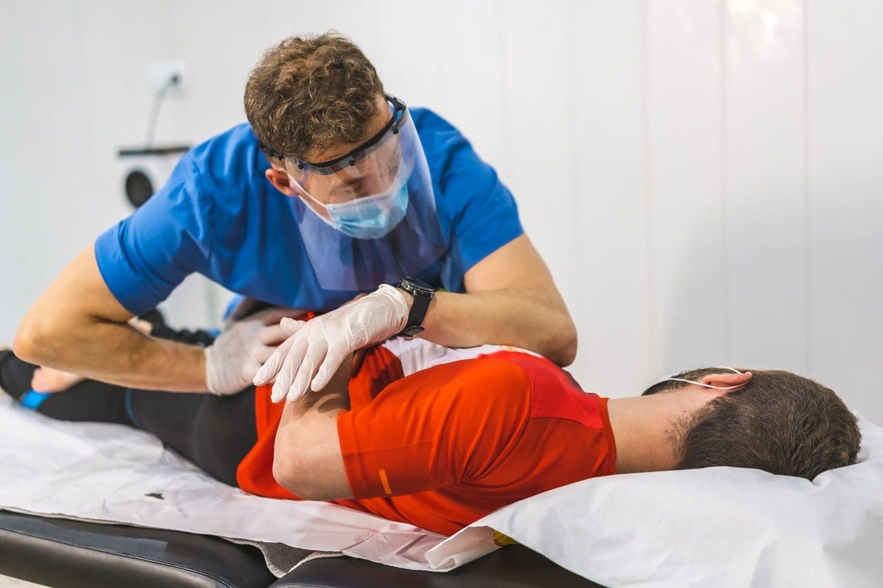
Back Clinic Work-Related Injuries Chiropractic and Physical Therapy Team. Work injuries and conditions can occur from a variety of circumstances, altering an individual’s lifestyle, however, those that occur in the work field can often also be debilitating and impairing, affecting an individual’s work performance. Work-related injuries can include bone fractures and muscle strains/sprains to conditions causing degeneration of many structures of the body, such as arthritis.
Also referred to as occupational injury, repetitive and constant motions of the hands, arms, shoulders, neck and back, among others, can gradually wear out the tissues, increasing the risk of injury which could eventually lead to further complications. A collection of articles depicts the causes and effects of many work-related injuries, carefully describing each variety. For more information, please feel free to contact us at (915) 850-0900 or text to call Dr. Jimenez personally at (915) 540-8444.
Sitting through a coding block, a dashboard review, a rack ticket, or a long El Paso drive can leave the low back stiff the moment you stand. This article explains why the first steps feel awkward after prolonged sitting: less joint motion, hip flexor tightness, tired trunk muscles, and a short-term rise in lumbar stiffness. It also covers simple movement resets, when stiffness is just a warm-up issue, and the nerve or pain signs that warrant an evaluation. Care at El Paso Back Clinic is framed as a choice you direct, with non-surgical options considered before medication dependence or surgery.
The meeting ends. You push the chair back and stand, and the first three steps feel like your low back forgot how to walk. Programmers know the feeling after a long merge. Analysts feel it after a dashboard review. Remote workers feel it when they move between the kitchen and the laptop. Drivers feel it when they step out of the car. Data center staff feel it after a seated ticket queue, then a crouch at a rack. The stiffness is real. It is also often mechanical, not a sign that your spine suddenly failed.
El Paso Back Clinic treats that first-stand moment as a function problem: easier steps, less guarded movement, and a plan that keeps you working without jumping to surgery.
What Prolonged Sitting Does to a Working Spine
Sitting is not poisonous. Unbroken sitting is the problem. Hips stay bent. The low back often rounds. Joints that should glide stay parked. Muscles that should share the load quiet down, then tire.
A laboratory study found that passive flexion stiffness of the lumbar spine rose in men after only one hour of sitting, with a higher chance of strain if someone then bends fully forward (Beach et al., 2005). You sit for a sprint, stand, and then reach for a bag, a cable, or a child seat.
Harvard Health notes that long sitting shortens the hip flexors, which attach near the pelvis and low back (Harvard Health Publishing, 2024). When you stand, tight hip flexors can tug the pelvis and shorten the first steps. Drivers add a twist when getting out.
Both prolonged sitting and prolonged standing raised low-back discomfort in a two-hour computer-task study (Park & Srinivasan, 2021). Standing all day is not the cure. Changing position is.
Why the First Steps Feel Awkward
The awkward walk has a few stacked reasons.
Joints have not moved. Facet joints and the hips need a few cycles of motion before they feel smooth.
Hip flexors shorten from the chair or car seat (Harvard Health Publishing, 2024).
Glute and deep trunk muscles are underused, so the low back bears the load during the first steps.
Passive spinal stiffness can rise after a long sit, especially in men, within the first hour (Beach et al., 2005).
Fatigue makes the body guard, so the first steps become shorter.
A programmer who has not left the chair since the last build can stand fine and still walk stiffly to the printer. An analyst feels the fatigue as they cross the lot. A technician feels it after alerts, then a cable pull. The spine goes from parked to loaded in one second.
A Two-Minute Reset Before You Walk
Active breaks that change posture have moderate-quality evidence for easing pain and discomfort, without hurting work output (Waongenngarm et al., 2018). You do not need a gym. You need a sequence.
Before you leave the chair
Scoot to the edge. Plant both feet.
Tip the pelvis forward and back five times. Small range. No bouncing.
Squeeze both glutes for five seconds, twice.
Stand by hinging at the hips, not by yanking with the low back.
The first thirty seconds on your feet
Hold the desk or the car door. Step one foot back into a gentle hip-flexor stretch. Keep the ribs stacked. Twenty seconds on each side is enough to start (Harvard Health Publishing, 2024).
March in place for ten slow steps before you walk away.
If you just drove, stand fully before you twist to grab a bag.
During the shift
Break a long sit before the hour mark when you can. Beach and colleagues saw stiffness changes by sixty minutes in men (Beach et al., 2005).
Alternate sitting and standing, but do not trade one frozen posture for another (Park & Srinivasan, 2021).
After a rack crouch, stand tall and take five easy steps before you lift.
Skip a heavy forward bend right after a long sit, when passive stiffness may be highest (Beach et al., 2005).
When Stiffness Is Only a Warm-Up
Many people loosen within a few minutes of walking. A short warm-up ache differs from pain that spreads or lingers. If the first steps are awkward and then walking feels normal, movement practice and a spinal check are often the right first path.
Benefits patients notice when motion returns:
Less grab in the low back on the first stand
Smoother steps to the car or the rack
Easier sitting through the next block
Better sleep because the evening is not spent guarding
Signs That Deserve an Examination
Some patterns should not be watched for weeks.
Pain that shoots into the buttock, thigh, or foot
Numbness, tingling, or a foot that slaps or drags
Weakness when you rise from a chair or climb a step
Pain that does not ease after several minutes of easy walking
Stiffness that lasts the whole shift, not just the first steps
Night pain that wakes you, fever, or unexplained weight loss
Bowel or bladder changes, or saddle numbness — seek urgent care
A new problem after a crash, a fall, or a lift you felt immediately
Those signs are information, not a verdict that you need surgery. They are a reason to map the joint, the nerve, and the work pattern.
How a Back-Focused Visit Uses the Whole Team
El Paso Back Clinic sits inside a collaborative model at Injury Medical Clinic PA. Dr. Alex Jimenez, DC, APRN, FNP-BC, CFMP, IFMCP, is a doctor of chiropractic and a board-certified family nurse practitioner (Texas APRN license #1191402, prescriptive authority #59628, NPI 1205907805). He pairs spinal alignment and mechanical care with medical diagnostics under collaborative oversight. Dr. Maria Guadalupe Cardenas, MD, board-certified in internal medicine (Texas license #J2933, NPI 1164426748), is the medical director and collaborative physician, with more than forty years as an internist. She oversees medical risk, labs, and how structural care fits into a safe medical plan.
That split serves beneficence: the visit is built for your safety and for the cause of the stiffness, not a single technique. Non-maleficence shows up in the order of care. Joint motion, decompression when indicated, and movement coaching come before long medication use or an operation. Autonomy means you choose. Findings are explained in plain language, and care can coordinate with the clinician you already trust.
For this pattern, the plan starts with how you sit, stand, and load the hips. Spinal decompression may be used when disc pressure and guarded motion keep the first steps painful. Adjustments aim to restore motion so the walk does not start from a locked joint. If nerve pain is sharp, image-guided epidural care can be a window for rehab, not a cure by itself. Shockwave or MLS laser may support irritated soft tissue. Peptides, PRP, or hormone therapy are not the default for simple stand-up stiffness. Consider them only when the exam and labs, guided by Dr. Cardenas, show a separate recovery problem.
Many programmers, analysts, drivers, and data center employees here carry strong group health benefits. An evaluation and non-surgical spinal care are often the covered path worth using before symptoms take over the shift.
If the first steps after sitting stay awkward, or if nerve symptoms have joined the stiffness, book a function-focused visit. Call Injury Medical Clinic PA at 915-850-0900 or visit https://www.elpasobackclinic.com. Bring how long you sit, whether you drive, and whether the pain fades or travels. You leave with a plan you understand, built to help you stand, walk, and finish the shift with less risk of an avoidable procedure. Start with the two-minute reset today.
Can Nutrition Change How Well an El Paso Spine Recovers?
Abstract: Mechanical care remains central to spinal recovery. This article explains why protein, glucose control, hydration, sleep, and smart loading still matter for shift workers recovering from disc, ligament, muscle, or spinal injuries. Food does not heal a herniated disc. Nutrition can support collagen remodeling, muscle preservation, and rehab tolerance. Poor meals, glucose swings, dehydration, and sleep loss can slow the same plan. El Paso Back Clinic pairs non-surgical decompression with medical oversight so patients can use group insurance and choose next steps with clear information.
The server room is quiet at 2:17 a.m. A data center technician finishes a rack job that required a crouch and a twist. An Amazon associate on the same clock has already walked miles of concrete. A programmer covering an overnight deploy has been in one chair since dinner. All three later need care for a disc, ligament, or muscle injury that will not settle.
Someone always offers the same advice. Eat cleaner. Drink collagen. Cut sugar. The advice is incomplete. At El Paso Back Clinic, the sentence is this: your disc does not eat dinner.
A herniated disc doesn’t tuck itself back in because breakfast included eggs. Ligaments do not restitch because a shake lists twenty grams of protein. Mechanical load, joint motion, nerve protection, and a graded return to work decide most of the outcome. Nutrition is the supply line, not the decompression table.
Two Different Kinds of “Disc Nutrition”
People use nutrition for two jobs. Mixing them creates false hope.
The disc’s own grocery route
An intervertebral disc has almost no direct blood supply. Glucose, oxygen, and waste move mostly by diffusion through the endplates. Slow, cyclic loading—the kind seen in walking and well-dosed rehab—can help that exchange. Long static postures and sudden heavy flexion can work against it (Gullbrand et al., 2015; Belavy et al., 2016). That is why non-surgical spinal decompression, alignment work, and movement coaching stay central. They change the mechanical environment the disc lives in. A protein bar cannot.
The tissues that do use your plate
The muscles that brace the spine, the tendons that transmit force, and the ligaments that limit end-range motion rebuild with amino acids, energy, and time. They waste away when nights get long, meals get irregular, and loading stops. Shift work fills that gap. Night schedules change when you eat, how you sleep, and how your body handles glucose. Food did not cause the herniation. The construction crew around the injured segment may still be underfed, under-rested, and asked to work a full shift (Matre et al., 2021; Oosterman et al., 2020).
Why Mechanical Care Still Comes First
Beneficence starts with the step most likely to help and least likely to harm. For many people with severe lumbago or disc-related pain, that means a non-surgical plan:
Examination of posture, hip motion, nerve tension, and work tasks
Imaging review when symptoms and function do not match
Adjustments and flexion-distraction or decompression when indicated
MLS laser or shockwave for surrounding soft tissue when appropriate
A loading plan that protects the segment while the body stays useful
Conservative care can reduce the rush toward surgery or long medication courses when those steps are not required. You should know what the spine needs mechanically before anyone sells a disc-healing diet.
Dr. Alex Jimenez, DC, APRN, FNP-BC, CFMP, IFMCP, leads the structural side and, with collaborative medical oversight, can add diagnostics and selected procedures. Dr. Maria Guadalupe Cardenas, MD, Board Certified in Internal Medicine, directs laboratory interpretation, metabolic risk, and coordination with existing physicians.
Protein: Building Material, Not a Reset Button
After injury or reduced loading, muscle protein breakdown rises. Rehab then asks those same muscles to stabilize a sore spine. Injury-nutrition reviews commonly place protein needs near 1.6 to 2.2 grams per kilogram per day, spread across meals (Smith-Ryan et al., 2020; Papadopoulou, 2020). Energy drinks and one burrito are a weak supply line.
What protein actually supports
Muscle preservation when you sit, stand, or protect a painful segment
Rehab quality, because weak, underfed muscle fails first
Amino acids for remodeling tissue, not a disc reset overnight
Collagen peptides plus training have some evidence for tendon morphology and joint comfort, but the effect depends on loading (Khatri et al., 2021; Aussieker et al., 2023; Bischof et al., 2024).
Shift-work protein problems we see in El Paso
One large meal after the shift, then almost nothing for ten hours
Protein pushed to 3 a.m., when appetite is low
“I’ll eat when I get home,” which becomes sleep
A practical target is protein every few waking hours: eggs, yogurt, leftover chicken, beans with rice, or a shake if food is not available on the floor. That supports recovery. It will not replace decompression.
Glucose: The Quiet Load on Collagen
High or swinging blood sugar does not cause every herniated disc. It can change collagen quality over time. Excess glucose helps form advanced glycation end-products (AGEs). AGEs cross-link collagen, reduce fiber slide, and make some tissues stiffer and more brittle. Research links diabetes and AGE burden with disc and tendon changes (Kakadiya et al., 2020; Rosenberg et al., 2023; Li et al., 2024).
For a programmer, NOC operator, or Amazon lead, the pattern is familiar: fasting through half the shift, a sweet drink for alertness, a heavy meal at the wrong clock, and poor sleep, which worsens glucose handling (Oosterman et al., 2020). That can raise inflammation and lower rehab tolerance. That doesn’t mean an MRI will reverse if you switch drinks. It means the same mechanical program may work better when glucose is steadier.
This is where MD and NP lab work earns its place. A1C, fasting glucose, lipids, and related markers help Dr. Cardenas see whether recovery is inflamed, insulin-resistant, or under-fueled. Treating numbers without treating the spine is incomplete. Treating the spine while ignoring uncontrolled glucose is also incomplete.
Sleep, Water, and the Night-Shift Recovery Tax
Shift work is associated with higher odds of chronic musculoskeletal pain, and low-grade inflammation is one proposed link (Matre et al., 2021). Circadian misalignment also reduces muscle insulin sensitivity and disturbs repair timing (Oosterman et al., 2020). A technician who decompresses at noon and then sleeps five broken hours is asking collagen to work nights too.
Hydration is simpler and still ignored. Caffeine-only nights and hot warehouse aisles do not dry a disc like a sponge on a stove, but they increase fatigue and make people move worse.
A short list that fits a night schedule
Keep water at the station; pair each energy drink with water
Protect a dark, cool sleep block even if it starts at 9 a.m.
Eat protein before the shift, not only after
Do not save the entire recovery plan for days off
Loading Still Writes the Script—Including After PRP
If a ligament, tendon, or joint later needs platelet-rich plasma (PRP), the injection is a signal, not a substitute. Mechanical loading after the procedure shapes the tissue response. Skipping rehab or returning to a poor lift can waste the appointment (Neph et al., 2020; Gremeaux et al., 2026). The same logic applies to image-guided epidurals. A calmer nerve is a window for movement, not a cure on its own.
The clinic keeps that hierarchy visible: protect the segment and restore motion; reload muscle in a way the job can survive; support protein, glucose, micronutrients, and sleep so the plan is tolerable; and add procedures only when they serve that sequence.
Using Group Insurance Without Guessing
Many data center, IT, and fulfillment employers in El Paso carry strong group insurance. That benefit can cover evaluation, chiropractic and decompression visits, medical assessment, and indicated labs. Using the plan early is often cheaper than waiting until lifting limits or sleep collapse.
A visit may include a mechanical exam tied to rack height, pallet work, chair time, or night rotation; a plain-language imaging review; a decompression plan with home loading rules; and labs when fatigue or slow tissue response is part of the story. You remain the decision-maker.
Call El Paso Back Clinic at 915-850-0900 or schedule through Injury Medical Clinic PA at Mission Plaza. Bring your work schedule, insurance card, and the movements that still scare you. The spine needs mechanics first. The body still has to show up with materials.
The Programmer Who Forgot to Breathe: Can Shallow Screen-Time Breathing Contribute to Rib, Mid-Back, and Low-Back Stiffness?
Abstract: Long coding sessions can change how you sit, move, and breathe. When the rib cage stays compressed and the trunk barely changes position, some people notice rib stiffness, mid-back tightness, low-back fatigue, or a deep breath that feels restricted. This article explains how the diaphragm, ribs, thoracic spine, abdominal wall, and lumbar stabilizers work together, what rehabilitation may address, and which breathing or chest symptoms need medical evaluation first.
A programmer can spend two hours solving one problem and barely notice anything below the neck. Then the task ends. The shoulders drop. The person sits back, tries to inhale deeply, and suddenly feels the ribs resist, the mid-back ache, or the low back feel tired.
That experience does not prove that “bad breathing” caused back pain. Pain is rarely that simple. However, prolonged flexed sitting, low movement variety, and shallow breathing can change how the chest wall and trunk share mechanical work. Research has linked prolonged sitting with immediate increases in low-back discomfort in some adults. A small laboratory study also found that greater posterior pelvic tilt reduced thoracic expansion and respiratory measures in healthy young men (De Carvalho et al., 2020; Aramaki et al., 2021).
The Diaphragm Is Also Part of Trunk Control
The diaphragm is the dome-shaped muscle beneath the lungs. When it contracts, it descends and helps draw air into the chest. It also works with the abdominal wall, pelvic floor, multifidus, and other trunk muscles to manage pressure and support the spine during movement.
That dual role matters at your desk. Your trunk must remain stable enough for typing, reaching, standing, and lifting while still allowing the ribs and abdomen to expand. Research on chronic low-back pain suggests that breathing and diaphragm function may interact with trunk control, but the evidence does not show that diaphragm dysfunction causes every back problem. Breathing is one variable in a much larger mechanical system (Chen et al., 2026; Li et al., 2026).
What a Long Screen Session Can Change
Picture a developer leaning toward a laptop during a difficult debugging session. The pelvis rolls backward, the low back rounds, the thoracic spine stays flexed, and the lower ribs move closer to the pelvis. That position may continue for long periods with little movement outside the hands.
Several changes can follow:
Rib movement can feel limited. A compressed trunk position may make comfortable expansion harder.
Thoracic motion narrows. The mid-back spends less time extending and rotating.
Support muscles stay active. Spinal and abdominal muscles continue to provide low-level control.
Breathing may become smaller. Some people brace the abdomen, briefly hold their breath, or breathe mainly into the upper chest when concentrating.
One position becomes the problem. There is rarely one perfect posture; low movement variety may be more important than a single snapshot of alignment.
These changes can help explain why the first deep breath after a long work block feels stiff, but they do not establish a simple cause-and-effect relationship between shallow breathing and pain.
Why Rib, Mid-Back, and Low-Back Symptoms Can Travel Together
The ribs attach to the thoracic spine, so every comfortable breath includes small movements through rib joints, chest-wall tissues, the thoracic spine, the diaphragm, and abdominal muscles. If the upper body stays flexed and still, those tissues may feel temporarily stiff.
The low back can fatigue at the same time because trunk control is shared. The diaphragm helps regulate pressure while the abdominal wall and spinal muscles manage load. That is why a useful examination should look beyond “take deeper breaths.”
A clinician may assess:
thoracic and rib mobility;
lumbar control and sitting tolerance;
hip mobility and trunk endurance;
symptom reproduction with breathing or movement;
neurological findings; and
whether breathing changes during reaching, lifting, or exercise.
The goal is to identify what is actually limiting function rather than assigning every symptom to posture.
Can Breathing Exercises Help?
Possibly, as an adjunct to rehabilitation.
A 2026 systematic review of 17 randomized trials found that breathing exercises may improve pain and function in chronic nonspecific low-back pain, but the evidence was low to very low certainty because studies varied and several had a high risk of bias (Chen et al., 2026). A separate 2026 meta-analysis found moderate-certainty evidence that diaphragmatic training may reduce pain and disability when added to care, while longer-term effects still need clarification (Li et al., 2026).
The practical message is not “breathe correctly and fix your back.” You can train breathing alongside mobility, strength, coordination, and graded activity when the examination suggests it matters.
A Five-Step Mechanical Reset
For ordinary screen-related stiffness, and only when no medical warning signs are present, a simple reset can restore movement options:
Change position. Sit taller, stand, or walk briefly, then take a deep breath.
Let the lower ribs widen. Place your hands around the lower rib cage and allow gentle expansion without straining.
Move the mid-back. Add comfortable extension, rotation, or reaching instead of repeatedly stretching only the low back.
Release unnecessary bracing. If you have been gripping your abdomen, let it soften enough for normal breathing.
Reload the trunk. Follow mobility with walking, light resistance, or prescribed stabilization exercises so the change transfers into function.
The target isn’t perfect posture. It improves variability, trunk capacity, and coordination.
When Chiropractic and Rehabilitation Fit
For mechanical rib, thoracic, or lumbar complaints, chiropractic and rehabilitation may be appropriate after excluding dangerous or non-musculoskeletal causes. Care may combine spinal or rib-joint assessment, manual treatment when indicated, mobility work, progressive trunk training, ergonomic changes, and exercise.
A 2026 review found that thoracic manual techniques and breathing exercises may improve pain or disability in some low-back-pain populations, but the certainty was low and treatment effects varied (Seyedhoseinpoor et al., 2026). That supports personalized care, not a one-size-fits-all breathing plan.
At El Paso Back Clinic, the useful outcome is physical function: sitting with less fatigue, moving more comfortably, tolerating work demands, and building capacity without rushing toward invasive treatment.
Red Flags: When Breathing Is Not a Posture Problem
Difficulty taking a satisfying deep breath can be musculoskeletal, but unexplained shortness of breath can also reflect heart, lung, vascular, infectious, or other medical conditions.
Seek emergency care for sudden severe shortness of breath, unexplained chest pain or pressure, fainting, blue lips or nails, confusion, or major breathing difficulty. New breathlessness after prolonged immobility, surgery, illness, or a long trip also deserves prompt medical attention because a blood clot is one possible cause (Mayo Clinic, 2025).
A persistent cough, fever, wheezing, worsening exercise tolerance, or breathlessness that doesn’t match your activity level deserves medical evaluation. Chiropractic treatment should not delay that workup.
Integrated Care, Informed Choices
When symptoms appear mechanical, Dr. Alex Jimenez, DC, APRN, FNP-BC, CFMP, IFMCP, can connect chiropractic assessment, rehabilitation, and medical evaluation within one coordinated plan. Dr. Maria Guadalupe Cardenas, MD, a board-certified internal medicine physician with over 40 years of experience, provides medical direction when cardiopulmonary, metabolic, medication-related, or other non-musculoskeletal factors need consideration.
This approach supports beneficence, non-maleficence, and autonomy: use care that is likely to help, avoid unnecessary risk, and give patients enough information to choose among reasonable options. Prioritize non-invasive treatment when appropriate, and add imaging, laboratory testing, referral, or medical treatment when findings justify it. Care can also coordinate with the patient’s existing medical team.
The Bottom Line
Screen-time breathing is not a diagnosis. Prolonged flexion and low movement variety may reduce rib and thoracic motion and alter how the diaphragm, abdominal wall, and spinal muscles share work. For some people, that pattern can accompany rib stiffness, mid-back tightness, or low-back fatigue.
Do not obsess over every breath. Restore movement options, strengthen the trunk, vary posture, and investigate symptoms that do not behave like ordinary mechanical stiffness. If pain keeps returning or breathing feels genuinely abnormal, get evaluated before assuming the screen is the only factor.
A practical starting point is simple: notice when concentration makes your body rigid, then change position before discomfort becomes the only signal you hear. Small, repeated movement choices throughout the workday can support comfort without turning posture or breathing into another performance task that creates unnecessary worry during demanding coding sessions.
The Hip That Hurts After Sitting: Is Your Low-Back Problem Actually Being Driven by the Hip?
Abstract: Prolonged sitting holds the hip in flexion, reduces usable hip extension, and can force the lumbar spine to move more than it should. Programmers, engineers, data analysts, gamers, and remote workers often feel these symptoms as front-of-hip tightness, buttock discomfort, or low-back pain after a long session. This article explains those mechanics and when structural care, mobility, strengthening, or decompression belong in the plan.
The call ended an hour ago. You are still in the chair. When you stand, the front of one hip feels pinched, the buttock feels thick, and the low back won’t straighten. A few steps later, the back eases. That pattern is common in people who sit to think. It doesn’t prove that every desk worker has tight hip flexors. Sitting changes the hip, but a stretch does not automatically repair the spine.
What Sitting Does to the Hip
Sitting is a flexed-hip posture. The thigh stays bent toward the trunk, often near 90 degrees, for hours, and the tissues around the hip adapt to that demand (Chamberlain, 2021).
The hip is designed for large ranges of motion. The lumbar spine is built for a smaller, more controlled range.
In a typical sit, the pelvis tilts backward slightly and lumbar lordosis decreases. That sit-to-stand strategy is normal, not a diagnosis by itself (Devin et al., 2012).
Perching on the chair edge or gaming with a collapsed trunk keeps the hip bent while the low back flexes or twists.
Front-of-hip tissues can feel short because they have been held short, because the joint does not tolerate end-range flexion, or because a lumbar nerve refers symptoms to the groin or buttock.
The useful question is not “Are the hip flexors tight?” It is “Where is motion missing, and what is setting the limit?”
When the Hip Cannot Extend, the Low Back Often Pays
Hip extension takes the thigh behind the body. Walking needs only about 10 to 15 degrees of that motion. If the hip does not extend, the pelvis and lumbar spine often finish the job by extending or rotating more than they should (Gómez-Hoyos et al., 2020).
That is the core of hip-spine thinking. Offierski and MacNab described coexisting hip and lumbar problems that can mimic, amplify, or hide each other (Offierski & MacNab, 1983). Later reviews add a practical rule: if the hip cannot supply sagittal motion, the lumbopelvic region often will (Devin et al., 2012).
For a desk-based engineer or analyst, the day can look like hours of hip flexion, brief standing with a hip that no longer extends easily, extra lumbar motion to keep the trunk upright, and a return to the chair before the hip practices the missing motion. People with low back pain often show reduced hip range of motion and weaker hip extensors and abductors than pain-free adults (Pizol et al., 2024). That does not make the hip the villain in every case. It does show that the hip and low back share a job.
Reduced Gluteal Capacity Is a Loading Problem
The gluteal muscles extend and stabilize the hip. In sitting, they are lengthened, unloaded, and rarely asked to produce force. Capacity drops because they are undertrained for controlling the pelvis when you stand or walk. If the hip cannot create or control extension, lumbar extensors often work harder. The back starts doing a hip job. That can feel like a tight belt after a long coding block or a late gaming session.
Adding hip strengthening to a low-back plan can reduce pain and disability in nonspecific low back pain (Santamaría et al., 2023). That is not a reason to load the glutes on day one. If the hip joint is irritated, aggressive bridging can increase groin pinch instead of building capacity.
Hold four targets at once: the hip needs motion, the gluteals need force and endurance, the trunk needs to stabilize instead of steal extra motion, and the workday needs more than one posture.
Tight Muscle, Stiff Joint, or Nerve Irritation?
A stretch that feels good in the front of the hip does not identify the source.
Adaptive muscle stiffness
The front-of-hip muscles have spent the day in flexion. They may resist lengthening when you stand. Symptoms often ease after walking, without catching or a sharp groin pinch at the end of range. This is the usual tight-flexor pattern, not the only one.
Hip joint limits
Intra-articular problems such as femoroacetabular impingement, labral irritation, or early degenerative change often prefer the groin. Sitting, crossing the legs, or tying a shoe can pinch. Clicking or limited internal rotation raises concern that the joint, not just the muscle, is setting the barrier (Chamberlain, 2021; Enseki et al., 2023).
Extra-articular hip pain
Lateral hip pain with side-lying or stair climbing can point toward gluteal tendon irritation. Deep buttock pain can come from posterior hip muscles, the sacroiliac region, or referred lumbar symptoms.
Nerve-related pain
A lumbar disc or irritated nerve root can send symptoms into the buttock, thigh, or groin. Numbness, tingling, shooting pain, or true weakness are not flexor tightness. They need a spinal and neurological exam.
Seek prompt evaluation for fever, unexplained weight loss, unchanging night pain, progressive weakness, or bowel or bladder changes. Those signs are not stretch problems.
A Direct Plan That Respects Both Regions
El Paso Back Clinic treats this as a mechanical problem first. Dr. Alex Jimenez, DC, APRN, FNP-BC, CFMP, IFMCP, evaluates spinal motion, hip motion, strength, and nerve signs in the same visit. Dr. Maria Guadalupe Cardenas, MD, Board Certified in Internal Medicine, provides medical direction when findings suggest more than postural overload.
A useful exam compares hip and lumbar motion, sit-to-stand quality, single-leg control, and nerve tension tests. The goal is to decide whether the low back is primary, the hip is primary, or both share the load. That is beneficence: treat the driver, not only the loudest complaint.
Chiropractic structural care can improve joint mechanics in the lumbar spine, pelvis, and hips when restriction is present. The intent is to give each region its own range so one area stops compensating for the other. Gentle hip mobilization may help when the capsule, not the muscle, is the barrier. Aggressive end-range stretching is a poor first choice if groin pinching suggests impingement.
Mobility work should look like the missing task: hip extension without extra lumbar arching, rotation without twisting through the lowest lumbar joints, and sit-to-stand without a lurch. Movement variability—standing, walking, sitting, and changing sides—reduces the hours the hip spends in one angle.
Once pain allows, training should include quiet hip hinges or bridges, side-lying or standing abduction, split-stance work that asks one hip to extend, and trunk endurance that resists motion instead of creating it. A programmer who sits for ten hours does not need a max squat in week one. They need a hip that can stand and walk without handing the job to the low back.
Non-surgical spinal decompression is a targeted tool for disc-related compression and selected nerve findings. It is not a default treatment for a stiff hip. If an exam and imaging support a disc or stenosis driver, decompression may reduce neural irritation while rebuilding capacity. If the spine is quiet and the hip is the limiting factor, the plan should focus on the hip. Choosing a non-invasive path first can reduce the rush toward long-term medication or surgery when those steps are not required. That is how the plan avoids adding risk the patient does not need.
What You Gain When Both Regions Share the Work
The outcomes patients want are practical: stand after a long review without grabbing the chair, walk without a chopped stride, and sit through focused work without a pinched groin or a locked low back. The hip should supply motion and force. The low back should supply controlled motion and stability.
Autonomy means you leave with a map: which symptoms are reasonable to train through, which signs should stop a stretch, and how this plan can sit alongside the care you already receive from your medical team.
Next Step With the El Paso Back Clinic Team
If sitting leaves a mark on the hip or low back, a focused exam can separate a short, adapted muscle from a joint or nerve problem. That is the difference between stretching what is loud and treating what is driving the load. Schedule an evaluation with Dr. Alex Jimenez, DC, APRN, FNP-BC, CFMP, IFMCP, and the collaborative medical team directed by Dr. Maria Guadalupe Cardenas, MD, so structural care, hip mobility, progressive strengthening, and—only when spinal findings justify it—decompression can be matched to your exam, not to a generic desk-stretch list.
Devin, C. J., McCullough, K. A., Morris, B. J., Yates, A. J., & Kang, J. D. (2012). Hip-spine syndrome. Journal of the American Academy of Orthopaedic Surgeons, 20(7), 434–442.
When the Standing Desk Starts Hurting: Why Standing All Day Is Not the Opposite of Sitting All Day
Abstract
Buying a standing desk feels like a solution. For many programmers, engineers, remote workers, and analysts, it is not. This article explains why standing all day is not the opposite of sitting all day. It covers static loading, lumbar compression, hip position, foot fatigue, and movement variability, then shows how a structural exam, chiropractic care, decompression when indicated, and trunk-hip conditioning shift the load away from the furniture.
You did the responsible thing. After months of low-back tightness at the keyboard, you raised the desk and promised to stand through stand-ups and tickets. For two weeks it felt like progress. Then the low back felt heavy by afternoon, one hip locked, and the feet ached. By dinner, standing no longer felt like relief.
That pattern is common among programmers, engineers, remote workers, and analysts who treat furniture as the solution. A standing desk can be useful. It is not a cure. The spine needs a changing load, not one perfect posture.
The Myth of the Perfect Desk Posture
The sales pitch is simple: sitting compresses the discs, and standing unloads them, so more standing should mean less pain. The body does not work that way.
Sitting and standing are both static postures. In both, the pelvis, lumbar segments, and hips hold a relatively fixed shape while the eyes stay on a screen. The muscles that keep you upright do not rest. They hold. After enough minutes, they fatigue, the pelvis drifts, and the lumbar curve collapses or over-arches. Pain follows the drift, not the desk brand.
Research on sit-stand workstations is mixed. Some reviews show a modest drop in low-back discomfort when people alternate positions (Agarwal et al., 2018). A Cochrane review of workplace standing and walking interventions found no clear reduction in musculoskeletal symptoms (Parry et al., 2019). Changing position tends to matter more than choosing one “better” position and staying there.
What Static Loading Actually Does
Static loading means a tissue is compressed, stretched, or contracted without enough change in length or force. Holding a grocery bag with a straight arm is the same idea. The bag is not heavy. The hold is.
In a standing workday, that holds stacks up:
The lumbar discs and facet joints accept a steady compressive force.
Hip and gluteal muscles co-contract to keep the pelvis from tipping.
Calves and foot muscles lock to keep you from swaying.
Blood return from the lower legs slows because the muscle pump is not cycling during walking.
Prolonged constrained standing has been linked to low-back discomfort, leg fatigue, foot pain, and venous pooling (Waters & Dick, 2015; EU-OSHA, 2021). Standing more than about four hours a day, especially without walking breaks, is also associated with a higher risk of chronic venous problems (Hirsch et al., 2024). The standing desk concentrated these loads into eight hours of “optimal posture.”
Lumbar Compression: Sitting Is Not Always Worse
Classic studies suggested sitting raised disc pressure compared with standing. A 2022 systematic review found that sitting often produces higher pressure in healthy discs, but later studies and studies of degenerated discs often show little difference between the two postures (Li et al., 2022).
That matters if you already have disc irritation or chronic lumbago. Switching desks may not unload the segment that hurts. Locked knees, a forward head, and a distant mouse can raise lumbar shear even as you feel “more active.” Laboratory work found standing produced the most fidgeting and the highest front-to-back shear, while both prolonged sitting and prolonged standing increased low-back discomfort (Le & Marras, 2016; Park & Srinivasan, 2021). Alternating sit-stand blocks changed trunk stiffness and muscle activity more favorably than either posture alone.
If your low back hurts after two hours of sitting, two hours of standing is not automatically medicine. It is a different static load.
Hips, Feet, and the Standing Fatigue Pattern
Desk workers who stand for long blocks often describe a cluster:
A dull lumbar ache below the belt line
One hip that feels pinched or stuck
Tightness across the front of the thighs
Heaviness in the calves
Tired, hot feet by mid-afternoon
Standing with the pelvis tucked under flattens the lumbar curve and keeps the hip flexors short. Standing with an exaggerated arch jams the lumbar facets. Neither version includes the motion those joints were built for: flexion, extension, rotation, and weight shift. Without walking, the calf pump is quiet, fluid pools, and the plantar fascia takes a continuous load. A thick mat can delay the complaint. It does not remove the need to move.
Movement Variability Beats Longer Standing
Tissues tolerate changing forces better than identical forces. Discs like cycles of load and unload. Hips like a changing angle. Feet like a push-off.
A useful work block is not “stand until it hurts, then sit until it hurts.”
Sit for focused typing with the lumbar curve supported.
Stand for shorter review or call blocks with soft knees and weight shifting.
Walk two to five minutes between tickets or meetings.
Change the mouse side or foot position before symptoms peak.
The goal is not to collect standing minutes. The goal is to interrupt the hold.
Change the Load, Don’t Just Change the Furniture
A standing desk is a tool. So is a chair. Neither one evaluates a stiff thoracic spine, a rotated pelvis, a weak hip abductor, or a disc that does not tolerate axial load.
El Paso Back Clinic starts with the structure, not the catalog. A focused exam looks at lumbar motion, hip extension and rotation, trunk endurance, foot and ankle stiffness, and nerve tension if pain travels into the leg. Imaging and medical screening are added when red flags appear.
Dr. Alex Jimenez, DC, APRN, FNP-BC, CFMP, IFMCP, evaluates both the mechanical pattern and the medical context. His dual licensure as a Doctor of Chiropractic and a board-certified Family Practice Nurse Practitioner allows structural care to work alongside appropriate diagnostics. Dr. Maria Guadalupe Cardenas, MD, Board-Certified in Internal Medicine, provides medical direction and laboratory oversight when metabolic or vascular issues could be amplifying fatigue or delaying recovery.
That collaboration is the ethical core of the visit. Beneficence means the plan exists to restore function and spare a larger intervention the patient may not need. Non-maleficence means starting with non-invasive, drug-sparing options instead of unnecessary surgery or long-term medication dependence. Autonomy means the patient sees the findings and chooses the next step with their existing medical team still in the loop.
What Care Looks Like When the Desk Is Not the Diagnosis
Chiropractic structural care
Adjustments and mobilization restore motion where the lumbar spine, pelvis, and hips have locked into a standing or sitting pattern so one joint is not doing the work of three.
Non-surgical decompression when indicated
If exam and imaging point to disc-related lumbago or radicular irritation, computer-guided non-surgical spinal decompression may be appropriate. In selected patients, adding decompression to standard physical care has improved pain, motion, endurance, and function compared with physical therapy alone (Amjad et al., 2022; Schueren et al., 2025). It is a targeted option when disc load, not furniture, is the limiter.
Mobility and progressive trunk-hip conditioning
Remote workers and engineers also need hip extension they can use at a desk: split-stance hip-flexor and calf lengthening, thoracic rotation, ankle rocks, and pain-free lumbar motion. Once irritability drops, the program targets weak areas with hip abduction, anti-extension trunk work, sit-to-stand control, and short carries.
A Practical Desk Reset
You do not need a new desk to start changing the load.
Cap uninterrupted standing at 20 to 30 minutes until hips and feet tolerate more.
Cap uninterrupted sitting the same way.
Use shoes with a stable heel if you stand at home.
Raise the monitor so you don’t fold your neck.
Walk during calls that do not require a shared screen.
Stop standing the moment the low back starts to brace. Bracing is a warning, not a badge.
If pain, numbness, or leg heaviness persists after you rotate positions, bring that data to a structural exam.
The Point
Desk workers did not fail the standing desk. Sitting overloads the lumbar spine in one pattern. Standing overloads it in a different way. The solution is a spine, pelvis, and hip complex that can change shape, share load, and recover. Change the load. Do not just change the furniture.
If standing still leaves a heavy low back, a locked hip, or tired legs, schedule a structural evaluation at El Paso Back Clinic. Dr. Alex Jimenez, DC, APRN, FNP-BC, CFMP, IFMCP, and Dr. Maria Guadalupe Cardenas, MD, coordinate chiropractic alignment, decompression when indicated, mobility training, and medical oversight on one plan. Bring your desk-setup notes and leave with a load strategy for Monday.
The Server-Rack Crouch: How Mid-Back Stiffness, Hip Restriction, and Awkward Reaching Overload the Lumbar Spine
Abstract: Data center technicians and network engineers crouch behind racks, kneel on hard floors, pull cables, and lift gear in tight aisles. When the mid-back cannot rotate, and the hips cannot flex well, the lumbar spine takes on extra bend, twist, and shear. This article maps that kinetic chain, shows what a structural exam should measure, and outlines alignment, mobility work, mechanical rehab, lifting strategy, and spinal decompression to keep a mechanical problem from becoming a disc or nerve injury.
The job does not look like heavy construction. You slide a switch into a dim rack, kneel to dress fiber, and then stand when a ticket hits. By day three, the low back feels thick and unreliable. Calling that “bad posture” does not tell you what to change.
The real issue is load sharing. The lumbar spine is built to carry compression and resist excess rotation. It is not built to supply the turn your mid-back should provide, or the hip bend your hips should provide, while you also reach and lift. When those neighbors stiffen, the low back becomes the available joint—and available joints get overloaded.
The Kinetic Chain Behind the Rack
Treat the trunk as a three-part system.
The thoracic spine and rib cage should rotate and extend so the arms can reach without dragging the pelvis.
The hips should flex and extend so you can drop or rise without folding the lumbar discs to their end range.
The lumbar spine should stay relatively stable while those two regions move.
When thoracic rotation is limited, the trunk borrows from the lumbar segments. Mobilizing restricted thoracic joints increases thoracic rotation and reduces lumbar rotation during the same turn (Yasuda et al., 2023). Modeling also shows that a stiffer thoracic spine raises lumbar disc stress and L5 pars stress (Morimoto et al., 2024). That is the server-rack crouch: you rotate to see a port, the ribs do not turn, and L4–L5 or L5–S1 supplies the missing degrees.
Hip restriction writes the same story in the sagittal plane. If hip flexion is short, a squat or kneel-to-stand uses extra lumbar flexion. Available hip flexion is linked to how much the lumbar spine flexes during a squat lift (Patterson et al., 2022). People with poorer hip and lumbar mobility also place higher bending stress on the spine during everyday lifts (Dolan & Adams, 1993). On the floor, this means hinging to pick up a box rather than rounding through the belt line.
Awkward reaching multiplies both problems. A reach around a rack post is flexion plus rotation plus a long lever. Confined-space research shows spinal load stays high in restricted height, though kneeling can cut shear compared with stooping because the trunk can stay more upright (Weston et al., 2020). Behind a rack, you often experience the worst mix: a stooped torso, rotated shoulders, and a load at arm’s length, then a sudden stand.
Why This Pattern Progresses
A single shift rarely herniates a disc. The pattern does. Repeated end-range lumbar flexion and rotation under load stress the annulus, facets, and ligaments. Over months, you may notice morning stiffness, a catch when standing from a kneeling position, or buttock pain after a long pull. Those are mechanical warnings. If the segment keeps moving past its useful range while a disc is already irritated, nerve roots can become inflamed. That is a load problem, not “tight muscles.”
What a Structural Assessment Should Measure
A useful exam maps missing motion and excess motion.
Mid-back and rib cage
Seated and quadruped thoracic rotation with the pelvis held still
Thoracic extension, watching for lumbar substitution
Rib and segmental joint play through the levels used in reaching
Hips and pelvis
Hip flexion, extension, and rotation
Hip-hinge quality from standing and from a half-kneel
Pelvic control during sit-to-stand and floor-to-stand
Lumbar spine and nerves
Segmental motion and tenderness
Repeated-movement testing to see whether flexion or extension changes symptoms
Nerve tension signs if pain or tingling travels below the knee
Imaging and a neurologic exam when weakness or progressive numbness appears
Dr. Alex Jimenez, DC, APRN, FNP-BC, CFMP, IFMCP, uses that map with medical screening. Dual licensure lets structural findings sit next to a review of inflammation and medication risk. Dr. Maria Guadalupe Cardenas, MD, Board Certified in Internal Medicine, provides collaborative oversight so care stays coordinated with your existing medical team. That is beneficence: the plan exists to protect the disc and the person.
Alignment First: Restore the Joints That Should Move
If the mid-back is locked and the hips are stiff, adjusting only the sore lumbar joints may provide temporary relief, but the improvement often fades by the next session.
Alignment at El Paso Back Clinic restores motion where it belongs:
Thoracic and rib work to return rotation and extension to the cage
Pelvic and hip work so the femur can flex without flattening the lumbar curve
Specific lumbar adjustments after neighboring regions can share load
Patients keep autonomy here. You should know which joint is being treated, why, and how you’ll measure change at the next visit.
Non-maleficence sits in the same step. Skilled, non-invasive adjustments and graded loading can reduce the rush toward long-term medication or surgery while the problem is still mechanical. Surgery has a place for true neurologic emergencies. Most back problems become that case only after the same compensation runs for years.
Mobility Restoration You Can Own
Clinic work does not replace the shift.
Thoracic rotation in a half-kneel, ribs turning, pelvis quiet
Hip-flexor length after long kneeling, so standing does not yank the lumbar spine
Hip-hinge rehearsal so the crease is at the hips, not at the belt
Ankle motion, because a stiff ankle forces an extra lumbar fold when you drop
Short sessions beat heroic stretching after the damage is done. Two minutes before a rack walk and two minutes after is a plan most crews can keep.
Mechanical Rehabilitation and Lifting Strategy
Mobility without strength leaves a new joint unprotected.
Hip-dominant hinges and split-stance pulls so the glutes lift the torso
Anti-rotation holds so the lumbar spine can resist twist while the arms reach
Tall-kneeling and half-kneeling work that mirrors floor-to-stand cable tasks
Gradual loading of lift height and mass so the hips take the extra demand
Lifting rules:
Get the load close before it leaves the floor or the cart
Turn the feet instead of twisting the belt line
Prefer a kneel with an upright trunk over a full stoop when the aisle is low
Split the task: slide, then lift, then turn
Use a partner or lift aid for awkward chassis
Confined work will never be perfect. The goal is fewer cycles at end-range lumbar flexion and twist.
When Spinal Decompression Belongs in the Plan
If the disc is already irritated, alignment and exercise may not be enough in the first weeks. Non-surgical spinal decompression uses controlled traction to reduce load on an injured disc while you rebuild the kinetic chain. In lumbar radiculopathy, adding decompression to routine physical therapy improved pain, lumbar motion, endurance, and disability more than therapy alone over four weeks (Amjad et al., 2022).
Decompression is not a substitute for finding the stiff thorax or the restricted hip. It is a tool to calm a loaded segment so those corrections can stick. At Injury Medical Clinic PA in El Paso, decompression is paired with alignment and, when indicated, medical review by Dr. Cardenas so inflammation and nerve status are not ignored.
A Direct Plan for the Next Cutover
If your low back catches when you stand from behind a rack, treat it as a mechanical diagnosis.
Get a structural exam that measures thoracic rotation, hip motion, and lumbar control.
Restore motion to the mid-back and hips, then protect the lumbar segments.
Rebuild hinge strength and anti-rotation capacity for kneeling and reaching.
Change the lift: closer load, feet turn first, upright kneel in a low aisle.
Use decompression when disc or nerve signs are present, under a plan you understand.
You remain the decision-maker. Integrative care at El Paso Back Clinic is designed to work with your current physicians, not around them. The aim is simple: keep the lumbar spine from doing a job it was never meant to do.
Reversing Severe Lumbago and Disc Compression from Amazon Fulfillment Shifts
Abstract: Severe low-back pain after fulfillment work or delivery routes can feel like a one-way road toward surgery. Yet many cases deserve a careful conservative evaluation first. This guide explains how repetitive bending, sorting, vehicle vibration, and lifting can aggravate lumbago and disc-related pain; where mechanical alignment and selected non-surgical decompression may fit; how medical oversight screens red flags; and how Amazon workers can use practical lifting limits to reduce repeat flares.
When a Shift Starts Loading the Lumbar Spine
A warehouse associate may begin a shift feeling stiff. Hours later, repeated low-bin reaches, package twisting, sorting, pallet work, or lifting away from the body can turn stiffness into severe lumbago. Delivery drivers add prolonged sitting, road vibration, repeated cabin exits, carrying, and awkward curbside lifts.
The problem is often cumulative. When the trunk bends and rotates while a load stays away from the body, spinal muscles must generate more force to control movement. Repetition can fatigue those tissues and degrade movement quality. NIOSH treats load weight, reach distance, vertical height, twisting, lifting frequency, duration, and grip quality as factors that change lifting risk (NIOSH, 2024).
That matters because “safe lifting” is not one universal number. A box manageable at waist height may be too demanding on the floor, far from the body, poorly gripped, or lifted repeatedly near the end of a long shift.
Why Pain Can Spike After the Shift
Symptoms may intensify after clocking out because fatigued muscles provide less support, sitting stiffens the hips, and irritated tissues remain sensitive after repeated loading. Delayed soreness doesn’t mean a disc suddenly worsened. Track which movements, loads, positions, and recovery habits consistently increase or decrease symptoms across workdays.
What “Disc Compression” Really Means
Between the lumbar vertebrae, intervertebral discs distribute load and allow movement. A disc is not a jelly doughnut that simply pops out and can be pushed back into place. It is a load-bearing structure with a fibrous outer ring and softer inner region. Repeated flexion, compression, rotation, aging, prior injury, and anatomy can contribute to disc changes.
Pain may come from several overlapping sources:
Irritated lumbar joints or surrounding soft tissues
Protective muscle spasm and reduced hip motion
Disc-related inflammation or mechanical sensitivity
A disc herniation that irritates a nerve root
Deconditioning after repeated pain episodes and activity avoidance
This is why a structural examination matters. The goal is to match symptoms with movement findings, neurologic testing, work demands, and imaging when indicated. Low-back guidelines support exercise, education, and selected manual or mobilization approaches as part of conservative care rather than a single passive treatment (George et al., 2021).
Step One: Restore Motion Without Forcing the Spine
At El Paso Back Clinic, targeted mechanical care begins with a practical question: which movements are limited, painful, unstable, or poorly coordinated?
One worker may have stiff hips that force repeated lumbar bending. Another may have painful lumbar joints that limit extension. A driver can show a different pattern after hours of seated flexion and vibration.
Chiropractic adjustments and non-thrust joint mobilization may improve mobility and reduce pain when the examination supports them. Guidelines report benefit from joint mobilization for many people with chronic low-back pain, including some with leg symptoms (George et al., 2021). Care should pair manual treatment with rehabilitation so improved motion becomes usable strength.
The Functional Target
The patient gains something measurable: easier standing, smoother walking, less guarded bending, better sleep positions, safer transitions from a delivery van, and more confidence handling daily tasks. Those functional gains matter more than trying to make every spinal image look “perfect.”
Step Two: Use Non-Surgical Decompression Carefully
Non-surgical spinal decompression is commonly described as controlled lumbar traction or unloading. The practical aim is to reduce mechanical stress temporarily and create a position in which painful tissues or irritated nerve roots may become less sensitive.
The evidence requires precision. A systematic review found short-term improvements in pain and disability when certain mechanical traction forms were added to treatment for lumbar radiculopathy, but study quality and results varied (Vanti et al., 2021). WHO guidance for chronic primary low-back pain advises against routine traction for most patients, so decompression should not become an automatic protocol for every painful back (World Health Organization, 2023).
For a selected patient, decompression may be one tool within movement retraining, strengthening, work modification, and medical evaluation. It should never be sold as a guaranteed way to “reinflate” a disc or permanently reverse degeneration.
Preventing Unnecessary Surgery Without Delaying Necessary Surgery
Avoiding surgery is valuable when recovery is safe with conservative care. Lumbar discectomy can be appropriate for selected patients, yet it still carries risks such as recurrent herniation, wound complications, dural tears, reoperation, and neurologic complications (Bombieri et al., 2022).
The safer message is not “never have surgery.” It is “earn the decision with the right evaluation.” Recent spine recommendations support surgery when conservative treatment fails and emphasize urgent or early surgery for cauda equina syndrome or progressive severe neurologic deficits (Costa et al., 2024).
Seek urgent assessment for new bowel or bladder dysfunction, saddle-region numbness, rapidly worsening leg weakness, or serious neurologic changes. Fever, major trauma, unexplained weight loss, or concern for infection, fracture, or cancer also deserves prompt review.
This is non-maleficence in practice: use the least invasive reasonable option when it is safe, but never let a desire to avoid surgery delay care for a true emergency.
Step Three: Set Proactive Lifting Limits
For Amazon associates, prevention must survive the workday. “Lift with your legs” is not enough.
Use these rules:
Bring the package close before standing up.
Turn with the feet instead of twisting under load.
Reduce load size when the object starts low, high, or far away.
Use team lifting or mechanical assistance for demanding or awkward items.
Break up repeated lifting when possible with task rotation or brief recovery periods.
Stop treating pain as a test of toughness; a changing neurologic symptom deserves evaluation.
The Revised NIOSH Lifting Equation is useful because it does not assume every worker should lift the same weight in every position. It calculates a recommended weight limit using task conditions and a lifting index that reflects physical demand. NIOSH recommends keeping the lifting index or composite lifting index at or below 1.0 when evaluating two-handed lifting tasks (NIOSH, 2024).
Integrated Oversight: Structure, Medicine, and Patient Choice
Dr. Alex Jimenez, DC, APRN, FNP-BC, CFMP, IFMCP, bridges chiropractic structural alignment, mechanical rehabilitation, and advanced medical diagnostics. He holds Texas Advanced Practice Nursing License #1191402, Prescriptive Authority #59628, and NPI #1205907805. His dual licensure lets the clinic coordinate musculoskeletal examination with broader medical assessment and appropriate advanced interventions.
Dr. Maria Guadalupe Cardenas, MD, is Board Certified in Internal Medicine, has more than 40 years of experience, and holds Texas Medical License #J2933 and NPI #1164426748. As Medical Director, Clinical Director, and Collaborative Physician at Injury Medical Clinic PA in El Paso, she provides internal medicine oversight, risk stratification, interpretation of indicated laboratory tests, and comorbidity coordination.
That collaboration supports beneficence: the plan centers on the patient’s best functional outcome. It supports non-maleficence by screening for medical risks before aggressive treatment and using non-invasive, drug-sparing options when appropriate. It also protects autonomy. Patients are taught what the examination shows, what each option can and cannot reasonably do, and when another specialist or their existing medical team should be involved.
A Better Goal Than “Toughing It Out”
Severe lumbago after warehouse or delivery work is not a character test. The goal is to interrupt the overload cycle before pain, guarding, deconditioning, and poor mechanics reinforce one another.
A conservative plan can include targeted joint care, selected decompression, progressive trunk and hip rehabilitation, work-specific lifting education, and medical oversight. The target is direct: more comfortable movement, safer lifting, better sleep, and the ability to work without organizing each day around the next flare.
Multidisciplinary Call to Action
If repeated bending, sorting, driving, or lifting causes severe low-back pain, start with a coordinated evaluation. El Paso Back Clinic can assess mechanical function, neurologic findings, lifting demands, and medical risks, then explain conservative options and when to refer. Bring your imaging, medication list, job demands, and questions. The final decision remains yours, supported by clear information and coordinated chiropractic and medical care.
IFM's Find A Practitioner tool is the largest referral network in Functional Medicine, created to help patients locate Functional Medicine practitioners anywhere in the world. IFM Certified Practitioners are listed first in the search results, given their extensive education in Functional Medicine