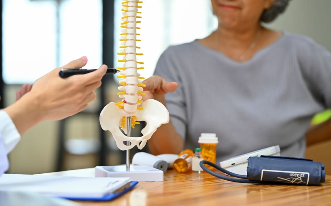
For individuals trying to retrain their body movements for back health improvement, what is the spinal area that helps the body twist, bend, and stand upright?
Lumbosacral Joint L5-S1
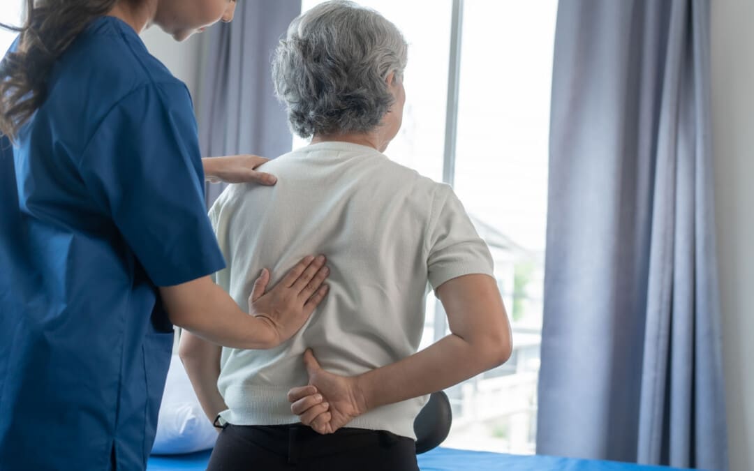
The L5-S1, also called the lumbosacral joint, is a term used to describe a part of the spine. It is where the lumbar spine ends and the sacral spine begins, and it connects these bones. The lumbosacral joint is also susceptible to misalignment and injury, such as disc herniation or a spinal disorder called spondylolisthesis.
The spinal column is the structure that allows the body to stand upright and helps you twist, bend, and alter trunk and neck position. Typically, 24 movable bones in the spine connect to the sacrum and the coccyx, or the tailbone. The sacrum and the coccyx each have multiple bones that fuse over time. L5-S1 consists of the last bone in the lumbar spine, called L5, and the triangle-shaped bone under it, known as the sacrum. S1 is at the top of the sacrum and comprises five fused bones.
Risk of Injury
Each area of the spine has a curve that goes in opposite directions. The places where the spinal curve directions change are junctional levels. The risk of injuries may be higher at junctional levels because the body weight shifts direction as the curves shift. The L5-S1 junction is located between the lumbar curve and the sacral curve. The lumbar curve sweeps forward, and the sacral curve goes backward.
The lumbosacral joint L5-S1 junction is highly vulnerable to misalignment, wear and tear, and injury. This is because the top of the sacrum is positioned at an angle for most individuals. Aging and injury increase the vulnerability of the L5-S1 junction even more. Pain coming from L5-S1 is usually treated with:
Heat and/or ice
Over-the-counter anti-inflammatory medications
Prescription pain medications
Muscle relaxers
Physical therapy
Chiropractic adjustments
Epidural steroid injections
If these therapies do not help, surgery may be recommended. L5-S1 is one of the two most common sites for back surgery.
Conditions
Disc herniation at L5-S1 is a common injury and cause of sciatica, which can cause pain and other issues (MedlinePlus, 2024). The L5-S1 junction is often the site of a condition known as spondylolisthesis.
Disc Herniation
Discs separate the vertebrae, cushioning the spinal column and allowing movement between vertebrae. A disc herniation means the disc slips out of place. (MedlinePlus, 2022) A disc herniation at L5-S1 is a common cause of sciatica. Symptoms of sciatica include:
Burning
Numbness
Pain or tingling that radiates from the buttock down the leg to the knee or foot.
Disc herniation can also cause chronic back pain and stiffness and trigger painful muscle spasms. Bowel problems are also possible with disc issues at L5-S1. Research links irritable bowel syndrome to herniated discs in the lower back. (Bertilson BC, Heidermakr A, Stockhaus M. 2015) Additional studies found disc problems at L5-S1 can lead to difficulty with sphincter control. (Akca N. et al., 2014) Initial treatments for disc herniation include rest and pain relievers to reduce inflammation and swelling, then physical therapy. Most recover with conservative interventions, and those who don’t may require a steroid injection or surgery. (MedlinePlus, 2022)
Spondylolisthesis
Spondylolisthesis occurs when a vertebra slips forward relative to the bone below it. The most common form of this condition is degenerative spondylolisthesis, which generally begins when the spine wears down with age. Isthmic spondylolisthesis is another common variation and starts as a tiny fracture in the pars interarticularis, a bone that connects the adjoining parts of the facet joint. (American Academy of Orthopaedic Surgeons, 2020) These fractures often occur before age 15, but symptoms do not develop until adulthood. Degeneration of the spine in later adulthood can further worsen the condition.
The angle of the sacrum can also contribute to spondylolisthesis. This is because the S1 tips down in the front and up in the back rather than being horizontal. Individuals with a greater tilt are usually at a higher risk of spondylolisthesis. (Gong S. et al., 2019) However, individuals with spondylolisthesis may not have any symptoms. Those who do may experience: (American Academy of Orthopaedic Surgeons, 2020)
Back stiffness
Standing difficulties
Walking difficulties
Lower back pain
Hamstring tightness
Spondylolisthesis is typically treated with non-surgical interventions that can include:
Pain medications
Heat and/or ice application
Physical therapy
Epidural steroid injections
Usually, non-surgical care is tried for at least six months. If pain and symptoms persist, surgery may be an option. Spinal fusion surgery can be effective but requires a long recovery time and can have additional risks.
Injury Medical Chiropractic and Functional Medicine Clinic
Injury Medical Chiropractic and Functional Medicine Clinic works with primary healthcare providers and specialists to develop an optimal health and wellness solution. We focus on what works for you to relieve pain, restore function, and prevent injury. Regarding musculoskeletal pain, specialists like chiropractors, acupuncturists, and massage therapists can help mitigate the pain through spinal adjustments that help the body realign itself. They can also work with other medical professionals to integrate a treatment plan to resolve musculoskeletal issues.
Chiropractic Healing After Trauma
References
MedlinePlus. (2024). Sciatica. Retrieved from https://medlineplus.gov/sciatica.html
MedlinePlus. (2022). Herniated disk. Retrieved from https://medlineplus.gov/ency/article/000442.htm
American Association of Neurological Surgeons. (2024). Herniate disc. https://www.aans.org/patients/conditions-treatments/herniated-disc/
Bertilson, B. C., Heidermark, A., & Stockhaus, M. (2015). Irritable Bowel Syndrome–a Neurological Spine Problem. Journal of Advances in Medicine and Medical Research, 4(24), 4154–4168. https://doi.org/10.9734/BJMMR/2014/9746
Akca, N., Ozdemir, B., Kanat, A., Batcik, O. E., Yazar, U., & Zorba, O. U. (2014). Describing a new syndrome in L5-S1 disc herniation: Sexual and sphincter dysfunction without pain and muscle weakness. Journal of craniovertebral junction & spine, 5(4), 146–150. https://doi.org/10.4103/0974-8237.147076
American Academy of Orthopaedic Surgeons. (2020). Spondylolysis and spondylolisthesis. https://orthoinfo.aaos.org/en/diseases–conditions/spondylolysis-and-spondylolisthesis/
Gong, S., Hou, Q., Chu, Y., Huang, X., Yang, W., & Wang, Z. (2019). Anatomical factors and pathological parts of isthmic fissure and degenerative lumbar spondylolisthesis.
For many individuals, getting on the floor to do crunches or other exercises can be difficult due to medical conditions, age, and injuries. Can engaging in chair abdominal exercises at home, work, or anywhere a chair is available help strengthen the upper, lower, and oblique abdominal muscles?
Chair Abdominal Exercises
Chair abdominal exercises are essential for strengthening the core, maintaining healthy flexibility, mobility, posture, and injury prevention. Incorporating abdominal exercise into one’s day can be easy, regardless of schedule or fitness level. Various exercises are designed to be performed while seated, making them accessible to everyone, including those with mobility issues or new to exercise.
Benefits
Building and maintaining a solid core is essential for overall health, posture, and daily activities, especially as the body ages. A strong core maintains body balance and stability, prevents falls, protects individuals from back pain, and makes lifting, bending, and walking easier. It allows individuals to stay independent and active. The abdominal muscles are one part of the core, which includes muscles in the back, glutes, pelvic floor, and diaphragm. These are important for keeping the body upright and stable, protecting the spine and organs, and assisting with movements between the ribcage and pelvis. Adding abdominal exercise workouts to a fitness routine is one way to improve overall core strength and stability. Chair-based exercises are convenient and accessible as individuals don’t need specialized machines or equipment, just a sturdy chair and some space. (Frizziero A. et al., 2021)
Chair Workout
Chair abdominal exercises are ideal gentle exercises for older individuals who need to increase their strength and mobility. Because chairs provide stability and safety, these exercises focus on seated movements and are intended for those with limited mobility. Chair abdominal exercises can be adapted and modified to individual preferences and needs, preparing the body for more advanced standing exercises. They improve the ability to perform daily tasks and increase joint mobility, muscle strength, and coordination. To prevent injuries, each exercise session should begin with a warm-up and end with a cool-down.
A warm-up could be gentle seated marches, shoulder rolls, and deep breathing exercises.
A cool-down can include seated stretches, focusing on the back, neck, and legs to help the body recover and prevent stiffness.
It is recommended that patients talk to a healthcare provider first if they have limited mobility or chronic health issues. Here are some sample exercises.
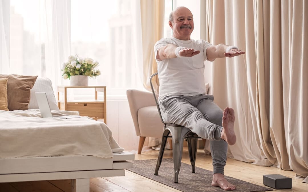
Sitting Knee Lifts
Sit on the edge of the chair with your back straight.
Lift your knees toward your chest, engaging the upper abs.
Lower them slowly back down.
Repeat for 10 to 15 reps.
Beginners lift one knee at a time.
Lower it and repeat with the other leg.
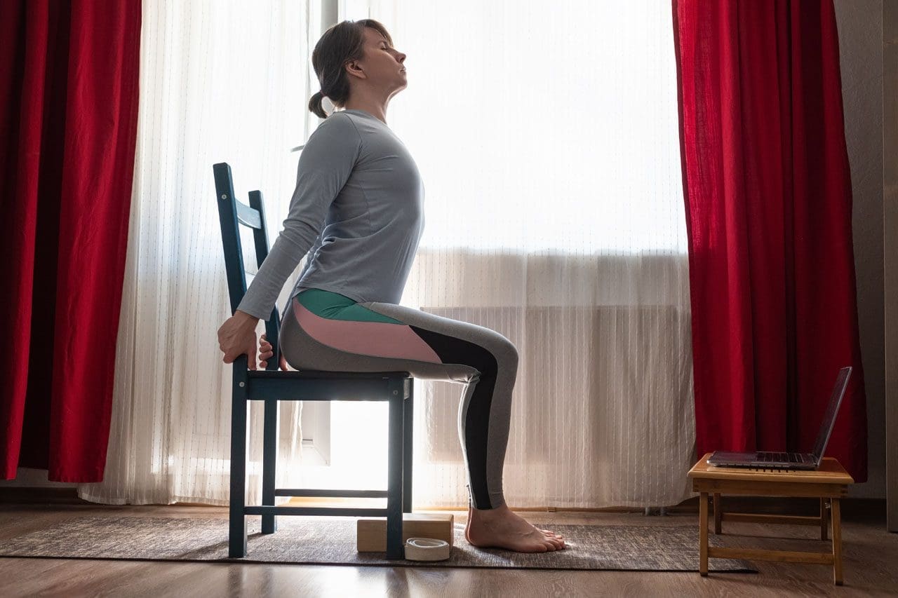
Sitting Crunches
Sit on the edge of the chair.
Lean back slightly.
Place hands behind your head.
Crunch forward, bringing the chest toward your knees.
Return to the starting position.
Repeat for 10 to 15 reps.
Beginners can sit more on the chair and hold the seat for stability.
Reduce the range of motion when crunching the chest toward the knees.
Sitting Leg Lifts
Sit on the edge of the chair with the back straight.
Extend your legs in front of you and lift them together, engaging the lower abdominals.
Lower them back down slowly.
Repeat for 10 to 15 repetitions.
Beginners sit back on the chair and grip the sides.
Lift one leg and hold for a count before lowering.
Switch legs.
Sitting Scissor Kicks
Sit on the edge of the chair and lean back slightly.
Extend your legs in front.
Alternate lifting each leg up and down in a scissor motion.
Perform for 30 seconds to one minute.
Beginners tap one heel on the floor between lifts to reduce abdominal tension.
Sitting Side Crunches
Sit on the edge of the chair with the back straight.
Lift one knee toward the chest while bringing the opposite elbow toward the knee, engaging the obliques.
Alternate for 10 to 15 reps per side.
Beginners, keep your feet flat on the floor with your hands behind your head.
Gently bend to the side, return to the center, and bend to the other side.
Sitting Bicycle Crunches
Sit on the edge of the chair and lean back slightly.
Lift the feet off the ground.
Alternate bringing each knee toward the opposite elbow in a pedaling motion.
Perform for 30 seconds to one minute.
For beginners, keep the heel gently on the ground while you crunch on the opposite side.
For a challenge, slow the motion and hold each twist a few seconds longer to create more tension. (Krzysztofik M. et al., 2019)
Progress
For beginners, start with the easier versions and perform fewer repetitions and shorter durations if needed. Focus on maintaining proper posture and form. As the body gets stronger, increase the number of repetitions or extend the duration of each exercise. Once comfortable, try more challenging chair exercises. Chair abdominal exercises are a simple, effective way to strengthen the core, improve posture, and support overall health. Regularly doing these exercises as part of a well-rounded routine can build a solid core without special equipment. Remember to listen to the body and progress at your own pace to more challenging variations over time to keep building strength.
Injury Medical Chiropractic and Functional Medicine Clinic
Achieving and maintaining fitness requires consistent work and development. Retraining the body and maintaining its optimal health requires daily efforts through exercise. Injury Medical Chiropractic and Functional Medicine Clinic works with primary healthcare providers and specialists to develop an optimal health and wellness solution. We focus on what works for you to relieve pain, restore function, and prevent injury. Regarding musculoskeletal pain, specialists like chiropractors, acupuncturists, and massage therapists can help mitigate the pain through spinal adjustments that help the body realign itself. They can also work with other medical professionals to integrate a treatment plan to resolve musculoskeletal issues.
Core Exercises and Back Pain
References
Frizziero, A., Pellizzon, G., Vittadini, F., Bigliardi, D., & Costantino, C. (2021). Efficacy of Core Stability in Non-Specific Chronic Low Back Pain. Journal of functional morphology and kinesiology, 6(2), 37. https://doi.org/10.3390/jfmk6020037
Krzysztofik, M., Wilk, M., Wojdała, G., & Gołaś, A. (2019). Maximizing Muscle Hypertrophy: A Systematic Review of Advanced Resistance Training Techniques and Methods. International journal of environmental research and public health, 16(24), 4897. https://doi.org/10.3390/ijerph16244897
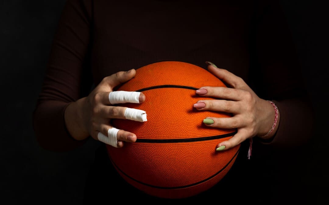
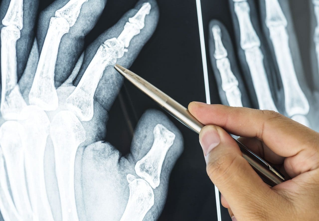
For individuals dealing with finger injuries, which can occur from various causes, including overuse, jobs, sports, and more, can knowing the cause of finger pain help healthcare providers determine what steps to take for treatment?
Finger Injuries
Finger injuries are common and can range from minor to serious. (van Veenendaal L. M. et al., 2014) Symptoms can result from an acute injury, including broken fingers and sprains, or chronic conditions like arthritis.
Fractures
Finger fractures can vary and can be serious and lead to permanent damage, deformity, and loss of function if not treated properly. What is important is that fractures are appropriately diagnosed so the proper treatment plan can be initiated. Most finger fractures can be addressed with simple treatments, while others may require surgery. (Oetgen M. E., and Dodds S. D. 2008)
Sprain and Dislocation
Sprains and dislocations are common finger injuries. (Prucz R. B. and Friedrich J. B. 2015) Both damage the ligaments that support the finger joints. In more severe injuries, a dislocation can occur, necessitating the finger to be put back into place or reduced. Individuals with a sprain or dislocation often notice finger swelling or stiffness for months after the injury.
Ligament Damage
Some call this injury skier’s or gamekeeper’s thumb, which results from a specific type of thumb dislocation. Here, the ulnar collateral ligament of the thumb is damaged. This ligament helps keep the thumb stable and supports grip and hand strength. However, this type of ligament injury often requires surgery. (Christensen T. et al., 2016)
Arthritis
Arthritis causes damage to normal joint surfaces where two bones come together. Fingers are one of the most common locations where arthritis occurs. (Spies C. K. et al., 2018) Two types of arthritis commonly affect the fingers: osteoarthritis and rheumatoid arthritis.
Arthritis of The Thumb
Arthritis of the thumb usually occurs at the joint where the thumb meets the wrist. This joint called the carpometacarpal/CMC joint, helps with gripping and pinching. Thumb arthritis is more common in women than men and increases in frequency over 40. (Deveza L. A. et al., 2017)
Trigger Finger
Trigger finger or stenosing tenosynovitis, is a common injury that causes pain and snapping of the fingers’ tendons, resulting in a sensation of locking or catching when bending and straightening the digits. (Makkouk A. H. et al., 2008) Other symptoms include pain and stiffness in the fingers and thumb. Treatments can vary from observation, rest, splinting, injections, and surgery.
Tendon Injuries
Mallet finger
A mallet finger is an injury to the tip of the finger. Usually, it occurs when the end of a straightened finger or thumb is hit, jamming the finger. After the injury, the individual may notice that they cannot fully straighten the tip of the finger. Treatment almost always uses a splint that has to stay on for about six weeks without removal. (Alla, S. R., Deal, N. D., and Dempsey, I. J. 2014) Very rarely is a surgical procedure necessary.
Jersey Finger
This is an injury to the finger flexor tendon. The flexor tendon pulls the finger into the palm when contracting the forearm flexor muscles. The injury occurs at the tip of the finger; typically, the tendon snaps back to the finger’s base or into the palm.
Ring Injuries
Injuries to the finger while wearing wedding bands or other finger jewelry can lead to serious complications. Even minor injuries can have devastating complications if the severity of the injury is not recognized and addressed. If an injury occurs while wearing the jewelry and there is soft tissue damage, including blood circulation being cut off, immediate medical attention is necessary.
Other Injuries
Bruises
The most common finger injury is caused by direct trauma to the skin and muscles. Symptoms include pain, swelling, tenderness, and discoloration of the skin.
Cuts and Scrapes
These can range from minor to more serious, such as injuries that cut through blood vessels, nerves, and tendons.
Injury Medical Chiropractic and Functional Medicine Clinic
After the initial inflammation and swelling have subsided, a doctor will recommend a treatment plan that usually involves physical therapy, self-performed physical rehabilitation, or supervision by a physical therapist or team. At Injury Medical Chiropractic and Functional Medicine Clinic, our areas of practice include Chronic Pain, Personal Injury, Auto Accident Care, Work Injuries, Back Injury, Low Back Pain, Neck Pain, Migraine Headaches, Sports Injuries, Severe Sciatica, Scoliosis, Complex Herniated Discs, Fibromyalgia, Chronic Pain, Complex Injuries, Stress Management, Wellness & Nutrition, Functional Medicine Treatments, and in-scope care protocols. We focus on what works for you to relieve pain and restore function. If other treatment is needed, individuals will be referred to a clinic or physician best suited to their injury, condition, and/or ailment.
Sports Injury Rehabilitation
References
van Veenendaal, L. M., de Klerk, G., & van der Velde, D. (2014). A painful finger as first sign of a malignancy. Geriatric orthopaedic surgery & rehabilitation, 5(1), 18–20. https://doi.org/10.1177/2151458514522125
Oetgen, M. E., & Dodds, S. D. (2008). Non-operative treatment of common finger injuries. Current reviews in musculoskeletal medicine, 1(2), 97–102. https://doi.org/10.1007/s12178-007-9014-z
Prucz, R. B., & Friedrich, J. B. (2015). Finger joint injuries. Clinics in sports medicine, 34(1), 99–116. https://doi.org/10.1016/j.csm.2014.09.002
Christensen, T., Sarfani, S., Shin, A. Y., & Kakar, S. (2016). Long-Term Outcomes of Primary Repair of Chronic Thumb Ulnar Collateral Ligament Injuries. Hand (New York, N.Y.), 11(3), 303–309. https://doi.org/10.1177/1558944716628482
Spies, C. K., Langer, M., Hahn, P., Müller, L. P., & Unglaub, F. (2018). The Treatment of Primary Arthritis of the Finger and Thumb Joint. Deutsches Arzteblatt international, 115(16), 269–275. https://doi.org/10.3238/arztebl.2018.0269
Deveza, L. A., Hunter, D. J., Wajon, A., Bennell, K. L., Vicenzino, B., Hodges, P., Eyles, J. P., Jongs, R., Riordan, E. A., Duong, V., Min Oo, W., O’Connell, R., & Meneses, S. R. (2017). Efficacy of combined conservative therapies on clinical outcomes in patients with thumb base osteoarthritis: protocol for a randomised, controlled trial (COMBO). BMJ open, 7(1), e014498. https://doi.org/10.1136/bmjopen-2016-014498
Makkouk, A. H., Oetgen, M. E., Swigart, C. R., & Dodds, S. D. (2008). Trigger finger: etiology, evaluation, and treatment. Current reviews in musculoskeletal medicine, 1(2), 92–96. https://doi.org/10.1007/s12178-007-9012-1
Alla, S. R., Deal, N. D., & Dempsey, I. J. (2014). Current concepts: mallet finger. Hand (New York, N.Y.), 9(2), 138–144. https://doi.org/10.1007/s11552-014-9609-y
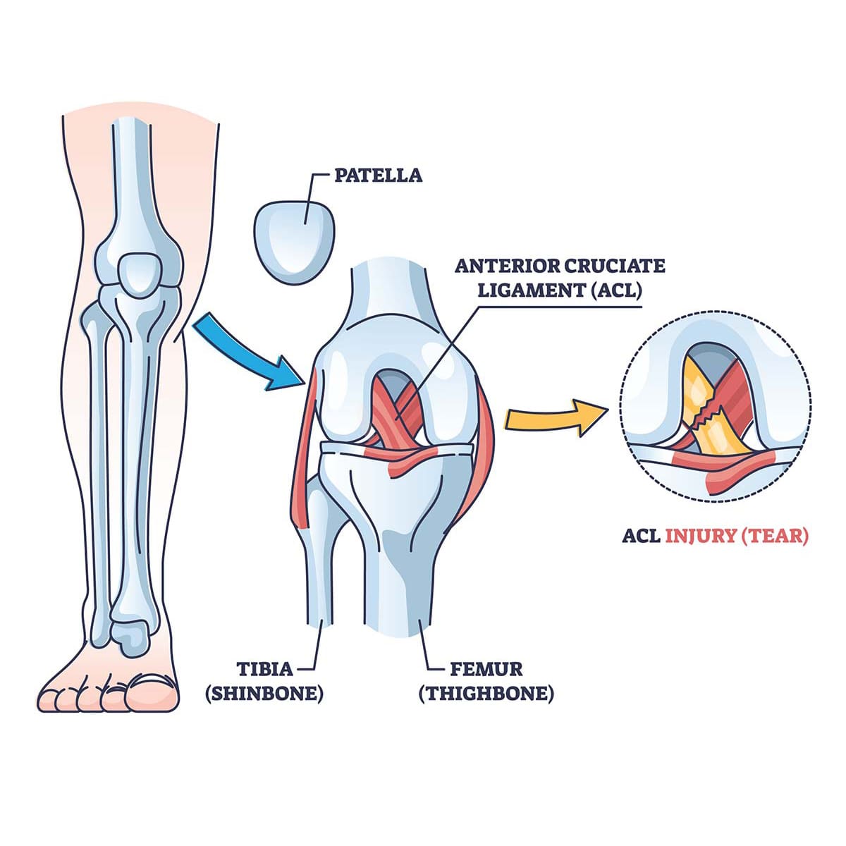
Can athletic individuals with ACL injuries find relief through non-surgical treatments to restore knee mobility?
Introduction
The body’s lower extremities help the individuals to be mobile but also help stabilize the body’s upper weight. From the hips to the feet, many people are on their feet and using every muscle group to allow functionality. Athletic individuals use their lower extremities to do various physical activities and are susceptible to injuries. An ACL injury is one of the most common and feared injuries that can impact an athletic person’s performance. These types of injuries affect the knees of the individual and can make a person feel miserable. However, numerous surgical and non-surgical treatments can help the recovery process of an ACL injury while helping the individual restore their motion to their lower extremities. Today’s article looks at what an ACL injury is, how it affects the knees, and how non-surgical treatments can help restore knee mobility from ACL injuries. We discuss with certified associated medical providers who consolidate our patients’ information to assess ACL injuries affecting their mobility. We also inform and guide patients while asking their associated medical provider intricate questions to integrate and provide them with numerous non-surgical treatments to be incorporated into their personalized treatment plan. Dr. Jimenez, D.C., includes this information as an academic service. Disclaimer.
What Is An ACL Injury?
Do you feel aches or pains around your knees after a long exercise regime? Do you feel or hear a loud popping sensation in your knees? Or do you experience pain and swelling affecting your ability to be mobile? Many of these pain-like scenarios are correlated with ACL injuries, that is amongst the most common and feared injuries for athletic individuals and non-athletic individuals. However, we must look at the ACL itself to better understand ACL injuries. The ACL (anterior cruciate ligament) plays an important role as it helps with knee joint stabilization, prevents excessive forward movements from the tibia (shin bone), and limits rotational knee movements. (Yoo & Marappa-Ganeshan, 2024) This ligament is one of the most injured structures affecting athletic performance. ACL injuries and tears can lead to many individuals having knee instability and an increased risk of future knee osteoarthritis. (Atik, 2024) This is because ACL injuries typically occur during physical activities involving sudden stops, jumps, or directional impacts to the knees.
How Does It Affect The Knees?
So, how do ACL injuries affect the knees of the individual? As stated earlier, the ACL is a crucial ligament that stabilizes the knee joint during movement. When that ligament is injured, it can cause pain-like symptoms like:
Pain
Limited range of motion
Knee instability
Altered biomechanics
This causes many people to have reduced physical activity levels, which can become a great economic burden to their daily routine. (Wang et al., 2020) When dealing with ACL injuries, it can also affect the meniscus in the knees as cartilage erosion often accelerates and can potentially lead to early osteoarthritis, which correlates with ACL injuries. (Key et al., 2022) However, when a person is dealing with ACL injuries, there are numerous treatments to reduce the pain-like symptoms caused by ACL injuries and help restore knee mobility.
Overcoming An ACL Injury-Video
Non-Surgical Treatments For ACL Injuries
When finding the right treatment for ACL injuries, many individuals can incorporate non-surgical treatments as part of their customized treatment plan. Non-surgical treatments can vary and may be suitable for individuals with partial ACL tears and knee instability and who have been involved in low-impact sports. When athletic individuals are dealing with ACL injuries, by incorporating non-surgical treatments, they can address the impairments, achieve functional stability, and safely return to their physical activities while improving the neuromuscular system to achieve functional knee stability. (Diermeier et al., 2020) Non-surgical treatments can positively impact many individuals by relieving the overlapping pain-like issues affecting the knees and the severity of ACL injuries.
Chiropractic Care
Chiropractic care is one of the many non-surgical treatments that can benefit individuals dealing with ACL injuries. Chiropractic care incorporates mechanical and manual manipulation to diagnose and treat any musculoskeletal issues associated with ACL injuries and emphasizes the body’s natural ability to heal itself. For many athletic and non-athletic individuals with ACL injuries, chiropractic care can offer several benefits:
Pain management
Enhancing mobility and flexibility
Improving balance
Strengthening supporting muscles
Chiropractic care can help individuals by stretching and strengthening weak muscles and soft tissues that can help break down scar tissues that may have surrounded the knee while improving blood flow to the injured area. Chiropractors can also incorporate specific rehabilitation exercises and physical therapy for the individual, focusing on strength, flexibility, and stability in the knees and surrounding muscles.
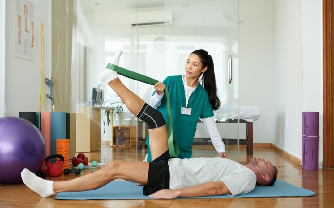
Physical Therapy
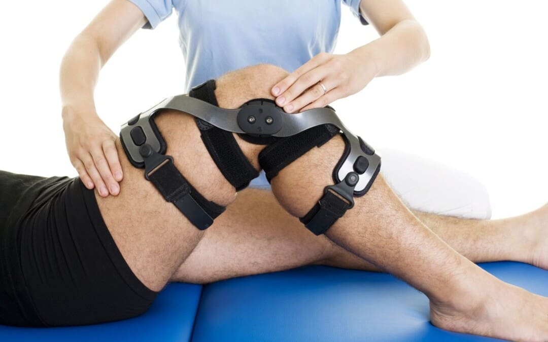
Another form of non-surgical treatment is through physical therapy. Physical therapy can help many individuals with ACL injuries through strength training, balance, and range of motion exercises that are catered to strengthen the surrounding muscles and help maintain the knee’s stability, flexibility, and mobility. Stretching exercises like Pilates and Tai Chi are favorable for ACL rehabilitation as they are important for functional outcomes and ACL stability. (Giummarra et al., 2022) Additionally, many individuals can utilize a functional knee brace to provide additional support to the knees when doing any physical therapy, as they can help stabilize the knee and prevent unwanted movements that could exacerbate the ACL injury. While ACL injuries are serious, non-surgical treatments offer viable alternatives for many athletes. Individuals can effectively manage their injuries and lead active, fulfilling lives by focusing on physical therapy, utilizing supportive braces, and adopting lifestyle modifications.
References
Atik, O. S. (2024). The risk factors for second anterior cruciate ligament (ACL) tear after ACL reconstruction. Jt Dis Relat Surg, 35(2), 255-256. https://doi.org/10.52312/jdrs.2024.57920
Diermeier, T., Rothrauff, B. B., Engebretsen, L., Lynch, A. D., Ayeni, O. R., Paterno, M. V., Xerogeanes, J. W., Fu, F. H., Karlsson, J., Musahl, V., Svantesson, E., Hamrin Senorski, E., Rauer, T., Meredith, S. J., & Panther Symposium, A. C. L. T. C. G. (2020). Treatment after anterior cruciate ligament injury: Panther Symposium ACL Treatment Consensus Group. Knee Surg Sports Traumatol Arthrosc, 28(8), 2390-2402. https://doi.org/10.1007/s00167-020-06012-6
Giummarra, M., Vocale, L., & King, M. (2022). Efficacy of non-surgical management and functional outcomes of partial ACL tears. A systematic review of randomised trials. BMC Musculoskelet Disord, 23(1), 332. https://doi.org/10.1186/s12891-022-05278-w
Key, S., Baygin, M., Demir, S., Dogan, S., & Tuncer, T. (2022). Meniscal Tear and ACL Injury Detection Model Based on AlexNet and Iterative ReliefF. J Digit Imaging, 35(2), 200-212. https://doi.org/10.1007/s10278-022-00581-3
Wang, L. J., Zeng, N., Yan, Z. P., Li, J. T., & Ni, G. X. (2020). Post-traumatic osteoarthritis following ACL injury. Arthritis Res Ther, 22(1), 57. https://doi.org/10.1186/s13075-020-02156-5
Can understanding the nucleus pulposus help in body positioning and prevention for individuals wanting to practice spinal hygiene and protect their discs from injury?
Nucleus Pulposus
The spinal discs are located between the spine’s vertebrae and are the body’s natural impact and shock absorbers. Within the disc is the nucleus pulposus, which plays a major role in providing the spine with shock absorption during movement. (Zhou Z. et al., 2014) The discs have a tough outer portion and a soft inner core. They are the:
It forms the tough circular exterior and comprises concentric sheets of collagen fibers or lamellae surrounding the inner core.
It has cartilaginous endplates that firmly attach to the vertebrae above and below.
Nucleus Pulposus
The nucleus pulposus is the inner core soft filling of the discs.
It contains a network of fibers suspended in a mucoprotein gel with a water base to maintain strength and pliability.
The near-liquid consistency makes it responsive to movement to handle the body’s axial load.
It helps maintain spinal suspension to prevent pressure on the bones and prevent bone-to-bone contact, reducing the potential for injuries and pain.
Shock Absorber
Each intervertebral disc is a shock-absorbing cushion, with the nucleus pulposus providing shock-absorbing properties (Zhou Z. et al., 2014). The intervertebral discs move as the body moves. For example, when arching the back, the disc moves forward slightly, and when twisting, the disc twists as well.
Spinal Action
The intervertebral disc supports spinal movements. When bending, twisting, arching, or tilting the spine, the nucleus pulposus swivels to accommodate these actions. These repeated spinal actions, which occur throughout the day and night, contribute to shifting positions while sitting, working, playing sports, carrying groceries, performing house chores, etc. An example is bending forward to pick something up. This action involves forward spinal flexion, which is bending the spine forward, flattening, or rounding. When bending using flexion, the spinal bones come closer together, pushing the nucleus pulposus toward the back.
Injuries
The disc can be pushed too far back with persistent or excessive spinal flexion. If the fibers of the annulus fibrosus become weak, they can tear, causing the nucleus pulposus to leak out and disc herniation. Generally, the nucleus pulposus will leak to the side and back; however, this corresponds to the location of the very sensitive nerve root/s with which it can come into contact, causing pain and other symptoms. The most common causes of disc herniation are degenerative wear and tear changes of the disc and trauma. Disc degeneration occurs as the body ages; it weakens the annulus fibers, allowing the nucleus pulposus to distend, bulge, or herniate.
Aging
Disc degeneration occurs with age but can also occur with injuries to the area. In young individuals, the nucleus pulposus is mostly water. For this age group, a herniation from trauma is more likely than in older individuals. (Ucar, D. et al., 2021) But as the body ages, the discs, especially the nucleus pulposus, begin to dry out. This dehydration leads to a significant loss of disc height. (UCLA Health, 2024) By age 60 or 70, the discs may be composed entirely of fiber, which can cause the shock absorption function not to work and disappear.
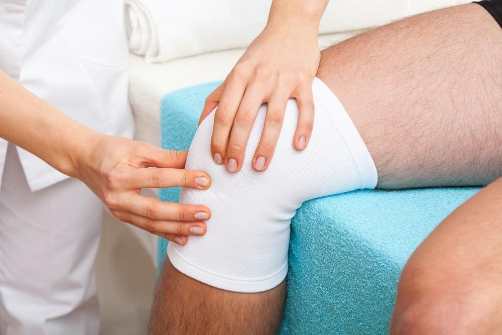
Chiropractic therapy is among the more conservative treatment options for a herniated disc and may be tried first before proceeding with more invasive treatments. Injury Medical Chiropractic and Functional Medicine Clinic works with primary healthcare providers and specialists to develop an optimal health and wellness solution that fully benefits the individual to get back to normal.
The Science of Functional Healing
References
Zhou, Z., Gao, M., Wei, F., Liang, J., Deng, W., Dai, X., Zhou, G., & Zou, X. (2014). Shock absorbing function study on denucleated intervertebral disc with or without hydrogel injection through static and dynamic biomechanical tests in vitro. BioMed research international, 2014, 461724. https://doi.org/10.1155/2014/461724
Nosikova, Y. S., Santerre, J. P., Grynpas, M., Gibson, G., & Kandel, R. A. (2012). Characterization of the annulus fibrosus-vertebral body interface: identification of new structural features. Journal of anatomy, 221(6), 577–589. https://doi.org/10.1111/j.1469-7580.2012.01537.x
Ucar, D., Duman, S., Bayram, Y., & Ucar, B. Y. (2021). Extruded disc herniations are experienced earlier by inactive young people in the high-tech gaming era. Journal of medicine and life, 14(3), 402–407. https://doi.org/10.25122/jml-2021-1059
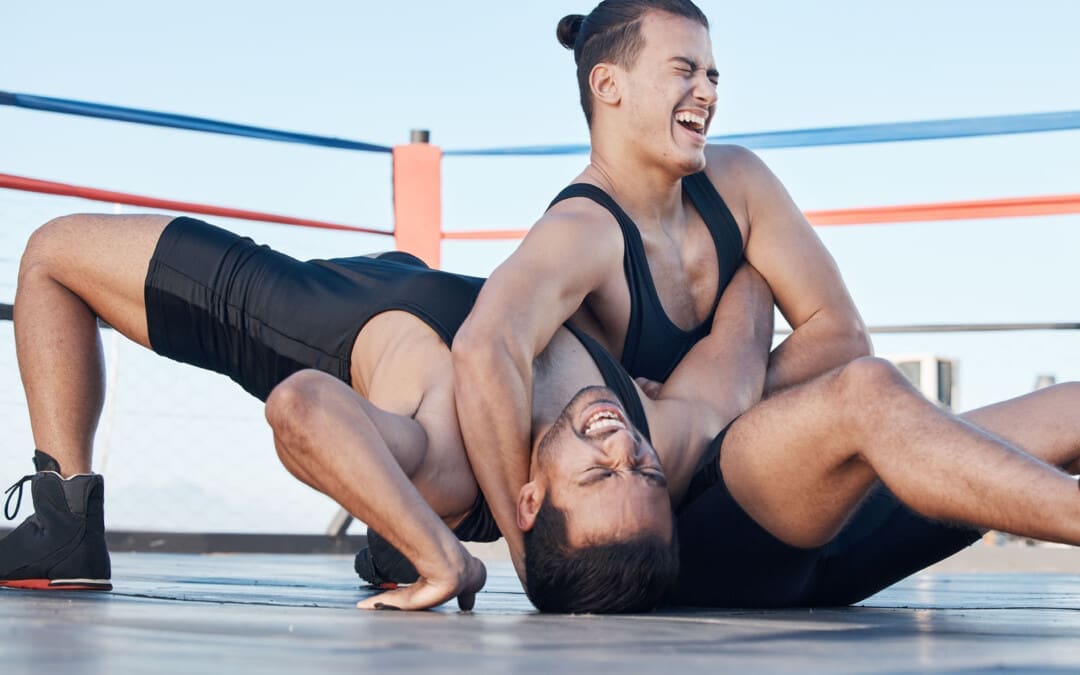
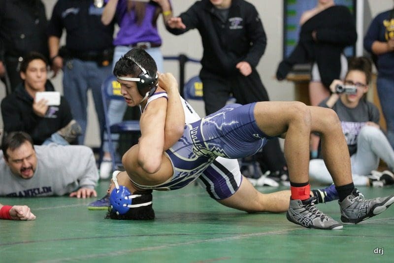
For wrestling athletes or those thinking about getting into the sport, can knowing about common injuries help in rehabilitation and prevention?
Wrestling Injuries
Wrestling is an intense and demanding sport. Studies have found that football and wrestling are the two high school sports with the highest risk of serious injury to athletes (Center for Injury Research and Policy, 2009). The injury rate for college wrestlers is 9 injuries per 1,000 athlete exposures. (Kroshus, E. et al., 2018) While most wrestling injuries include strains and sprains, there can also be serious traumatic and unusual injuries. Using proper safety gear and learning correct techniques can significantly reduce the risk of injuries. The majority occur during competition.
Common
The most common wrestling injuries are similar to those in other sports and include:
Muscle Soreness
Muscle soreness that is experienced 12 to 48 hours after an intense workout or competition.
Resting is often all that is needed to recover.
Bruises and Contusions
Sparring, take-downs, and hard landings can result in various bruises and contusions.
Sprains and Strains
Rest, ice, compression, and elevation are recommended to treat sprains and strains immediately.
Ankle Sprains
Ankle sprains occur when surrounding ligaments stretch and tear around the joint.
Wrist Sprains
Typically, it occurs when stretching or tearing the ligaments.
Falling or landing on the hands is a common cause.
Overtraining Syndrome
Frequently occurs in athletes who train beyond the body’s ability to recover.
Dehydration
When trying to make weight, dehydration can be a serious health problem that many wrestlers experience.
Other Injuries
Other injuries common in wrestling:
Wrist tendinitis
Finger fractures
Iliotibial band syndrome
Meniscus tears
Groin pull
Hamstring pull or tear
Pulled calf muscle
Achilles tendonitis
Achilles tendon rupture
Clavicle/Collarbone fracture
Concussion
Serious
The forcing of a joint beyond its normal range of motion is the most common cause of serious injuries. The most serious wrestling injuries affect the neck, shoulder, elbow, and knee and include:
Neck
The cervical vertebrae are often forced into vulnerable positions during various techniques and movements, which can result in a neck injury. Common types include:
Neck Strain
Whiplash
Cervical Fracture
Shoulder
A combination of leverage and twisting causes most upper body and shoulder injuries during competition. Types of shoulder injuries include:
Rotator cuff injury
Shoulder separation
Shoulder dislocation
Elbow Dislocation
Elbows are under tremendous strain when maneuvering.
Dislocations of the radial head are often related to the athlete bracing for a fall on an outstretched arm during take-downs.
Knee
Most knee injuries occur to the ligaments of the knee joint.
These include anterior and posterior cruciate ligament or ACL/PCL injuries.
Safety
Wrestling requires flexibility, strength, and proper technique to prevent injury, combined with thorough instruction and coaching and following basic safety precautions. Some tips include.
Safety Gear
Wear appropriate headgear and mouthguards during practices, meets, and tournaments.
Improving Joint Flexibility
Wrestlers with a high degree of shoulder flexibility have fewer injuries.
The flexibility of the lower back, hamstrings, elbows, and cervical spine should also be worked on.
Gain or Lose Weight Safely
Avoid dramatic weight loss and weight-gaining strategies by maintaining healthy nutrition and hydration during the season.
Avoiding Dangerous Holds and Slam Moves
Safe wrestling techniques need to be followed as these can generate severe injuries.
Regardless of how common or seemingly not serious an injury or medical condition is, it’s important to rest and recover and tell a coach and health care professional, as some injuries and conditions can become serious if left untreated. Injury Medical Chiropractic and Functional Medicine Clinic focuses on and treats injuries and chronic pain syndromes through personalized care plans that improve ability through flexibility, mobility, and agility programs to relieve pain. Our providers use an integrated approach to create personalized care plans for each patient, including Functional Medicine, Acupuncture, Electro-Acupuncture, and Sports Medicine principles. Our goal is to relieve pain naturally by restoring health and function to the body. If other treatment is needed, Dr. Jimenez has teamed up with top surgeons, clinical specialists, medical researchers, and rehabilitation providers to provide the most effective treatments.
Perseverance and Power
References
Nationwide Children’s Hospital. (2024). Center for Injury Research and Policy. https://www.nationwidechildrens.org/research/areas-of-research/center-for-injury-research-and-policy
Kroshus, E., Utter, A. C., Pierpoint, L. A., Currie, D. W., Knowles, S. B., Wasserman, E. B., Dompier, T. P., Marshall, S. W., Comstock, R. D., & Kerr, Z. Y. (2018). The First Decade of Web-Based Sports Injury Surveillance: Descriptive Epidemiology of Injuries in US High School Boys’ Wrestling (2005-2006 Through 2013-2014) and National Collegiate Athletic Association Men’s Wrestling (2004-2005 Through 2013-2014). Journal of athletic training, 53(12), 1143–1155. https://doi.org/10.4085/1062-6050-154-17
What are the healing times of common sports injuries for athletes and individuals who engage in recreational sports activities?
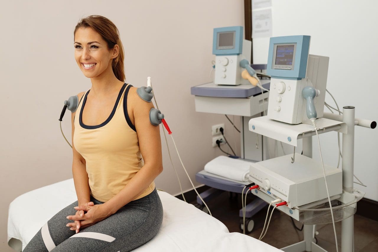
A young, happy sportswoman is getting tens-electrotherapy treatments at a medical clinic.
Healing Times for Sports Injuries
Healing time from sports injuries depends on various factors, such as the location and extent of the injury and the health of the skin, joints, tendons, muscles, and bones. It is also important to take the time to recuperate or not rush back into physical sports activities before the bones or tissues have fully healed. To prevent re-injury, ensure the doctor clears health before returning to sports or strenuous physical activity.
According to CDC research, an average of 8.6 million sports and recreation-related injuries occur annually. (Sheu, Y., Chen, L. H., and Hedegaard, H. 2016) However, most sports injuries are superficial or caused by low-grade strains or sprains; at least 20% of injuries result from bone fractures or more serious injuries. Bone fractures take longer than sprains or strains, and complete tendon or muscle ruptures can take months before one can fully return to activities. Individuals in decent physical shape with no underlying illness or impairment, here is what they can expect when recovering from the following sports injuries:
Bone Fractures
In sports, the highest rate of bone fractures occurs with football and contact sports. Most are centered around the lower extremities but can involve the neck and shoulder blades, arms, and ribs.
Simple Fractures
Depends on the individual’s age, health, type, and location.
Generally, takes at least six weeks to heal.
Compound Fractures
In this case, a bone is broken in several places.
It may require surgery to stabilize the bone.
Healing time can take up to eight months.
Fractured Clavicle/Collarbone
It may require the immobilization of the shoulder and upper arm.
It can take five to ten weeks to heal fully.
Fractured fingers or toes can heal in three to five weeks.
Fractured Ribs
Part of the treatment plan includes breathing exercises.
Painkillers may be needed short term.
Usually, it takes around six weeks to heal.
Neck Fractures
It may involve any one of the seven neck vertebrae.
A neck brace or a halo device that is screwed into the skull for stability may be used.
A sprain is the stretching or tearing of ligaments or the tough bands of fibrous tissue that connect two bones at a joint.
A strain is the overstretching or tearing of muscles or tendons.
Sprained Ankles
It can heal in five days if there are no complications.
Severe sprains involving torn or ruptured tendons can take three to six weeks to heal.
Calf Strains
Classified as grade 1 – a mild strain can heal in two weeks.
A grade 3 – severe strain may require three months or more to heal completely.
The use of calf suppression sleeves can expedite the recovery of strains and sprains in the lower leg.
Acute Neck Strain
A tackle, impact, fall, quick shifting, or whipping motion can cause a whiplash injury.
Healing time can take a couple of weeks to six weeks.
Other Injuries
ACL Tears
Involving the anterior cruciate ligament.
Usually, it requires months of recuperation and rehabilitation, depending on several factors, including the type of sports activity.
Full recovery from surgery takes six to 12 months.
Without surgery, there is no specific timeline for rehabilitation.
Achilles Tendon Ruptures
It is a serious injury.
These occur when the tendon is either partially or completely torn.
Individuals will more than likely require surgery.
Recovery time is four to six months.
Cuts and Lacerations
Depends on the depth and location of the injury.
It can take anywhere from a week to a month to heal.
If there are no accompanying injuries, stitches can be removed within two to three weeks.
If a deep cut requires stitches, more time is necessary.
Mild Contusions/Bruises
Are caused by a trauma to the skin, causing blood vessels to break.
In most cases, a contusion will take five to seven days to heal.
Shoulder Separations
When treated properly, it usually takes around two weeks of rest and recovery before the patient returns to activity.
Multidisciplinary Treatment
After the initial inflammation and swelling have subsided, a doctor will recommend a treatment plan that usually involves physical therapy, self-performed physical rehabilitation, or supervision by a physical therapist or team. Fortunately, athletes and individuals who regularly exercise tend to have a faster healing time because they are in top physical shape, and their cardiovascular system provides a stronger blood supply that speeds up the healing process. At El Paso’s Chiropractic Rehabilitation Clinic & Integrated Medicine Center, we passionately focus on treating patients’ injuries and chronic pain syndromes. We focus on improving ability through flexibility, mobility, and agility programs tailored to the individual. We use in-person and virtual health coaching and comprehensive care plans to ensure every patient’s personalized care and wellness outcomes.
Our providers use an integrated approach to create personalized care plans that include Functional Medicine, Acupuncture, Electro-Acupuncture, and Sports Medicine principles. Our goal is to relieve pain naturally by restoring health and function to the body.
If the chiropractor feels the individual needs other treatment, they will be referred to a clinic or physician best suited for them. Dr. Jimenez has teamed up with the top surgeons, clinical specialists, medical researchers, and premier rehabilitation providers to provide the top clinical treatments for our community. Providing highly noninvasive protocols is our priority, and our personalized patient-based clinical insight is what we provide.
Lumbar Spine Injuries in Sports: Chiropractic Healing
References
Sheu, Y., Chen, L. H., & Hedegaard, H. (2016). Sports- and Recreation-related Injury Episodes in the United States, 2011-2014. National health statistics reports, (99), 1–12.
IFM's Find A Practitioner tool is the largest referral network in Functional Medicine, created to help patients locate Functional Medicine practitioners anywhere in the world. IFM Certified Practitioners are listed first in the search results, given their extensive education in Functional Medicine