Chronic neck pain may stem from ligamentous injuries. Uncover insights and solutions to relieve your pain and improve mobility.
Introduction: The Persistent Shadow of Chronic Neck Pain
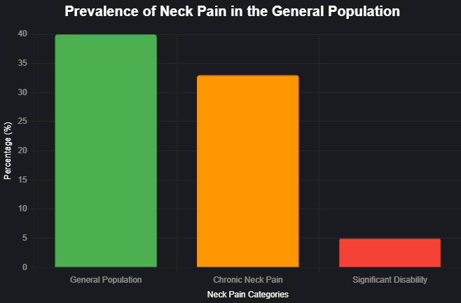
Chronic neck pain is like a grim companion that lingers far too long, much like a gloomy Wednesday Addams brooding over a particularly dreary day. It’s not just a minor annoyance; it’s a widespread condition affecting 30–50% of the general population, with women over 50 bearing the brunt (The Open Orthopaedics Journal, 2014). About one-third of these individuals endure pain lasting longer than six months, and for 5%, it becomes a debilitating force, slashing their quality of life. Imagine trying to enjoy a sunny day when your neck feels like it’s plotting a mutiny.
What’s behind this misery? Often, it’s not just a stiff muscle or a pinched nerve but something deeper: ligamentous injuries. These tough bands of tissue, which hold your spine together like the strings of a grim puppet, can be damaged in motor vehicle accidents (MVAs), falls, or even by the slow torture of poor posture. When ligaments falter, the cervical spine loses stability, setting off a cascade of overlapping pain symptoms that can make life feel like a gothic novel.
In this guide, we’ll unravel the connection between ligamentous injuries and chronic neck pain, focusing on their relevance to personal injury practices, particularly in motor vehicle accident (MVA) cases. We’ll explore prolotherapy, a treatment that might banish the pain like a well-timed quip from Wednesday herself. And we’ll spotlight Dr. Alexander Jimenez, a dual-licensed chiropractor and nurse practitioner in El Paso, whose expertise in treating MVA victims is as sharp as a guillotine’s edge. So, let’s dive into this tale of pain, recovery, and the hope of a pain-free existence.
The Role of Ligamentous Injuries in Chronic Neck Pain
Ligaments are the unsung heroes of your body, silently keeping your bones in line like loyal but underappreciated butlers. In the neck, the cervical spine depends on these structures, especially the capsular ligaments, to stabilize the facet joints—the small hinges between vertebrae that allow you to turn your head without it rolling off like a scene from a Tim Burton film. When these ligaments are injured, they can stretch or tear, leading to cervical instability —a condition in which the vertebrae move more than they should, causing discomfort and chronic pain.
The symptoms depend on where the injury strikes. In the upper cervical spine (C0-C2), you might face nerve irritation, leading to migraines, dizziness, vertigo, tinnitus, facial pain, or arm pain—basically, a full cast of unpleasant characteristics. In the lower cervical spine (C3-C7), expect muscle spasms, a grinding or popping sensation (crepitation), tingling (paresthesia), and that relentless neck pain that feels like it’s auditioning for a horror movie role.
Whiplash, often from MVAs, is a prime suspect in these injuries. Research indicates that whiplash can stretch ligaments up to 275% of their normal length, leaving them lax and unable to return to their original position (The Open Orthopaedics Journal, 2014). Even low-speed collisions (7–8 mph) can generate forces strong enough to cause this damage, with the head whipping through 18 inches at 7G in less than a quarter of a second. It’s like your neck is starring in its high-speed chase scene but without the Hollywood glamour.
Other culprits include falls, sports injuries, or the slow, gradual strain of carrying heavy loads improperly. Without proper care, these injuries can turn chronic neck pain into a lifelong nemesis, lurking in the shadows of every movement.
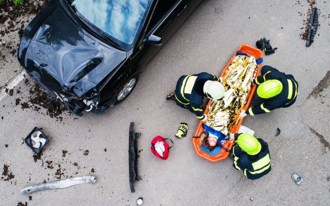
Motor Vehicle Accidents: The Catalyst for Neck Injuries
Motor vehicle accidents are the modern equivalent of a medieval joust—sudden, violent, and likely to leave you worse for wear. Rear-end collisions are notorious for causing whiplash-associated disorders (WAD), a collection of symptoms ranging from neck pain and stiffness to headaches and cognitive fog. It’s as if your body decides to throw a tantrum after being rudely jostled.
The sneaky thing about WAD is its delayed debut. You might walk away from a crash feeling like you’ve dodged a bullet, only to find symptoms creeping in days or weeks later, like an unwelcome plot twist. This delay happens because initial inflammation and improper ligament healing can take time to manifest as chronic pain. A 2019 study explained that early spinal manipulation therapy (SMT) can reduce the risk of long-term issues, emphasizing the need for prompt care (El Paso Back Clinic).
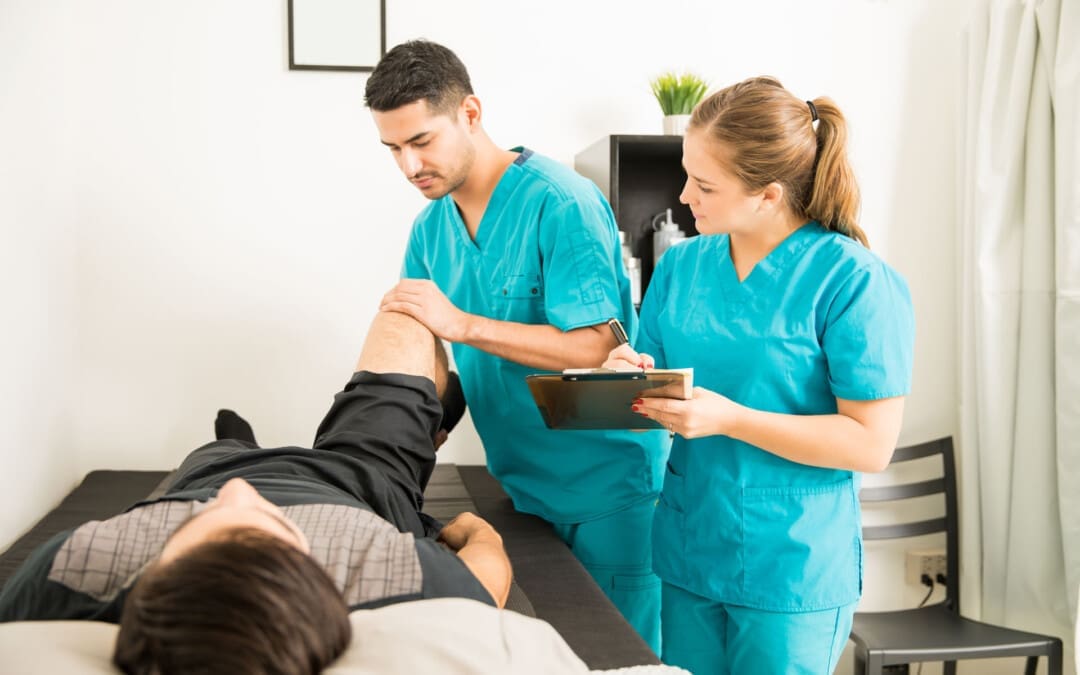
At El Paso Back Clinic, Dr. Alexander Jimenez specializes in treating musculoskeletal injuries related to motor vehicle accidents (MVAs). His team understands that ligaments, muscles, and tendons bear the brunt of these collisions, leading to conditions like sprains, strains, and even disc herniations. Early intervention, through chiropractic adjustments and other therapies, can prevent these injuries from becoming a lifelong saga of pain.
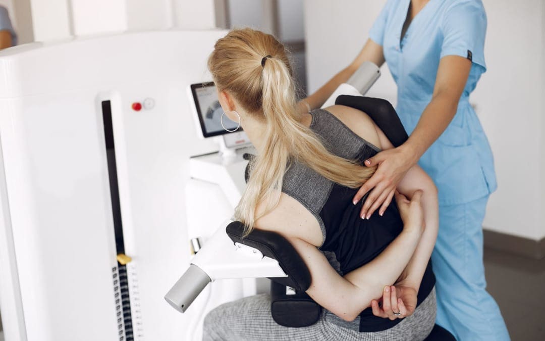
Prolotherapy: A Ray of Hope for Ligament Repair
Prolotherapy is like a dark ritual for healing, but instead of candles and chants, it uses injections to summon your body’s repair crew. This regenerative therapy involves injecting a solution—typically a mixture of dextrose, saline, and a local anesthetic—into damaged ligaments or tendons. The irritation sparks a healing response, encouraging the growth of new tissue to strengthen the area. It’s as if your body is tricked into rebuilding its crumbling architecture.
For chronic neck pain, prolotherapy targets the lax ligaments that cause cervical instability. Unlike conventional treatments like NSAIDs, narcotics, or physical therapy, which often mask the pain, prolotherapy aims to fix the root cause. The 2014 report highlights that over 85% of patients with cervical ligament injuries, including those with WAD or headaches, reported minimal to no residual pain after prolotherapy. Another study showed a significant improvement in the Neck Disability Index (NDI) scores 12 months post-treatment, with a change of 13.77 (p < 0.001) (The Open Orthopaedics Journal, 2014).
Dr. Jimenez offers prolotherapy at El Paso Back Clinic, integrating it into his holistic approach. This minimally invasive procedure, performed outpatient, is gaining traction as an alternative to surgery or long-term medication, offering hope to those tired of living under pain’s grim shadow.
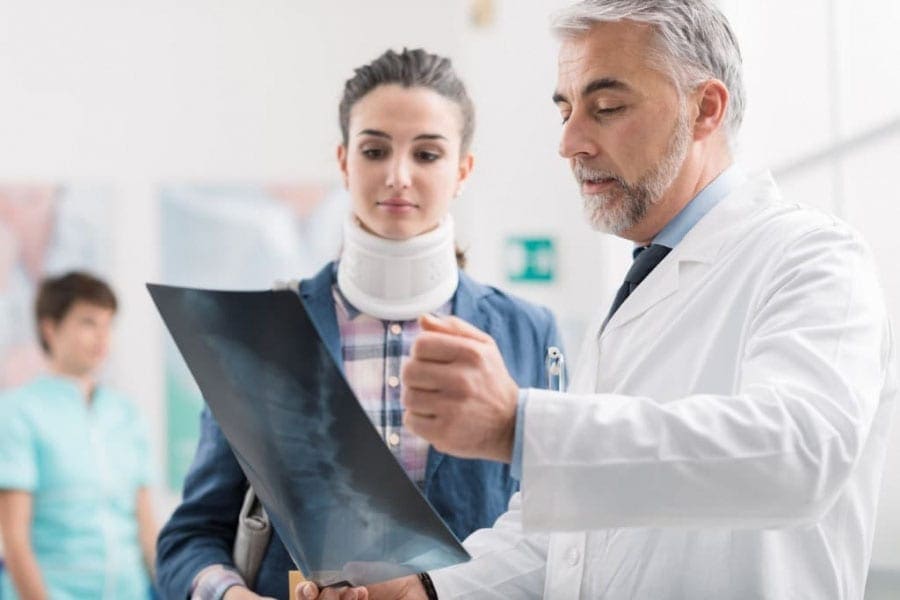
Dr. Alexander Jimenez: El Paso’s Trusted Expert
In El Paso, Dr. Alexander Jimenez is the equivalent of a lighthouse in a storm for MVA victims. With over 30 years of experience, his dual licensure as a chiropractor and nurse practitioner allows him to blend holistic and traditional medical care, creating a comprehensive treatment plan that’s as precise as Wednesday Addams’ aim with a crossbow. At El Paso Back Clinic, he’s known for clinically correlating injuries, ensuring that personal injury attorneys and medical providers understand the full scope of a patient’s condition.
Dr. Jimenez’s approach goes beyond symptom relief. He delves into the root causes, employing techniques such as chiropractic adjustments, manual manipulations, and prolotherapy to restore function. His collaboration with personal injury attorneys ensures proper documentation, which is crucial for legal cases. Patients praise his compassionate care, with many regaining their quality of life after chronic pain (El Paso Back Clinic).
Injury Medical Clinic offers a range of services, from wellness and nutrition to specialized care for auto accident injuries, making it a one-stop shop for recovery. Whether it’s a sprained ligament or a complex herniated disc, Dr. Jimenez’s expertise shines through.
The Science Behind Ligamentous Injuries
To understand why ligamentous injuries are such a pain—literally—let’s dive into the science. The cervical spine’s capsular ligaments are designed to keep vertebrae in check, but they’re not invincible. A force as low as 5 N can cause failure, although most studies report a force of around 100 N (The Open Orthopaedics Journal, 2014). In MVAs, even low-speed impacts can generate sufficient force to stretch or tear these ligaments, resulting in instability.
This instability triggers a domino effect. The vertebrae move excessively, irritating nerves and causing inflammation. Within the upper cervical spine, this can lead to vertebrobasilar insufficiency, resulting in symptoms like vertigo or tinnitus. In the lower spine, it’s primarily characterized by muscle spasms and chronic pain. Up to 25% of severe neck trauma lesions, especially with rotation, involve the C0-C2 ligaments alone, making them a critical focus for treatment.
Why Conventional Treatments Fall Short
Conventional treatments like NSAIDs, narcotics, cervical collars, and physical therapy are like putting a Band-Aid on a broken bone—they might help temporarily. Still, they don’t fix the underlying issue. The 2014 report notes that these methods have limited long-term success, particularly for WAD, which is notoriously resistant to standard care (The Open Orthopaedics Journal, 2014). Patients often find themselves in a cycle of temporary relief followed by recurring pain, like a grim rerun of a bad sitcom.
This is where prolotherapy stands out. By stimulating ligament repair, it addresses the instability at the heart of chronic neck pain, offering a potential cure rather than a temporary fix. Dr. Jimenez’s use of prolotherapy, combined with chiropractic care, aligns with this evidence-based approach, providing a path to lasting relief.
The Importance of Early Intervention
Time is not your friend when it comes to MVA injuries. The longer you wait, the more likely it is that ligament damage will lead to chronic pain. Early chiropractic care, as Dr. Jimenez provides, can realign the spine and reduce inflammation, preventing the progression to chronic syndromes. The 2019 European Spine Journal study underscores this, showing that early SMT can significantly lower the risk of long-term issues (El Paso Back Clinic).
Dr. Jimenez’s clinic emphasizes immediate care, utilizing diagnostic tools to assess ligament damage and tailor treatments accordingly. This proactive approach is crucial for personal injury cases, where timely intervention can significantly impact a patient’s recovery and legal outcome.
Chiropractic Care for Healing After Trauma- Video
Case Studies: Real-Life Recovery Stories
While specific patient stories from Dr. Jimenez’s clinic aren’t detailed here, his website highlights testimonials from patients who have regained their lives after overcoming chronic pain (El Paso Back Clinic). Imagine a patient, let’s call her Morticia, who suffered whiplash in a rear-end collision. Initially, she dismissed the stiffness, but weeks later, headaches and neck pain began to take over. After conventional treatments failed, she turned to Dr. Jimenez. Through a combination of chiropractic adjustments and prolotherapy, her ligaments healed, and her pain faded, allowing her to return to her gothic gardening with a smirk.
Another hypothetical case: Gomez, a construction worker, experienced neck pain after a minor MVA. Dr. Jimenez’s thorough assessment revealed ligament laxity and prolotherapy sessions restored stability. Gomez was back to lifting heavy loads, his only complaint being the lack of a good cigar to celebrate.
These stories, while fictionalized for humor, reflect the real impact of Dr. Jimenez’s care, as evidenced by patient reviews averaging 5.0 stars (Healthline FindCare).
The Legal Angle: Documenting Injuries for Personal Injury Cases
In personal injury cases, documentation is king. Dr. Jimenez’s expertise in clinically correlating injuries ensures that attorneys have the evidence needed to build strong cases. Ligamentous injuries, though not always visible on standard imaging, can be assessed through clinical evaluations and specialized tests. Dr. Jimenez’s reports detail the extent of the damage, linking it to the MVA and justifying treatments such as prolotherapy. This collaboration is vital for securing fair compensation for victims (El Paso Back Clinic).
Holistic and Traditional Care: Dr. Jimenez’s Dual Approach
Dr. Jimenez’s dual licensure sets him apart. As a chiropractor, he employs techniques such as spinal manipulation, cranial therapy, and prolotherapy to address physical injuries. As a nurse practitioner, he can incorporate medical diagnostics and treatments to ensure a comprehensive approach to patient care. This blend is like mixing Wednesday’s dark wit with Gomez’s fiery passion—effective and unique. His clinic’s focus on wellness, nutrition, and functional medicine further enhances recovery, addressing not just the injury but the whole patient (El Paso Back Clinic).
The Broader Impact of Chronic Neck Pain
Chronic neck pain doesn’t just affect the neck; it ripples through life, impacting work, relationships, and mental health. 5% of sufferers with significant disability face reduced productivity and quality of life, making effective treatment critical. Conditions like disc herniation, cervical spondylosis, and post-concussion syndrome often coexist with ligament injuries, complicating recovery. Dr. Jimenez’s holistic approach tackles these complexities, offering hope where conventional methods fall short.
Prevention: Avoiding the Pain Before It Starts
While not always possible, preventing chronic neck pain involves maintaining good posture, practicing safe driving habits, and seeking prompt care after injuries. Strengthening neck muscles through exercises and avoiding repetitive strain can help alleviate symptoms. Dr. Jimenez’s clinic offers wellness programs to build resilience, reducing the risk of future injuries (El Paso Back Clinic).
The Dark Humor of Pain: A Wednesday Addams Perspective
Living with chronic neck pain is like being stuck in a never-ending Addams Family reunion—grim, uncomfortable, and full of unexpected twists. Your neck might creak like the mansion’s floorboards, and every turn of the head feels like a plot twist in a gothic novel. But fear not; there’s a way out of this dreary tale. With treatments like prolotherapy and experts like Dr. Jimenez, you can send that pain packing, leaving you free to brood over more interesting things, like the perfect shade of black.
Conclusion: A Serious Call to Action
Chronic neck pain, especially from ligamentous injuries, is no laughing matter despite our grim humor. It’s a condition that demands attention, particularly after MVAs, where timely care can prevent a lifetime of suffering. Dr. Alexander Jimenez at El Paso Back Clinic offers a beacon of hope, combining chiropractic expertise, prolotherapy, and a deep understanding of personal injury cases. If you’re grappling with neck pain, don’t let it define you. Seek expert care, explore options like prolotherapy, and take the first step toward recovery. Your neck—and your sanity—will thank you.
What are the advantages of having a team of nurse practitioners and chiropractors help maintain the health of your spine after a car accident?
Benefits of Chiropractic and Nurse Practitioners for Motor Vehicle Collisions
One of the main causes of spinal injuries, such as whiplash, herniated discs, and soft tissue injury, which can cause severe pain and impair movement, is motor vehicle collisions (MVCs). For both short-term symptoms and long-term rehabilitation, these injuries frequently necessitate a multimodal therapy strategy. While nurse practitioners, as advanced practice registered nurses, conduct medical evaluations, write prescriptions, and oversee overall health management, chiropractors focus on musculoskeletal care, including spine adjustments and manual therapies. These professionals’ collaboration aims to provide a comprehensive, patient-centered strategy for spine health following MVC. (Kent, R., et al., 2023)
For those recuperating from auto accident injuries, a chiropractic and nurse practitioner team can offer thorough spinal health care with an emphasis on pain management and increased mobility.
A chiropractic and nurse practitioner team can offer a comprehensive approach to spinal health after a car accident by addressing pain, improving mobility, and facilitating faster recovery.
Chiropractors focus on spinal alignment and joint mobility.
Nurse practitioners provide broader medical oversight and patient education.
The team approach can lead to more effective and personalized care for individuals recovering from car accident injuries. (Riva, J. J., et al., 2010)
Key advantages of this collaborative approach
A chiropractor and nurse practitioner (NP) therapy team can combine their skills to provide comprehensive care for spine health following a motor vehicle collision (MVC) and address acute and long-term requirements.
Care that is multidisciplinary and holistic
Collaboration between chiropractors and NPs to address structural and systemic issues enhances treatment outcomes, particularly for spine injuries related to motor vehicle collisions (MVC), as well as for chronic headaches and neck discomfort. (Riva, J. J., et al., 2010)
Plans for Treatment That Are Unique to You
Chiropractors and NPs create personalized patient treatment plans, focusing on their specific injuries and overall health, including pre-existing conditions and medication needs. This approach enhances outcomes by tailoring care to the patient’s unique circumstances.
Managing Pain Without Relying Too Much on Drugs
By using non-invasive methods to alleviate pain, chiropractic therapy may help reduce the use of opioids. NPs can prescribe short-term pain relief and monitor side effects, ensuring safe use and reducing dependency risks. Natural pain management combined with medical supervision lessens dependence and side effects. (Prater, C., Tepe, M., & Battaglia, P. 2020)
Quicker Recuperation and Rehabilitation
As demonstrated in the treatment of auto accidents, chiropractic adjustments can lessen muscle spasms and restore joint function. By referring patients to physical therapy and tracking their progress, NPs can hasten recovery and reduce the likelihood of developing persistent back pain. This integrated therapy not only reduces chronic back pain and other long-term problems, but it also accelerates healing.
Help with Insurance and the Law
Chiropractic and medical providers must carefully record injuries and treatments for insurance claims or legal cases after an MVC to ensure just reimbursement and coverage for care.
Why It Works After MVC
Following a motor vehicle collision (MVC), a chiropractor and nurse practitioner team offers a patient-centered approach to spine health. This team enhances recovery, lowers chronic risks, and improves patient outcomes by fusing NP’s medical management with chiropractic knowledge. This method ensures rapid alleviation and long-term health, especially helpful for complex spine injuries due to MVC.
Injury, Chiropractic, and Functional Medicine Clinic
Dr. Jimenez, a nurse practitioner, uses medical knowledge and chiropractic care to treat various conditions. The clinic provides tailored care programs incorporating functional medicine, acupuncture, electroacupuncture, and sports medicine. The clinic focuses on strength, agility, and flexibility for treating chronic pain syndromes and injuries. Patients of all ages and abilities benefit from comprehensive care plans and in-person and virtual health coaching, ensuring tailored treatment and wellness outcomes.
Personal Injury Rehabilitation
References
Kent, R., Cormier, J., McMurry, T. L., Johan Ivarsson, B., Funk, J., Hartka, T., & Sochor, M. (2023). Spinal injury rates and specific causation in motor vehicle collisions. Accident; analysis and prevention, 186, 107047. doi.org/10.1016/j.aap.2023.107047
Riva, J. J., Muller, G. D., Hornich, A. A., Mior, S. A., Gupta, A., & Burnie, S. J. (2010). Chiropractors and collaborative care: An overview illustrated with a case report. The Journal of the Canadian Chiropractic Association, 54(3), 147–154.
Prater, C., Tepe, M., & Battaglia, P. (2020). Integrating a Multidisciplinary Pain Team and Chiropractic Care in a Community Health Center: An Observational Study of Managing Chronic Spinal Pain. Journal of primary care & community health, 11, 2150132720953680. doi.org/10.1177/2150132720953680
Optimizing Recovery from Work-Related Back Injuries: The Interdisciplinary Approach of Dr. Alexander Jimenez, DC, APRN, FNP-BC
Introduction
Back injuries sustained at work are a leading cause of disability and lost productivity, affecting diverse professions such as truck drivers, construction workers, healthcare providers, and office employees. These injuries often result from improper lifting, repetitive motions, or prolonged sitting, leading to conditions like herniated discs, muscle strains, or chronic pain. For those injured, securing workers’ compensation benefits and accessing effective treatment are critical steps toward recovery. Dr. Alexander Jimenez, a dual-licensed chiropractor and nurse practitioner with over three decades of experience, specializes in treating work-related back injuries, particularly those from personal injury cases and auto accidents. His integrative approach, detailed on www.dralexjimenez.com and www.chiromed.com, combines chiropractic care, advanced medical interventions, and collaboration with trusted medical and legal professionals to deliver comprehensive care.
This article outlines the benefits of chiropractic care in managing work-related back injuries, the expanded treatment options enabled by Dr. Jimenez’s nurse practitioner licensure, and the interdisciplinary strategies that optimize recovery. It also highlights how Dr. Jimenez collaborates with legal providers to ensure patients receive the support needed to navigate workers’ compensation claims effectively.
The Burden of Work-Related Back Injuries
Work-related back injuries are prevalent across industries, with the Bureau of Labor Statistics reporting over 900,000 cases of nonfatal occupational injuries involving the back in 2020. High-risk occupations include:
Truck Drivers: Prolonged sitting and unloading heavy cargo increase spinal stress.
Construction Workers: Lifting, twisting, and repetitive motions contribute to injury.
Healthcare Workers: Patient handling and prolonged standing elevate risk.
Manual Laborers: Heavy lifting and improper techniques lead to strains.
Office Workers: Poor posture and sedentary behavior cause chronic pain.
These injuries often result from improper lifting, repetitive turning, twisting while lifting, or holding heavy objects overhead. Symptoms may include acute pain, limited mobility, or chronic conditions requiring long-term management. Workers’ compensation programs exist to cover medical expenses and lost wages, but the process can be complex, necessitating expert medical and legal guidance.
Benefits of Chiropractic Care for Work-Related Back Injuries
Chiropractic care is a cornerstone of non-invasive treatment for back injuries, focusing on restoring spinal alignment, reducing pain, and improving function. Dr. Jimenez leverages evidence-based chiropractic techniques to address work-related injuries, supported by randomized controlled trials (RCTs) and cohort studies.
Pain Reduction and Functional Improvement
Chiropractic adjustments, or spinal manipulations, realign the spine to alleviate pressure on nerves and muscles. A 2018 RCT published in The Spine Journal (Goertz et al.) found that chiropractic care combined with usual medical care significantly reduced pain and disability in patients with low back pain compared to medical care alone. Dr. Jimenez employs manual adjustments, flexion-distraction techniques, and soft tissue therapies to target specific injury sites, promoting natural healing.
Non-Invasive and Drug-Free Approach
Chiropractic care offers a drug-free alternative to pain management, reducing reliance on opioids, which is critical given the opioid crisis. A 2020 cohort study in Pain Medicine (Whedon et al.) demonstrated that patients receiving chiropractic care for low back pain had a lower likelihood of opioid prescriptions compared to those receiving only medical care. Dr. Jimenez integrates therapies like myofascial release and therapeutic exercises to manage pain without pharmacological interventions.
Prevention of Chronic Conditions
Early chiropractic intervention can prevent acute injuries from becoming chronic. A 2019 study in Journal of Manipulative and Physiological Therapeutics (Eklund et al.) showed that maintenance chiropractic care reduced the recurrence of low back pain episodes. Dr. Jimenez designs personalized treatment plans that include corrective exercises and ergonomic counseling to minimize re-injury risk.
Support for Workers’ Compensation Claims
Chiropractors play a vital role in documenting injuries for workers’ compensation claims. Dr. Jimenez provides detailed clinical notes and treatment plans, ensuring compliance with workers’ compensation requirements. His expertise in occupational health allows him to assess whether injuries are work-related, facilitating accurate reporting to employers and insurance carriers.
Expanded Scope of Practice: Nurse Practitioner Expertise
As a board-certified Family Nurse Practitioner (FNP-BC), Dr. Jimenez’s dual licensure enhances his ability to provide comprehensive care, particularly in personal injury and auto accident cases. Nurse practitioners (NPs) have an expanded scope of practice, allowing them to diagnose, treat, and prescribe medications, which complements chiropractic interventions.
Advanced Diagnostics and Treatment
NPs can order and interpret diagnostic imaging, such as X-rays, MRIs, and CT scans, to assess the extent of spinal injuries. A 2021 study in Journal of General Internal Medicine (Mafi et al.) highlighted the accuracy of NPs in managing musculoskeletal conditions through diagnostics and treatment planning. Dr. Jimenez uses these tools to develop precise treatment strategies, ensuring timely interventions for conditions like herniated discs or vertebral fractures.
Medication Management
In cases where pain or inflammation requires pharmacological support, Dr. Jimenez can prescribe medications like non-steroidal anti-inflammatory drugs (NSAIDs) or muscle relaxants. His NP training ensures judicious use of medications, aligning with evidence-based guidelines to avoid over-reliance. This is particularly beneficial in auto accident cases, where soft tissue injuries often require short-term pharmacological support alongside chiropractic care.
Coordination of Multidisciplinary Care
NPs are trained to coordinate care across specialties, making Dr. Jimenez uniquely positioned to oversee interdisciplinary treatment plans. For complex injuries, he collaborates with orthopedic surgeons, neurologists, and physical therapists to ensure holistic care. A 2020 RCT in BMJ Open (Côté et al.) demonstrated improved outcomes when NPs coordinated multidisciplinary care for back pain, reducing recovery time and costs.
Enhanced Patient Education
NPs emphasize patient education, empowering individuals to manage their recovery. Dr. Jimenez provides guidance on posture, lifting techniques, and lifestyle modifications, drawing on his NP training to address comorbidities like obesity or diabetes that may complicate recovery. This integrative approach aligns with the philosophies outlined on www.chiromed.com, emphasizing patient-centered care.
Interdisciplinary Treatment Strategies
Dr. Jimenez’s practices integrate chiropractic and NP services with interdisciplinary treatments tailored to work injury trauma and auto accident cases. These strategies address the physical, emotional, and legal challenges of recovery.
Comprehensive Treatment Modalities
Chiropractic Adjustments: Correct spinal misalignments to reduce pain and restore mobility.
Physical Therapy: Strengthen core muscles and improve flexibility through targeted exercises.
Hydrotherapy: Use water-based therapies to reduce inflammation and enhance circulation.
Spinal Injections: Administer corticosteroid injections for severe inflammation, guided by diagnostic imaging.
Rehabilitation Programs: Design long-term plans to restore function and prevent re-injury.
A 2017 cohort study in Spine (Blanchette et al.) found that multidisciplinary care combining chiropractic, physical therapy, and medical interventions resulted in faster return-to-work rates for back injury patients. Dr. Jimenez’s integrative approach mirrors these findings, ensuring patients receive tailored care.
Focus on Personal Injury and Auto Accidents
Personal injury cases, including auto accidents, often involve whiplash, soft tissue injuries, and spinal trauma. Dr. Jimenez’s dual expertise allows him to address these conditions comprehensively. For example, whiplash may require chiropractic adjustments to restore cervical alignment, physical therapy to strengthen neck muscles, and short-term NSAIDs to manage pain. His ability to order MRIs ensures accurate diagnosis of soft tissue damage, which is critical for workers’ compensation and insurance claims.
Collaboration with Medical Specialists
Dr. Jimenez collaborates with a network of trusted medical providers, including:
Orthopedic Surgeons: For surgical interventions like discectomy or spinal fusion.
Neurologists: To address nerve-related symptoms like radiculopathy.
Pain Management Specialists: For advanced pain relief techniques, such as epidural injections.
This collaborative model ensures seamless care, with Dr. Jimenez overseeing the treatment plan to maintain continuity. His practice websites emphasize this integrative philosophy, highlighting partnerships with specialists who share a commitment to patient outcomes.
Collaboration with Trusted Legal Providers
Navigating workers’ compensation and personal injury claims requires legal expertise, particularly when employers or insurance carriers dispute claims. Dr. Jimenez partners with trusted legal providers specializing in workers’ compensation and personal injury law to support his patients.
Role of Legal Providers
Legal providers assist with:
Claim Filing: Ensuring timely and accurate submission of workers’ compensation claims, adhering to state-specific statutes of limitations (e.g., one year in many states).
Dispute Resolution: Representing patients in disputes over injury causation or benefit denials.
Maximizing Benefits: Advocating for coverage of all necessary treatments, including chiropractic care, diagnostics, and rehabilitation.
Auto Accident Claims: Handling insurance negotiations and litigation for injuries sustained in work-related auto accidents.
Dr. Jimenez refers patients to attorneys who understand the medical complexities of back injuries, ensuring alignment between clinical documentation and legal arguments. This collaboration is critical in gray-area cases, such as injuries sustained at work-related events or while working from home.
Streamlined Communication
Dr. Jimenez maintains active communication with legal providers, sharing clinical notes and treatment plans to substantiate claims. Workers’ compensation is exempt from HIPAA privacy regulations for injury-related records, allowing seamless information exchange. This ensures legal providers have the evidence needed to advocate effectively, reducing delays in treatment or compensation.
Patient Advocacy
By connecting patients with reputable attorneys, Dr. Jimenez empowers them to focus on recovery without the stress of legal battles. His websites, www.dralexjimenez.com and www.chiromed.com, emphasize this patient-centered approach, highlighting resources for legal support alongside medical care.
Workers’ Compensation: Coverage and Process
Workers’ compensation is an employer-funded insurance program that covers medical expenses and lost wages for work-related injuries. Coverage typically includes:
Emergency department visits
Diagnostic testing (e.g., X-rays, MRIs)
Follow-up care with specialists
Surgical interventions
Rehabilitation and physical therapy
Dr. Jimenez ensures patients understand their rights under workers’ compensation, advising them to report injuries immediately to their employer’s human resources department. Prompt reporting strengthens claims and prevents disputes over injury causation.
Challenges and Solutions
Challenges in securing workers’ compensation include:
Gray-Area Injuries: Injuries at work-related events or while working remotely may be contested. Dr. Jimenez provides detailed documentation to clarify work-related causation.
Employer Pressure: Some employers may pressure workers to return before medical clearance. Dr. Jimenez collaborates with legal providers to protect patients’ rights.
Wage Replacement Issues: Workers reliant on overtime may face reduced compensation. Dr. Jimenez advises patients to consult attorneys to maximize benefits.
A 2019 study in Journal of Occupational and Environmental Medicine (Mueller et al.) found that early legal intervention improved outcomes in contested workers’ compensation cases, underscoring the value of Dr. Jimenez’s legal partnerships.
Maximizing Recovery: Patient-Centered Strategies
Dr. Jimenez’s integrative approach prioritizes patient compliance and transparency to optimize recovery. Key strategies include:
Adherence to Treatment Plans: Keeping appointments and following recommendations ensures consistent progress.
Open Communication: Patients are encouraged to report symptoms accurately, enabling adjustments to treatment plans.
Ergonomic Training: Guidance on posture, lifting, and workplace modifications reduces re-injury risk.
Holistic Care: Addressing comorbidities and mental health to support overall well-being.
His websites provide resources like exercise guides and ergonomic tips, reinforcing these strategies and empowering patients to take an active role in their recovery.
Conclusion
Work-related back injuries and auto accident trauma require a comprehensive, interdisciplinary approach to achieve optimal recovery. Dr. Alexander Jimenez, DC, APRN, FNP-BC, combines his expertise as a chiropractor and nurse practitioner to deliver evidence-based treatments, from spinal adjustments to advanced diagnostics and medication management. His collaboration with trusted medical and legal providers ensures patients receive holistic care and robust support through the workers’ compensation process. By integrating chiropractic care, medical interventions, and legal advocacy, Dr. Jimenez empowers patients to recover fully and return to work with confidence. Visit www.dralexjimenez.com and www.chiromed.com to learn more about his integrative approach and commitment to patient-centered care.
References
Goertz, C. M., et al. (2018). Effect of usual medical care plus chiropractic care vs usual medical care alone on pain and disability among US service members with low back pain: A comparative effectiveness clinical trial. The Spine Journal, 18(8), 1391–1399.
Whedon, J. M., et al. (2020). Association between chiropractic care and use of prescription opioids among patients with low back pain. Pain Medicine, 21(6), 1087–1095.
Eklund, A., et al. (2019). The Nordic maintenance care program: Effectiveness of chiropractic maintenance care versus symptom-guided treatment for recurrent and persistent low back pain. Journal of Manipulative and Physiological Therapeutics, 42(7), 477–487.
Mafi, J. N., et al. (2021). Nurse practitioners and physician assistants in primary care: A systematic review of scope of practice and patient outcomes. Journal of General Internal Medicine, 36(5), 1423–1432.
Côté, P., et al. (2020). Management of neck pain and associated disorders: A clinical practice guideline from the Ontario Protocol for Traffic Injury Management (OPTIMa) Collaboration. BMJ Open, 10(3), e035366.
Blanchette, M. A., et al. (2017). Effectiveness of multidisciplinary rehabilitation programs for work-related low back pain: A systematic review. Spine, 42(13), 1007–1016.
Mueller, K. L., et al. (2019). Impact of early legal representation on workers’ compensation claim outcomes. Journal of Occupational and Environmental Medicine, 61(4), 312–318.
What are some leg-strengthening exercises that will expedite recovery for athletes and physically active individuals who have undergone leg surgery?
Post Surgery Leg Strengthening
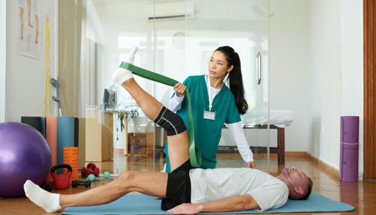
Leg muscles may weaken after hip, knee, ankle, or foot surgery. This happens because those muscles are not used as much during recovery. Gaining back strength and muscle endurance after an injury or surgery is an important step in recovery. Leg exercises can help regain mobility and prevent complications like blood clots and pressure sores after surgery or an injury, which is why engaging in post-surgery leg strengthening is important once the doctor gives the ok.
Benefits
Post-surgery leg strengthening exercises have several benefits, including
Rebuilds strength and confidence
Retraining for optimal mobility and flexibility
Prevents pressure sores
Prevents blood clots
When the leg muscles are contracted, they move blood like a pump, maintaining proper circulation. Moving in a bed after surgery also helps prevent pressure sores from forming and blood clots. A physical therapy team will determine the right leg exercises for each individual and their injury/condition. This is an important step when moving forward after surgery. (Hoogeboom T. J. et al., 2014)
This isometric exercise means the muscles contract while no motion occurs at the hip joints. To perform:
Lie on your back and tighten your buttock muscles.
Hold the muscles tight for 5 seconds, then relax.
Repeat 10 or 15 times.
Gluteal sets can be done several times per day.
Heel Slide
Heel slides can help regain strength in the major muscles of the leg. To perform:
Lie on your back.
Bend the knee of the surgical leg and slowly slide the heel toward your butt.
Slide as far as possible and hold for 5 seconds.
Slowly return to the starting position and repeat.
Short Arc Quad
The short arc quad, or SAQ, is a simple way to get the quadricep muscles working. To perform:
Lie on your back with a towel roll, small ball, or something similar under the knee.
Slowly straighten the knee.
Tighten the quad muscle on the top of the thigh.
Hold for 3 seconds, then relax.
Repeat 10 to 15 times.
Quad Set
This exercise helps get the quad muscles working. It also helps control the position of the kneecap. To perform:
Lie on your back.
Place a small towel roll under the knee.
Try to press the back of the knee flat against the floor.
Hold for 10 seconds and release.
Repeat 10 to 15 times.
Individuals can complete quad sets bilaterally or with both knees simultaneously. This makes the stronger leg help strengthen the weaker side.
Straight Leg Raise
To perform:
Lie on your back.
Lift your leg straight off the floor until it is at the height of the opposite bent knee.
Hold for 10 seconds and slowly lower.
Repeat 10 to 15 times.
Be sure to keep the knee straight for the entire exercise. Keep the opposite knee bent for comfort. To ensure the knee is straight, individuals can complete a quad set first and then the straight leg raise. The exercise can be more challenging by increasing repetitions or adding a 2- to 3-pound ankle weight on the thigh. For even more challenge, add the ankle weight to the ankle.
Hamstring Strengthening
Working out the hamstrings after injury or surgery is important. The hamstring muscles bend the knee and extend the hip backward. To perform:
Lie on your stomach.
Bend one knee to raise the lower limb straight in the air.
Hold for 5 seconds and lower slowly.
Repeat 10 to 15 times.
Once the exercise is easy to do, increase the repetitions to 30. Individuals can also try adding a 2- to 3-pound ankle weight.
Physical therapy can help individuals regain mobility after injury or surgery. A therapist may prescribe exercises as part of an at-home exercise program. Over time, progress will go from simple exercises to more challenging ones to improve balance and mobility. (Madara K. C. et al., 2019)
Injury Medical Chiropractic & Functional Medicine Clinic
Before starting this or any other exercise program, consult a doctor and a physical therapist to find the right exercises for your situation. Injury Medical Chiropractic and Functional Medicine Clinic works with primary healthcare providers and specialists to develop an optimal health and wellness solution. We focus on what works for you to relieve pain, restore function, and prevent injury. Regarding musculoskeletal pain, specialists like chiropractors, acupuncturists, and massage therapists can help mitigate the pain through spinal adjustments that help the body realign itself. They can also work with other medical professionals to integrate a treatment plan to resolve musculoskeletal issues.
Are You Recovering From Ankle Sprains?
References
Hoogeboom, T. J., Dronkers, J. J., Hulzebos, E. H., & van Meeteren, N. L. (2014). Merits of exercise therapy before and after major surgery. Current opinion in anaesthesiology, 27(2), 161–166. doi.org/10.1097/ACO.0000000000000062
Madara, K. C., Marmon, A., Aljehani, M., Hunter-Giordano, A., Zeni, J., Jr., & Raisis, L. (2019). PROGRESSIVE REHABILITATION AFTER TOTAL HIP ARTHROPLASTY: A PILOT AND FEASIBILITY STUDY. International Journal of Sports Physical Therapy, 14(4), 564–581.
Finger pulley injuries are unique digital injuries distinct from sprains or dislocations. They occur specifically in rock climbers and occasionally in baseball pitchers. What are the symptoms, diagnoses, and treatments available?
Finger Pulley Injury
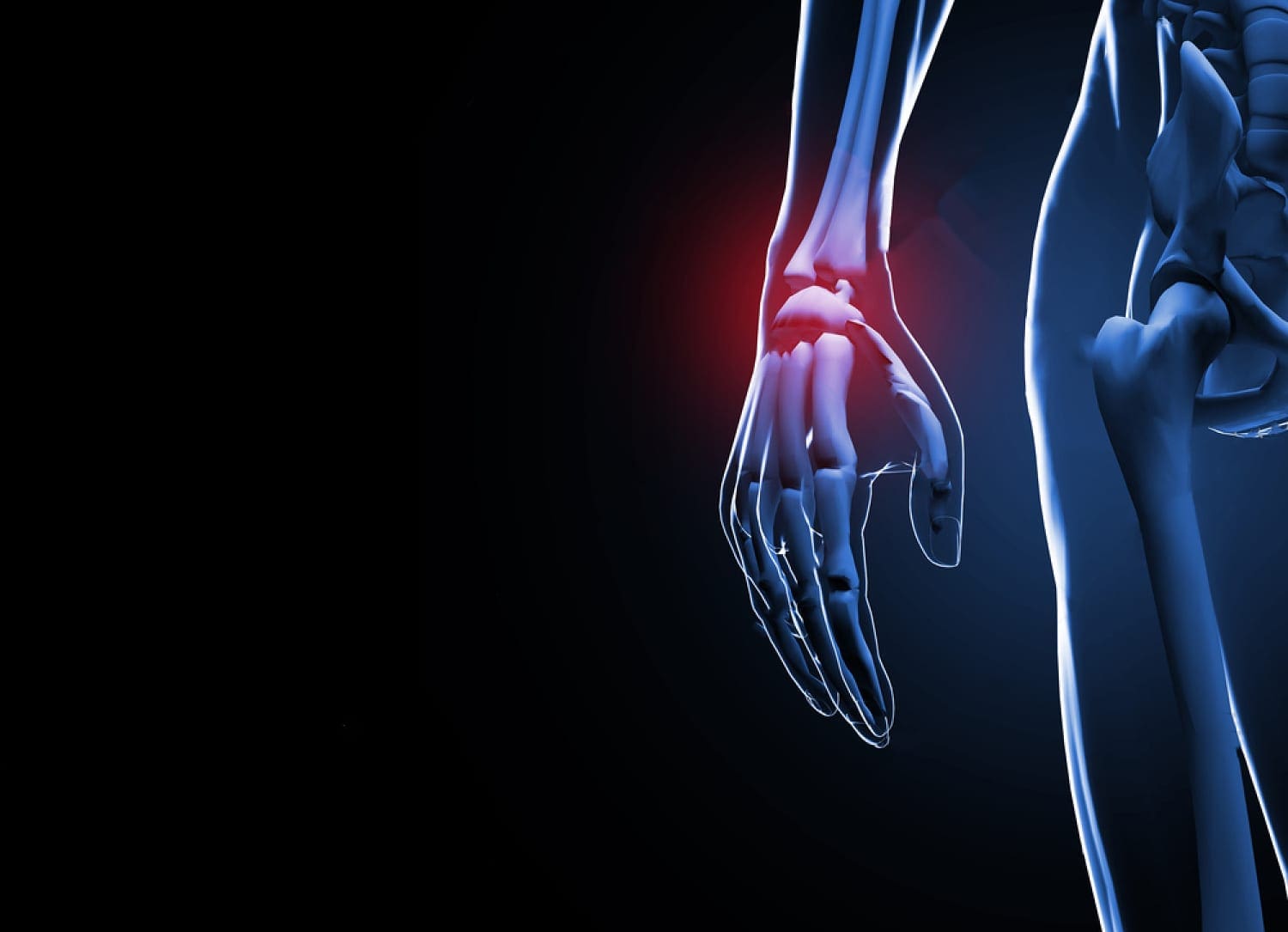
A finger pulley injury, common in activities like climbing, involves damage to the fibrous bands (pulleys) that hold tendons against bones. This causes pain, swelling, and potentially bowstringing of the tendons.
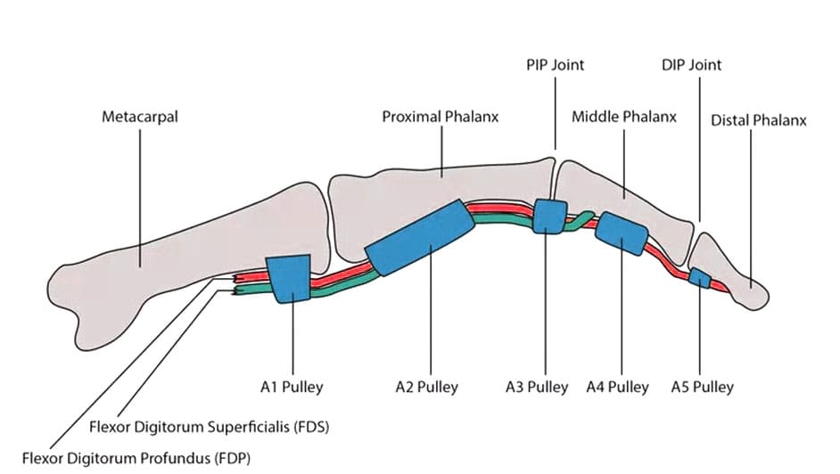
Finger pulleys are structures that hold tendons against the bones of the fingers.
Injury symptoms include pain, swelling, and a popping sound heard at the time of the injury.
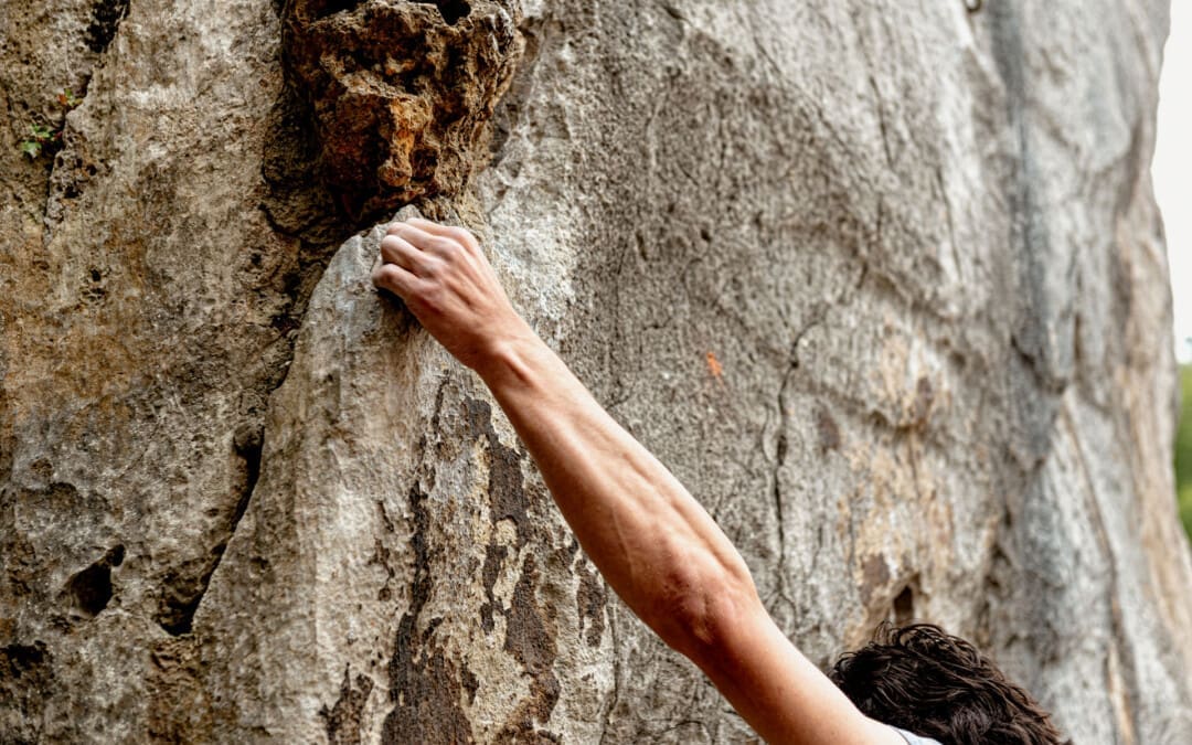
Finger pulley injuries, or ruptures of the digital pulley, are seen almost exclusively in rock climbers. (Miro P. H. et al., 2021)
This activity stresses the digits when maneuvering along uneven surfaces while supporting the entire body’s weight. The injuries result from the mechanics of the finger tendons and joints and the position the fingers hold while rock climbing. Rock climbing has grown in popularity. The only other sport in which this injury has been described is baseball, in pitchers. The forces acting on the finger are very different in these activities, but both place high stress on the finger pulleys.
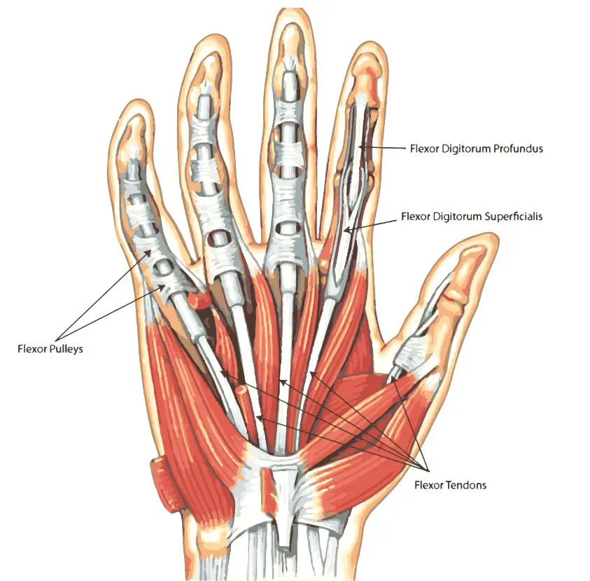
Digital Pulleys
Everyone has structures in their fingers called digital pulleys. These pulleys hold the tendons against the bones of the fingers. Each finger has eight pulleys, but only two are considered critical to prevent the finger tendons’ bowstringing (when one pulley gives out or ruptures). This can result in various injury outcomes, from a simple strain of the pulley to ruptures of multiple pulleys in a single digit. Pain, stiffness, and an inability to fully flex the finger can occur. (Carruthers K. H., Skie M., & Jain M. 2016) In severe situations, when the tendons are bowstringing, the tendon may lift away from the finger when making a fist.
Symptoms
Pain and Tenderness
Localized pain and tenderness at the finger’s base, particularly when gripping or bending. Pain on the palm side of finger and tenderness with pressure
Swelling
Swelling and bruising around the affected finger joint, especially on the palm side.
Stiffness and pain when bending the fingers or difficulty gripping. Difficulty forming a fist
Bowstringing
Visible displacement of the tendon from its normal position, causing a bulge at the finger’s base.
Most commonly, the middle or index digit is the injured finger. The two critical pulleys in the finger are designated the A2 and A4. (Carruthers K. H., Skie M., & Jain M. 2016) Individuals may see swelling, redness, and inflammation at the base of the finger (A2) and/or in the space between the two finger joints closest to the tip of the finger (A4). In rock climbers, either or both of those pulleys may be injured. In baseball pitchers, the injury is typically isolated to the A4 pulley.
Causes
Overuse and Repetitive Strain: Frequent or intense gripping or crimping, common in rock climbing and other activities, can cause pulley injuries.
Dynamic or Sudden Movements: Desperate or dynamic moves or poor technique can lead to injury.
Excessive Force: Pulleys can rupture when the force exerted on them is too great.
Mechanism of injury: The A2 pulley is the most commonly injured, followed by the A4 pulley.
Diagnosis
Emergency treatment is generally unnecessary. However, it is important to have suspected digital pulley injuries examined by a specialist within several days to a week after the injury. The most important aspect of an evaluation is determining whether the injury has caused the bowstringing of the tendons. Imaging tests may be performed to help with the diagnosis and plan treatment. An ultrasound is recommended as the initial imaging technique. (Miro P. H. et al., 2021)
If an ultrasound is inconclusive, an MRI may be advised. Sometimes, an MRI is performed with the finger held straight and then bent to see if the tendons are bowstringing. An X-ray can also help exclude other causes of finger pain, including sprains and fractures.
Treatment
Conservative Care
Immobilization, physical therapy, and pulley-protective measures, such as splints or taped fingers, are often used.
Surgery
Surgery may be necessary for severe grade IV injuries where conservative care fails.
Only in situations where there are multiple pulley ruptures or if there is delayed treatment should surgery be necessary.
Rehabilitation
Focuses on regaining flexibility, strength, and grip function through exercises and physical therapy.
If the tendons do not bowstring, treatment usually protects the injured finger until swelling and pain subside. If there is bowstringing of the tendons, more careful management of the injury is needed. Individuals who suspect a pulley injury rest or splint the finger and use nonsteroidal anti-inflammatory drugs as necessary for pain until they can get a medical evaluation. (Carruthers K. H., Skie M., & Jain M. 2016) Physical therapy, along with immobilization, the H-tape method, and a protective pulley splint, are recommended for most injuries. (Miro P. H. et al., 2021) Specialized splints and therapy techniques can allow the pulleys to heal properly.
Returning to activity varies significantly with the severity of the injury. With mild pulley strains, full activity can be resumed as soon as swelling and pain have subsided. Treatment for full ruptures being treated non-surgically is typically between one and three months. For individuals requiring surgical reconstruction of a pulley injury, restrictions may apply up to a year after the surgery.
Injury Medical Chiropractic & Functional Medicine Clinic
To prevent complications, a healthcare provider should evaluate pulley injuries as soon as possible. Treatment most often consists of physical therapy, but surgery may be necessary. Injury Medical Chiropractic and Functional Medicine Clinic works with primary healthcare providers and specialists to develop an optimal health and wellness solution. We focus on what works for you to relieve pain, restore function, and prevent injury. Regarding musculoskeletal pain, specialists like chiropractors, acupuncturists, and massage therapists can help mitigate the pain through spinal adjustments that help the body realign itself. They can also work with other medical professionals to integrate a treatment plan to resolve musculoskeletal issues.
Sports Injury Treatments
References
Miro, P. H., vanSonnenberg, E., Sabb, D. M., & Schöffl, V. (2021). Finger Flexor Pulley Injuries in Rock Climbers. Wilderness & environmental medicine, 32(2), 247–258. doi.org/10.1016/j.wem.2021.01.011
Carruthers, K. H., Skie, M., & Jain, M. (2016). Jam Injuries of the Finger: Diagnosis and Management of Injuries to the Interphalangeal Joints Across Multiple Sports and Levels of Experience. Sports Health, 8(5), 469–478. doi.org/10.1177/1941738116658643
Individuals who have fractured their scaphoid bone may experience pain and swelling in the wrist just below the thumb. Can immobilization with a cast and physical therapy help?
Scaphoid Fracture
A scaphoid fracture is a break in one of the wrist’s small or carpal bones. This type of fracture occurs most often after a fall onto an outstretched hand. Symptoms typically include swelling and pain in the wrist just below the base of the thumb. These fractures can be difficult to diagnose since they don’t always appear on an X-ray. If the X-ray is negative and the healthcare provider suspects a scaphoid fracture, an MRI may be necessary. Surgery may be required in more severe cases or when the injury is not healing correctly. (American Academy of Orthopaedic Surgeons, 2023)
A Break In The – Navicular Bone
The scaphoid is one of eight carpal bones in the wrist. It is located just below the thumb’s base and is shaped like a kidney bean. This bone can be identified by holding a thumbs-up position and feeling for the hollow between the two tendons below your thumb. The scaphoid is located at the base of the hollow. A break in the scaphoid bone most commonly occurs in the middle of the bone but can also happen at either end. A scaphoid fracture can be displaced or non-displaced (American Academy of Orthopaedic Surgeons, 2023)
Displaced Fracture
It is when the bone fragments have moved out of alignment.
Non-displaced Fracture
It is when the fragments are still in their normal location in the hand.
The scaphoid’s blood supply comes from a small vessel that enters the most distant part of the bone and flows back through the bone. Because of this one small blood supply, a fracture in the center can stop the circulation to the proximal portion of the bone. Because of this, scaphoid fractures need immediate diagnosis and treatment.
Symptoms
Pain or deep aching on the thumb-side of the wrist, typically after a fall on an outstretched arm, could be a scaphoid fracture. Other symptoms experienced include: (American Academy of Orthopaedic Surgeons, 2023)
A healthcare provider will evaluate the hand for tenderness and pain in the hollow and/or the bone. If a break is suspected, they will order an X-ray. (Clementson M., Björkman A., & Thomsen N. O. B. 2020) Many patients are diagnosed with a wrist sprain when they have a fracture. Diagnosis can be difficult because the fracture often doesn’t appear on X-rays until weeks after the healing process starts. Physicians commonly treat a wrist injury as a scaphoid fracture initially and then repeat X-rays within two weeks. (American Academy of Orthopaedic Surgeons, 2023) If the injury doesn’t show on an X-ray, the provider may order an MRI, as these fractures can be easier to see on an MRI. An MRI can help ensure appropriate treatment immediately. (Wong S. B. S., & Peh W. C. G. 2019)
Treatment
If a wrist fracture is diagnosed, the wrist will be immobilized in a cast. However, a healthcare provider may also put the wrist in a cast if the X-ray is negative but they suspect a fracture. This will stabilize the injury until an MRI can be performed. With immobilization and follow-up treatment, scaphoid fractures often heal without surgery. Repeat X-rays are taken over several weeks or months so the provider can make sure the injury is healing correctly. If it is not healing correctly, surgery may be recommended. (Clementson M., Björkman A., & Thomsen N. O. B. 2020) If the fracture is displaced, healing correctly may be a challenge. In this case, a physician may recommend initial surgery to reposition the bones. (Clementson M., Björkman A., & Thomsen N. O. B. 2020) This type of surgery involves pinning the bone in place with screws.
Rehabilitation is an important part of healing because immobilization takes a long time. Wrist range-of-motion exercises can be started, followed by strengthening exercises for the wrist flexors and extensors. Supination, pronation, and grip exercises are also part of physical therapy.
This condition causes degeneration of the cartilage in the joint.
Avascular Necrosis
This is when the blood supply to the bone is reduced or cut off, causing the bone to die.
Injury Medical Chiropractic and Functional Medicine Clinic
Injury Medical Chiropractic and Functional Medicine Clinic works with primary healthcare providers and specialists to build optimal health and wellness solutions. We focus on what works for you to relieve pain, restore function, prevent injury, and mitigate issues through adjustments that help the body realign itself. The clinic can also work with other medical professionals to integrate a treatment plan to resolve musculoskeletal problems.
Clementson, M., Björkman, A., & Thomsen, N. O. B. (2020). Acute scaphoid fractures: guidelines for diagnosis and treatment. EFORT open reviews, 5(2), 96–103. doi.org/10.1302/2058-5241.5.190025
Wong, S. B. S., & Peh, W. C. G. (2019). The role of magnetic resonance imaging in the evaluation of scaphoid fractures. Journal of Medical Radiation Sciences, 66(1), 3–4. doi.org/10.1002/jmrs.316
Almigdad, A., Al-Zoubi, A., Mustafa, A., Al-Qasaimeh, M., Azzam, E., Mestarihi, S., Khair, Y., & Almanasier, G. (2024). A review of scaphoid fracture, treatment outcomes, and consequences. International orthopaedics, 48(2), 529–536. doi.org/10.1007/s00264-023-06014-2
For individuals trying to retrain their body movements for back health improvement, what is the spinal area that helps the body twist, bend, and stand upright?
Lumbosacral Joint L5-S1
The L5-S1, also called the lumbosacral joint, is a term used to describe a part of the spine. It is where the lumbar spine ends and the sacral spine begins, and it connects these bones. The lumbosacral joint is also susceptible to misalignment and injury, such as disc herniation or a spinal disorder called spondylolisthesis.
The spinal column is the structure that allows the body to stand upright and helps you twist, bend, and alter trunk and neck position. Typically, 24 movable bones in the spine connect to the sacrum and the coccyx, or the tailbone. The sacrum and the coccyx each have multiple bones that fuse over time. L5-S1 consists of the last bone in the lumbar spine, called L5, and the triangle-shaped bone under it, known as the sacrum. S1 is at the top of the sacrum and comprises five fused bones.
Risk of Injury
Each area of the spine has a curve that goes in opposite directions. The places where the spinal curve directions change are junctional levels. The risk of injuries may be higher at junctional levels because the body weight shifts direction as the curves shift. The L5-S1 junction is located between the lumbar curve and the sacral curve. The lumbar curve sweeps forward, and the sacral curve goes backward.
The lumbosacral joint L5-S1 junction is highly vulnerable to misalignment, wear and tear, and injury. This is because the top of the sacrum is positioned at an angle for most individuals. Aging and injury increase the vulnerability of the L5-S1 junction even more. Pain coming from L5-S1 is usually treated with:
Heat and/or ice
Over-the-counter anti-inflammatory medications
Prescription pain medications
Muscle relaxers
Physical therapy
Chiropractic adjustments
Epidural steroid injections
If these therapies do not help, surgery may be recommended. L5-S1 is one of the two most common sites for back surgery.
Conditions
Disc herniation at L5-S1 is a common injury and cause of sciatica, which can cause pain and other issues (MedlinePlus, 2024). The L5-S1 junction is often the site of a condition known as spondylolisthesis.
Disc Herniation
Discs separate the vertebrae, cushioning the spinal column and allowing movement between vertebrae. A disc herniation means the disc slips out of place. (MedlinePlus, 2022) A disc herniation at L5-S1 is a common cause of sciatica. Symptoms of sciatica include:
Burning
Numbness
Pain or tingling that radiates from the buttock down the leg to the knee or foot.
Disc herniation can also cause chronic back pain and stiffness and trigger painful muscle spasms. Bowel problems are also possible with disc issues at L5-S1. Research links irritable bowel syndrome to herniated discs in the lower back. (Bertilson BC, Heidermakr A, Stockhaus M. 2015) Additional studies found disc problems at L5-S1 can lead to difficulty with sphincter control. (Akca N. et al., 2014) Initial treatments for disc herniation include rest and pain relievers to reduce inflammation and swelling, then physical therapy. Most recover with conservative interventions, and those who don’t may require a steroid injection or surgery. (MedlinePlus, 2022)
Spondylolisthesis
Spondylolisthesis occurs when a vertebra slips forward relative to the bone below it. The most common form of this condition is degenerative spondylolisthesis, which generally begins when the spine wears down with age. Isthmic spondylolisthesis is another common variation and starts as a tiny fracture in the pars interarticularis, a bone that connects the adjoining parts of the facet joint. (American Academy of Orthopaedic Surgeons, 2020) These fractures often occur before age 15, but symptoms do not develop until adulthood. Degeneration of the spine in later adulthood can further worsen the condition.
The angle of the sacrum can also contribute to spondylolisthesis. This is because the S1 tips down in the front and up in the back rather than being horizontal. Individuals with a greater tilt are usually at a higher risk of spondylolisthesis. (Gong S. et al., 2019) However, individuals with spondylolisthesis may not have any symptoms. Those who do may experience: (American Academy of Orthopaedic Surgeons, 2020)
Back stiffness
Standing difficulties
Walking difficulties
Lower back pain
Hamstring tightness
Spondylolisthesis is typically treated with non-surgical interventions that can include:
Pain medications
Heat and/or ice application
Physical therapy
Epidural steroid injections
Usually, non-surgical care is tried for at least six months. If pain and symptoms persist, surgery may be an option. Spinal fusion surgery can be effective but requires a long recovery time and can have additional risks.
Injury Medical Chiropractic and Functional Medicine Clinic
Injury Medical Chiropractic and Functional Medicine Clinic works with primary healthcare providers and specialists to develop an optimal health and wellness solution. We focus on what works for you to relieve pain, restore function, and prevent injury. Regarding musculoskeletal pain, specialists like chiropractors, acupuncturists, and massage therapists can help mitigate the pain through spinal adjustments that help the body realign itself. They can also work with other medical professionals to integrate a treatment plan to resolve musculoskeletal issues.
Bertilson, B. C., Heidermark, A., & Stockhaus, M. (2015). Irritable Bowel Syndrome–a Neurological Spine Problem. Journal of Advances in Medicine and Medical Research, 4(24), 4154–4168. doi.org/10.9734/BJMMR/2014/9746
Akca, N., Ozdemir, B., Kanat, A., Batcik, O. E., Yazar, U., & Zorba, O. U. (2014). Describing a new syndrome in L5-S1 disc herniation: Sexual and sphincter dysfunction without pain and muscle weakness. Journal of craniovertebral junction & spine, 5(4), 146–150. doi.org/10.4103/0974-8237.147076
Gong, S., Hou, Q., Chu, Y., Huang, X., Yang, W., & Wang, Z. (2019). Anatomical factors and pathological parts of isthmic fissure and degenerative lumbar spondylolisthesis.
IFM's Find A Practitioner tool is the largest referral network in Functional Medicine, created to help patients locate Functional Medicine practitioners anywhere in the world. IFM Certified Practitioners are listed first in the search results, given their extensive education in Functional Medicine
 Digital Pulleys