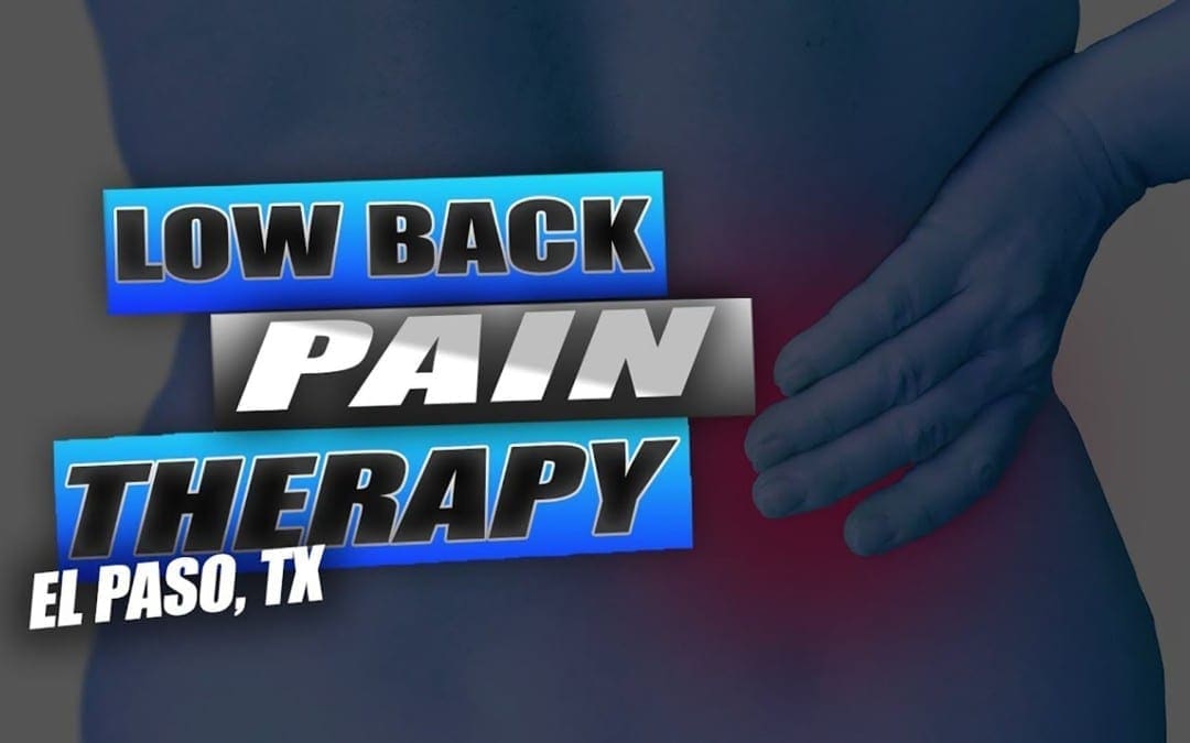
Low back pain which gradually influenced his quality of life was developed. David Garcia was unable to walk as his symptoms worsened and his back pain became excruciating. He first visited Dr. Alex Jimenez, chiropractor in El Paso, TX, following a recommendation from his sister. Dr. Jimenez managed to supply David Garcia with all the aid he deserved for his low back pain, restoring his well-being. David Garcia clarifies the wonderful service Dr. Alex Jimenez and his team have given him to offer him relief from his painful symptoms and he highly recommends chiropractic care as the non-surgical pick for low back pain, among other health problems.
Chiropractic Non-Surgical Relief
We are blessed to present to you�El Paso�s Premier Wellness & Injury Care Clinic.
As El Paso�s Chiropractic Rehabilitation Clinic & Integrated Medicine Center,�we passionately are focused on treating patients after frustrating injuries and chronic pain syndromes. We focus on improving your ability through flexibility, mobility and agility programs tailored for all age groups and disabilities.
We want you to live a life filled with more energy, positive attitude, better sleep, less pain, proper body weight and educated on how to maintain this way of life.
I assure you, I will only accept the best for you�
If you have enjoyed this video and we have helped you in any way, please feel free to subscribe and recommend�us.
Chiropractic care is enjoying an upsurge in popularity as more people are veering away from invasive procedures and pharmaceuticals in favor of more natural treatment options for their pain. A recent Gallup poll for Palmer Chiropractic College confirms this. Of the people surveyed:
More than 35.5 million people said they sought chiropractic care within the 12 months (the Gallup survey was conducted from February 8, 2016, through March 11, 2016).
95% said chiropractic was effective in treating their condition.
89% said they would recommend chiropractic to friends and family.
97% said that they would likely see a chiropractor for neck or back pain.
88% said that chiropractic care is a good value for the money.
Non-Drug Treatments for Back Pain should be Sought First
In April 2017, the American College of Physicians published their updated guidelines for managing and treating low back pain. In it, they recommended seeking non-drug treatments which include the application of heat, exercise, stress reduction, and spinal manipulation before turning to medications. The purpose is to steer patients away from unnecessary medicating and toward healthier, more natural options as the first line of defense in managing low back pain.
Another study published in2017 in the Journal of the American Medical Association supported spinal manipulation as a preferred first treatment option over pharmaceuticals for patients with acute low back pain. While these recommendations are primarily in response to the opioid epidemic, it is also in response to the numerous studies that show chiropractic care is safe and effective for back pain management.
Chiropractic is a Healthier, Safer Option
The Centers for Disease Control (CDC) reports that opioid abuse and overdose has reach epidemic proportions. An estimated 91 Americans die every day from an opioid overdose. This includes opioids obtained by a prescription from a doctor.
Popular pain medications like oxycodone and hydrocodone have seen steadily increasing abuse, overdose, and death rates over the past two decades. In fact, deaths caused by an overdose of these drugs as well as methadone and others, have quadrupled since 1999.
These medications are highly addictive and have many unpleasant and even dangerous side effects. While most states are taking aggressive steps to curb the over-prescribing of these medications, patients are still finding ways around the prescribing guidelines and restrictions. This is a compelling reason for patients to seek natural, non-medicinal pain relief options first � and for their doctors to recommend them.
More People are Turning to Natural Remedies
Many patients are simply getting fed up with invasive treatments and numerous pills, causing them to turn to remedies that are more natural. One benefit of chiropractic and other natural remedies is that they rarely treats only the symptom.
Instead, it addresses the root of the problem to treat the cause. This approach has many benefits beyond being medication free and non-invasive. When the cause of the condition or problem is corrected, it can eliminate other troubling symptoms as well.
Chiropractic is a Healthier, Whole Body Option
A patient who sees their chiropractor for low back pain may find that after a few treatments their headaches, constipation, and digestive issues are also resolved. This is because chiropractic treats the body as a whole, unlike traditional medicine that tends to treat it in parts.
In the body, everything is connected, so a problem in one area could easily cause problems in other areas. By correcting the root of the problem, the patient receives more rounded healthcare.
Where traditional medicine will often opt to prescribe a pain pill for lower back pain, chiropractic care looks for the cause of the back pain and treats the pain symptom from the cause. The chiropractor may advise lifestyle changes, changes in diet, and even recommend supplements. With chronic health conditions like diabetes, heart disease, and obesity increasing at an alarming rate, chiropractic care offers a solution that treats the body as a whole in a more natural, safer way.
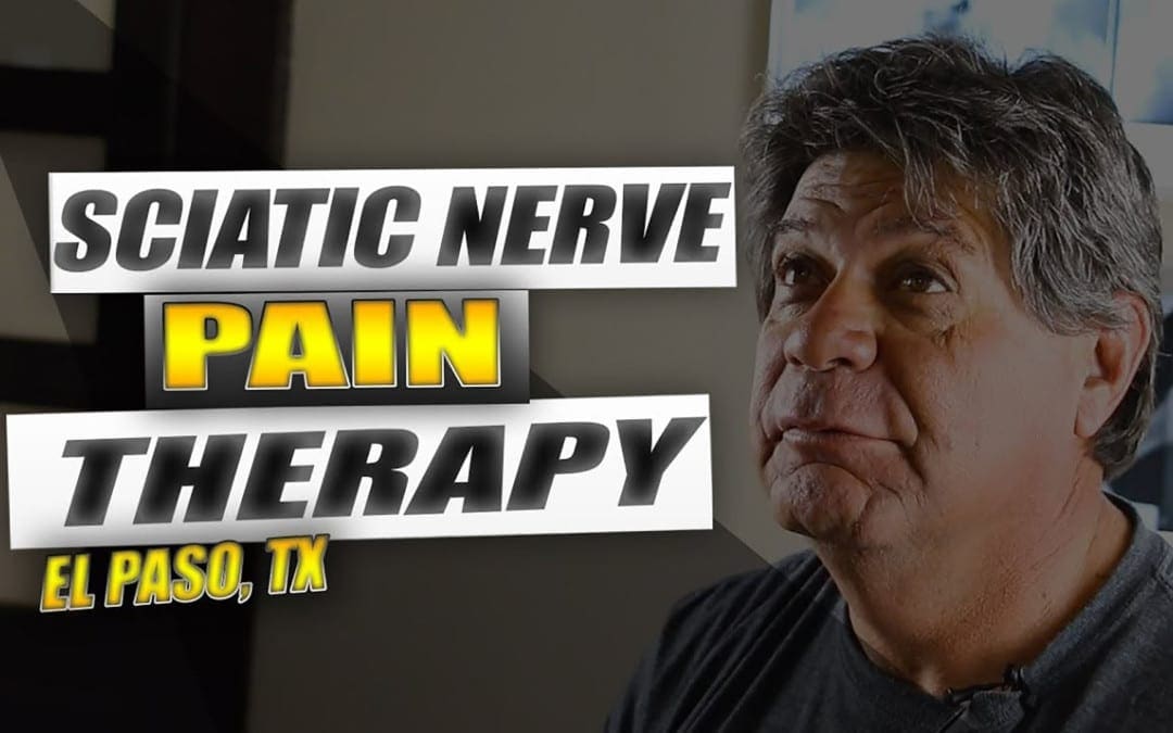
Sciatica is a set of symptoms characterized by discomfort and pain along the length of the sciatic nerve, which runs down the buttocks, hips, and thighs, into the feet and the legs. Also known as sciatic nerve pain, sciatica is brought on by the compression or impingement of the nerve through harms and/or ailments such as a herniated disc. The patients in the following video describe the way their quality of life has influenced. After getting chiropractic care with Dr. Alex Jimenez, chiropractor, patients talk how treatment has helped them achieve pain relief from their sciatica. Sciatic nerve pain is one of the most frequent health issues. The people highly recommend Dr. Alex Jimenez as the non-invasive pick for sciatica.
Nerve Pain Therapy
We are blessed to present to you�El Paso�s Premier Wellness & Injury Care Clinic.
As El Paso�s Chiropractic Rehabilitation Clinic & Integrated Medicine Center,�we passionately are focused on treating patients after frustrating injuries and chronic pain syndromes. We focus on improving your ability through flexibility, mobility and agility programs tailored for all age groups and disabilities.
We want you to live a life filled with more energy, positive attitude, better sleep, less pain, proper body weight and educated on how to maintain this way of life.
I assure you, I will only accept the best for you�
If you have enjoyed this video and we have helped you in any way, please feel free to subscribe and recommend�us.
Being involved in an automobile accident can influence the quality of life of an individual. Whiplash, pain, and neck pain, among other frequent automobile accident injuries, can severely limit a victim’s capacity to participate and engage in their regular activities. Dr. Alex Jimenez, a doctor of chiropractic, focuses on the identification, therapy, and prevention of a number of accidents and/or aggravated conditions associated with the musculoskeletal and nervous system, including auto accident injuries. Chiropractic care is an alternative treatment option which utilizes manual manipulations and adjustments, one of methods and other treatment procedures, to help treat many different health issues. Patients describe how Dr. Alex Jimenez has helped them find relief from their neck pain and back pain after an auto accident. Patients highly recommend Dr. Alex Jimenez as the non-surgical selection for automobile accident injuries, among other health problems such as whiplash.
Personal Chiropractic Care
We are blessed to present to you�El Paso�s Premier Wellness & Injury Care Clinic.
As El Paso�s Chiropractic Rehabilitation Clinic & Integrated Medicine Center,�we passionately are focused on treating patients after frustrating injuries and chronic pain syndromes. We focus on improving your ability through flexibility, mobility and agility programs tailored for all age groups and disabilities.
We want you to live a life filled with more energy, positive attitude, better sleep, less pain, proper body weight and educated on how to maintain this way of life.
I assure you, I will only accept the best for you�
If you have enjoyed this video and we have helped you in any way, please feel free to subscribe and recommend�us.
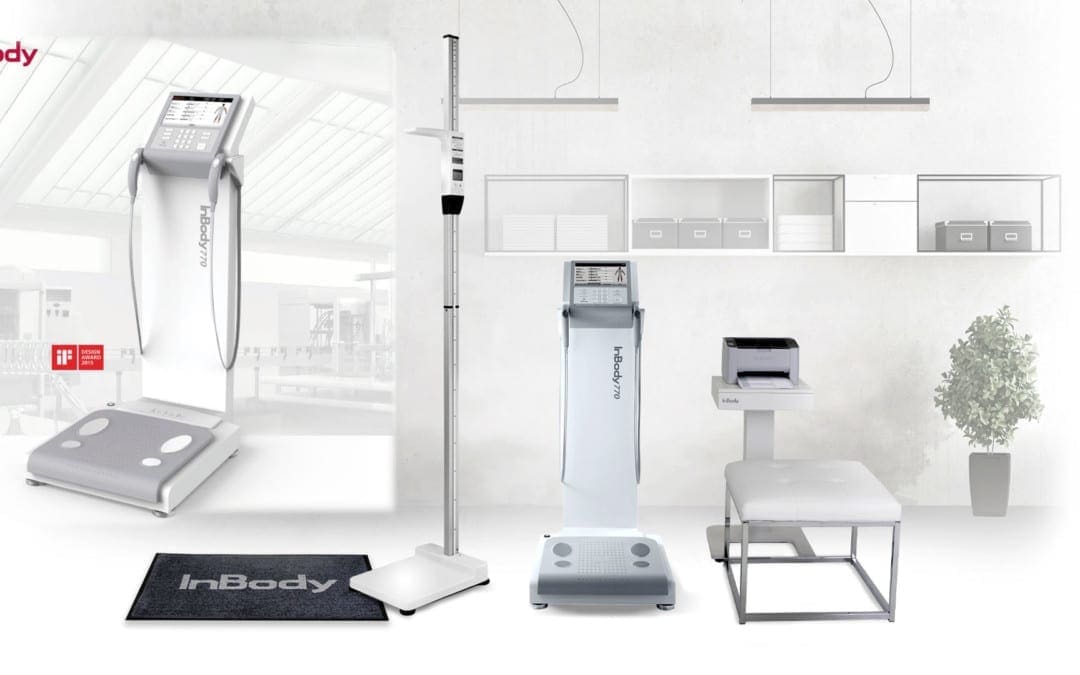
The InBody770 represents the apex of BIA technology. The InBody770 reports on body composition and also reports a complete breakdown of body water. There are 6 frequencies, which create a total of 30 impedance values. There is no other machine on the planet that can match the accuracy and dependability of the InBody770.
The InBody 770
Quick Tests
Measure fat mass, muscle mass, and body water levels in under a minute. No dunking, pinching, or discomfort. Stand on the device and hold the hand electrodes.
No Predicting
Just impedance is used to determine your body composition; No estimations like age and gender are needed or required to predict your composition.
Cloud Software
Automatically save all of the information from InBody to Lookin’Body Web, InBody’s cloud database management system. Easily view and manage customer’s results and see progress anytime.
Check Out The InBody 770 Result Sheet
To find out how to use this data in practice? Download the free guide here.
The knee is the largest, complicated joint in the human body. The knee joint is exposed to many different wellness issues. Knee injuries related to sports are a common issue that can affect playing time. Dr. Jimenez helps many athletes recover from knee injuries along with a variety of other sports-related injuries. Individuals share their experience of knee injury rehabilitation and treatment Dr. Jimenez has provided them. As an added benefit it has enhanced their�sports performance. Chiropractic rehabilitation is an alternative treatment option focusing on spinal adjustments and manual manipulations to deal with many different accidents and/or conditions related to the musculoskeletal and nervous system. Patients say Dr. Jimenez is the non-surgical, chiropractic choice.
Non-Surgical Injury Rehabilitation
We are blessed to present to you�El Paso�s Premier Wellness & Injury Care Clinic.
As El Paso�s Chiropractic Rehabilitation Clinic & Integrated Medicine Center,�we passionately are focused on treating patients after frustrating injuries and chronic pain syndromes. We focus on improving your ability through flexibility, mobility and agility programs tailored for all age groups and disabilities.
We want you to live a life filled with more energy, positive attitude, better sleep, less pain, proper body weight and educated on how to maintain this way of life.
I assure you, I will only accept the best for you�
If you have enjoyed this video and we have helped you in any way, please feel free to subscribe and recommend�us.
Painful symptoms may influence a person’s everyday physical actions. Patients with neck pain and spine pain caused by an assortment of health problems, including automobile accidents, have found tremendous relief from their symptoms with Dr. Alex Jimenez. Chiropractic care is a safe and effective, alternative treatment option which concentrates on the diagnosis, treatment, and prevention of injuries and/or conditions related to the musculoskeletal and nervous systems. Dr. Alex Jimenez has enhanced the quality of life of many patients by carefully restoring the integrity of their spine and improving debilitating symptoms. Patients highly recommend Dr. Alex Jimenez since the non-surgical selection for neck pain and spine pain, among other health problems, in El Paso, Tx.
Non-Surgical Care
�
We are blessed to present to you�El Paso�s Premier Wellness & Injury Care Clinic.
As El Paso�s Chiropractic Rehabilitation Clinic & Integrated Medicine Center,�we passionately are focused on treating patients after frustrating injuries and chronic pain syndromes. We focus on improving your ability through flexibility, mobility and agility programs tailored for all age groups and disabilities.
We want you to live a life filled with more energy, positive attitude, better sleep, less pain, proper body weight and educated on how to maintain this way of life.
I assure you, I will only accept the best for you�
If you have enjoyed this video and we have helped you in any way, please feel free to subscribe and recommend�us.
Assembly lines have long been the standard for factory workers. Henry Ford got the ball rolling on December 1, 1913, when he created the very first assembly line to mass produce a car. Workers stood for hours, doing the same tasks over and over. Although Ford took steps to reduce at least some of the damage, many factory workers still went home with aching backs and feet, migraines, fallen arches, and repetitive motion injuries.
Now, more than a hundred year later, some things have changed. According to the United States Department of Labor Bureau of Labor Statistics, there were 1,834,000 assembly line and fabrication jobs in 2014. Technology is better, and some tasks can be automated making some people�s jobs more manageable � and, unfortunately, eliminating some as well.
Despite the great strides in technology, there are still some things that haven�t changed all that much. The working conditions in many factories are often still not as healthy as they could be. Many workers are still required to stand for long periods of time and perform repetitive motions for hours without a break. This can lead to injuries, pain, and certain conditions that can cause immobility, inflexibility, and even disability. The good news is, chiropractic can help.
Working in a Standing Position Can Be Bad For Your Health
Many assembly line jobs require that the worker stand for long periods of time. While standing is a natural posture for humans and, by itself does not pose any real harm or health problem, working in a standing position every day isn�t good for you. It can lead to muscle fatigue, stiff shoulders and neck, swelling of the legs and feet, low back pain, varicose veins, fallen arches, and sore feet � to name a few.
Another problem with standing for extended or frequent periods of time without any breaks (such as walking or stretching) can cause the joints in the feet, knees, hips, and spine to become locked or immobilized temporarily. If the behavior continues, it can cause degenerative damage, leading to rheumatic diseases because the ligaments and tendons become damaged.
Other Assembly Line Related Health Problems
Barring traumatic injury due to an accident, working in a factory environment can cause problems with mobility, pain, and flexibility. The nature of the job places specific demands on the human body that can lead to certain types of injuries and health conditions, which include:
Repetitive motion injury � When a worker performs the same task that involves the same movements over and over, it can lead to certain types of injuries. Carpal tunnel is common repetitive motion injury.
Overexertion � Lifting, pulling, even standing can take a toll on the body, especially when it is done without adequate breaks. The person can get muscle fatigue, pulled muscles, and pulled tendons.
Body movement injuries � When the worker is continuously reaching, twisting, crawling, and bending, it can cause problems with the muscles and joints.
Chiropractic can Help Assembly Line Workers
Chiropractic care can help keep bodies flexible and help with range of motion. It is a very effective, non-invasive treatment for pain and can help with joint and muscular problems as well. Regular chiropractic treatments can help you better manage your body�s response to your work environment. It can also undo many of the ill effects that that type of work can cause.
You can enjoy more pain-free days without invasive surgeries or medications that leave you groggy, nauseous, or worse. When you sit down with your chiropractor, he or she will talk to you about your medical history as well as your current lifestyle. After a complete evaluation, you will be given a plan of action that may include lifestyle changes, dietary changes, and recommended supplements in addition to spinal manipulation. Chiropractic is all about whole body wellness, and that is what will help you perform better on your job and recover faster.
James Hill came to Injury Medical & Chiropractic Clinic after his daughter Madison suffered a sports injury. Through a variety of chiropractic treatment methods and techniques, Dr. Jimenez helped Madison find relief from her ankle pain so she could return-to-play as soon as possible. Madison regained the confidence she needed with Dr. Jimenez�and is recovering properly. James Hill and Madison highly recommend Dr. Alex Jimenez as the non-surgical choice for sports injuries. Chiropractic treatment is an�alternative treatment that works through rehabilitation procedures.
Sports Injury Chiropractic
We are blessed to present to you�El Paso�s Premier Wellness & Injury Care Clinic.
As El Paso�s Chiropractic Rehabilitation Clinic & Integrated Medicine Center,�we passionately are focused on treating patients after frustrating injuries and chronic pain syndromes. We focus on improving your ability through flexibility, mobility and agility programs tailored for all age groups and disabilities.
We want you to live a life filled with more energy, positive attitude, better sleep, less pain, proper body weight and educated on how to maintain this way of life.
I assure you, I will only accept the best for you�
If you have enjoyed this video and we have helped you in any way, please feel free to subscribe and recommend�us.
Today we will begin to discuss how the spectrum which medicine currently considers �normal� may not actually be optimal towards your overall health and wellness. These reference ranges can change based on age, gender, physical activity, and more. As a matter of fact, if we were to evaluate an individual�s weight in the United States, it would be considered �normal� to be overweight, simply because 70 percent of the population is overweight. Reference ranges for lab tests today are based on a sick population when we should aspire for optimal well-being.
Then, I will demonstrate how this knowledge can apply to the most basic medical measurements: your vital signs. Everyone knows that when you first visit a doctor, they take your vital signs, including your weight, your blood pressure, your heart rate, and your temperature. However, does your doctor tell you what your results mean? How can you tell if you�re healthy?
What�are�Vital�Signs?
Hello everyone, it�s Dr. Alex Jimenez. Welcome to part 2 of �Taking Control of Your Healthcare�. Today, we will discuss laboratory reference ranges associated with your vital signs. It may not sound like an interesting topic, but it�s really important for your well-being.
Most doctors generally define a patient�s test results as either �normal� or �abnormal�. But what exactly does �normal� and �abnormal� mean? Normal is when 95 percent of the population falls within a common range while abnormal is when the remaining percent of the population falls outside that common range. Whether you�re healthy or sick, young or old, although there can be some variations for children, normal and abnormal are simply statistical numbers which fall within two standard deviations.
However, these standard deviations don�t necessarily mean it�s optimal or not. It�s only a statistical number, after all. As a matter of fact, disease can occur along healthy and unhealthy individuals. These lab reference ranges should be defined according to what is best for a human being.
By way of instance, vitamin D levels are classified as normal if they�re over 20, however, the ideal levels are over 50. So why is 20 considered �normal�? This is because approximately 80 percent of the population is deficient in vitamin D, therefore, they fall under what is considered to be the �normal� range. However, this doesn�t mean that these levels are best for your overall health and wellness. Furthermore, �normal� blood sugar levels have been classified to be under 100, although we know �optimal� blood sugar levels have been classified to be between 70 to 80. Blood sugar levels over 80 can have an increased risk of disease. And unfortunately, our �normal� laboratory reference ranges are not optimal because we�ve become a sick population. In the United States it can be considered �normal� to be overweight because 70 percent of our population is overweight. But, although being overweight or obese is considered normal in the United States, that�s not something we would want to aspire to. We want to aspire to achieve overall health and wellness.
Now let�s continue to discuss the meaning of normal. Many patients often visit the doctor only to be told that their lab tests have returned normal, however, they may still be feeling sick. What does that mean? Does it mean you�re sick? Does it mean you�re healthy? As I�ve mentioned previously, either your doctor is missing something or you�re crazy, and I�m pretty sure your doctor is missing something. This is one of the main differences between conventional medicine and functional medicine. Through functional medicine, many doctors focus on health care rather than sick care. We�re looking for more subtle deviations from optimal.
Until your liver function is considered abnormal according to the current standard, your liver cells may already be dying. A functional medicine doctor may review your lab tests differently than a conventional doctor. This is primarily because the reference ranges that we�re focusing on aim towards optimal health, not disease. Many conventional doctors evaluate lab tests differently than functional medicine doctors, and then they either follow a �watch-and-wait approach�, or they label you as �not sick� after the most minimal amount of lab tests. As a matter of fact, one patient who visited me had blood sugar levels of 120, where a blood sugar of 126 is already considered type 2 diabetes. And I said, �Did you see your doctor regarding this?� And he said, �Yeah.� I asked him what the doctor said. And he finally said, �Well, he said to wait until I actually had diabetes and then to come back for medication.� And that is the last thing we want to be doing as healthcare professionals.
Reference ranges give us an average number of values which have been recorded among the general population. But let�s not forget, �normal� reference ranges are relative. They change based on age, gender, physical activity, and more. If you were to evaluate an individual�s weight in the United States today, it would be normal to be overweight, as I mentioned, only because 70 percent of people are overweight. And unfortunately, we keep changing our reference ranges based on our sick population. This is not what we should aspire to do as doctors. This is why functional medicine treats the individual, not only the numbers.
Additionally, reference ranges which were once considered normal can also change over time. One instance of how reference ranges change was demonstrated by a known global laboratory company called LabCorp, where they recently changed their reference ranges for male testosterone levels. Previously, LabCorp considered normal testosterone levels for an adult male to be between 348 to 1,197. This value was based on a population of lean adult males. However, in 2017, they lowered normal testosterone levels for an adult male to be between 264 to 916. Moreover, overweight men, excluding obese men, were probably included in the cohort study, ultimately changing reference ranges for male testosterone levels. Research studies have found that excess abdominal fat can cause lower testosterone levels. By changing reference ranges, however, this demonstrates that conventional medicine is considering overweight individuals to be a part of the norm. But this isn�t what we want. We want to strive for overall well-being.
This is why you need to start taking control of your own healthcare. As one in two people have some type of chronic disease, we have to evaluate how we interpret lab tests as well as how �normal� may not necessarily mean health and wellness but simply an average for a growing sick population in the United States.
Taking Control of Your Vital Signs
The primary goal of this series of videos is to encourage you to become the leader of your own well-being by understanding what your lab tests mean, understanding what optimal looks like, and understanding which lab tests are designed to help you achieve overall health and wellness rather than focusing on the disease. I would also like to educate you in order for you to make an informed decision on who you choose to be your doctor, or partner, in your journey to well-being. Now let�s look into the most common medical measurements: your vital signs.
Your vital signs are initially taken by the nurse when you visit the doctor. These vital signs generally include blood pressure, weight, heart rate, temperature, and even oxygen saturation. However, are you aware of what these numbers mean? Has your doctor discussed these numbers with you? Why would they take your vital signs if they�re just going to record them and never discuss them with you? Do the numbers actually demonstrate your health and wellness? If you have high blood pressure or a heart rhythm problem, your healthcare professional is most likely going to tell you, but otherwise, you may not find out what the value of your vital signs is.
Your heart rate is probably one of the most important vital signs taken during a doctor visit. The pulse is a measure of how fast your heart is beating. The human heart beats more than 115,000 times per day. So, if your heart rate is above 100, then we define that as having a high heart rate. But, if you have a heart rate higher than 80, that can increase your risk of developing heart disease. What causes this increase? Although many factors can lead to cardiovascular disease, stress is one of the most common causes because it raises your adrenaline and causes increased heart rate and blood pressure. Drinking too much coffee, stimulating medicines like Adderall, or an overactive thyroid, heart or lung health issue can also increase the risk of heart disease.
The fight-or-flight response, a physiological reaction which becomes activated during times of stress, can also cause an increase in heart rate. When an individual�s heart rate is frequently above 80, it might be time to begin incorporating some stress management techniques into their life, by way of instance, mindfulness meditation and other forms of meditation. Stress is not the only cause of an increased heart rate. Anxiety, magnesium deficiencies, being out of shape and dehydration can also cause an increased heart rate. Ideally, we want to achieve a lower heart rate, optimally under 70.
On the other side of the spectrum, a decreased heart rate below 60 might also demonstrate the presence of thyroid gland dysfunction or low thyroid function. Athletes and distance runners actually have low heart rates because they�re so well-conditioned. Their heart rates can be as low as 50 or even 45. But, if you have a low heart rate and you�re not an athlete or a distance runner, it may be time for you to go talk to your doctor.
While heart rate is one of the most important vital signs taken during a doctor visit, there�s another vital sign which can be just as important, your heart rate variability. This reflects the health of your automatic or autonomic nervous system, which is in charge of controlling all of the unconscious elements of your nervous system, such as digestion and breathing. Heart rate variability has frequently been associated with longevity and even death. The less variable the heart rate is, the higher the mortality rate. Many doctors don�t measure a patient�s heart rate variability, but fortunately, you can manage it yourself. Hot-and-cold therapies, saunas, exercise, yoga and meditation, can all help improve a patient�s heart rate variability.
Now let�s move on to the next most important of the vital signs taken during a doctor visit, blood pressure. If you were to line up all the blood vessels in your body, they would extend approximately 59,000 miles. That�s almost seven times around the earth. These same blood vessels carry over 7,500 liters of blood throughout your entire body on a regular basis. With each heartbeat, blood is pushed against the artery walls, which causes an increase in pressure.
Medical measurements for blood pressure have two numbers. The top number is known as systolic, or the pressure when the heart is contracting, and the bottom number is known as diastolic, or the pressure when the heart is relaxing or at rest. Normal reference ranges for blood pressure continue to change because we keep finding out that the reference ranges we used to think of as normal, which were first 140/90, then 130/80, were still associated with an increased risk of stroke and heart attack. Many doctors today may mention a problem with your blood pressure only if it�s over 130/80.
The reason why blood pressure is so important is because when it�s elevated, it can place additional pressure on the heart and arteries, potentially leading to heart disease, or stroke, heart failure, or even kidney failure. And it�s not only the heart and arteries which are affected by blood pressure: the brain, kidneys and even the eyes can all be tremendously affected, leading to strokes, dementia, kidney failure, and blindness, among other health issues. Maintaining your blood pressure at an optimal level is fundamental towards your overall health and wellness. As a matter of fact, normal blood pressure is currently believed to be under 120/80, however, it may turn out to be even lower.
While high blood pressure is bad, low blood pressure can also be just as bad. A good functional medicine doctor will discuss with you the risks of both high blood pressure and low blood pressure. Blood pressure below 100/60 may cause problems but not necessarily. Blood vessels in the human body function just like pistons in a car. If not enough pressure is built up, it can become really difficult for the blood to flow against gravity. And because the human brain is positioned higher than the heart, we depend on our blood pressure to supply our brain with the necessary amount of oxygen and nutrients required to function accordingly.
If you have low blood pressure, you might experience other symptoms, such as fatigue. Other symptoms associated with low blood pressure include dizziness when standing, weakness and even brain fog. Also, both chronic high blood pressure and chronic low blood pressure may contribute to an increased risk of dementia.
Now we�ve discussed the importance of heart rate and blood pressure. But, how about we discuss another important vital sign: your body temperature? A fever or an elevated body temperature can often be a sign of infection. Temperature can also provide an insight into the function of our metabolism. The lower an individual�s metabolism, the less heat they produce, which may manifest as a slightly lower-than-normal body temperature. The thyroid gland plays a big role in metabolism and regulating your temperature. So, if you frequently feel cold, you might want to discuss ordering the right thyroid panel with your doctor. But, what tests should you ask your doctor to determine this? Don�t worry, we will discuss which thyroid tests you should take in the video on hormones. The optimal body temperature should be approximately 98.6 degrees Fahrenheit. If it�s lower than 97.7 degrees Fahrenheit, however, it may indicate that you have a thyroid problem.
The final medical measurements we�re going to discuss are your height and your weight. Doctors utilize your height and weight to calculate your body mass index, or BMI. At our clinic, by way of instance, we utilize the InBody 770, a body composition and body water analyzer, to help easily determine your body mass index and more. However, body mass index doesn�t always factor in body composition, or the percentage of fat versus muscle in the human body. By way of instance, a pro football player who is 6�6� and 265 pounds has a body mass index of over 30, which puts them in the obese category.
But if you were to take a look at this individual�s body, they would never be categorized as obese. This demonstrates that BMI isn�t an accurate measurement, especially for athletes. Also, a 65-year-old woman may have more fat than muscle in their body while their BMI measurements may appear �optimal�. Instead, many functional medicine doctors use waist-to-hip medical measurements. This is a simple measurement you can do at home to determine body fat distribution, which can also help demonstrate the risk of metabolic dysfunction. Obesity, type 2 diabetes, and heart disease are caused by excess belly fat or fat accumulated around the organs. Excess fat around the midsection can ultimately increase the risk for heart disease and metabolic issues, such as diabetes, dementia, cancer and many other health problems.
But first, let�s discuss how you can calculate your waist-to-hip ratio. To measure your waist, you simply take the measurements of the widest area around your waist, which is generally the biggest part around your belly button. To measure your hip, you then take the measurements of the widest area around your hip, which is generally where your hip bones are on your sides. So, you take these measurements and then you divide the measurements of your waist by the measurements of your hip. And this is the most fundamental number you have to look at.
In men, a waist-to-hip ratio of less than 0.9 is considered optimal. If the ratio is greater than one, meaning that your belly is bigger than your hip, it can put men at higher risk of developing metabolic syndrome, heart disease, diabetes, stroke, cancer, and dementia. In women, a waist-to-hip ratio of less than 0.8 is considered optimal. If the ratio is greater than 0.85, it can put women at higher risk of developing metabolic syndrome as well as the other health issues mentioned above.
Medical measurements, including heart rate, temperature, respiration rate, and blood pressure, are several vital signs which help indicate doctors the state of a patient’s fundamental body functions. Reference ranges today are utilized to determine “normal” health and wellness spectrums, however, research studies have demonstrated that these reference ranges may actually not be optimal spectrums. Understanding the most basic medical measurements, or vital signs, is important towards a patient’s well-being, as it can help people recognize whether they are feeling healthy or sick, regardless of the standards.
Understanding Your Vital Signs
And those were your vital signs, your most basic medical measurements. These numbers are very essential as they are fundamental towards your overall health and wellness. Understanding how your body functions as a whole is important to optimize your well-being. Therefore, next time you visit your doctor, ask about your vital signs and discuss these reference ranges with them. I truly believe that a combination of your own research and having a good relationship with a qualified healthcare professional can lead you on the right path to overall health and wellness.
Finding a doctor that will work with you is essential towards achieving the results you deserve. If your doctor is not willing to have a conversation with you about your well-being or the lab tests that are needed to track your results, then you might want to consider finding another doctor.
If you learned about the simplest medical measurements then you will definitely enjoy the next video, where we will discuss the blood tests utilized to determine nutritional deficiencies. Over 90 percent of individuals in the United States are deficient in nutrients at the RDA level. That�s the minimum amount necessary to prevent diseases caused by nutritional deficiencies, such as scurvy and rickets.
We�re going to discuss how a doctor who specializes in functional medicine evaluates results and what other tests your doctor might be unaware of that can tell us a lot about your nutritional status. Thanks again for joining me so far and I�ll see you later. The scope of our information is limited to chiropractic and spinal health issues as well as functional medicine topics and discussions. To further discuss the subject matter, please feel free to ask Dr. Alex Jimenez or contact us at�915-850-0900�.
Curated by Dr. Alex Jimenez
Additional Topic Discussion:�Acute Back Pain
Back pain�is one of the most prevalent causes of disability and missed days at work worldwide. Back pain attributes to the second most common reason for doctor office visits, outnumbered only by upper-respiratory infections. Approximately 80 percent of the population will experience back pain at least once throughout their life. The spine is a complex structure made up of bones, joints, ligaments, and muscles, among other soft tissues. Injuries and/or aggravated conditions, such as�herniated discs, can eventually lead to symptoms of back pain. Sports injuries or automobile accident injuries are often the most frequent cause of back pain, however, sometimes the simplest of movements can have painful results. Fortunately, alternative treatment options, such as chiropractic care, can help ease back pain through the use of spinal adjustments and manual manipulations, ultimately improving pain relief.
XYMOGEN�s Exclusive Professional Formulas are available through select licensed health care professionals. The internet sale and discounting of XYMOGEN formulas are strictly prohibited.
Proudly,�Dr. Alexander Jimenez makes XYMOGEN formulas available only to patients under our care.
Please call our office in order for us to assign a doctor consultation for immediate access.
If you are a patient of Injury Medical & Chiropractic�Clinic, you may inquire about XYMOGEN by calling 915-850-0900.
For your convenience and review of the XYMOGEN products please review the following link.*XYMOGEN-Catalog-Download
* All the above XYMOGEN policies remain strictly in force.
IFM's Find A Practitioner tool is the largest referral network in Functional Medicine, created to help patients locate Functional Medicine practitioners anywhere in the world. IFM Certified Practitioners are listed first in the search results, given their extensive education in Functional Medicine