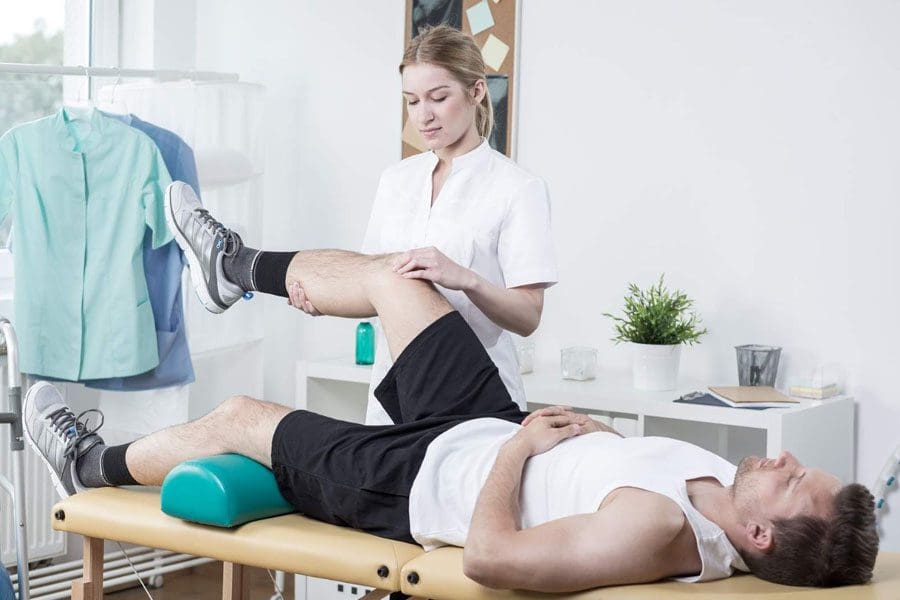
For wrestling athletes or those thinking about getting into the sport, can knowing about common injuries help in rehabilitation and prevention?
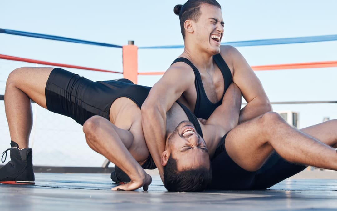
Wrestling Injuries
Wrestling is an intense and demanding sport. Studies have found that football and wrestling are the two high school sports with the highest risk of serious injury to athletes (Center for Injury Research and Policy, 2009). The injury rate for college wrestlers is 9 injuries per 1,000 athlete exposures. (Kroshus, E. et al., 2018) While most wrestling injuries include strains and sprains, there can also be serious traumatic and unusual injuries. Using proper safety gear and learning correct techniques can significantly reduce the risk of injuries. The majority occur during competition.
Common
The most common wrestling injuries are similar to those in other sports and include:
Muscle Soreness
Muscle soreness that is experienced 12 to 48 hours after an intense workout or competition.
Resting is often all that is needed to recover.
Bruises and Contusions
Sparring, take-downs, and hard landings can result in various bruises and contusions.
Sprains and Strains
Rest, ice, compression, and elevation are recommended to treat sprains and strains immediately.
Ankle Sprains
Ankle sprains occur when surrounding ligaments stretch and tear around the joint.
Wrist Sprains
Typically, it occurs when stretching or tearing the ligaments.
Falling or landing on the hands is a common cause.
Overtraining Syndrome
Frequently occurs in athletes who train beyond the body’s ability to recover.
Dehydration
When trying to make weight, dehydration can be a serious health problem that many wrestlers experience.
Other Injuries
Other injuries common in wrestling:
Wrist tendinitis
Finger fractures
Iliotibial band syndrome
Meniscus tears
Groin pull
Hamstring pull or tear
Pulled calf muscle
Achilles tendonitis
Achilles tendon rupture
Clavicle/Collarbone fracture
Concussion
Serious
The forcing of a joint beyond its normal range of motion is the most common cause of serious injuries. The most serious wrestling injuries affect the neck, shoulder, elbow, and knee and include:
Neck
The cervical vertebrae are often forced into vulnerable positions during various techniques and movements, which can result in a neck injury. Common types include:
Neck Strain
Whiplash
Cervical Fracture
Shoulder
A combination of leverage and twisting causes most upper body and shoulder injuries during competition. Types of shoulder injuries include:
Rotator cuff injury
Shoulder separation
Shoulder dislocation
Elbow Dislocation
Elbows are under tremendous strain when maneuvering.
Dislocations of the radial head are often related to the athlete bracing for a fall on an outstretched arm during take-downs.
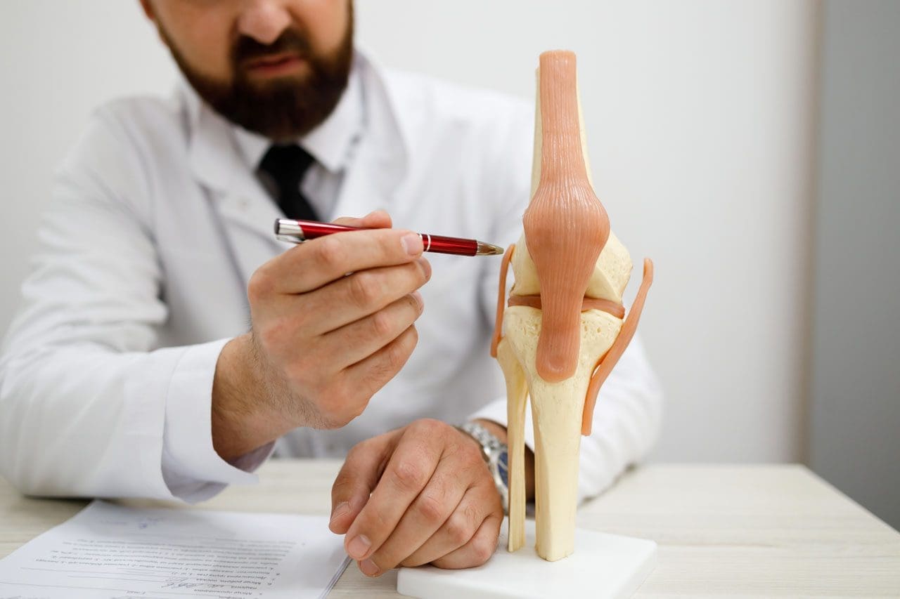
Knee
Most knee injuries occur to the ligaments of the knee joint.
These include anterior and posterior cruciate ligament or ACL/PCL injuries.
Safety
Wrestling requires flexibility, strength, and proper technique to prevent injury, combined with thorough instruction and coaching and following basic safety precautions. Some tips include.
Safety Gear
Wear appropriate headgear and mouthguards during practices, meets, and tournaments.
Improving Joint Flexibility
Wrestlers with a high degree of shoulder flexibility have fewer injuries.
The flexibility of the lower back, hamstrings, elbows, and cervical spine should also be worked on.
Gain or Lose Weight Safely
Avoid dramatic weight loss and weight-gaining strategies by maintaining healthy nutrition and hydration during the season.
Avoiding Dangerous Holds and Slam Moves
Safe wrestling techniques need to be followed as these can generate severe injuries.
Regardless of how common or seemingly not serious an injury or medical condition is, it’s important to rest and recover and tell a coach and health care professional, as some injuries and conditions can become serious if left untreated. Injury Medical Chiropractic and Functional Medicine Clinic focuses on and treats injuries and chronic pain syndromes through personalized care plans that improve ability through flexibility, mobility, and agility programs to relieve pain. Our providers use an integrated approach to create personalized care plans for each patient, including Functional Medicine, Acupuncture, Electro-Acupuncture, and Sports Medicine principles. Our goal is to relieve pain naturally by restoring health and function to the body. If other treatment is needed, Dr. Jimenez has teamed up with top surgeons, clinical specialists, medical researchers, and rehabilitation providers to provide the most effective treatments.
Kroshus, E., Utter, A. C., Pierpoint, L. A., Currie, D. W., Knowles, S. B., Wasserman, E. B., Dompier, T. P., Marshall, S. W., Comstock, R. D., & Kerr, Z. Y. (2018). The First Decade of Web-Based Sports Injury Surveillance: Descriptive Epidemiology of Injuries in US High School Boys’ Wrestling (2005-2006 Through 2013-2014) and National Collegiate Athletic Association Men’s Wrestling (2004-2005 Through 2013-2014). Journal of athletic training, 53(12), 1143–1155. doi.org/10.4085/1062-6050-154-17
For individuals who are getting older, can increasing bone strength help prevent fractures and optimize bone health?
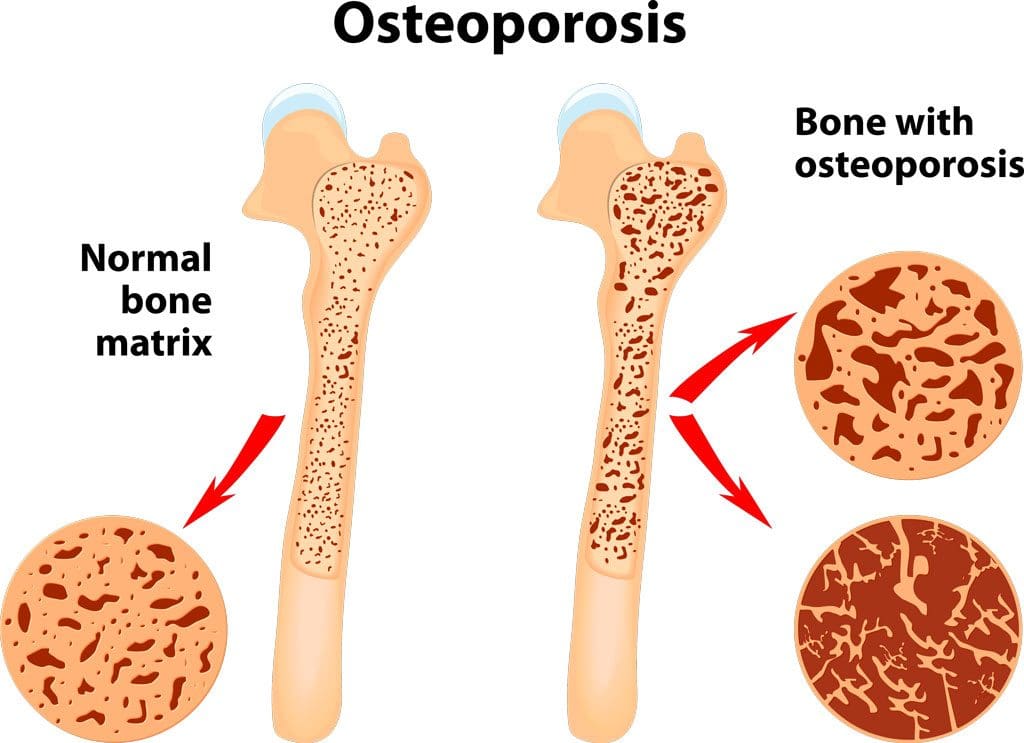
Bone Strength
Bone strength is important, as a fractured hip can be serious for older individuals. A study found that for individuals in their 60s who had a hip fracture, 6.5% of women and 9.4% of men died within a year. Among individuals in their 80s, 13.1% of women and 19.6% of men died within a year. (Dimet-Wiley, et al., 2022)
Increasing bone strength can help prevent various issues. A small increase in bone mineral density has been shown to help reduce the risk of fractures, especially hip fractures. A decades-long study found that just a 3% increase in bone strength helps lower the chance of breaking a hip. Researchers enrolled two groups of individuals aged 60 and older, one in 1989 and the second in 1999.
The bone mineral density of each subject’s femoral neck joint at the top of the thigh bone near the hip was measured.
They then followed the subjects for years to see who experienced hip fractures.
While the bone mineral density of the second group was only 3% higher than the first group, these subjects experienced a 46% reduction in hip fractures. (Tran, T. et al., 2023)
Bone Loss
Bone loss is progressive in men and women and increases as the body ages. Osteoporosis is a condition in which bone tissue deteriorates. (Department of Health and Human Services Office of Disease Prevention and Health Promotion. 2020) Bones constantly break down and reform as a normal remodeling process. If the balance of this process is impaired, osteoporosis develops, resulting in more bone breakdown than formation. While men and women experience bone loss, it’s more common in females. (National Institute of Arthritis and Musculoskeletal Diseases. 2022) Menopause is a risk factor because of the decline of estrogen (National Library of Medicine, Medline Plus, 2022). Estrogen reinforces bone strength by protecting against bone breakdown; with estrogen loss, bone breakdown increases. However, anyone of any age or background can experience bone loss due to the following:
While some loss of bone strength is common, several strategies exist to maintain bone health. Exercise, specifically weight-bearing activities, can increase bone strength. When bones and muscles are used to hold a position against gravity, this mechanically stresses the bone, causing it to reform stronger. Movement and physical exercise as medicine and the forces transmitted through the bones generate mechanical signals that tell the cells to increase bone formation relative to breakdown. Exercises focusing on posture, balance, gait, and coordination are recommended for individuals with osteoporosis to strengthen the core, quadriceps, and hip flexors. Different types of exercises can include:
Walking to strengthen the spine and hips.
Walking outside or on a treadmill provides more loading force to the bone.
Planks and push-ups can strengthen the forearm and wrist bones.
Holding a water bottle in each hand and lifting up and down 10 times together or alternating a few times a day.
Side leg lifts can strengthen the hip and forearm bones simultaneously.
Weight training provides the bones with a workout by having them support a weight load.
Any exercise therapy program should be designed by a healthcare provider, physical therapist, and trainer according to the individual’s condition and appropriate for them.
Diet
What goes into the body definitely affects bone health. Calcium and vitamin D are key to bone building, but both are needed as vitamin D is needed to absorb the calcium ingested. Calcium can be found in:
Dairy
Dairy products and non-dairy alternatives are fortified with calcium.
Leafy greens.
Beans.
Almonds.
The recommended daily calcium intake for adults over 50 is 1,200 milligrams.
Vitamin D can come from:
Sunlight
Fish.
Mushrooms.
Fortified milk.
Supplements.
The recommended daily vitamin D intake for adults aged 70 is 15 micrograms and 20 micrograms for individuals over 70.
Studies have found that increasing calcium and vitamin D intake with supplements can help maintain bone health. Talk to a healthcare provider about whether supplements could be beneficial.
Hormone Therapy
Females also naturally produce testosterone, which promotes bone formation. As levels drop with age and negatively impact bone strength, hormone therapy could be recommended. Declining testosterone levels start with women in their 20s and men in their 30s. The typical drop in women is 1% to 3% yearly before menopause and stabilizes somewhat afterward. Female patients at risk of bone loss may be prescribed testosterone in various forms that continuously emit the hormone. The dosage is low, so patients do not experience unwanted hair growth or skin changes. Combined with estrogen, testosterone effectively increases bone growth in female patients. Not everyone is a candidate for hormone therapy, like individuals with a history of breast cancer, heart disease, blood clots, or liver disease. (National Library of Medicine. Medline Plus, 2019)
Making small adjustments can optimize bone health and overall well-being
At Injury Medical Chiropractic and Functional Medicine Clinic, we passionately focus on treating patients’ injuries and chronic pain syndromes to create personalized care plans that improve ability through flexibility, mobility, and agility programs tailored to the individual. Using an integrated approach, our goal is to relieve pain naturally by restoring health and function to the body through Functional Medicine, Acupuncture, Electro-Acupuncture, and Sports Medicine protocols. If the individual needs other treatment, they will be referred to a clinic or physician best suited for them, as Dr. Jimenez has teamed up with the top surgeons, clinical specialists, medical researchers, and premier rehabilitation providers to provide the most effective clinical treatments. We focus on what works for you and strive to better the body through researched methods and total wellness programs.
Chiropractic Care: Movement Medicine
References
Dimet-Wiley, A., Golovko, G., & Watowich, S. J. (2022). One-Year Postfracture Mortality Rate in Older Adults With Hip Fractures Relative to Other Lower Extremity Fractures: Retrospective Cohort Study. JMIR aging, 5(1), e32683. doi.org/10.2196/32683
Tran, T. S., Ho-Le, T. P., Bliuc, D., Center, J. R., Blank, R. D., & Nguyen, T. V. (2023). Prevention of Hip Fractures: Trade-off between Minor Benefits to Individuals and Large Benefits to the Community. Journal of bone and mineral research : the official journal of the American Society for Bone and Mineral Research, 38(11), 1594–1602. doi.org/10.1002/jbmr.4907
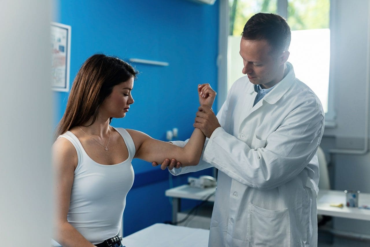
During a fall individuals tend to automatically outstretch their hands to help break a fall, which can slam onto the ground causing a falling onto an outstretched hand or FOOSH injury. Should individuals get checked by a healthcare provider if they believe there is no injury?
FOOSH Injuries
Falling down usually results in minor injuries. A FOOSH injury occurs when falling down and trying to break the fall by reaching out with the hand/s. This can result in an upper extremity injury like a sprain or a fracture. But sometimes, falling on one’s hands can lead to serious injuries and/or create future musculoskeletal issues. Individuals who have fallen or suffered a FOOSH injury should consult their healthcare provider and then a physical therapist or chiropractor to safely develop a treatment plan to rehabilitate, strengthen, and expedite recovery.
After The Injury
For individuals who have fallen down and landed on their hand, wrist, or arm, here are a few things to ensure the proper care for the injury, including:
Follow the R.I.C.E. protocol for acute injuries
Visit a healthcare provider or local emergency clinic
Contact a physical therapist
A FOOSH injury could be or become serious, so to avoid letting small issues become big problems, get examined by a musculoskeletal specialist. The healthcare provider will obtain an imaging scan of the injured and surrounding areas. They will perform a physical examination to determine the type of injury, like a sprain or muscle strain. Not getting appropriate medical treatment after a fall can result in chronic pain and loss of function. (J. Chiu, S. N. Robinovitch. 1998)
Common Injuries
A FOOSH injury can injure different areas. These usually involve the wrist and hand, but the elbow or shoulder can also be injured. Common injuries include:
Colles’ fracture
A wrist fracture where the end of the arm bone is displaced backward.
Smith’s fracture
A wrist fracture, similar to a Colles’ fracture, is where the end of the arm bone is displaced towards the front of the wrist.
Boxer’s fracture
A fracture of the small bones in the hand.
Typically, it occurs after punching something, but it can happen from falling on an outstretched fist.
Elbow dislocation or fracture
The elbow can pop out of the joint or can break a bone in the elbow.
Collarbone fracture
The force from falling with the hands and arms outstretched can travel up to the collarbone, causing a fracture.
Proximal humeral fracture
Falling onto an outstretched hand injury can cause the arm bone to get jammed into the shoulder, causing a proximal humeral fracture.
Shoulder dislocation
The shoulder can pop out of the joint.
This can cause a rotator cuff tear or labrum injury.
Regardless of the injury, individuals should visit a healthcare provider to evaluate the damage. If the injury is serious, the practitioner can make an accurate or differential diagnosis and develop a treatment plan. (William R. VanWye et al., 2016)
Physical Therapy
Individuals can benefit from physical therapy to help recover and return to their previous level of function. Physical therapy varies depending on the specific injury, but generally, a physical therapist can help individuals return to function after a fall on an outstretched hand. (William R. VanWye et al., 2016) Common treatments can include:
Treatments and modalities to decrease pain, inflammation, and swelling.
Instruction on how to wear an arm sling properly.
Exercises and stretches to improve the range of motion, strength, and functional mobility.
Balance exercises.
Scar tissue management if surgery was necessary.
The therapy team will ensure the proper treatment is utilized to quickly and safely return to normal activities.
Chiropractic Care For Healing After Trauma
References
Chiu, J., & Robinovitch, S. N. (1998). Prediction of upper extremity impact forces during falls on the outstretched hand. Journal of biomechanics, 31(12), 1169–1176. doi.org/10.1016/s0021-9290(98)00137-7
VanWye, W. R., Hoover, D. L., & Willgruber, S. (2016). Physical therapist screening and differential diagnosis for traumatic-onset elbow pain: A case report. Physiotherapy theory and practice, 32(7), 556–565. doi.org/10.1080/09593985.2016.1219798
A nerve becomes pinched/compressed when added pressure is placed on it by surrounding structures that can include muscles, bones, ligaments, tendons, or a combination. This injures and damages the nerve causing function problems and symptoms and sensations in that area or other parts of the body that are supplied by that nerve. Medical practitioners refer to this as nerve compression or entrapment. Although compressed nerves are more commonly associated with the neck, arms, hands, elbows, and lower back, any nerve in the body can experience irritation, spasms, inflammation, and compression. The causes and treatment of a compressed nerve in the knee.
Compressed Nerve In The Knee
There’s only one nerve that goes through the knee that has an increased risk of getting compressed. It’s a branch of the sciatic nerve called the peroneal nerve. The nerve goes around the outside of the knee before traveling down the outside of the lower leg. At the bottom of the knee, it lies between the bone and skin, making it vulnerable to irritation or compression by anything that can put pressure on the outside of the knee.
Causes
Traumatic injuries over time can lead to pressure on the nerve from inside the knee. Common causes of a compressed nerve in the knee include:
Frequently Crossing Legs
Compression by the opposite knee, while the legs are crossed is the most common cause.
Knee Brace
A too-tight or strong brace can compress the leg and nerve.
Thigh-High Compression Stockings
Designed to maintain pressure on the legs, if too tight these stockings can compress the nerve.
Squatting Posture For Long Periods
This position places pressure on the side of the knee.
Fractures
A fracture of the large lower leg bone/tibia or sometimes the small bone/fibula near the knee can entrap the nerve.
Lower Leg Cast
The portion of the cast around the knee can be tight and compress the nerve.
Tell the doctor if a cast or brace feels tight or is causing numbness or pain in the leg.
Knee-High Boots
The top of a boot can land right below the knee and be too tight pinching the nerve.
Knee Ligament Injury
The nerve can become compressed due to bleeding or inflammation from an injured ligament.
Knee Surgery Complications
This is rare, but the nerve can inadvertently get pinched during knee replacement surgery or an arthroscopic procedure.
Prolonged Bed Rest
When lying down the legs tend to rotate outward and the knees flex.
In this position, the mattress can place pressure on the nerve.
Tumors or Cysts
Tumors or cysts can develop right on top or next to a nerve irritating and compressing the area.
Abdominal or Gynecologic Surgery
The equipment used to keep the legs rotated outward and the knees flexed for gynecologic and abdominal surgeries can compress the nerve.
Symptoms
The peroneal nerve supplies sensation and movement to the outside of the lower leg and the top of the foot. When compressed, it becomes inflamed, which causes the symptoms of a compressed nerve. Usually, only the lining/myelin sheath around the nerve is what gets injured. However, when the nerve gets damaged, the symptoms are similar but more severe. Common symptoms include:
Weakness that limits the ability to lift the foot toward the leg aka dorsiflexion.
This causes dragging the foot when walking.
The ability to turn the foot outward and extend the big toe is also affected.
Symptoms can be felt on the outside of the lower leg and on the top of the foot and include:
Tingling or pins and needles sensations.
Numbness.
Loss of sensation.
Pain.
Burning.
For individuals that have had a pinched nerve for two or more weeks, the muscles supplied by the nerve can begin to waste away or atrophy.
Symptoms can be intermittent or continuous depending on the cause.
The other common cause is a pinched nerve in the lumbar/lower spine.
When this is the cause, sensations, and pain will present in the lower back or the back and outside of the thigh.
Diagnosis
A doctor will look at medical history and perform an examination to make a diagnosis, determine the cause, and lay out a personalized treatment plan. The nerve in the knee can be felt as it travels around the top of the tibia, so a doctor may tap on it. If there is shooting pain down the leg, a pinched nerve may be present. Tests a doctor may order can include:
Knee X-ray
Shows any bone fractures or abnormal masses.
Knee MRI
Can confirm the diagnosis
Shows masses within the nerve.
Shows details of fractures or other problems in the bones.
Electromyogram – EMG
Tests electrical activity in the muscles.
Nerve Conduction Test
Tests the signal speed of the nerve.
Treatment
Treatment is aimed at reducing pain and improving mobility.
Over-the-Counter Pain Medication
OTC medication can reduce inflammation and improve symptoms short term.
Ice and Heat
Applying either heat or ice for 15 to 20 minutes at a time can provide relief from the symptoms.
An ice pack can make symptoms worse if it adds more pressure on the nerve.
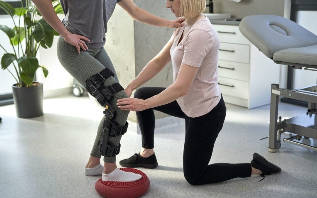
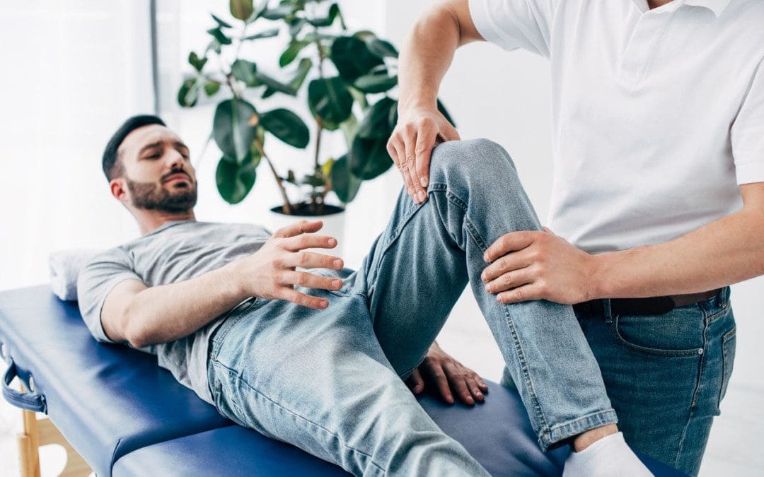
Chiropractic and Physical Therapy
Chiropractic and physical therapy can release the compressed nerve, realign the structures, strengthen the muscles, and provide gait training.
Orthotic Boot
If walking gait is affected because the foot cannot bend, an orthotic boot can help.
This is a support that maintains the foot in a neutral position to walk normally.
Corticosteroid Injection
A corticosteroid injection can reduce inflammation and relieve pressure on the nerve.
Surgery
The nerve can suffer permanent damage if it has been pinched for a long time.
If that happens, surgery cannot repair the damage.
A doctor can perform surgery to correct a fracture, tumor, or other invasive problem causing a compressed nerve.
If conservative treatment doesn’t work, a peroneal nerve decompression procedure can be done to remove the pressure.
If surgery is needed, symptoms can disappear immediately, but it takes around four months to recover and rehabilitate.
Injury Rehabilitation
References
Krych, Aaron J et al. “Is peroneal nerve injury associated with worse function after knee dislocation?.” Clinical orthopedics and related research vol. 472,9 (2014): 2630-6. doi:10.1007/s11999-014-3542-9
Lezak B, Massel DH, Varacallo M. Peroneal Nerve Injury. [Updated 2022 Nov 14]. In: StatPearls [Internet]. Treasure Island (FL): StatPearls Publishing; 2023 Jan-. Available from: www.ncbi.nlm.nih.gov/books/NBK549859/
Soltani Mohammadi, Sussan, et al. “Comparing the squatting position and traditional sitting position for ease of spinal needle placement: a randomized clinical trial.” Anesthesiology and pain medicine vol. 4,2 e13969. 5 Apr. 2014, doi:10.5812/aapm.13969
Stanitski, C L. “Rehabilitation following knee injury.” Clinics in sports medicine vol. 4,3 (1985): 495-511.
Xu, Lin, et al. Zhongguo gu Shang = China Journal of Orthopedics and Traumatology vol. 33,11 (2020): 1071-5. doi:10.12200/j.issn.1003-0034.2020.11.017
Yacub, Jennifer N et al. “Nerve injury in patients after hip and knee arthroplasties and knee arthroscopy.” American Journal of physical medicine & Rehabilitation vol. 88,8 (2009): 635-41; quiz 642-4, 691. doi:10.1097/PHM.0b013e3181ae0c9d
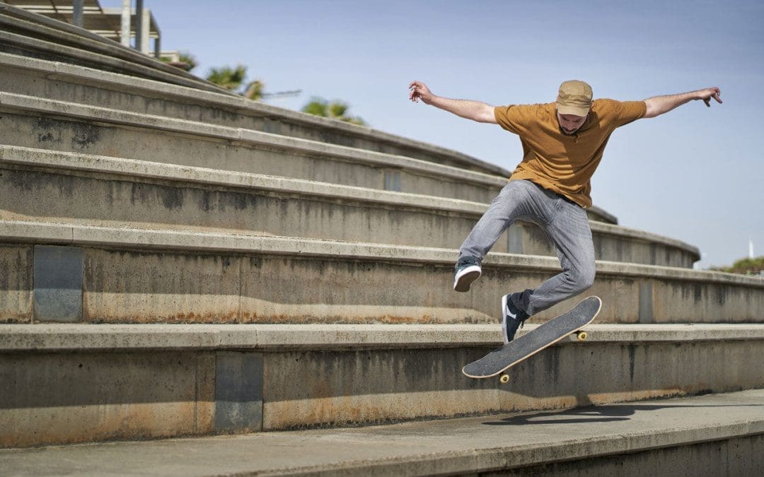
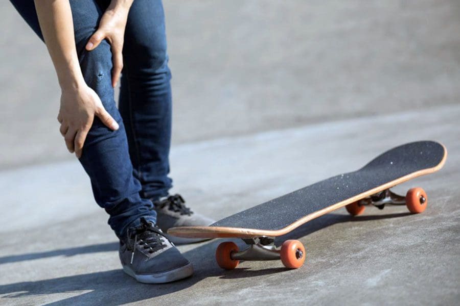
Skateboarding is a popular activity among children, teenagers, and young adults. It is recreational, competitive, fun, and exciting but, like any sport, carries a risk of injury. There are around 70,000 skateboarding injuries requiring a visit to the emergency room every year. The most common injuries involve the shins, ankles, forearms, wrists, elbows, face, and skull, with many left untreated that worsen as they heal improperly, leading to further damages and complications. Chiropractic can treat the injuries, rehabilitate the muscles and joints, and strengthen the body to get the skater back on their board.
Skateboarding Injuries
Skateboarding injuries can range from scrapes, cuts, and bruises to sprains, strains, broken bones, and concussions.
Shin injuries often happen during flip/twist tricks where the board or axle hits the shin causing bruising and swelling.
Shoulder, wrist, and hand injuries are common when skaters lose their balance and fall with outstretched arms.
Ankle injuries include rolls/sprains, as well as dislocations and fractures.
Dislocations usually happen to the shoulders, wrists, and fingers.
Facial injuries include teeth knocked out, broken nose, or jaw are typically caused by fast forward hard falls.
Severe injuries include concussions and head injuries.
Injury causes
Skateboarding injuries typically occur from:
Skating on irregular surfaces locks up wheels and affects balance, causing falls.
Losing balance or losing control of the board and falling hard/slamming into the pavement.
Inexperience, slow reaction times, and less coordination lead to falls and slams.
Skating into another skater, a person walking or cycling, a car, or a road hazard.
Trying an advanced trick/maneuver too soon and beyond their skill level.
Help prevent further injuries and long-term effects.
Chiropractic Skateboarding Injury Treatment
References
Forsman, L, and A Eriksson. “Skateboarding injuries of today.” British journal of sports medicine vol. 35,5 (2001): 325-8. doi:10.1136/bjsm.35.5.325
Hunter, Jamie. “The epidemiology of injury in skateboarding.” Medicine and sport science vol. 58 (2012): 142-57. doi:10.1159/000338722
Partiali, Benjamin, et al. “Injuries to the Head and Face From Skateboarding: A 10-Year Analysis From National Electronic Injury Surveillance System Hospitals.” Journal of oral and maxillofacial surgery: official journal of the American Association of Oral and Maxillofacial Surgeons vol. 78,9 (2020): 1590-1594. doi:10.1016/j.joms.2020.04.039
Shuman, Kristin M, and Michael C Meyers. “Skateboarding injuries: An updated review.” The Physician and sportsmedicine vol. 43,3 (2015): 317-23. doi:10.1080/00913847.2015.1050953
Individuals drive to jobs, to school, run errands, take road trips, spending a lot of time on the road. Accidents and crashes happen more frequently with all kinds of injuries. The National Highway Traffic Safety Commission has found that 37% of car accidents and crashes involve leg injuries and damage. Chiropractic physical rehabilitation and functional medicine can help heal injuries getting the individual back to everyday life.
Bruising and cuts are typical from the impact and the body getting slammed around. Lacerations can be noticed right away, but bruising comes from blood pooling underneath the skin and can take time to present, possibly 24 to 48 hours. Most bruises and cuts heal independently from home first aid care. A standard recovery used to take care of bruising is R.I.C.E or rest, ice, compression, and elevation. This helps the healing process; however, if the injury/s are more severe, chiropractic can help with therapeutic massage to relieve pain and strengthen the injured muscles, tendons, and ligaments.
ACL Injuries
The femur or thigh bone has several bands of tissue connecting it to the patella or kneecap and tibia or shin bone. One of the bands is the anterior cruciate ligament or ACL. Injuries to this band of tissue are common in sports. Car accidents and crashes are another common cause, specifically tearing the ligament. Individuals experiencing a tear may notice some or all of the following symptoms:
A cracking or popping sound when the accident or crash took place.
Swelling in and around the knee.
Severe pain in and around the knee.
Unstable and unsteady when walking or standing.
Reduced range of motion that makes walking or moving difficult.
A chiropractor can help treat the injury and help correct any muscular imbalances.
Meniscus Tears
Tears to the meniscus are also common in car accidents and crashes. The meniscusis a part of the knee. Two wedge-shaped pieces of cartilage provide a cushion where the femur and tibia meet to absorb shock. The wedges are called menisci.
When the meniscus tears, individuals might feel or hear a pop and could feel the leg suddenly give out.
Swelling in the knee.
Some pain but still be able to walk.
The knee will be stiff for the next few days.
More difficulty bearing weight or walking.
The RICE method is a recommended method of self-care. Many meniscus tears do not require surgery to improve knee function. Mild to moderate meniscus tears can be successfully treated with chiropractic techniques like soft tissue work, corrective stretches, and exercises. Surgery could eventually be necessary for severe cases to repair the meniscus to prevent long-term complications.
Chiropractic care can help the body heal and recover from a bone fracture. A patient’s bone density is evaluated and tested with an individualized treatment plan to help regain and maintain optimal bone strength. The treatments strengthen the muscles, reduce stiffness, improve nutrition, and relieve pain. Manipulation adjustments, rehabilitation, relaxation techniques, and dietary health coaching help individuals heal faster and strengthen their bones. The objective is to help regain increased mobility and range of motion.
Sciatica
Car accidents and crashes are one instance where the spine can be damaged enough to bring on sciatic pain where no back problems were present before. The impact from a car accident can cause the discs to be knocked out of place, damaged, and/or rupture around the surrounding tissue. Any of these results can pinch the sciatic nerve, leading to pain and other sciatica symptoms. Chiropractic can realign the spine and relieve pressure from the nerve/s.
DOC Spinal Decompression Table
References
Atkinson, T, and P Atkinson. “Knee injuries in motor vehicle collisions: a study of the National Accident Sampling System database for the years 1979-1995.” Accident; analysis and prevention vol. 32,6 (2000): 779-86. doi:10.1016/s0001-4575(99)00131-1
Foulk, David M, and Brian H Mullis. “Hip dislocation: evaluation and management.” The Journal of the American Academy of Orthopaedic Surgeons vol. 18,4 (2010): 199-209. doi:10.5435/00124635-201004000-00003
Wilson, L S Jr et al. “Foot and ankle injuries in motor vehicle accidents.” Foot & ankle international vol. 22,8 (2001): 649-52. doi:10.1177/107110070102200806
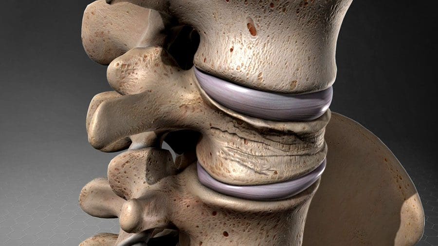
Sometimes the bones or vertebrae of the spine can crack and collapse under their weight. This is known as a compression fracture, vertebral compression fracture, or VCF. There are almost 1 million compression fractures every year, usually because the bones become weakened and crack under the weight of the vertebrae above them. These fractures can cause spinal weakness affect posture and the ability to stand up straight. They are often the cause for individuals to hunch over, also called kyphosis.
Compression Fracture
Compression fractures are small breaks or cracks in the vertebrae. The breaks occur in the vertebral body, the thick rounded part on the front of each vertebra. These fractures cause the spine to weaken and collapse. With time, these fractures affect posture as the spine curves forward. The fractures are often found in the middle/thoracic spine in the lower area. They often result from osteoporosis but can also happen after a trauma like an automobile accident, work, sports injury, or a tumor on the spine.
Symptoms
Compression fracture symptoms range from mild to severe or no symptoms. Many individuals can stand or walk without pain. They are often discovered when X-rays are taken for another condition. Symptoms include:
Back pain can come on suddenly and last for a significant time, often diagnosed as chronic back pain.
It usually develops between the shoulders and the lower back.
The pain and discomfort decrease when lying down and worsen when standing or walking.
Decreased mobility or flexibility in the spine. Individuals are unable to twist or bend.
Hunched over appearance, known as dowager’s hump or hunchback.
Loss of height from the vertebrae compression and the spine curving.
Pinched nerves
Nerve damage can cause tingling, numbness, and difficulty walking.
Loss of bladder or bowel control with severe, untreated fractures.
Individuals At Risk
Individuals who have had a compression fracture are more likely to have another one.
Women over 50 have a higher risk due to osteoporosis.
With age, the risk increases for men and women.
Diagnosis
A doctor will perform an examination and ask about symptoms. The exam will include:
Checking spinal alignment.
Posture analysis.
Gently palpates different areas of the back to identify the source of pain.
Examine for signs of nerve damage that include numbness, tingling, or muscle weakness.
A doctor will order imaging studies to examine the backbones, muscles, and soft tissues. Imaging studies include:
CT scan, X-ray, or MRI of the spine.
DEXA scan is a type of X-ray that measures bone loss bone density.
A myelogram is a procedure used along with imaging studies. A contrast dye is injected into the spine before the scan making the images easier to see.
Compression fracture treatment focuses on relieving pain, stabilizing the vertebrae, and ongoing fracture prevention. Treatment depends on the severity of the fracture and the individual’s overall health. Treatment can include:
Pain Medication
A doctor can recommend over-the-counter non-steroidal anti-inflammatory medication.
A doctor may prescribe muscle relaxers or prescription medication.
Follow instructions carefully when taking medications.
Back Brace
A special type of back brace helps to support the vertebrae.
The brace can also relieve pain by reducing how much the spine moves.
Strengthening Meds
Medications known as bisphosphonates can help slow down bone loss, stabilize the bones and prevent fractures.
This minimally invasive procedure relieves pain, stabilizes the bones, and improves mobility.
During vertebroplasty, the doctor inserts a needle in the vertebra and injects bone cement.
During kyphoplasty, the doctor inserts an inflatable device that they fill with cement.
Both are outpatient procedures allowing the individual to go home the same day.
Individuals over 65 or that have osteoporosis or a history of cancer are recommended to see their doctor. Individuals who present with sudden back pain that doesn’t get better after a day or two are advised to see a doctor and evaluate for back pain so the doctor can determine the cause and develop a treatment plan.
Body Composition
Vitamin D To Build Muscle
Skeletal Muscle Mass decreases as the body ages, primarily due to decreased physical activity. Vitamin D has been reported to influence muscle quality. This could be helpful for adults as they age. Muscle loss diminishes functional performance on activities that require strength and coordination. When this loss of muscle mass becomes significant, it becomes a condition known as sarcopenia. Treatments include:
Bischoff-Ferrari, H A et al. “Vitamin D receptor expression in human muscle tissue decreases with age.” Journal of bone and mineral research: the official journal of the American Society for Bone and Mineral Research vol. 19,2 (2004): 265-9. doi:10.1359/jbmr.2004.19.2.265
Donnally III CJ, DiPompeo CM, Varacallo M. Vertebral Compression Fractures. [Updated 2021 Nov 21]. In: StatPearls [Internet]. Treasure Island (FL): StatPearls Publishing; 2022 Jan-. Available from: www.ncbi.nlm.nih.gov/books/NBK448171/
Hassan-Smith, Zaki K et al. “25-hydroxyvitamin D3, and 1,25-dihydroxyvitamin D3 exert distinct effects on human skeletal muscle function and gene expression.” PloS one vol. 12,2 e0170665. 15 Feb. 2017, doi:10.1371/journal.pone.0170665
McCarthy, Jason, and Amy Davis. “Diagnosis and Management of Vertebral Compression Fractures.” American family physician vol. 94,1 (2016): 44-50.
IFM's Find A Practitioner tool is the largest referral network in Functional Medicine, created to help patients locate Functional Medicine practitioners anywhere in the world. IFM Certified Practitioners are listed first in the search results, given their extensive education in Functional Medicine