A good attitude about weight loss will go a long way in helping you to achieve success. Whether your weight problem has resulted from eating the wrong foods, lack of routine physical exercise, using food to resist anxiety, age, or genetics �you can help defeat that by setting reasonable goals and expectations that are realistic.
The initial step to take would be to discuss your set for weight loss and general health by means of your physician. Your physician can assist you to make informed choices about treatments that contain weight loss plans and exercise suitable to your needs.
Therapies include dietary, behavioral, drug, and for some patients, surgical alteration of the digestive system to reduce the quantity of food consumed. A safe and realistic weight reduction plan may result in success.
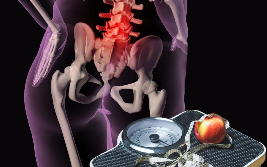
Evaluating your body weight is more involved than stepping on the scale. This info is assessed to find out your risks due to extra weight (eg, high blood pressure).
Nutrition Means To Feed Your Body
In the event you haven’t detected, the ‘D’ word (Diet) hasn’t been used in this post as it relates to weight reduction. Granted, caloric reduction will be required by a weight loss program. Yet, for many overweight or obese folks, a weight loss program means exercising, handling anxiety, and making lifestyle changes, which might comprise relearning how to eat.
It’s vitally crucial that you feed your body the nutrients it needs to be healthy and live. No one food contains all the essential nutrients �it takes combining a wide variety of foods to help meet your body’s needs. If you have been heavy or obese for a long time, your body may really be starving for necessary nutrients!
Nutrients Their Food Sources & Activities In The Human Body
*Fats are essential in taking the fat-soluble vitamins A, D, E, and K. There are just three types of fats:
Saturated Fat will raise blood cholesterol levels. These fats are found mostly in meat and diary products.
Polyunsaturated Fat tends to lower blood cholesterol levels. It’s mainly found in plant sources such as safflower, sunflower, soybean, corn, and cottonseed.
Monounsaturated Fat tends to lower the bad cholesterol or LDL (low density cholesterol). Examples include canola oil, olive oil, peanut oil, and avocados.
Although this amount is exceeded by most Americans, dietary ingestion of fat shouldn’t exceed 30% per day.
Remove the skin from poultry, trim visible fat from meat, an easy method to cut back fat consumption is to choose lean cuts of meat, choose water-packed tuna, and pick dairy products made from skim or low-fat milk.
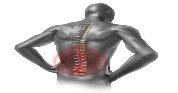
Back pain may be common, but it is not normal. It is so common that half of all working Americans admit to having back pain symptoms each year. That means there are potentially 150,000 people who suffer from back pain in the city of St. Louis alone. Low back pain is the single leading cause of disability worldwide per the Global Burden of Disease 2010 and it is also one of the most common reasons for missed work. In fact, back pain is the second most common reason for visits to the doctor�s office, outnumbered only by upper-respiratory infections.
Imaging will show all my back problems
Imaging may not reveal everything. Back pain is often a complex and multi-factorial issue, meaning that a spinal diagnosis isn�t always straightforward. To uncover what�s causing your back pain and what�s necessary for recovery requires a full clinical diagnostic process. This typically requires three steps. First you need a review of your medical history. To get to the root of your back pain, your physician should spend time asking you a series of questions about your symptoms, history, activities, positions, treatments, and more. After that is explored you will need a physical examination. A competent physician should check your spine to determine function, strength, discomfort in certain positions, and more. Lastly, you will go through diagnostic testing. Only after a physician has reviewed your medical history and given you a physical examination is imaging appropriate. Everything from an X-ray to CT scans and MRI scans can be appropriate to assess certain conditions.
Back pain that comes and goes isn�t a problem
Our bodies are incredible at adapting. However, what we know is that in nature nothing stays the same.� When someone tells me that their problem comes and goes it concerns me because it due to something that they are doing or not doing that�s causing them to either notice the problem or not notice the problem. Either way the underlying problem is still there and needs to be corrected. When the pain comes and goes, it�s usually the lead up to your back �going out� without warning. The reality for many individuals is that back pain is a result of a cumulative effect from simple movements.�� There�s always a cause for back pain and seeing a physician is your best chance for appropriate diagnosis and recovery
In fact if you find yourself saying, �back pain runs in my family� or �I just learned to live with it,� those are concerning statements because it means that the current approach you�re taking to correct the problem isn�t working, and you should make a change in order to actually correct the problem.
Drugs and Surgery are the only way to correct your back pain
In 2013 The Journal of the American Medical Association suggested chiropractic care as an option for people suffering from low back pain and noted that surgery is usually not needed and should only be tried if other therapies fail. In fact, after an extensive study of all available care for low back problems, the federal Agency for Health Care Policy and Research recommended that low back pain sufferers choose the most conservative care first. They also recommended spinal manipulation as the only safe and effective, drugless form of initial professional treatment for acute low back problems in adults
�Back pain is a normal part of aging
While we see back pain in older populations we also see it in younger populations as well.� We are seeing more and more people starting to have more back pain and arthritis at younger ages.� There are even times where I hear patients ask if arthritis in their spine is normal at their age. Again this may be common, but it is not normal.� If it were normal it would likely be seen in the entire spine not just in specific segments of the spine.
Tips to Prevent Back Pain
�Maintain a healthy diet and weight.
�Remain active�under the supervision of your doctor of chiropractic.
�Avoid prolonged inactivity or bed rest.
�Warm up or stretch before exercising or physical activities, such as gardening.
�Maintain proper posture.
�Wear comfortable, low-heeled shoes.
�Sleep on a mattress of medium firmness to minimize any curve in your spine.
� Lift with your knees, keep the object close to your body, and do not twist when lifting.
�Quit smoking. Smoking impairs blood flow, resulting in oxygen & nutrient deprivation to spinal tissues.
�Work with your doctor of chiropractic to ensure that your
Dr. Vidan�is�a private practice Chiropractor in St. Louis. He�is very passionate about helping athletes from peewee�s to pro�s get back on the field after an injury and stay on the field. This allows athletes to enjoy the most out of their sporting experience. Dr. Vidan provided chiropractic care for the St. Louis Cardinals players and staff during the 2011 World Championship season, and continues to enjoy the opportunity to help athletes and organizations at the highest levels.
Sciatica is a disabling condition characterised by pain in the leg along the distribution of the sciatic nerve. It can be accompanied by back pain, tingling, numbness, reduced strength and reflex changes in the leg.
Sciatica is most commonly caused by irritation of the nerve roots emerging from the lower spine. For this reason it is often considered a type of nerve pain.
It is estimated that around 5 to 10% of people with low back pain have sciatica, equating to around 200,000 to 400,000 Australians. It is notoriously difficult to treat sciatica with over-the-counter medications and complementary therapies.
Our study released today examines the commonly prescribed nerve pain treatment pregabalin for acute and chronic sciatica. The results show that pregabalin does not improve pain symptoms or function, but is associated with unwanted side effects such as dizziness when compared to a placebo.
Huge Uptake Of New Drug
Medicines that have shown to be effective for treating nerve pain were considered to be an exciting new treatment option for sciatica.
Pregabalin became subsidised by the Australian government for nerve pain in 2013 and quickly became widely prescribed for conditions such as sciatica. In its first year of listing, nearly 1.4 million prescriptions were written and in its second year, this figure increased to 2.4 million. This was 32% more than the government predicted.
Since its first approval in 2004 pregabalin has become the most widely prescribed medicine for nerve pain globally, with worldwide sales of between US$3-5 billion annually. The astonishing growth is likely to be a consequence of many factors but may partly be a reflection of the lack of effective treatments for sciatica.
But while pregabalin has been shown to be effective for other types of nerve pain, there was little evidence it helped patients with sciatica. There were also emerging concerns of increased harmful effects, including risk of suicidality and misuse.
We designed our study to examine whether pregabalin is effective and has tolerable side effects in patients with sciatica.
Pregabalin Does Not Work For Sciatica
The research compared the effects of pregabalin against placebo (identical inactive capsules) in 207 patients with sciatica.
Patients were randomly assigned to take up to eight weeks of pregabalin or placebo, prescribed and monitored by a general practitioner or a medical specialist. To keep the results as unbiased as possible, patients, doctors and study staff were kept blinded to who was treated with pregabalin and who received placebo capsules.
This study found after eight weeks there was no difference in the severity of leg pain between those who took pregabalin and those who took placebo capsules. The same result was seen at one year. There were also no differences in other relevant outcomes, such as back pain severity and function, at either eight weeks or one year.
However, people who took pregabalin reported more adverse effects. The most common adverse effect reported in the trial was dizziness.
The study shows that taking pregabalin does not improve your sciatic symptoms when compared with placebo, but you are more likely to have adverse effects when taking pregabalin.
Treatment Options For Sciatica
Few alternative treatment options exist for people suffering from sciatica.
There is limited data describing the effects of nonsurgical treatments such as exercise, spinal manipulation or acupuncture on sciatica.
There is also no convincing evidence to show medicines such as anti-inflammatory drugs, oral corticosteroids or opioid analgesic medicines are effective. Epidural corticosteroid injections have been shown to have a small benefit in the short-term only.
Surgery confers a short-term effect in selected patients with sciatica, but after a year people with sciatica who have not had surgery do just as well as people who�ve had the procedure.
The good news is that sciatica does get better with time. It�s important to stay as active as possible and to avoid prolonged bed rest (as this can delay recovery).
If you�re currently taking pregabalin, speak to a doctor about your condition, and mention any improvement or adverse effects you�ve experienced since starting pregabalin. It�s important not to stop pregabalin abruptly � usually doses should be reduced slowly over a few weeks. Abruptly stopping pregabalin can have some ill effects and should be done with care, close monitoring and advice from a doctor.
It�s unfortunate, but we do not currently have a lot of effective treatment options for people with sciatica. Speak to your doctor or treating clinician (such as a physiotherapist) about what may be appropriate for you, including specific advice on how you can stay as active as possible.
Although their main method of treatment is the spinal manipulation, many chiropractors also use other therapies to treat their patients. The following is a brief description of some of the most common therapies chiropractors offer.
Therapeutic Exercise
Chiropractors commonly prescribe specific strengthening exercises for their patients with back, neck, and extremity problems. These exercises can decrease pain, prevent muscle deterioration, promote joint health, increase strength, stability and range of motion, and protect against new or recurring injuries.
Your chiropractor will show you how to do the exercises and supervise you until you are comfortable doing them on your own. It’s important to keep up with your exercises as prescribed (similarly to drug prescriptions). Studies show that individuals who follow their exercise instructions heal faster than those who do not.
Therapeutic Stretches
Following an injury, therapeutic stretching is an important way to prevent scar tissue from forming. Even after the injury has healed, maintaining a regular stretching program helps keep tissues flexible, increases mobility, and protects you from new injuries. As with exercise, your chiropractor will instruct you on proper stretching techniques and will supervise you until you are comfortable enough to do them on your own.
Traction
Many chiropractors use traction, in which traction devices are applied to distract areas of the spine. This treatment helps separate the vertebrae resulting in disc decompression, reduced nerve root pressure, and decreased.
Soft Tissue Manual Therapy
Chiropractors use a variety of hands-on soft tissue therapies to improve the function of the soft tissues (muscles, ligaments, tendons, and joint capsules).�These include pin and stretch, also known under a proprietary name Active Release Technique (ART) and instrument-assisted soft tissue mobilization (Graston Technique).
Physical Therapy Modalities
Muscle Stimulation
This type of therapy uses light electrical pulses that are transmitted to specific areas of the body through electrodes placed on the skin. There are many different types of electrical stimulation. Some are more beneficial for pain relief or to reduce inflammation, some best treat muscle spasm, and some actually cause muscles to contract in order to reduce muscle atrophy. Some forms of electrical stimulation have combination effects.
TENS
A TENS (transcutaneous electric nerve stimulation) unit is a small, battery-powered, portable muscle stimulation machine that can be used at home to help control pain. Variable intensities of electric current are used to control pain. This treatment is recommended to help patients get through periods of severe (acute) pain. TENS units are typically not recommended for chronic pain. In fact, a 2009 �report from the American Academy of Neurology found that TENS units are not effective at treating chronic low back pain.1
Ultrasound
Therapeutic ultrasound is a form of deep heat therapy created by sound waves. When applied to soft tissues and joints, the sound waves are a form of micro-massage that help reduce swelling, increase blood flow, and decrease pain, stiffness, and spasms.
Ice and Heat Therapy
Ice and heat have long been used to treat many painful conditions. Ice therapy is often used to reduce swelling and help control pain immediately after an injury. Heat therapy is used to relax the muscles, increase circulation, and can provide relief to patients with chronic pain. Depending on the patient’s condition, a combination of ice and heat can be used.
Diet and Nutritional Counseling
Studies have shown that poor diet and nutritional imbalances contribute to a number of serious illnesses, such as heart disease, stroke, diabetes, and cancer. Chiropractors are specifically trained in diet and nutritional counseling. Your chiropractor can design a nutritional program specific to your needs that can help you maintain good health and minimize the risk of developing these serious health conditions.
Lifestyle Modification Counseling
Good health is much more than the absence of pain or disease. The lifestyle choices you make on a daily basis can greatly affect your long-term health. We now know that years of seemingly small unhealthy lifestyle choices can, over time, turn into very large health problems. Examples of lifestyle choices and behaviors that can have negative effects on your health include:
lack of regular exercise
smoking
poor diet
excessive mental stress
over-reliance on medication
excessive consumption of alcohol
poor posture
improper lifting
Your chiropractor will talk to you about your lifestyle choices, help you sort through and identify unhealthy health habits, and give you practical strategies to deal with and manage them.
As you can see, chiropractic medicine is more than just spinal manipulations. Chiropractors use a variety of treatment modalities to help the body to heal itself and return the patient to a pain-free and healthy life.
What is a Herniated Disc?
Herniation of the nucleus pulposus (HNP) occurs when the nucleus pulposus (gel-like substance) breaks through the anulus fibrosus (tire-like structure) of an intervertebral disc (spinal shock absorber).
A herniated disc occurs most often in the lumbar region of the spine especially at the L4-L5 and L5-S1 levels (L = Lumbar, S = Sacral). This is because the lumbar spine carries most of the body’s weight. People between the ages of 30 and 50 appear to be vulnerable because the elasticity and water content of the nucleus decreases with age.
The progression to an actual HNP varies from slow to sudden onset of symptoms. There are four stages: (1) disc protrusion (2) prolapsed disc (3) disc extrusion (4) sequestered disc. Stages 1 and 2 are referred to as incomplete, where 3 and 4 are complete herniations. Pain resulting from herniation may be combined with a radiculopathy, which means neurological deficit. The deficit may include sensory changes (i.e. tingling, numbness) and/or motor changes (i.e. weakness, reflex loss). These changes are caused by nerve compression created by pressure from interior disc material.
Progression of Herniated Disc
The extremities affected are dependent upon the vertebral level at which the HNP occurred. Consider the following examples:
Cervical – Pain in the neck, shoulders, and arms Thoracic – Pain radiates into the chest Lumbar – Pain extends into the buttocks, thighs, legs
Cauda Equina Syndrome occurs from a central disc herniation and is serious requiring immediate surgical intervention. The symptoms include bilateral leg pain, loss of perianal sensation (anus), paralysis of the bladder, and weakness of the anal sphincter.
Diagnosis of a Herniated Disc
The spine is examined with the patient laying down and standing. Due to muscle spasm, a loss of normal spinal curvature may be noted. Radicular pain (inflammation of a spinal nerve) may increase when pressure is applied to the affected spinal level.
A Lasegue test, also known as Straight-leg Raising Test, is performed. The patient lies down, the knee is extended, and the hip is flexed. If pain is aggravated or produced, it is an indication the lower lumbosacral nerve roots are inflamed.
Other neurological tests are performed to determine loss of sensation and/or motor function. Abnormal reflexes are noted as these changes may indicate the location of the herniation.
Radiographs are helpful, but Computed Axial Tomography (CAT) or Magnetic Resonance Imaging (MRI) provides more detail. The MRI is the best method enabling the physician to see the soft spinal tissues unseen in a conventional x-ray.
Radiographic Evidence of HNP
The findings from the examination and tests are compared to make a proper diagnosis. This includes determining the location of the herniation so treatment options can be reviewed with the patient.
Chiropractor, Dr. Alexander Jimenez examines being able to have sex despite having back pain.
The results of SpineUniverse’s national survey on Sexual Satisfaction and Back Pain (read the article Back Pain and Its Impact on Sexual Satisfaction for survey results) indicate that back pain is ruining the sex lives of many people.
It is vital to consider that behind the numbers are real individuals, while the statistical results of the survey are very important. People who care about their partner’s and about their sexual gratification satisfaction. People who are now frustrated and even depressed regarding the impact of back pain on their sex lives.
What exactly can they do to better their situation?
Most specialists agree that three tips can allow you to have better sex� even with back pain:
Tip # 1: Talk It Out
For many people, talking about sex comes for others, their faces turn red even thinking about possibly referring to sex.
Nonetheless, you as well as your partner have to locate a method to discuss your back malady, and the way that it will impact-� or already does change�your relationship.
Take the time to talk through the five dilemmas below:
Back pain: How intense is the pain? Where does it hurt? What moves or increase or positions alleviate the pain?
Sex drive: Is your back pain killing your sex drive? Then you’ve got to discuss this, if it is. If you do not clarify why and simply begin avoiding having sex, you�re your relationship with your partner can be damaged. It’s more straightforward to identify that it is a problem, and never simply theirs �and then find a solution together.
Emotional Impact: What does back pain do to your emotions? Do you feel less appealing to your own partner? Depressed?
Physical Limitations: Living with back pain means living with physical constraints in multiple facets of your daily life. What physical constraints would you now need to work about during sex?
Intimacy: What physical and nonphysical steps can enhance familiarity? (Yes, familiarity means more than sex.) Within the limits caused by back pain, what else can you do to feel close and connected?
Tip # 2: Practical Changes
Here we go, the nitty gritty details of what to do (or not do). (It is ok in case you skipped ahead to this part, but make sure to return and browse the remaining post.)
Position Matters
It may not be the sexiest thing to think about, but you have to remember your diagnosis. Have you got spinal stenosis? A herniated disc? Degenerative changes in your spine? Because what is causing your pain affects how your body reacts to different positions, your analysis is vitally important during sex. For example:
If you have spinal stenosis, your back pain will probably get worse if you arch your back during sex.
Your pain will probably improve should you bend forward during sex when you have a disc herniation or degenerative disk disorder.
So if you’re able to identify which positions naturally lessen your back pain, you can then accommodate your position during sex to help make the experience less painful, given your specific state. For example:
Men that have degenerative disk disease may locate their back pain is decreased by lying with a pillow placed under their low back, while their partner straddles them.
Change The Place
As we’ve learned from Hollywood films, sex does not occur merely in a bed. And perhaps being out of bed will actuality help lower your back pain. For example:
In case you like lying in your back during sex, a solid surface, such as a rug on the floor, may be more comfy for you.
But remember, back pain is individual, if you are on a soft mattress, and perhaps your pain is less during sex. You are required to figure out what’s best for you and your partner.
Rest Your Back
Back pain is frequently made worse by your muscles becoming tense as well as knotted around the region that was painful. Going in a hot tub before sex, having a soothing massage, and sometimes even just using heat or ice packs on the affected region can all ease away muscle pains just before sex.
For more practical tips about sex and back pain and more details on sexual positions go to Sex & Back Pain.
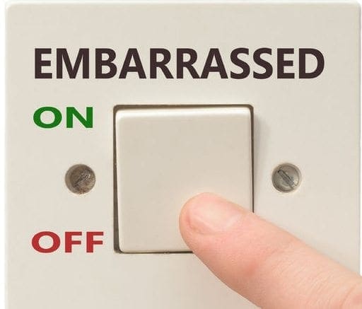
Tip # 3: Speak To A Medical Specialist
We know, talking about sex together with your doctor isn’t the most appealing notion (unless your doctor is Dr. Ruth). But think of this: When Viagra first became available, many men were too embarrassed to talk about erectile dysfunction using their doctor. Subsequently Bob Dole appeared in among their advertisements, and that made it more easy to talk to your physician about sex. (Maybe the thinking was�’If Bob Dole, a politician, can declare he has a problem, maybe I can, too!’)
Besides, physicians have heard it all and they’re prepared to help. Your physicians care about all facets of your physical and emotional well-being; they won’t pity, judge or mock you. So take a deep breath, push past the potential embarrassment, and confer with your physician about how back pain is affecting your sex life. Often, physicians can give advice that is really useful. By way of example, even just a modest change in a medication can make a major difference to your pain.
Because Sex Is More Than�
Sex is more than just the sum of its own physical parts�it’s more than a formula of physical steps that lead to the “perfect” experience. Lots of that which we see in films and on TV these days makes sex the pinnacle of a relationship, the one thing that clearly defines you as a couple (think Grey’s Anatomy).
However, for the vast bulk of people, sexual satisfaction depends on numerous variables, not just physical performance. Factors for example emotional connectedness, a bouquet of flowers sent for no reason, attentive listening, saying thank you for the small things, or sending the kids to Grandma can add to sexual gratification.
And your back pain limits none of those things. You can still have a satisfying, intimate relationship�back pain or not.
Let�s chat about chiropractic and pregnancy. For those of you that didn�t read my last post, What I Wish I Would�ve Known the First Time Around, I am the daughter of a chiropractor. My dad, Dr. Robert J. Natusch, Jr, DC, recently retired after near 40 years with offices in Northern New Jersey. My brother and sister-in-law are both licensed chiropractors at their family owned and operated practice, Upper Valley Chiropractic in Lebanon, New Hampshire (you can find them on Facebook).
Chiropractic � a Lifestyle of Health
My two siblings and I grew up vaccine-free, drug-free, and antibiotic-free. We never took over-the-counter pain meds or pills, even for headaches. If we had an issue, my dad would give us an adjustment and my mom gave us an herbal or holistic remedy and sent us on our way. To me, that was normal!
I was born at home. Those in attendance for my birth aside from my mom and dad? My older brother (21 months older) and my aunt, to tend to my brother while my mom and dad labored with me. I was mere minutes old when I received my first adjustment. It was our way of life and continues to be for my husband, our two sons, and I. It is our weekly preventative healthcare maintenance. Dr. John Tenpenny is our �primary� care doctor and if you�re in Florida, I highly recommend him.
Why the Two Go Together
Since you�re reading this, I�m guessing you�re curious how chiropractic care and pregnancy can be in the same sentence. Some feel those two are taboo and many were shocked when I told them I�d continue to see my chiropractor weekly through my 9 months of pregnancy. The gasps came when I told them my chiropractor adjusted me while I was in labor with my childrenand he came to my house again after my children were born to give them their first adjustments, too! I�m sure I�m a smidge biased, but thankfully, this has become much more mainstream. And personally, I feel moms and babes are all the better for it!
I don�t have many official references for this post, except what I�ve heard my dad repeat in his lectures my entire life.
�It is vital for moms to get adjusted through their pregnancy and beyond. Since you�re gradually adding weight to your abdomen, that causes lower back stress. Your whole posture and skeletal structure slowly alters for the duration of the pregnancy. This can amplify current misalignments.�
But, let�s get a little more in depth as to why this is important while you�re growing another human being inside you.
(Please remember, I am not a licensed chiropractor. This post is not intended to diagnose, cure, or treat any disease or health concern. This information is for educational purposes only.)
This image shows a few vertebrae within your spine. The vertebrae are your bones. The blue and red slivers are your sponge-like discs that cushion your vertebrae so your bones don�t rub on each other and cause pain. The yellow parts are the nerves nestled safely in between your vertebrae which send and receive signals to and from your brain to your organs. When that perfect structure is altered by things like bad posture, carrying your baby on your hip, sleeping on a pillow, crossing your legs, tripping, falling, carrying a purse on one shoulder, car accidents, sports, etc., etc. those messages are not able to be sent or received properly. It�s like a garden hose with the water on full blast, but there�s a kink in the line. The water won�t be at its fullest potential because there is a blockage.�
That�s the red nerve and disc within this picture. The organs which are connected to that nerve are not sending and receiving the complete message to and from the brain because the vertebra is putting pressure (a kink) in that nerve. That causes pain which may even be felt elsewhere in the body and not felt within the spine. You may treat the symptom of pain unsuccessfully until you get to the source which lies within the spine.
Just think for a moment, does that nerve have anything to do with the organ that helps grow your baby? Maybe it�s the one that represents your uterus. If left unadjusted, your body is not able to operate to its fullest potential and ability when growing your little one. Messages are not being sent or received fully. The consequences of not rectifying the issue may not reveal itself until delivery!
A simple adjustment (or series of adjustments depending on the severity and years of your life without chiropractic care) can aid in rectifying the issue and allowing your body to operate at its fullest potential and health while growing your baby.
Call Today!
How The Pelvis Benefits From Chiropractic
Let�s take a moment to look at how God designed our birthing pelvis.
Our pelvis is designed with a cartilage substance to connect our pubic (pubis) bone. During pregnancy, labor, and birth, the body sends signals to control that cartilage. When it softens, it�s time for the baby to soon make his arrival! If there is a communication kink somewhere within the spine, those signals would not be clearly communicated. You�d be so much better off addressing the misalignment (know as a subluxation) now � well before delivery day � so your body can relax in its designed location.
When people ask me if it�s safe to be adjusted during pregnancy, the analogy of the kinked water hose comes to mind. Our female bodies are created and designed to grow and carry a baby. A chiropractor�s job is to keep my spine in the correct alignment so my body can function to its fullest potential. During the most spine-altering 9 months in my life, I would never think to neglect my weekly chiropractic adjustments! That�s like deciding to stay home from the grocery store when you need food.
Every parent can say they only want the best, optimal health for their child once they are born. Being under a chiropractor�s care allows you the ability to give your child the best, optimal health while they are in your womb.
Though the most visible kinesiology tape users might be professional athletes, Olympians, or weekend warriors � a much smaller clientele is also proving the advantages of this unique treatment. With a special line designed for sensitive skin and playful prints and colors that will speak to the sticker and Band-Aid loving child, it�s not surprising that kinesiology tape is quickly becoming more than an athlete�s aid.
While the uses and applications of kinesiology tape for pediatric treatment are growing daily, here is a quick run down of how kinesiology tape is being used in pediatric care today:
Pain &�Swelling Relief
� When kinesiology tape is applied to an injured or inflamed area, children can enjoy some relief without having to take pain medication or sit through icing and therapy treatments.
Orthopedic Treatment
� Children often don�t understand the purpose behind rehabilitation exercises, so kinesiology tape provides an additional or alternative treatment for children with orthopedic injuries, weak or underdeveloped muscles, gait abnormalities, paralysis � even poor posture.
Neuromuscular Disorders
� Kinesiology tape has proven effective to activate weak muscles and inhibit overactive muscles.� With a simple and safe taping application, children suffering from neuromuscular conditions like cerebral palsy, or muscular dystrophy could see improvements in symptoms and movement ability. It has also been shown to improve muscle tone in genetic disorders like Downs Syndrome and other conditions causing either spasticity, atrophy or poor muscle tone.
For children dealing with serious medical conditions, a colorful and painless treatment that can be worn for several days, even while playing and bathing, can make a big difference in their comfort level.
Interested in learning more about pediatric kinesiology taping? Dr. Kenzo Kaze, the creator of Kinesio Tape, provides step by step guidelines for taping infants and children in his manual, Kinesio Taping in Pediatrics available at Theratape.com.
For a list of successful case studies where kinesiology tape has provided improvement for children, check out Theratape�s research compilation as well as this case report (pdf) from Novel Physiotherapies. Hopefully, as more and more case studies document positive results, the use of kinesiology tape in pediatric care will continue to expand and increase.
IFM's Find A Practitioner tool is the largest referral network in Functional Medicine, created to help patients locate Functional Medicine practitioners anywhere in the world. IFM Certified Practitioners are listed first in the search results, given their extensive education in Functional Medicine