Regenerative Spine Care and Sciatica Relief in El Paso: How Epidural Injections, PRP, mFAT, and Shockwave Therapy Work Together
Sciatica and chronic back pain can affect almost every part of daily life. Sitting can hurt. Walking can feel limited. Sleep may be broken. Work, exercise, driving, and family time can become harder than they should be.
At El Paso Back Clinic, the goal is to look deeper than the pain signal. Pain is important, but it is often only the warning light. The real problem may involve an irritated nerve, a damaged disc, a strained ligament, a weak core, poor spinal motion, scar tissue, inflammation, or a past injury that never healed correctly.
This is why a modern spine care plan may combine chiropractic care, rehabilitation, medical oversight, functional medicine, epidural spinal injections, regenerative therapies, and shockwave therapy. Each part has a different job. Together, they may help calm nerve irritation, support tissue repair, improve movement, and help the body return to better function.
What Is Sciatica?
Sciatica is pain that travels along the sciatic nerve. This nerve starts in the lower back and travels through the buttock, hip, leg, and foot. When a spinal nerve root becomes irritated or compressed, pain can travel down the leg.
Common sciatica symptoms may include:
Low back pain
Buttock or hip pain
Burning pain down the leg
Numbness or tingling
Weakness in the leg or foot
Pain that worsens with sitting
Pain that improves when lying down or changing position
Sciatica is not always caused by the same problem. It may come from a herniated disc, disc degeneration, spinal stenosis, facet arthritis, muscle tension, pelvic imbalance, scar tissue, or inflammation. This is why a complete exam matters.
Why Chronic Back Pain Needs More Than Temporary Relief
Chronic back pain is pain that lasts longer than expected. It often continues for more than 12 weeks. By that time, the body may start to change how it moves. Muscles tighten. Joints stiffen. Nerves become more sensitive. The patient may avoid activity, which can lead to weakness and more pain.
Traditional care often focuses on short-term pain relief. That can help during a flare-up, but it may not be enough when the deeper problem is structural or inflammatory.
A more complete plan may look at:
Spinal alignment and joint motion
Disc health
Nerve irritation
Ligament and tendon stress
Muscle weakness
Core control
Inflammation
Nutrition
Sleep
Blood sugar and metabolic health
Prior auto, work, or sports injuries
This whole-person view is important because healing is not only about one painful spot. The spine is part of a larger system.
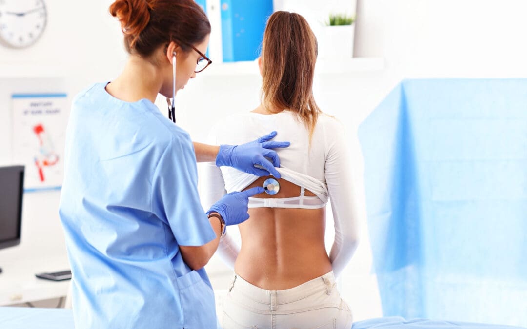
How Epidural Spinal Injections May Help Sciatica
An epidural spinal injection places medication or biologic material near an irritated spinal nerve. The goal is to reduce inflammation around the nerve root and help calm leg pain.
For a patient with strong nerve pain, this can be helpful. When pain is severe, the patient may not be able to move, stretch, exercise, or sleep well. If an epidural injection reduces the pain enough, the patient may be able to begin rehabilitation and chiropractic care more safely.
Epidural steroid injections are commonly used for spinal stenosis and nerve-related back and leg pain. However, long-term outcomes may vary. In one PCORI-supported report on lumbar spinal stenosis, epidural injections with corticosteroid plus lidocaine did not show long-term benefits over lidocaine alone for pain, function, opioid use, or surgery rates in the studied group (Friedly et al., 2019).
This does not mean epidural injections are useless. It means they should be used carefully and as part of a larger care plan.
Why Some Patients Look Beyond Repeated Steroid Injections
Steroids can reduce inflammation. That is why they are often used during painful flare-ups. But repeated steroid use may carry risks. Cortisone injections can have side effects, including cartilage damage, tendon weakening, blood sugar changes, infection risk, and bone thinning, especially when used too often or in high amounts (Mayo Clinic, 2026).
For some patients, this raises an important question:
Can we reduce pain while also supporting tissue repair?
This is where regenerative therapies may enter the conversation. Regenerative care does not simply try to hide symptoms. It aims to support the body’s natural healing response.
What Are Regenerative Spine Therapies?
Regenerative spine therapies use biologic materials, often from the patient’s own body, to support healing. These treatments may be considered for chronic spine pain, disc-related pain, ligament injury, facet joint pain, and nerve irritation when the patient is a proper candidate.
Common regenerative options include:
PRP: platelet-rich plasma
PFP: platelet-fibrin plasma or platelet-fibrin products
Platelet lysate: a platelet-derived fluid rich in growth factors
mFAT: microfragmented adipose tissue
These therapies are often called orthobiologics. “Ortho” refers to bones, joints, muscles, ligaments, and spine structures. “Biologics” refers to healing materials that come from living tissue.
The University of Iowa Health Care describes regenerative medicine as care that may use a person’s own cells, tissues, or biologic materials to support healing and repair (University of Iowa Health Care, n.d.).
PRP: Platelet-Rich Plasma for Spine and Nerve-Related Pain
PRP is made from a small sample of the patient’s blood. The blood is processed to concentrate platelets. Platelets are best known for helping blood clot, but they also carry growth factors and healing signals.
In spine care, PRP may be used to support damaged or irritated tissues, such as:
Disc-related pain areas
Facet joints
Ligaments
Tendons
Soft tissues around the spine
Research on PRP for low back pain is still growing. A narrative review on regenerative medicine for chronic low back pain described PRP and other biologic therapies as promising options, while also noting that more high-quality research is needed (Wang et al., 2023). A systematic review of PRP for low back pain found PRP was generally effective and safe for degenerative low back pain but also called for stronger studies and better treatment standards (Machado et al., 2023).
In simple terms, PRP is not a magic cure. But for selected patients, it may help support a better healing environment.
Platelet Lysate and Epidural Biologic Injections
Platelet lysate is made from platelets, but it is processed differently than PRP. The platelets are broken open, releasing growth factors into a thinner fluid. Because it is less thick than PRP, platelet lysate may be considered for nerve-related areas, including epidural use in some regenerative medicine settings.
A study of lumbar epidural platelet lysate for radicular pain reported improvements in pain and function through 24 months, with mild adverse events reported in a small percentage of patients (Centeno et al., 2017). More research is still needed, but this area is important because it examines biological support for nerve-related back and leg pain.
A 2025 meta-analysis also compared epidural PRP with steroid injections for lumbar disc disease with radiculopathy. The authors reviewed randomized controlled trials and examined pain and function outcomes over several time points (Muthu et al., 2025). This growing research shows why biologic epidural options are becoming a major topic in modern spine care.
PFP: A Natural Scaffold for Healing
PFP, or platelet-fibrin plasma, is similar to PRP but includes more fibrin activity. Fibrin is a natural protein involved in clotting and wound repair.
You can think of fibrin as a healing web. It may help hold platelets and growth factors in one area longer. This may be useful when the care plan is focused on damaged ligaments, tendons, or joint tissues.
PFP may support:
Local repair signaling
Tissue stability
Collagen remodeling
Longer contact time for healing factors
A more organized repair response
Like other regenerative options, PFP should be used after a detailed exam and proper diagnosis.
mFAT: Microfragmented Adipose Tissue
mFAT stands for microfragmented adipose tissue. Adipose tissue is fat tissue. In this treatment, a small amount of a patient’s own fat is collected, processed, and prepared for injection into a target area.
Fat tissue contains signaling cells and support structures that may help with tissue repair. mFAT is often discussed in regenerative medicine for joint, soft tissue, and orthopedic problems. It does not “regrow” a spine overnight. Instead, it may help support the local repair environment in selected cases.
For chronic spine problems, mFAT may be considered when there is deeper tissue degeneration, joint wear, or long-standing injury patterns. The key is proper patient selection, medical screening, imaging review, and follow-up care.
Shockwave Therapy: The Biological Catalyst
Shockwave therapy, also called extracorporeal shockwave therapy (ESWT), uses sound waves to stimulate tissue. It is non-surgical and does not involve medication.
Shockwave therapy may help painful tissues by creating a controlled healing signal. This process is called mechanotransduction. That means the body turns mechanical energy into a biological response.
ESWT may support healing by helping:
Increase local blood flow
Stimulate new small blood vessel formation
Improve cell activity
Reduce pain signaling
Break down scar-like tissue
Improve collagen remodeling
Support tissue repair pathways
A systematic review and meta-analysis found that ESWT improved pain and lumbar function in patients with chronic low back pain, with no serious adverse effects reported in the included studies (Liu et al., 2023). Another review described shockwave as a tool that may support tissue repair through blood vessel growth, anti-inflammatory effects, and cell signaling (Cheng & Wang, 2015).
Why Shockwave and Regenerative Injections May Work Well Together
Regenerative injections bring healing signals to damaged tissue. Shockwave therapy may help prepare the tissue to respond better.
This is why ESWT can be described as a biological catalyst. A catalyst helps a process move forward. Shockwave does not replace PRP, PFP, platelet lysate, or mFAT. It may help create a better local environment for healing.
A simple way to picture it is this:
PRP, PFP, platelet lysate, and mFAT bring healing signals.
Shockwave therapy helps wake up slow-healing tissue.
Chiropractic care improves joint motion and biomechanics.
Rehabilitation rebuilds strength, balance, and control.
Functional medicine looks for healing barriers inside the body.
When combined correctly, these tools may help the body repair itself more effectively than a single treatment alone.
The Role of Chiropractic Care at El Paso Back Clinic
Chiropractic care is often central to sciatica and back pain recovery because movement matters. If spinal joints, hips, pelvis, and soft tissues are not moving well, stress can build up around the nerves and discs.
At El Paso Back Clinic, chiropractic care may support:
Better spinal motion
Less joint stiffness
Improved posture
Better pelvic and hip mechanics
Reduced muscle guarding
Safer return to activity
Better rehab progress
Dr. Alexander Jimenez, DC, APRN, FNP-BC, CCST, CFMP, IFMCP, ATN, uses a dual-scope clinical view that connects chiropractic evaluation, injury care, functional medicine, and rehabilitation. His clinical observations often focus on how spinal structure, inflammation, metabolic health, and movement patterns work together.
This matters because many patients do not only have “a bad disc.” They may have a body system that is under stress.
Medical Oversight With Dr. Maria Guadalupe Cardenas, MD
At Injury Medical Clinic PA and within the larger integrative care model connected with El Paso Back Clinic, Dr. Maria Guadalupe Cardenas, MD, serves as Medical Director and Collaborative Physician. She is Board Certified in Internal Medicine, has over 40 years of experience as an internist, and is listed with NPI #1164426749 and Texas MD License #J2933.
This medical oversight is valuable because many spine patients have other health issues that can affect treatment safety and healing.
These may include:
Diabetes or blood sugar problems
High blood pressure
Autoimmune conditions
Medication use
Blood thinner use
Hormone changes
Infection risk
Poor sleep
Chronic inflammation
Older injuries or surgeries
A multidisciplinary clinic can help connect the dots between medical history, spine pain, nerve symptoms, and recovery goals.
Functional Medicine: Looking for Healing Barriers
Functional medicine asks a deeper question:
Why is this patient not healing well?
For chronic back pain and sciatica, the answer may lie beyond the spine. The body heals best when it has the right nutrients, blood flow, hormones, oxygen, sleep, and control of inflammation.
Functional medicine support may look at:
Vitamin D status
Blood sugar and insulin
Inflammation markers
Thyroid function
Hormone balance
Gut health
Nutrition
Weight management
Sleep quality
Stress load
This does not replace spine care. It supports spine care. A patient with poor blood sugar control, low protein intake, poor sleep, and high inflammation may heal more slowly. Improving these areas may help the patient respond better to chiropractic care, rehab, injections, and shockwave therapy.
Why Personal Injury Patients May Benefit
After a car crash, work injury, or sports injury, pain may not show up right away. Some symptoms appear hours or days later. Neck pain, back pain, headaches, sciatica, numbness, and stiffness can develop after the body’s stress response calms down.
Personal injury care needs careful documentation and a clear clinical plan. At El Paso Back Clinic, the care model may include:
Injury history
Orthopedic testing
Neurological testing
Range-of-motion findings
Imaging review when needed
Functional limits
Treatment response
Rehab progress
Referrals when needed
This matters because injury recovery is not only about pain relief. It is also about restoring function and documenting how the injury changed it.
A Step-by-Step Spine Recovery Plan
A patient-centered spine plan may include several phases.
Phase 1: Calm the Nerve
When sciatica is active, the first goal is to reduce irritation. This may include careful activity changes, decompression, gentle chiropractic care, targeted injection options, and pain-control strategies.
Phase 2: Improve the Healing Environment
Once pain is more controlled, regenerative therapies and shockwave therapy may be considered. The goal is to support tissue repair, improve circulation, and help chronic tissue move out of a stalled healing state.
Phase 3: Restore Motion
Chiropractic care, soft-tissue therapy, mobility work, and decompression may help the spine and pelvis move more freely.
Phase 4: Rebuild Strength
Rehabilitation helps the patient rebuild core strength, hip control, balance, posture, and endurance. This step helps protect the spine from future flare-ups.
Phase 5: Maintain Long-Term Function
The final goal is not just to feel better for a few days. The goal is to help the patient return to life with improved movement, strength, and awareness of how to prevent future problems.
Who May Be a Candidate?
A patient may be a candidate for this type of care if they have:
Sciatica
Chronic low back pain
Disc herniation
Disc degeneration
Annular tear
Facet arthritis
Ligament injury
Post-accident back pain
Pain that returns after basic care
Difficulty walking, sitting, or sleeping due to nerve pain
Not every patient is a candidate for every treatment. Severe weakness, loss of bowel or bladder control, fever, infection signs, cancer history, major trauma, or rapidly worsening nerve symptoms need urgent medical attention.
Final Thoughts
Sciatica and chronic back pain can be frustrating, but patients now have more options than short-term pain masking. Epidural spinal injections may help calm acute nerve irritation. Regenerative therapies such as PRP, PFP, platelet lysate, and mFAT may support repair in damaged or irritated tissues. Shockwave therapy may act as a biological catalyst by improving blood flow, stimulating cell activity, and helping chronic tissue respond.
At El Paso Back Clinic, this kind of care fits into a larger model that includes chiropractic care, medical oversight, functional medicine, personal injury care, and rehabilitation. With Dr. Alex Jimenez, DC, APRN, FNP-BC, working alongside Dr. Maria Guadalupe Cardenas, MD, Medical Director and Collaborative Physician, patients receive a team-based approach focused on structure, function, safety, and long-term healing.
The goal is simple: reduce pain, restore movement, support healing, and help patients return to the life they want.
Many adults notice extra weight creeping on, especially around the middle, even when they try to eat better and stay active. Hormone changes over time often play a quiet but powerful role in how the body stores fat, burns energy, and controls hunger. Bioidentical hormone replacement therapy (BHRT) offers a way to bring those internal messengers back into better balance. It is not a quick weight-loss fix or a magic pill. Instead, it helps remove some of the metabolic roadblocks that make diet and lifestyle efforts harder to sustain.
When hormone levels are optimized, many people find it easier to manage cravings, keep steady energy, and support lean muscle. This article explains how BHRT, and specifically the EvexiPEL method from Evexias Health Solutions, can work alongside smart eating and daily habits for longer-lasting results.
What Bioidentical Hormones Actually Do in the Body
Hormones act like chemical messengers. They tell the body when to store fat, when to burn it, how hungry to feel, and how well muscles can grow. Key players include estrogen, testosterone, insulin, cortisol, and thyroid hormones. When these get out of balance—often from aging, stress, or other life changes—metabolism can slow, fat can gather more easily around the belly, and cravings for sweets can grow stronger.
Bioidentical hormones are made to match the exact structure of the ones the human body produces naturally. They usually come from plant sources and are customized for each person after lab testing. The goal is to restore balance rather than force rapid change. Because they more closely match the body’s own chemistry, many patients experience smoother effects than with synthetic options.
How Balanced Hormones Help with Weight and Fat Control
Balanced hormones support weight management in several practical ways:
Fewer intense sugar cravings: When estrogen, progesterone, and cortisol signals stabilize, the brain’s hunger cues become easier to manage. People often report a less urgent desire for processed sweets or snacks.
Better insulin sensitivity: Improved insulin function helps the body use blood sugar for energy rather than store it as fat. This makes it easier to maintain a steady weight over time.
More consistent daily energy: Steady hormone levels reduce afternoon slumps. With more energy, it becomes easier to go for a walk, prepare a healthy meal, or stick to an exercise plan.
Support for lean muscle: Testosterone and other hormones help maintain or build muscle. Muscle tissue burns more calories even at rest, which supports a higher everyday metabolism.
Less stubborn abdominal fat: Hormone balance can influence where the body prefers to store fat. Many notice gradual improvement in midsection fat when levels are optimized alongside healthy habits.
These changes do not happen overnight. They create an internal environment where diet and movement efforts can finally show clearer results.
EvexiPEL Pellet Therapy: Steady Delivery Without the Roller Coaster
Evexias Health Solutions developed the EvexiPEL method as a form of BHRT that uses tiny, custom-made pellets. A trained provider places the pellets just under the skin during a short office visit. The pellets then release a steady, consistent dose of bioidentical hormones—such as testosterone or estradiol—over several months, usually three to six.
This steady release mimics the body’s natural rhythm far better than daily creams, gels, pills, or weekly shots. Many patients describe avoiding the ups and downs, or “roller coaster,” that can come with other delivery methods. Consistent levels often translate into more reliable energy, steadier moods, and fewer hormone-driven cravings throughout the day.
Because the delivery stays even, people can focus on building healthy routines instead of managing daily symptom swings. EvexiPEL is always paired with lab testing and a full wellness plan; it is never used alone.
Why Nutrition Matters Even More with BHRT
BHRT works best when paired with a diet built around fresh, whole foods. Think plenty of vegetables, quality proteins, healthy fats from avocados and nuts, and fiber-rich choices. These foods provide the body with the raw materials it needs for hormone production, detoxification, and stable blood sugar.
Cutting back on processed carbohydrates and added sugars helps too. These foods can spike blood sugar and work against the improvements in insulin sensitivity that BHRT supports. Many people find that once hormones stabilize, choosing whole foods feels more natural because energy stays higher and cravings quiet down.
Evexia’s providers often combine pellet therapy with targeted nutraceuticals—high-quality supplements designed to support metabolism, gut health, and mitochondrial energy. This root-cause approach to care addresses multiple systems at once rather than focusing on calories alone.
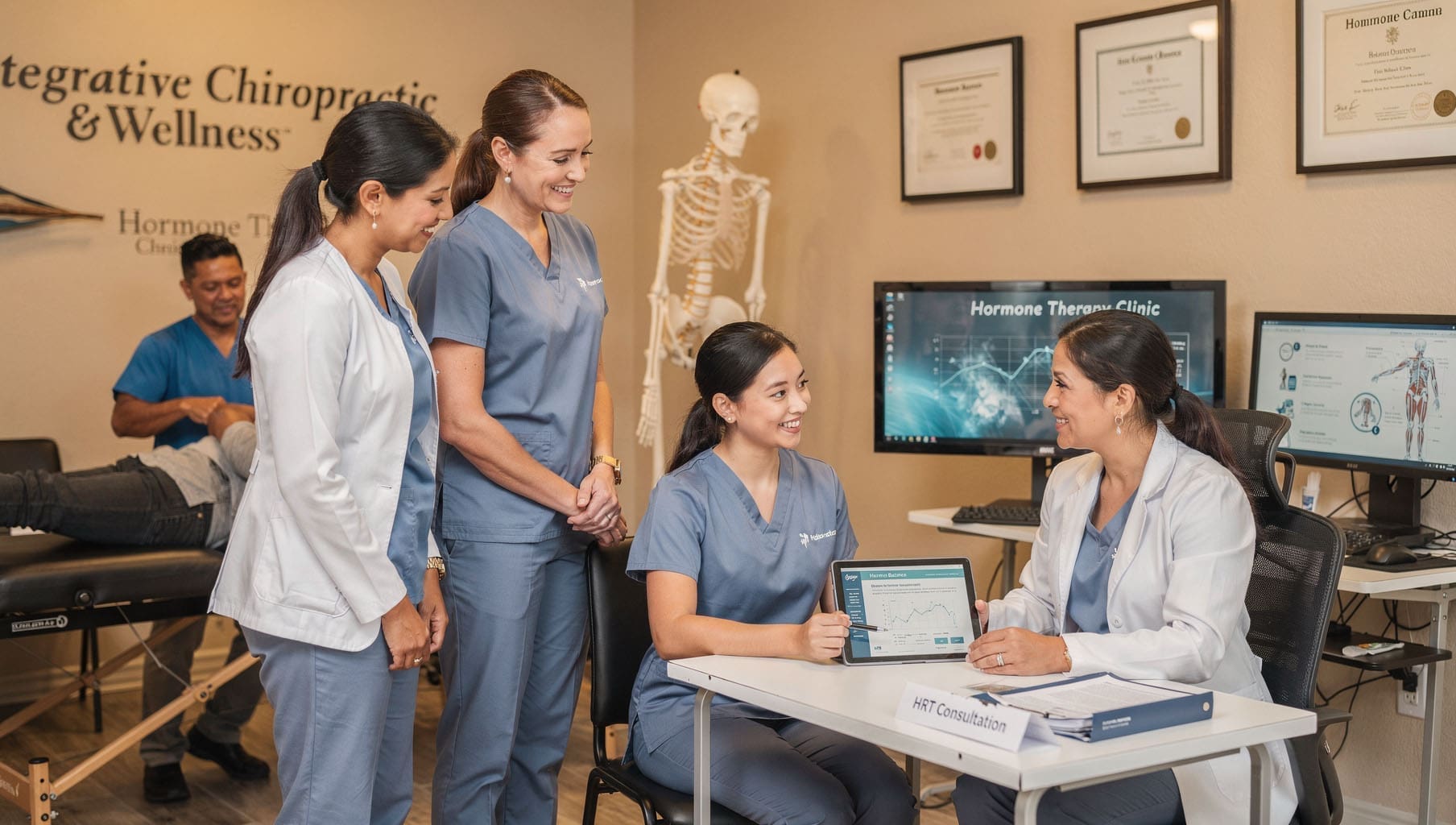
The Advantage of Multidisciplinary Integrative Care
Hormone balance does not exist in a vacuum. The nervous system, gut health, sleep, stress, and physical structure all influence how well hormones work. That is why care from a coordinated team often produces stronger, longer-lasting outcomes.
A clear example is the collaborative model at Injury Medical Clinic PA in El Paso, Texas. Dr. Alexander Jimenez, DC, APRN, FNP-BC, CFMP, IFMCP, ATN, CCST, brings chiropractic expertise, functional medicine insights, and advanced wellness protocols. He works directly with Medical Director Dr. Maria Guadalupe Cardenas, MD, a board-certified internal medicine physician with more than 40 years of experience (NPI #1164426749, Texas MD License #J2933).
In this setup:
Chiropractic care from Dr. Jimenez helps optimize nervous system function, posture, and mobility, so patients can move more comfortably and handle daily stress more effectively.
Dr. Cardenas provides medical oversight, reviews lab results, manages internal medicine needs, and ensures safe, appropriate hormone monitoring.
Functional medicine and nutrition support address gut health, inflammation, and lifestyle factors that affect metabolism.
Rehabilitation and personal injury services remove physical barriers that might otherwise limit activity and exercise.
Dr. Jimenez’s clinical observations in integrative settings show that patients achieve better metabolic and energy improvements when hormone optimization is combined with whole-person care. The spine and nervous system directly influence hormone signaling and stress responses. When both are supported, the body becomes more efficient at using the benefits of balanced hormones for weight and overall wellness.
This team approach makes BHRT one component of a larger, personalized strategy rather than an isolated treatment.
What Results Typically Look Like
People who combine EvexiPEL BHRT with whole-food nutrition and team-based support often describe:
More stable energy that lasts through the afternoon without relying on caffeine or sugar.
Reduced cravings that once derailed healthy eating plans.
Gradual improvements in body composition—less fat, better muscle tone—as insulin sensitivity and metabolism improve.
Easier adherence to daily movement because joints and energy feel better supported.
These changes build over weeks and months. The steady hormone delivery helps patients stay consistent long enough for new habits to stick. BHRT does not replace the need for healthy food choices and regular activity; it makes those efforts more effective by clearing hormonal interference.
Sample Report
Taking the Next Step Toward Balanced Health
If stubborn weight, low energy, or strong cravings have been ongoing challenges despite sincere efforts, checking hormone levels can be a useful step. A provider trained in EvexiPEL or similar BHRT methods will review full lab results, health history, and lifestyle before recommending a plan. Results vary, and therapy must always occur under proper medical supervision.
Clinics that blend chiropractic care, internal medicine oversight, functional nutrition, and regenerative approaches—like the model with Dr. Jimenez and Dr. Cardenas—can offer the coordinated support many people need. By addressing hormones, nervous system health, nutrition, and daily habits together, patients often move from frustration to steady, inside-out progress.
Balanced hormones alone will not create lasting change. But when they work in harmony with smart daily choices and a supportive care team, weight management becomes less of a constant struggle and more of a natural outcome of a body that is finally working with you instead of against you.
El Paso Personal Injury and Work Injury Chiropractor
Abstract
Personal injury and work injury recovery should focus on more than short-term pain relief. At an integrative chiropractic clinic in El Paso, the goal is to help the body heal, restore movement, reduce inflammation, and improve daily function. This article explains how integrative chiropractic care, functional medicine, rehabilitation, soft-tissue therapy, therapeutic ultrasound, and nutritional counseling may support recovery after car accidents, whiplash, slips and falls, work injuries, and muscle or ligament strains. It also explains why proper documentation is important in personal injury cases and why ethical care should always be based on medical need rather than referral pressure. When care is evidence-based, patient-focused, and well-documented, it can support both healing and clear communication between patients, healthcare providers, attorneys, and insurance companies.
El Paso Integrative Chiropractic Care for Injury Recovery
When a person is injured in a motor vehicle accident, workplace incident, or slip and fall, the body often reacts in several ways at once. Pain may start in the neck, back, shoulder, hip, or knee, but the injury can also affect the nervous system, soft tissues, spinal joints, ligaments, and muscles.
At El Paso Back Clinic, the approach to care is based on helping the whole person, not just chasing symptoms. This matters because pain is often only one part of the injury story. A patient may also have stiffness, headaches, poor sleep, muscle weakness, inflammation, nerve irritation, or fear of movement after trauma.
Integrative chiropractic care combines several tools to help the body recover, including:
Chiropractic adjustments to improve joint motion
Rehabilitation exercises to restore strength and coordination
Soft-tissue therapy to reduce muscle tightness and scar-like adhesions
Functional medicine support to address inflammation, nutrition, and recovery health
Nutritional counseling to support tissue healing
Objective documentation to track injuries, progress, and medical needs
El Paso Back Clinic describes integrative chiropractic care as a whole-person model that may include chiropractic care, exercise, nutrition, lifestyle support, and complementary therapies to address the root causes of pain and dysfunction (El Paso Back Clinic, n.d.).
Why Personal Injury and Work Injuries Need a Whole-Body Plan
After trauma, the body often enters a protective state. Muscles tighten to guard injured areas. Joints may stop moving normally. Inflammation increases as the immune system sends repair cells to damaged tissues. Nerves may become more sensitive. This is a normal healing response at first, but when it lasts too long, it may lead to chronic pain and poor movement.
This is why injury care should not only ask, “Where does it hurt?” It should also ask:
What tissue was injured?
What movement is limited?
Is there nerve involvement?
Is the pain caused by inflammation, joint restriction, muscle guarding, or all three?
What daily activities are affected?
What treatment is medically necessary?
Is imaging or referral needed?
In my clinical observations, many patients hurt after crashes or work injuries try to push through pain. Some wait days or weeks before getting evaluated. This can be a problem because untreated injuries may lead to more stiffness, poor posture, weaker muscles, and longer recovery times.
A careful exam helps identify the problem early. This may include checking range of motion, muscle strength, reflexes, sensation, joint movement, posture, walking patterns, and signs of nerve irritation.
Chiropractic Adjustments and Spinal Joint Motion
Chiropractic adjustments are used to help restore motion to spinal and extremity joints that are not moving well. After an injury, a joint may become restricted because of swelling, muscle guarding, or altered body mechanics. When one area stops moving properly, another area may overwork to compensate.
For example, after a rear-end collision, the neck may lose its normal range of motion because the muscles tighten to protect the cervical spine. The upper back may also become stiff. This can lead to headaches, shoulder tension, and pain with turning the head.
A proper chiropractic adjustment is a controlled treatment. The goal is not to “crack the spine” for quick relief. The goal is to improve joint mobility, reduce mechanical stress, and help the nervous system receive better movement signals from the body.
Chiropractic care may help support recovery from:
Whiplash-related neck pain
Low-back pain after a crash
Mid-back pain from seatbelt trauma
Hip or pelvic restriction after a fall
Headaches linked to neck dysfunction
Work-related lifting injuries
Shoulder and extremity movement problems
Research-based guidelines support the use of non-drug treatments, including spinal manipulation, exercise, massage, and multidisciplinary care, for many types of low-back pain when clinically appropriate (American College of Physicians, 2017).
Whiplash Injury Care and Neck Rehabilitation
Whiplash is one of the most common injuries after a motor vehicle accident. It happens when the head and neck move suddenly forward and backward or side to side. This rapid motion can strain muscles, ligaments, joints, discs, and nerves.
Whiplash symptoms may include:
Neck pain
Headaches
Upper-back tightness
Shoulder pain
Dizziness
Jaw tension
Numbness or tingling
Poor sleep
Pain with driving or computer work
Whiplash is not always visible on a basic X-ray. That does not mean the pain is not real. Many whiplash injuries involve soft tissues, which include muscles, ligaments, tendons, fascia, and joint capsules.
A strong whiplash care plan may include:
Gentle chiropractic adjustments or mobilization
Soft-tissue therapy
Neck-specific strengthening exercises
Posture training
Home exercise instruction
Gradual return to normal activity
Monitoring for neurological symptoms
Modern whiplash research supports multimodal care. This means combining manual therapy, exercise, education, and self-management rather than relying on a single treatment method (Bussières et al., 2016). This is important because whiplash recovery requires both pain control and movement retraining.
Soft-Tissue Therapy and Muscle Recovery After Injury
After trauma, muscles often tighten to protect the injured area. This is called muscle guarding. At first, guarding may help prevent further injury. Over time, however, it can create stiffness, trigger points, pain with movement, and poor posture.
Soft-tissue therapy may help improve tissue movement and reduce tightness. This may include hands-on therapy, stretching, myofascial work, instrument-assisted techniques, massage-style therapy, or therapeutic modalities.
Soft-tissue care is often used for:
Muscle strains
Ligament sprains
Scar tissue
Trigger points
Whiplash-related muscle guarding
Work-related overuse injuries
Back and neck stiffness
The goal is to prepare the body for better movement. Soft-tissue therapy may reduce pain enough for the patient to participate in rehabilitation exercises. This is important because long-term recovery depends on restoring strength and control, not only reducing soreness.
Therapeutic Ultrasound in Chiropractic Injury Care
Therapeutic ultrasound is a treatment tool that uses sound-wave energy to support soft-tissue care. It is often used in chiropractic and rehabilitation settings for muscles, tendons, ligaments, and joint stiffness.
The clinical goal of ultrasound may include:
Improving local tissue circulation
Reducing stiffness
Helping tight tissues relax
Supporting soft-tissue healing
Preparing tissues for stretching or movement
Decreasing pain in selected conditions
For personal injury care, therapeutic ultrasound may be considered for soft-tissue injuries such as whiplash strain, muscle spasm, sprains, or tendon irritation.
However, it should be used with clear reasoning. Ultrasound should not be added only to increase billing or create more treatment visits. It should match the patient’s exam findings and recovery goals.
In personal injury cases, ultrasound treatment notes may help show that care was provided and tracked. Still, the strongest documentation comes from the full clinical record, including the injury history, examination findings, diagnosis, functional limits, treatment plan, progress notes, and medical necessity.
Research on therapeutic ultrasound is mixed and depends on the condition being treated. Some studies show benefits for pain and function in certain musculoskeletal conditions, while other studies show limited or uncertain results. This is why ultrasound should be used as part of a broader evidence-informed plan, not as a stand-alone cure.
Functional Medicine and Nutrition for Better Healing
Injury recovery is not only mechanical. It is also biological. The body needs the right internal environment to heal. This includes proper protein, vitamins, minerals, hydration, sleep, and inflammation control.
Functional medicine looks at the body as a connected system. In personal injury care, this may include reviewing:
Inflammation
Blood sugar balance
Nutrient status
Digestive health
Sleep quality
Stress response
Energy levels
Recovery barriers
For example, a patient who eats poorly, sleeps badly, and has high stress may take longer to recover. A patient with low protein intake may struggle to rebuild muscle. A patient with high inflammation may feel more pain and stiffness.
Nutritional support may focus on:
Protein for tissue repair
Vitamin C for collagen support
Omega-3 fatty acids for inflammation balance
Vitamin D for muscle and immune function
Magnesium for muscle and nerve support
Hydration for circulation and tissue health
Whole foods to reduce processed-food inflammation
Clinical nutrition research continues to show that diet can affect immune function, recovery, tissue repair, and rehabilitation outcomes (Kozjek et al., 2025; Turnagöl et al., 2021).
Rehabilitation Exercises and Functional Movement
Pain relief is important, but it is not the final goal. The final goal is better function. A patient should be able to move, work, sleep, drive, lift, walk, and return to daily life with more confidence.
Rehabilitation exercises help rebuild the body after injury. These exercises may focus on:
Core stability
Neck strength
Hip and pelvic control
Balance
Posture
Mobility
Coordination
Safe lifting mechanics
Return-to-work movement patterns
After an injury, the nervous system may avoid certain movements because it expects pain. This can lead to weakness and stiffness. Guided rehabilitation helps the body learn that movement is safe again when done properly.
For example, a patient with low-back pain may need core and hip exercises. A whiplash patient may need deep neck flexor training. A worker with shoulder strain may need scapular stability and rotator cuff control.
This is why rehabilitation is often paired with chiropractic adjustments. The adjustment helps improve motion. The exercise helps the patient keep and control that motion.
Personal Injury Documentation and Attorney Communication
In personal injury cases, proper documentation is very important. Attorneys often look for healthcare providers who can clearly explain what happened, what was injured, what treatment was needed, and how the injury affected the patient’s life.
Strong chiropractic records may include:
Mechanism of injury
Date of injury
Pain location
Functional limitations
Orthopedic test findings
Neurological findings
Range-of-motion measurements
Diagnosis
Treatment plan
Patient response
Progress or setbacks
Referrals or imaging needs
This does not mean the chiropractor works for the attorney. The chiropractor works for the patient’s health. Good documentation simply helps show the truth of the injury and the care provided.
Personal injury attorneys often value chiropractors who use evidence-based care, maintain clear notes, provide objective findings, and develop reasonable treatment plans. These records may help explain the injury claim, but they must always be based on honest clinical findings.
Ethical Chiropractor and Attorney Referral Relationships
Attorney-chiropractor relationships can be helpful when they are built on patient care, communication, and honest documentation. Injured patients may need legal help, and attorneys may need medical records that clearly explain the injury.
But these relationships must be ethical.
A patient should avoid any system where treatment is driven mainly by money, referrals, or inflated bills. Some legal and healthcare experts warn about “settlement mill” patterns. In these situations, patients may be sent to the same providers over and over, receive unnecessary treatment, or end up with high medical bills that do not match their true medical needs.
Ethical care should be based on:
Medical necessity
Patient choice
Accurate diagnosis
Reasonable treatment frequency
Clear documentation
Progress-based care
Referral when needed
No hidden pressure
A reputable attorney may recommend providers, but the patient should still have the right to choose. A reputable chiropractor should make treatment decisions based on the patient’s condition, not because of a referral relationship.
The El Paso Back Clinic Approach to Injury Recovery
The El Paso Back Clinic model fits well with personal injury and work injury care because it focuses on whole-person recovery. A strong injury plan should not be random. It should follow a clear clinical path.
That path may include:
Step One: Careful Evaluation The provider reviews the accident or work injury, symptoms, medical history, movement, neurological signs, pain patterns, and red flags.
Step Two: Diagnosis and Clinical Reasoning The provider identifies likely injured tissues and explains why certain treatments may help.
Step Three: Chiropractic and Soft-Tissue Care Adjustments, mobilization, and soft-tissue therapy may be used to improve motion and reduce guarding.
Step Four: Rehabilitation and Functional Movement Exercises are added to restore strength, posture, balance, and safe movement.
Step Five: Functional Medicine and Nutrition The provider may review diet, inflammation, sleep, hydration, and recovery barriers.
Step Six: Documentation and Progress Tracking The care plan is updated based on patient response, objective findings, and functional improvement.
In my clinical observations, patients often do best when they understand the “why” behind care. When patients understand why they are doing exercises, why nutrition matters, and why follow-up is necessary, they are more likely to stay engaged in their recovery.
Telemedicine and Follow-Up Support in Injury Care
Telemedicine can also support modern injury care. It does not replace hands-on examination or treatment when those are needed, but it can help patients stay connected between visits.
Telemedicine may help with:
Reviewing symptoms
Updating home exercises
Discussing nutrition
Monitoring recovery
Reviewing red flags
Coordinating referrals
Supporting follow-up care
This can be useful for patients with transportation problems, work schedules, or ongoing pain that makes frequent travel difficult. El Paso Back Clinic has discussed telemedicine as part of integrative injury care and patient support (El Paso Back Clinic, n.d.).
Conclusion
Personal injury and work injury recovery should be based on more than short-term pain relief. A strong care plan should help restore movement, strength, nerve function, soft-tissue health, nutrition, and daily function.
At an integrative chiropractic clinic such as El Paso Back Clinic, care may include chiropractic adjustments, rehabilitation, soft-tissue therapy, therapeutic ultrasound when appropriate, functional medicine, and nutritional counseling. This approach helps address both the mechanical and physiological sides of healing.
For patients and attorneys, the best care is honest, ethical, well-documented, and medically necessary. When treatment is based on the patient’s real needs, it can support recovery while also creating clear records that explain the injury and the path toward better function.
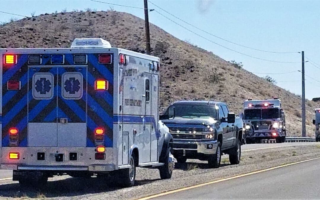
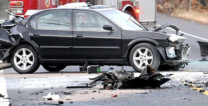
High Speed Accidents in El Paso, Texas: How Integrative Chiropractic Care at El Paso Back Clinic Helps Victims Heal
Excessive-speed accidents in El Paso, Texas, are high-impact collisions in which speed is the primary cause of the problem. These crashes often lead to serious injuries or even death. In 2025, speeding ranked as the leading cause of traffic accidents in the city, contributing to nearly 750 crashes. The good news is that El Paso is taking action with its Vision Zero plan, and victims can find real help through integrative chiropractic care at El Paso Back Clinic. This article takes you on a simple journey—from understanding the problem to finding lasting recovery.
What Exactly Are Excessive Speed Accidents?
Excessive-speed accidents occur when drivers go well above the posted limit or exceed the speed for the road conditions. In El Paso, this often happens on busy highways or city streets. These are not small bumps—they create powerful forces that damage cars and people.
The crashes usually look like this:
Rear-end hits, when a speeding car slams into the vehicle ahead.
T-bone crashes at intersections.
Rollovers when control is lost.
Hot spots in El Paso include the busy I-10 corridor, the area near Montana Avenue and McRae Boulevard, and roads close to the airport. Speed can quickly turn a normal drive into a dangerous one.
Why Speeding Is a Big Problem in El Paso Right Now
Speeding takes away reaction time and makes crashes much worse. In 2025, the city recorded its 32nd traffic death by mid-year, and speed was a leading factor in many of them. Even though some speeding tickets have dropped, local residents still see the danger on the roads every day.
Real stories show the pain. One deadly motorcycle crash on Montana Avenue involved high speed and a failure to yield. The rider did not survive. In another case, a teenager died in a high-speed single-car crash on Montana Avenue when his vehicle left the road and rolled over. These events remind everyone how quickly things can change.
Texas law is clear: drivers must stay at or below posted limits and slow down for weather, traffic, or construction (Texas Transportation Code § 545.352). Yet the problem continues, which is why El Paso is stepping up.
Dangerous Spots You Should Know About
Certain areas in El Paso see more speed-related crashes than others:
I-10 Corridor: Heavy truck traffic and fast lanes create risky conditions, especially near the airport exit.
Montana Avenue & McRae Blvd: Busy intersections and heavy traffic make this a high-crash zone.
Airport-Area Roads: Quick-access lanes and sudden turns increase danger.
Knowing these spots helps drivers stay alert and slow down.
The Serious Injuries Speed Causes
High-speed crashes often leave people with major injuries that affect daily life. Common problems include:
Whiplash from the sudden snap of the neck.
Traumatic brain injuries (TBIs) from head impact.
Internal injuries, broken bones, and torn muscles.
Pain, stiffness, headaches, or numbness may not show up right away. Without quick care, these issues can become long-term problems that make work and family time harder.
El Paso’s Vision Zero Plan Is Making Roads Safer
To fight these crashes, the city created the Vision Zero Action Plan. The goal is zero traffic deaths and serious injuries. The plan uses a “safe systems” approach—designing roads that protect people even when mistakes happen.
Here’s what the plan focuses on:
Lowering speeds through better road design, such as narrower lanes and rumble strips.
Adding brighter lights and clearer crosswalks.
Running education campaigns to remind everyone to slow down.
Creating safer paths for walkers and bike riders.
Speed control is the biggest tool in the plan. Cities that used it saw fewer serious crashes. El Paso is using grants and community ideas to build safer streets for everyone.
Your Recovery Journey Starts at El Paso Back Clinic
After a speed-related crash, the next step is healing. Integrative chiropractic care at El Paso Back Clinic offers a comprehensive, non-surgical approach to getting better. Led by Dr. Alexander Jimenez, DC, APRN, FNP-BC, the clinic combines traditional chiropractic with functional medicine, rehabilitation, and advanced therapies. Their large facilities in El Paso make care easy and effective for auto accident victims.
Dr. Jimenez has more than 25 years of experience treating crash injuries. His clinical observations show that high-speed accidents often cause hidden damage to the spine, nerves, and soft tissues. Symptoms can appear days later, so a full check-up is important. The clinic uses MRI scans, range-of-motion tests, and detailed exams to identify the exact problems early.
How Integrative Care Works at El Paso Back Clinic
The team at El Paso Back Clinic does not stop at one type of treatment. They create a full plan that helps the whole body heal. Services include:
Gentle spinal adjustments to fix misalignments caused by the crash.
Soft-tissue therapies such as massage and myofascial release help loosen tight muscles.
Spinal decompression to ease nerve pressure.
Targeted rehabilitation exercises to rebuild strength and balance.
Functional medicine support with nutrition advice to reduce inflammation.
This holistic approach helps patients recover faster without surgery or heavy pain pills. Many people return to work and normal activities sooner.
For whiplash, the clinic’s methods quickly reduce neck pain and headaches. Patients with back injuries or nerve issues often feel better mobility after just a few visits. Dr. Jimenez notes that early integrative care prevents chronic pain and long-term complications.
Getting the Right Paperwork for Your Claim
Healing is only half the battle. Victims also need solid proof for insurance companies or lawyers. El Paso Back Clinic provides clear, detailed documentation that helps personal injury claims succeed. Reports include:
Full medical records linking the crash to your injuries.
MRI results and range-of-motion studies.
Notes from Dr. Jimenez that explain how speed caused the damage.
This paperwork makes it easier to obtain fair payment for medical bills, lost wages, and pain. The clinic works smoothly with attorneys, so you can focus on getting well.
Real Benefits Patients Notice at the Clinic
People who choose El Paso Back Clinic often share these wins:
Faster relief from pain and stiffness.
Better movement and daily function.
Lower chance of ongoing problems.
Improved overall wellness through nutrition and stress management.
Personalized care that fits their exact injuries.
The clinic’s convenient locations and friendly team make the process simple. No long waits—just expert help when you need it most.
Simple Tips to Avoid Speeding Crashes
While recovery is available, prevention is still best. Slow down on I-10 and Montana Avenue. Watch for trucks and construction. Stay alert at every intersection. Support Vision Zero by speaking up for safer roads in your neighborhood.
Moving Forward After a Crash
Excessive-speed accidents in El Paso hurt many families each year, but help is available at El Paso Back Clinic. The city’s Vision Zero plan works to stop future tragedies, while the clinic’s integrative chiropractic care helps victims heal today.
If you or someone you love has been in a speed-related crash, do not wait. Visit El Paso Back Clinic at elpasobackclinic.com right away. Their team, led by Dr. Alexander Jimenez, offers the complete non-surgical care and documentation you need to get back on your feet. Recovery is possible, and safer roads are on the way—one careful choice at a time.
After an MVA: Delayed Injury Symptoms, Signs to Watch For, and the Role of Chiropractic Care
Imagine driving down the road on an ordinary day. Then, without warning, another car hits yours. The impact jars your body. Glass breaks. Metal bends. In the first moments, you check yourself and feel okay. You walk away from the scene thinking the worst is over. But a day or two later, a headache starts. Your neck feels stiff. Your back aches. These are delayed symptoms of injury after a car accident. They often appear because your body’s natural response hides the damage at first. This article walks you through what happens next, which signs matter most, and why quick care can stop small problems from becoming lifelong ones. You will see a clear path from the crash to full recovery.
Why Do Symptoms Show Up Later?
Right after a crash, your body releases a surge of adrenaline. This hormone kicks in to help you handle danger. It masks pain so you can move to safety. Shock also plays a role. Your mind and muscles stay tense at first. As the adrenaline fades and swelling begins, real problems surface. Inflammation builds slowly. Nerves get pressed. Soft tissues stretch or tear in ways you do not feel right away. Experts note that many injuries take hours or even days to cause noticeable pain (Burns Bryant, n.d.; South Atlanta Injury Lawyers, n.d.). The delay can fool people into thinking they are fine. But ignoring early clues can lead to worse trouble down the road.
Common Delayed Symptoms to Monitor
In the days after a crash, pay close attention to your body. Here are key signs that often appear later:
Persistent headaches: These can start mild and grow stronger. They may signal whiplash or a mild concussion. The sudden jolt to your head and neck strains muscles and irritates nerves (Chambers Medical, n.d.; Dr. Derek Day, n.d.).
Neck or back stiffness and soreness: Your head snaps forward and back in many crashes. This causes whiplash. Muscles tighten. Joints lose smooth movement. You might feel sore when turning your head or bending (South Atlanta Injury Lawyers, n.d.; Theneckandbackclinics, n.d.).
Numbness or tingling (pins and needles): A “pins and needles” feeling in your arms, hands, legs, or feet often means nerves are compressed. Swelling or a slight shift in your spine can pinch them (Burns Bryant, n.d.; McIntyre Law, n.d.).
Restricted movement: You find it hard to turn your neck or bend your back. Tight muscles and inflammation limit your range of motion. This protective response can become permanent if not addressed (Integrated Health & Injury Center, 2026).
Stomach pain or swelling: Pain in your belly, nausea, vomiting, or diarrhea can point to internal issues. Organs may bruise or bleed slowly (1800law1010, n.d.; Onmyside, n.d.).
Dizziness, confusion, or memory problems: Trouble with balance, forgetting recent events, or feeling “foggy” may indicate a concussion. The brain bounces inside the skull during impact (Chambers Medical, n.d.).
Mood changes: Sudden irritability, anxiety, or sadness can appear. The stress of the crash, plus brain or neck strain, affects emotions (Ruhmann Law Firm, n.d.; Total Vitality Medical, n.d.).
These symptoms do not always hit at once. They can creep in over several days.
Serious Injuries: These Signs May Reveal
Delayed symptoms are your body’s way of waving a red flag. They often point to bigger problems:
Whiplash and soft tissue injuries stretch or tear ligaments and muscles in the neck and back. Without care, scar tissue forms and movement stays limited (2Keller, n.d.).
Concussions or mild traumatic brain injuries change how your brain works. Headaches, dizziness, and memory loss are common clues (Chambers Medical, n.d.).
Spinal misalignment or disc problems press on nerves. This can cause ongoing pain, numbness, or weakness (McIntyre Law, n.d.; Smith & Hassler, n.d.).
Internal bleeding or organ injury may start small but grow dangerous. Abdominal pain is a key warning (1800law1010, n.d.).
Catching these early stops them from turning into chronic pain or permanent damage.
When to Seek Medical Attention Right Away
Do not wait if you notice any of these red-flag symptoms:
Dizziness or sudden loss of balance
Numbness in arms or legs
Memory loss or confusion
Extreme pain that keeps getting worse
Vomiting or severe stomach pain
Blurred vision or ringing in the ears
These signs mean you could have a concussion, spinal injury, or internal bleeding. Get checked immediately. A doctor can run scans and rule out life-threatening issues. Early action protects your long-term health (Plw.law, n.d.; Lorfing Law, n.d.).
How Integrative Chiropractic Clinics Offer Complete, Non-Invasive Help
Once serious issues are ruled out, many people turn to integrative chiropractic clinics for full recovery. These clinics combine gentle chiropractic adjustments with other natural therapies. The goal is simple: restore proper alignment, calm inflammation, improve movement, and prevent chronic problems.
Chiropractors use targeted adjustments to realign the spine. This takes pressure off nerves and lets the body heal naturally. Soft-tissue work eases tight muscles. Rehab exercises strengthen weak areas. Patients often feel better without relying on pain pills or surgery (Tarpon Total Healthcare, n.d.; Stumpff Chiro, n.d.).
Dr. Alexander Jimenez, DC, APRN, FNP-BC, brings a special integrative approach to car accident care. Practicing in El Paso, Texas, he combines chiropractic adjustments with functional medicine and advanced diagnostics. His clinical observations show that many patients develop delayed symptoms like neck stiffness, headaches, numbness, and back pain days or weeks after a crash. He notes that adrenaline initially hides the damage, but swelling and misalignment soon create ongoing issues. Dr. Jimenez stresses early evaluation. His non-invasive methods focus on spinal realignment, reducing inflammation, and supporting the body’s natural healing. Patient stories from his clinic highlight full recoveries from whiplash and soft-tissue injuries when care starts promptly (Jimenez, n.d.; Injury Medical Clinic, n.d.).
Integrative care also helps with documentation for insurance claims. Detailed records of your injuries and progress strengthen your case if needed. The journey feels supportive—each visit builds on the last until you move freely again.
Your Clear Path to Recovery
The road after a car accident need not be confusing. Start by listening to your body in the first few days. Note any new aches, even small ones. Get a medical check if red flags appear. Then consider an integrative chiropractic clinic for gentle, drug-free support. Clinics like those led by experts such as Dr. Alexander Jimenez offer comprehensive care that addresses the root cause rather than just masking symptoms. Alignment improves. Inflammation drops. Range of motion returns. Chronic pain stays away.
Many people who follow this path regain their active lives faster. They avoid long-term stiffness or headaches that steal joy from daily activities. The key is simple: do not ignore what your body tells you later.
Take that first step today. A quick exam can give you peace of mind and set you on the road to full healing. Your future self will thank you for acting early.
T-Bone Crashes from Left Turn Mistakes: Recovery at El Paso Back Clinic in Texas
Left turns at busy intersections or median openings seem simple, but they cause many serious crashes on Texas roads. One common type of accident occurs when a driver tries to turn left without waiting for clear traffic. This mistake lets another car slam into the side of the turning vehicle. People call this a “Failure to Yield Left Turn” accident. It usually ends in a “T-Bone” or side-impact crash because the front of the oncoming car hits the side of the car that is sticking out into the traffic lane.
These crashes bring pain, injuries, and stress for drivers and passengers in El Paso and across Texas. This article explains the type of accident, why it happens so often, who is usually at fault, and the common injuries. It also shows how El Paso Back Clinic uses a whole-person, noninvasive approach to help people recover from Failure to Yield Left-Turn (T-bone) accidents. The clinic’s main goals are to ease acute pain, reduce inflammation, and restore long-term mobility, enabling patients to return to daily life more quickly.
What Is a Failure to Yield Left Turn Accident?
A Failure to Yield Left Turn accident occurs when a driver making a left turn does not give the right of way to oncoming traffic. The turning car ends up partially in the path of straight-moving vehicles. This leads to a side-impact collision, often called a T-Bone crash. The name comes from the “T” shape the two cars form at the moment of impact. One car’s front hits the other car’s side.
Police and insurance experts use a few key terms to describe this situation:
Failure to Yield Right of Way: The driver making the turn broke the law by failing to wait until the path was completely clear.
T-Bone or Side-Impact Collision: This happens when the front of an oncoming car strikes the side of the turning car.
“Sticking Out” Accident: A common phrase for when a car does not fully clear the intersection or median opening and blocks active traffic lanes.
Improper Lane Usage / Positioning: This technical violation occurs when a driver does not line up properly in the median gap, also known as a “median break” or “crossover.”
These crashes are dangerous because the sides of cars have less protection than the front or back. A small mistake during a left turn can turn into a high-impact event, especially on busy El Paso roads.
Why These Accidents Happen So Often
Left turns require drivers to cross paths with oncoming cars, judge speed and distance, and find a safe gap in traffic. Many factors make this hard. Drivers often misjudge how fast an oncoming car is moving or how much space they need to complete the turn safely.
Common reasons for these mistakes include:
Inability to accurately judge the distance and speed of incoming vehicles.
Being in a hurry and rushing through the turn instead of waiting for a full clear path.
Not pulling far enough into the median area, which leaves the car “sticking out” into traffic.
Distractions like phones, passengers, or navigation systems that take attention away from the road.
Poor visibility from weather, parked cars, or heavy traffic that hides oncoming vehicles.
Safety experts note that left turns are among the riskiest moves because they cross opposing traffic lanes. Even at low speeds, a miscalculation can lead to a sudden crash on Texas highways or city streets.
Who Is Almost Always at Fault?
In most cases, the driver making the left turn is at fault. Traffic laws require that driver to wait until the intersection or median gap is completely clear before turning. The oncoming car usually has the right of way.
Legal resources explain that failure to yield is the main cause. The turning driver must give way to vehicles already in the intersection or approaching closely enough to create a hazard. If the turning driver misjudges speed, fails to yield to an oncoming vehicle, or does not position the car correctly, they break the rules and cause the crash.
Fault can sometimes be shared if the oncoming driver was speeding or distracted, but the left-turning driver bears the primary responsibility in most of these incidents. Evidence such as police reports, traffic camera footage, and witness statements helps insurance companies and courts determine responsibility.
Summary of Dangerous Turning Situations
Several common scenarios lead to these crashes. Here are the main ones:
Pulling out when the front end sticks out: This creates a Failure to Yield / T-Bone situation.
Turning before the median gap is clear: Known as an improper median crossover turn.
Making a left turn the wrong way: This includes turning without checking for oncoming traffic or ignoring yield signs.
These situations often happen at busy intersections, driveways, or parking lot exits in El Paso. They can involve cars, trucks, or even motorcycles, which are harder to see.
Common Injuries from T-Bone and Side-Impact Crashes
The sudden side hit in a T-Bone crash throws the body sideways. This causes injuries that differ from those in front-end collisions. The impact often causes lateral whiplash, in which the neck and spine twist sharply. Soft-tissue injuries, muscle strains, and spinal misalignments are very common.
Typical injuries include:
Neck and back pain from whiplash and disc issues.
Shoulder injuries, such as rotator cuff strains from bracing against the wheel.
Hip and pelvic problems from hitting the door or console.
Headaches, numbness in the arms or legs, and reduced mobility.
Bruising, swelling, and inflammation in muscles and ligaments.
Symptoms may not show up right away. Some people feel fine at first but develop pain, stiffness, or tingling hours or days later. Prompt care is important to prevent long-term problems.
How El Paso Back Clinic Helps After a Failure to Yield Accident
El Paso Back Clinic takes a whole-person, non-invasive approach to treating injuries from these crashes. Located in El Paso, Texas, the clinic provides local drivers with advanced rehabilitation for auto accident injuries. Instead of focusing on a single symptom, the team looks at the whole body. The main goals are to ease acute pain, reduce inflammation, and restore long-term mobility.
Chiropractic care works well for T-Bone injuries because it addresses the direct contact that causes lateral whiplash and misalignment. A typical treatment plan at El Paso Back Clinic includes:
Spinal adjustments to realign the spine and improve joint movement.
Physical therapy exercises to rebuild strength and coordination.
Massage therapy to relax tight muscles and improve blood flow.
Functional rehabilitation to help patients move safely again.
Spinal decompression and electro-acupuncture for deeper relief.
These methods help without surgery or heavy medication. They target soft tissue injuries and nerve irritation that often follow side-impact crashes. The clinic also offers functional medicine to address inflammation, nutrition, and lifestyle factors that affect healing.
Dr. Alex Jimenez, DC, APRN, FNP-BC, leads the care at El Paso Back Clinic. With dual licenses as a chiropractor and family nurse practitioner, he brings over 30 years of experience in personal injury and auto accident recovery. His clinical observations show that many patients from side-impact crashes have hidden neck misalignments that cause headaches, brain fog, and ongoing pain. He combines chiropractic adjustments with functional medicine, advanced imaging for clear diagnosis, and detailed records to support both healing and any legal needs. Dr. Jimenez stresses early intervention so patients reach Maximum Medical Improvement (MMI) faster and avoid chronic issues.
The clinic’s multidisciplinary team includes physical therapists and advanced trainers at facilities like Just Play Fitness. Patients receive personalized rehab programs that include strength training, flexibility exercises, and nutritional support. This full-body approach helps restore balance and function. Many El Paso patients report reduced pain and improved mobility after a few sessions at the East Side, Central, or Northeast locations.
Reaching Maximum Medical Improvement Quickly
Maximum Medical Improvement (MMI) is the point when a patient’s condition has improved as much as it can with current treatment. El Paso Back Clinic helps people get there sooner by treating the whole body. Early chiropractic care reduces inflammation, prevents scar tissue buildup, and retrains muscles to work properly.
Clinic reports indicate that combining adjustments, massage, exercise, and functional medicine leads to faster recovery from whiplash and soft-tissue injuries. Patients return to work and normal activities with less pain and fewer long-term problems.
Conclusion
Failure to yield at left turns is a common but preventable cause of accidents with careful driving and patience at intersections. Understanding terms like T-Bone collision, “sticking out” accident, and improper positioning helps drivers stay alert on El Paso roads. When these crashes do happen, the left-turning driver is usually responsible because of the legal duty to yield.
The good news is that injuries from these side-impact crashes do not have to define the future. El Paso Back Clinic offers safe, effective relief right here in Texas. The clinic focuses on full-body healing through spinal adjustments, therapy, rehabilitation, and functional medicine. This non-invasive care eases pain, reduces inflammation, and restores mobility, helping patients reach Maximum Medical Improvement and enjoy life again.
Safe driving starts with respect for left turns. If you or someone you know has been in a Failure to Yield Left Turn accident in El Paso, seek medical attention right away at El Paso Back Clinic. Proper care can make all the difference in recovery. Call 915-850-0900 or visit elpasobackclinic.com to start healing today.
How to Prove Car Accident Injuries in El Paso: Expert Medical Documentation at El Paso Back Clinic
Car crashes happen fast, but the pain can last for weeks or months. Many people in El Paso feel stiff or sore right after a wreck. Others notice problems days later. Insurance companies often push back and say your injuries are old problems or not related to the crash at all. The good news? You can build a rock-solid case with quick action and smart record-keeping. Getting medical help fast and keeping detailed notes creates a clear link between the accident and your injuries. This helps you heal and get fair payment for your bills, lost work, and pain.
This guide walks you through simple steps to prove your car accident injuries. You will see why seeing a doctor within 72 hours matters, how to build a strong paper trail, and why El Paso Back Clinic offers the best integrated care in El Paso to support your recovery and your claim.
Why Seek Immediate Medical Attention Within 72 Hours
The clock starts right after the crash. Medical professionals agree that you should seek a check-up within 72 hours. This quick step shows a direct connection between the accident and your injuries.
Waiting longer gives insurance adjusters a chance to claim your pain comes from something else. Early visits create official records that tie your symptoms straight to the wreck. Soft-tissue injuries like whiplash or back strain often feel mild at first but worsen over time. Even if you think you are okay, hidden damage can show up later.
Emergency room or clinic notes from the first few days become powerful proof.
Doctors can order X-rays or MRIs to catch problems early.
Starting treatment right away helps you heal faster and keeps your medical story clear.
Prompt care stops insurers from calling your injuries “pre-existing.” (Greater Texas Orthopaedics, 2025; Georgia Spine and Orthopaedics, n.d.)
Building a Detailed Paper Trail: Records, Photos, and Your Daily Journal
One doctor visit is not enough. You need a complete paper trail that shows exactly what happened to your body after the crash. Save every medical record: doctor notes, bills, prescriptions, and test results like X-rays and MRIs.
Take clear photos of bruises, cuts, and swelling as soon as possible. Snap pictures from different angles in bright light and update them as things change. These images are hard for anyone to argue against.
Stick to your full treatment plan and never skip appointments. Gaps in care can make it look like your pain is not serious or not crash-related. Keep receipts and notes about missed work or daily activities, too.
Your daily pain journal is one of the strongest tools you have. Write simple notes each day about how you feel. This personal record proves the real impact of your injuries over time and helps show pain and suffering.
Include these details every day in your journal:
Pain level on a scale of 1 to 10.
Where the pain is and what makes it better or worse.
How the injury limits walking, sitting, driving, sleeping, or working.
Emotional feelings like worry, sadness, or trouble focusing.
Any missed work, family time, or normal activities.
Consistent notes like these make it much harder for insurance companies to say your injuries are unrelated. (Reno Law Firm, n.d.; Darrell Castle Law, n.d.; Texas Injury Accident Lawyers, n.d.)
Why El Paso Back Clinic Delivers the Best Integrated Care for Accident Injuries
Not every injury shows up on a quick emergency room visit. Many people leave the ER with no broken bones but still have real pain from whiplash, muscle strains, or joint problems. El Paso Back Clinic, led by Dr. Alex Jimenez, DC, APRN, FNP-BC, CFMP, IFMCP, provides comprehensive care and the detailed records you need for your claim.
This El Paso clinic is part of the larger Injury Medical Clinic PA and offers a full multidisciplinary team right here in town. They specialize in auto accident care, whiplash, soft-tissue injuries, back pain, neck pain, and personal injury cases. The clinic blends chiropractic adjustments, advanced nursing, functional medicine, physical therapy, and rehabilitation in one place.
Dr. Alex Jimenez brings more than 25 years of experience as both a chiropractor and a board-certified Family Nurse Practitioner. He and his team provide prompt evaluations, advanced diagnostics, and personalized treatment plans that clearly link your injuries to the crash. Their approach includes digital motion X-rays, nerve tests, MRIs, and functional assessments to spot root causes that regular doctors might miss.
At El Paso Back Clinic, you get:
Immediate comprehensive exams and treatment plans that document the accident connection.
Chiropractic care focused on soft-tissue injuries and spinal alignment that emergency rooms often overlook.
APRN/FNP-BC support for pain management, functional testing, and full-body rehab.
Functional medicine tools that look at how the crash affects inflammation, energy levels, and overall health.
The clinic’s detailed records and progress notes help prove your injuries are new and accident-related. Patients in El Paso often share stories of faster healing and stronger claims due to clear documentation and coordinated care. Whether your crash caused whiplash, herniated discs, sciatica, or chronic pain, the team at El Paso Back Clinic creates the objective evidence insurers and courts respect. (Jimenez, n.d.; El Paso Back Clinic, n.d.)
How Strong Documentation Proves Causation in Your Claim
Causation simply means showing that the car accident caused your injuries. Good records and expert care make this link obvious. Insurance companies and courts want clear timelines, consistent symptoms, and professional notes.
Diagnostic images show new disc problems or swelling that started after the crash. The doctor reports tracking your condition from day one. Your pain journal captures the daily reality that no scan can.
When your case moves to settlement talks or court, these records become key evidence. They help calculate medical costs, lost wages, and fair payment for pain and suffering. Notes from a specialized clinic, such as El Paso Back Clinic, hold significant value because of their focus on soft-tissue injuries commonly encountered in accidents.
Common problems insurers raise include:
Claims that injuries are from aging or old sports issues.
Arguments that you waited too long to get help.
Questions about how bad the pain really is.
Your complete paper trail and El Paso Back Clinic records answer every doubt with facts. (Pendas Law, n.d.; Mitl Law, n.d.; PFFP Law, n.d.; Edwards Injury Law, n.d.)
Extra Tips to Make Your Motor Vehicle Accident Claim Stronger
Stay consistent with every part of your care. Go to every follow-up visit and report any new symptoms right away.
Share your journal notes with your doctor so they become part of your official file.
Ask for copies of every report, image, and treatment plan. Keep everything organized in one folder or on your phone.
If the injury changed your job or daily life, get a note from your employer regarding time missed. This adds another layer of proof.
Choosing El Paso Back Clinic early often means faster healing plus the strongest possible support for your legal case.
Take the Next Step: Protect Your Health and Your Claim at El Paso Back Clinic
Proving car accident injuries does not have to be hard. Start with medical care within 72 hours. Build a solid paper trail with records, photos, and a daily journal. Then turn to El Paso Back Clinic for expert integrated care that combines chiropractic, nursing, and functional medicine.
Dr. Alex Jimenez and the team at El Paso Back Clinic have helped countless El Paso residents recover from whiplash, back pain, and more while creating the documentation needed to win fair settlements. Their modern facilities, advanced diagnostics, and whole-person approach set them apart.
Do not wait. Your health and your case both improve when you act from day one. Call El Paso Back Clinic today at 915-850-0900 or visit https://elpasobackclinic.com/ to schedule your evaluation. Get the care you need and the proof your claim deserves.
IFM's Find A Practitioner tool is the largest referral network in Functional Medicine, created to help patients locate Functional Medicine practitioners anywhere in the world. IFM Certified Practitioners are listed first in the search results, given their extensive education in Functional Medicine