El Paso Back Clinic®: Your Path to Wellness and Recovery
At El Paso Back Clinic®, we believe in empowering our patients to live pain-free, active lives through comprehensive chiropractic care and integrative medicine. Led by Dr. Alex Jimenez, DC, APRN, FNP-BC, our clinic in El Paso, TX, specializes in treating a wide range of injuries, from motor vehicle accidents (MVAs) to sports mishaps, with a focus on restoring mobility, flexibility, and overall wellness. By addressing posture, spinal health, and musculoskeletal issues, we help patients recover from injuries, manage pain, and prevent future complications. This article explores how our holistic approach, combining chiropractic care, nutrition, and advanced therapies, transforms lives at El Paso Back Clinic®.
The Role of Posture in Wellness and Recovery
Good posture is the foundation of a healthy body, especially when recovering from injuries or managing chronic pain. Poor posture, often caused by accidents or prolonged sitting, can lead to muscle strain, joint stress, and increased discomfort. At El Paso Back Clinic®, we prioritize correcting posture to improve spinal alignment, which enhances mobility and reduces the risk of further injury. Proper posture allows the body to distribute weight evenly, supporting natural healing and optimal function (Optimal Spine Chiro, 2023).
Our chiropractic adjustments focus on restoring spinal alignment, relieving pressure on nerves, and improving overall body mechanics. This is particularly important for patients recovering from MVAs or sports injuries, where misalignments can worsen pain or delay recovery. By addressing posture, we help patients move better, feel better, and stay active (Zaker Chiropractic, 2023).
Dr. Alex Jimenez: Leading Wellness Care in El Paso
Dr. Alex Jimenez, a chiropractor and nurse practitioner, brings a unique combination of medical and chiropractic expertise to El Paso Back Clinic®. With years of experience treating severe pain, sciatica, whiplash, and sports injuries, Dr. Jimenez uses advanced diagnostics and integrative therapies to create personalized treatment plans. His dual training allows him to address both the structural and medical aspects of injuries, ensuring comprehensive care (Jimenez, 2025).
Advanced Diagnostics for Precise Treatment
At El Paso Back Clinic®, we use state-of-the-art diagnostic tools, such as X-rays and MRIs, to identify the root causes of pain and injury. Dr. Jimenez employs techniques like motion palpation and static palpation to assess spinal and joint function, ensuring treatments are tailored to each patient’s needs. This approach is critical for conditions like whiplash or spinal misalignments caused by MVAs, where precise diagnosis leads to faster recovery (El Paso Back Clinic, 2023).
Supporting Legal and Medical Needs
In personal injury cases, such as those from car accidents, Dr. Jimenez provides detailed medical reports to support insurance claims or legal proceedings. His ability to navigate both medical and legal aspects ensures patients can focus on healing while their cases are handled effectively. This expertise is especially valuable for injuries like back pain or joint dislocations, where documentation is key (Personal Injury Doctor, 2017).
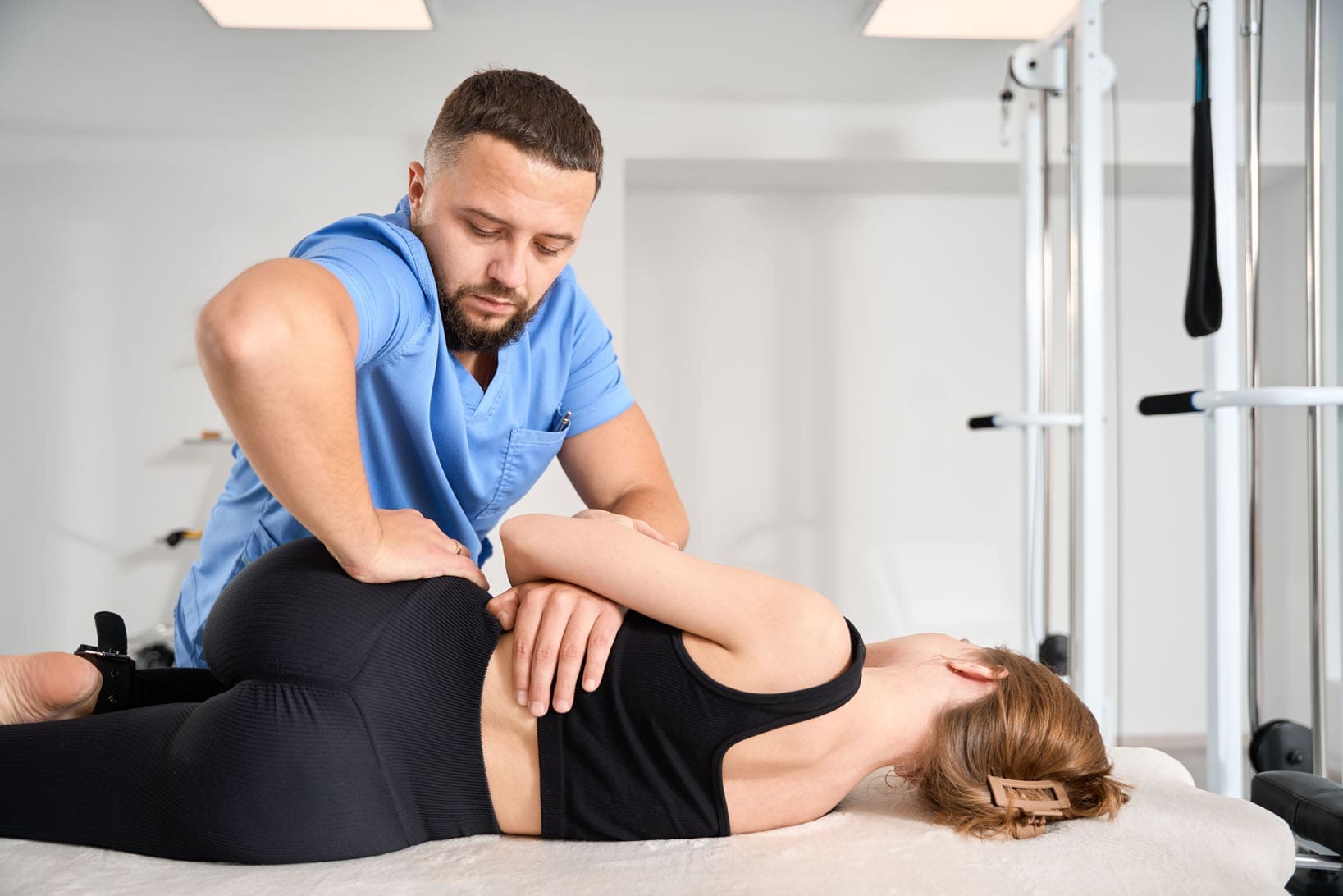
El Paso Back Clinic® specializes in treating musculoskeletal injuries caused by MVAs, workplace accidents, sports, and daily activities. Our non-invasive, drug-free approach focuses on restoring function and relieving pain through chiropractic care and integrative therapies.
Whiplash and Neck Pain
Whiplash, common in car accidents, results from sudden neck jolts that strain muscles and misalign the spine. Our clinic uses gentle spinal adjustments, trigger point therapy, and corrective exercises to reduce inflammation and restore mobility. These treatments help patients recover quickly and avoid chronic neck pain (El Paso Back Clinic, 2023).
Back Pain and Spinal Injuries
Back injuries, such as herniated discs or spinal misalignments, are frequent after MVAs or heavy lifting. Dr. Jimenez employs spinal manipulation and decompression techniques to alleviate pain and promote healing. We also incorporate mobility exercises to strengthen supporting muscles, ensuring long-term spinal health (El Paso Back Clinic, 2023).
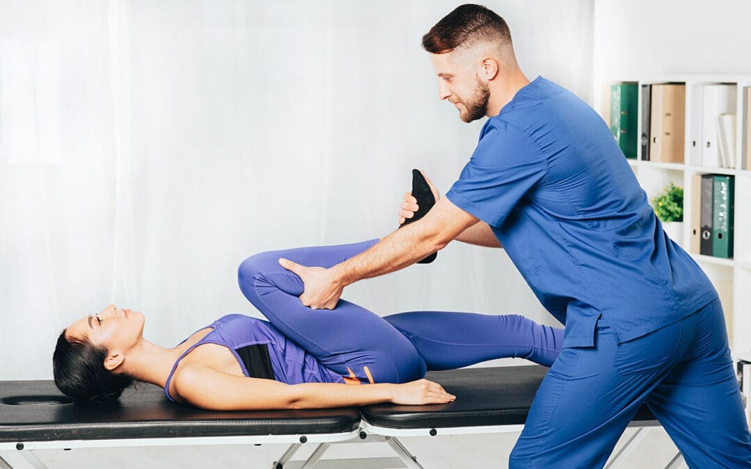
Sports and Joint Injuries
Athletes trust El Paso Back Clinic® for recovery from injuries like ACL tears, sprains, or joint dislocations. Our integrative approach combines chiropractic adjustments with physical therapy and nutritional guidance to restore strength and enhance performance. By improving posture and alignment, we help athletes return to their sport stronger than before (Square One Health, 2023).
Our clinic’s strength lies in combining chiropractic care with integrative therapies to address the whole person, not just the injury. This holistic approach promotes natural healing, reduces pain, and prevents long-term complications.
Targeted Exercise and Physical Therapy
Customized exercise programs are central to our treatment plans. These exercises strengthen muscles, improve flexibility, and support spinal health, helping patients recover from injuries like fractures or sprains. For example, after an 18-wheeler accident, we design specific stretches to restore mobility without aggravating spinal trauma (El Paso Back Clinic, 2023).
Massage Therapy and Acupuncture
Massage therapy relieves muscle tension and boosts circulation, aiding recovery from soft tissue injuries. Acupuncture reduces inflammation and stimulates natural pain relief, complementing chiropractic care. These therapies work together to provide comprehensive pain relief and support healing (Mountain Movement Center, 2023).
Nutritional Support for Healing
Nutrition is a key component of recovery at El Paso Back Clinic®. Dr. Jimenez provides dietary recommendations, such as anti-inflammatory foods and supplements, to support tissue repair and reduce pain. This approach strengthens the body’s ability to heal after MVAs or sports injuries, promoting long-term wellness (El Paso Back Clinic, 2023).
Preventing Long-Term Complications with Integrative Care
Untreated injuries can lead to chronic pain, reduced mobility, or conditions like degenerative arthritis. At El Paso Back Clinic®, our integrative approach prevents these issues by addressing the root causes of pain and injury. Regular chiropractic adjustments maintain spinal alignment, while therapies like acupuncture and exercise reduce inflammation and strengthen the body. This comprehensive care ensures patients recover fully and maintain optimal health (Current Physical Therapy, 2025).
For athletes, this means returning to their sport with improved performance and less risk of re-injury. For accident victims, it means reclaiming their daily lives without pain. Our focus on wellness empowers patients to live active, healthy lives long after their treatment ends (Tigard Chiropractic Auto Injury, 2023).
El Paso Back Clinic®, under the leadership of Dr. Alex Jimenez, is dedicated to helping patients achieve wellness through chiropractic care and integrative medicine. By focusing on posture, spinal health, and holistic therapies, we treat injuries from MVAs, sports, and daily life, while preventing long-term complications. Whether you’re recovering from whiplash, back pain, or a sports injury, our personalized approach ensures you return to a pain-free, active life. Visit El Paso Back Clinic® to start your journey to optimal health today.
Find out the benefits of carpal tunnel syndrome by incorporating chiropractic care for reducing pain and enhancing wrist health and mobility.
Contents
Chiropractic Care for Carpal Tunnel Syndrome: A Comprehensive Guide to Relief and Recovery
Imagine trying to text your best friend, but your fingers feel like they’re auditioning for a role as pins and needles in a sci-fi flick. Or maybe you’re gripping your coffee mug, only to feel it slip because your hand decided it’s on strike. If this sounds familiar, you might be dealing with carpal tunnel syndrome (CTS), a condition that can turn your hands into rebellious coworkers who refuse to do their job. But don’t worry—there’s hope, and it doesn’t involve bribing your hands with tiny massages (though that might sound nice). Chiropractic care, especially from experts like Dr. Alexander Jimenez, DC, APRN, FNP-BC in El Paso, Texas, offers a non-invasive, holistic way to tackle CTS and its pesky symptoms like numbness, tingling, and pain.
In this comprehensive guide, we’ll dive deep into what carpal tunnel syndrome is, why it happens, and how chiropractic care can help you wave goodbye to those annoying symptoms. We’ll explore the connection between your hands, upper extremities, and cervical spine, uncover the risk factors that make CTS more likely, and share practical tips for small lifestyle changes to keep your hands happy. Plus, we’ll highlight Dr. Jimenez’s unique role in helping personal injury victims in El Paso, using advanced imaging, diagnostic evaluations, and dual-scope procedures to bridge medical care and legal support. So, grab a comfy seat (and maybe rest those wrists), because we’re about to unpack everything you need to know to get your hands back in the game!
What Is Carpal Tunnel Syndrome?
Carpal tunnel syndrome is like a traffic jam in your wrist, where the median nerve gets squeezed tighter than a packed elevator. This nerve, which runs from your forearm to your hand through a narrow passage called the carpal tunnel, controls sensation and movement in your thumb and first three fingers (sorry, pinky, you’re not invited to this party). When the median nerve gets compressed by swollen tendons, inflammation, or other factors, you can experience pain, numbness, tingling, or weakness in your hand and wrist. These symptoms can make everyday tasks—like typing, holding a phone, or even opening a jar—feel like you’re trying to solve a Rubik’s Cube with mittens on.
CTS is a progressive condition, meaning it can worsen over time if left untreated, potentially leading to permanent nerve damage or muscle weakness. Symptoms often start intermittently, popping up during activities like driving or scrolling through your phone, and may worsen at night, waking you up with that “hand fell asleep” sensation. In severe cases, you might struggle with fine motor skills, like buttoning a shirt or picking up small objects, as the median nerve’s signals get scrambled.
Dr. Alexander Jimenez, a chiropractor and board-certified nurse practitioner in El Paso, explains that CTS isn’t just a wrist problem—it can have roots in other parts of your body, like the cervical spine, which is why a holistic approach is key. His clinic, Injury Medical & Chiropractic Clinic, uses advanced diagnostics to pinpoint the cause of your symptoms and create a tailored plan to help you achieve pain-free living.
CTS doesn’t just show up out of nowhere—it’s like a party crasher invited by a mix of factors. Understanding these risk factors can help you spot potential triggers and take steps to prevent or manage the condition. Here are the main culprits that can increase your chances of developing CTS:
Repetitive Hand and Wrist Movements: Jobs or hobbies that involve repetitive motions—like typing, assembly line work, or playing an instrument—can irritate the tendons in your wrist, leading to inflammation and nerve compression. Think of it as your wrist saying, “I’m tired of this repetitive playlist!”
Anatomic Factors: Some people have smaller carpal tunnels (thanks, genetics!), which can make the median nerve more prone to compression. Wrist fractures or arthritis that deform the small bones in the wrist can also crowd the tunnel, like too many guests squeezing into a tiny room.
Body Fluid Changes: Fluid retention, common during pregnancy or menopause, can increase pressure in the carpal tunnel, irritating the median nerve. The good news? CTS related to pregnancy often improves after delivery.
Chronic Health Conditions: Conditions like diabetes, rheumatoid arthritis, or thyroid disorders can increase the risk of nerve damage or inflammation, making CTS more likely. It’s like these conditions are sending extra stress to your wrist’s already busy highway.
Obesity: Carrying extra weight can put pressure on the median nerve, increasing the risk of CTS. Maintaining a healthy weight can help keep that tunnel clear.
Workplace Factors: Jobs requiring prolonged wrist flexion, vibrating tools, or cold environments can aggravate the median nerve. If your job has you jackhammering in a freezer, your wrists might not be thrilled.
Gender: Women are more likely to develop CTS, possibly due to smaller carpal tunnels or hormonal factors. Sorry, ladies, it’s like your wrists drew the short straw.
Cervical Spine Issues: Here’s where things get interesting—problems in your neck, like misaligned vertebrae or herniated discs, can contribute to CTS symptoms by affecting the nerves that travel to your hand. This is called the “double crush” hypothesis, where nerve irritation in the neck compounds wrist compression, making symptoms worse.
Dr. Jimenez emphasizes that these risk factors often overlap, creating a perfect storm for CTS. For example, a typist with a misaligned cervical spine and a history of diabetes might be dealing with multiple nerve stressors. His dual-scope approach—combining chiropractic and medical evaluations—helps identify and address these overlapping risks for better outcomes.
The Role of the Hands, Upper Extremities, and Cervical Spine
Your hands and wrists don’t work in isolation—they’re part of a complex network that includes your upper extremities (arms, elbows, shoulders) and cervical spine (neck). Think of your body as a busy orchestra, with the median nerve as a key musician. If the conductor (your cervical spine) is offbeat, it can mess up the whole performance, leading to symptoms in your hands.
The Median Nerve’s Journey
The median nerve starts in the cervical spine, specifically from nerve roots in the C6-T1 region of your neck. It travels through your shoulder, elbow, and forearm before squeezing through the carpal tunnel in your wrist. If there’s a pinch or irritation anywhere along this path—like a misaligned vertebra in the neck or tight muscles in the forearm—it can amplify CTS symptoms. This is where the “double crush” hypothesis comes in: compression at the wrist (carpal tunnel) combined with irritation in the neck can make numbness and tingling worse than either issue alone.
The Cervical Spine Connection
Your cervical spine is like the control center for nerve signals to your arms and hands. Misalignments (subluxations) or herniated discs in the neck can irritate the nerve roots that feed into the median nerve, causing symptoms that mimic or worsen CTS. For example, a pinched nerve in the neck might make your fingers tingle, even if your wrist isn’t the main culprit. Dr. Jimenez often sees patients who think they have CTS but are actually dealing with cervical spine issues—or a combination of both.
Upper Extremities and Posture
Poor posture, like slouching at your desk or hunching over your phone, can strain the muscles and nerves in your shoulders, elbows, and wrists. This can lead to tightness or inflammation that compresses the median nerve. Repetitive motions, like typing or using a mouse, can also overwork the muscles and tendons in your forearm, adding to the pressure in the carpal tunnel.
Dr. Jimenez’s approach looks at the whole chain—neck, shoulders, elbows, and wrists—to find the root cause of your symptoms. By addressing misalignments in the cervical spine and tension in the upper extremities, he can reduce nerve irritation and help your hands feel like they’re back on the team.
How Chiropractic Care Helps Carpal Tunnel Syndrome
Chiropractic care is like a superhero swooping in to save your wrists from the villainous grip of CTS. Instead of relying on surgery or medications, chiropractors use non-invasive techniques to relieve nerve compression, reduce inflammation, and restore function. Here’s how chiropractic care, particularly from Dr. Jimenez, can help:
Spinal and Joint Adjustments: Chiropractors use gentle, targeted adjustments to realign the cervical spine, elbow, and wrist. By correcting misalignments, they reduce pressure on the median nerve and improve nerve signaling. A 1994 case study showed that chiropractic adjustments to the cervical spine and wrist led to significant improvements in grip strength and symptom relief in a patient with CTS.
Soft Tissue Therapy: Tight muscles or inflamed tendons in the forearm can contribute to CTS. Dr. Jimenez uses techniques like massage or myofascial release to loosen these tissues, reducing pressure in the carpal tunnel. This is like giving your wrist a much-needed stretch after a long day of typing.
Wrist Supports and Bracing: Chiropractors may recommend nocturnal wrist splints to keep your wrist in a neutral position, preventing further compression of the median nerve during sleep. Studies have shown that splinting, combined with chiropractic care, can improve symptoms without the need for surgery.
Ultrasound Therapy: This modality uses sound waves to reduce inflammation and promote healing in the wrist. A 1998 study found ultrasound therapy effective for CTS, and Dr. Jimenez incorporates it into his treatment plans when appropriate.
Exercise and Rehabilitation: Dr. Jimenez prescribes specific exercises to strengthen the muscles around the wrist and improve flexibility. Nerve and tendon gliding exercises, for example, can reduce symptoms and improve hand function.
Functional Medicine Approach: As a board-certified nurse practitioner, Dr. Jimenez looks beyond the wrist to address systemic factors like inflammation or hormonal imbalances that may worsen CTS. Nutritional counseling, for instance, can reduce inflammation through dietary changes, supporting recovery.
Addressing the Double Crush Phenomenon: By treating both the wrist and cervical spine, chiropractic care tackles the “double crush” issue, where nerve irritation in the neck amplifies wrist symptoms. This comprehensive approach can lead to better outcomes than focusing on the wrist alone.
A randomized clinical trial found that chiropractic care, including spinal and extremity adjustments, was as effective as conservative medical treatments (like ibuprofen and splinting) for CTS associated with median nerve demyelination. Dr. Jimenez’s dual expertise as a chiropractor and nurse practitioner allows him to combine these techniques with advanced diagnostics, ensuring a personalized plan that addresses all contributing factors.
Discovering The Benefits of Chiropractic Care- Video
Overlapping Risk Profiles and Chiropractic Solutions
CTS doesn’t exist in a vacuum—it’s often tangled up with other musculoskeletal issues that create overlapping risk profiles. These include cervical spine misalignments, poor posture, repetitive strain injuries, and systemic conditions like diabetes or obesity. Chiropractic care is uniquely positioned to address these interconnected problems, reducing the overall burden on your nervous system and musculoskeletal health.
Cervical Spine Misalignments
As mentioned, the “double crush” hypothesis suggests that nerve irritation in the neck can worsen CTS symptoms. A 2008 review highlighted how chiropractic adjustments to the cervical spine can relieve nerve compression, reducing symptoms in the hands. Dr. Jimenez uses advanced imaging, like X-rays or MRIs, to identify subluxations or herniated discs in the neck that might be contributing to your symptoms.
Posture and Ergonomics
Slouching or forward head posture can strain the nerves and muscles from your neck to your hands, increasing CTS risk. Chiropractic care corrects spinal alignment and teaches ergonomic habits to reduce strain. For example, Dr. Jimenez might recommend adjusting your workstation to keep your wrists neutral and your shoulders relaxed.
Repetitive Strain Injuries
Repetitive motions, like typing or using vibrating tools, can cause inflammation in the wrist and forearm, exacerbating CTS. Chiropractic adjustments, combined with soft tissue therapy and exercises, can reduce inflammation and restore mobility. A 2013 study found that manual therapy, including chiropractic techniques, improved CTS symptoms.
Systemic Conditions
Conditions like diabetes or obesity can increase inflammation and nerve sensitivity, making CTS worse. Dr. Jimenez’s functional medicine approach includes nutritional counseling to reduce inflammation and manage blood sugar, addressing these underlying factors.
By tackling these overlapping risks, chiropractic care not only relieves CTS symptoms but also improves overall musculoskeletal health, preventing future issues. Dr. Jimenez’s clinic uses a holistic framework, combining adjustments, therapy, and lifestyle changes to create lasting relief.
Dr. Alexander Jimenez: A Leader in Personal Injury Care in El Paso
In El Paso, Texas, Dr. Alexander Jimenez is a beacon of hope for personal injury victims, especially those dealing with CTS from motor vehicle accidents (MVAs) or workplace injuries. With over 25 years of experience as a chiropractor and board-certified nurse practitioner, Dr. Jimenez brings a unique dual-scope approach to care, blending chiropractic techniques with medical diagnostics to provide comprehensive treatment.
Advanced Imaging and Diagnostics
Dr. Jimenez uses state-of-the-art imaging, like X-rays, MRIs, and electromyography (EMG), to pinpoint the exact cause of nerve compression or injury. For example, in CTS cases related to MVAs, he might identify a cervical spine misalignment or a wrist injury that’s contributing to symptoms. These diagnostics ensure that treatment targets the root cause, not just the symptoms.
Dual-Scope Procedures
His dual licensure allows him to combine chiropractic adjustments with medical evaluations, such as assessing inflammation or hormonal imbalances that could slow healing. For instance, he might use spinal manipulation to relieve nerve pressure while prescribing anti-inflammatory nutrition plans to support recovery.
Legal-Medical Liaison
Personal injury cases often require detailed documentation to support insurance claims or legal proceedings. Dr. Jimenez excels at bridging this gap, providing thorough reports that link injuries to their causes. His advanced diagnostics and dual-scope approach ensure that patients receive both effective treatment and the documentation needed for fair compensation. Whether it’s a whiplash-related CTS case or a workplace injury, Dr. Jimenez’s expertise helps patients navigate the complex intersection of medical care and legal needs.
Patient Success Stories
Patients rave about Dr. Jimenez’s compassionate, personalized care. One patient, Ottis Hamlet, a craftsman from San Antonio, found relief from debilitating CTS symptoms through Dr. Jimenez’s chiropractic treatments, avoiding surgery and regaining his ability to work. Testimonials highlight how Dr. Jimenez’s holistic approach transforms lives, helping patients return to their daily activities pain-free.
Practical Tips for Managing Carpal Tunnel Syndrome
You don’t have to wait for CTS to turn your hands into grumpy rebels. Here are some small changes, inspired by Dr. Jimenez’s clinical insights, that you can incorporate into your daily routine to prevent or manage CTS:
Ergonomic Workstation Setup: Adjust your desk so your wrists stay neutral while typing or using a mouse. Use a padded wrist rest and keep your keyboard at elbow height. Dr. Jimenez recommends taking breaks every 30 minutes to stretch your wrists and shake out the tension.
Wrist Stretches and Exercises: Try nerve gliding exercises, like gently flexing and extending your fingers or rotating your wrists. These can reduce tension in the carpal tunnel and improve flexibility. Dr. Jimenez often prescribes these as part of a rehab plan.
Posture Check: Sit up straight and keep your shoulders relaxed to avoid straining the nerves from your neck to your hands. Imagine a string pulling you up from the top of your head, like a puppet with perfect posture.
Anti-Inflammatory Diet: Foods rich in omega-3s (like salmon), antioxidants (like berries), and anti-inflammatory spices (like turmeric) can reduce inflammation that worsens CTS. Dr. Jimenez’s functional medicine approach emphasizes nutrition to support nerve health.
Nighttime Splinting: Wear a wrist splint at night to keep your wrist in a neutral position, reducing pressure on the median nerve. It’s like giving your wrist a cozy blanket to rest in.
Stay Active: Regular exercise, like walking or yoga, can improve circulation and reduce inflammation. Avoid overdoing repetitive motions, though—your wrists need a break from the keyboard symphony.
Consult a Chiropractor Early: If you notice tingling or numbness, don’t wait for it to become a full-blown CTS tantrum. Dr. Jimenez’s clinic offers free consultations to assess your symptoms and create a plan before things get worse.
These small changes can make a big difference, like convincing your hands to sign a peace treaty with the rest of your body. Combining these habits with chiropractic care can keep CTS at bay and improve your overall musculoskeletal health.
Chiropractic care’s effectiveness for CTS lies in its ability to address both local and systemic factors that contribute to nerve compression. Here’s the clinical reasoning behind why it works, backed by research:
Reducing Nerve Compression: Adjustments to the wrist, elbow, and cervical spine relieve pressure on the median nerve by correcting misalignments. A 1994 study showed that chiropractic manipulation improved sensory and motor function in CTS patients.
Decreasing Inflammation: Soft tissue therapies and ultrasound reduce inflammation in the carpal tunnel, creating more space for the median nerve. A 1998 study supported ultrasound’s role in CTS treatment.
Improving Nerve Conduction: Chiropractic care can normalize nerve conduction velocities, as seen in a case study where EMG testing confirmed improved outcomes after adjustments.
Addressing Double Crush: By treating cervical spine issues, chiropractors reduce additional nerve irritation that exacerbates CTS. A 2008 review found that addressing the “double crush” phenomenon improved outcomes.
Holistic Management: Dr. Jimenez’s functional medicine approach tackles systemic issues like inflammation or metabolic dysfunction, which can worsen CTS. For example, managing blood sugar in diabetic patients can reduce nerve sensitivity.
Recent studies further support chiropractic care for CTS. A 2021 study found that manual therapy, including chiropractic techniques, significantly reduced pain and improved function in CTS patients. Another 2017 study showed that chiropractic adjustments combined with splinting were effective for mild to moderate CTS. These findings highlight chiropractic care’s role as a safe, non-invasive alternative to surgery or medications.
Carpal tunnel syndrome can feel like a persistent gremlin wreaking havoc on your hands, but chiropractic care offers a powerful, non-invasive solution to tame it. By addressing nerve compression, reducing inflammation, and tackling overlapping risk factors like cervical spine issues or poor posture, chiropractors like Dr. Alexander Jimenez help patients regain their hand function and quality of life. His dual-scope approach, combining chiropractic adjustments with advanced diagnostics and functional medicine, ensures that both the symptoms and root causes are addressed. For personal injury victims in El Paso, Dr. Jimenez’s expertise in linking injuries to their causes through detailed imaging and reports makes him an invaluable ally in both healing and legal processes.
Disclaimer: This blog post is for informational purposes only and is not a substitute for professional medical advice. Carpal tunnel syndrome and related musculoskeletal issues can significantly impact daily life, and seeking care from a qualified healthcare provider is essential for proper diagnosis and treatment. Always consult a licensed professional, such as Dr. Alexander Jimenez, before starting any new treatment plan. Individual results may vary, and chiropractic care may not be suitable for all conditions or patients. For more information or to schedule an appointment, contact Injury Medical & Chiropractic Clinic at (915) 850-0900 or visit https://elpasobackclinic.com/.
Junk Food, Cravings, and Chiropractic Care: An Integrative Approach to Health
Introduction
For many people, resisting junk food feels like a constant battle. The more you try to cut it out, the harder it seems to avoid. That’s because unhealthy foods are not just tasty—they are scientifically engineered to be addictive. The mix of fat, sugar, and salt lights up pleasure centers in the brain, making it easy to overeat and difficult to stop.
At El Paso Back Clinic, Dr. Alexander Jimenez, DC, APRN, FNP-BC, takes an integrative approach to health. His team helps patients overcome pain, recover from injuries, and make healthier lifestyle choices. One overlooked part of recovery and wellness is diet—especially how cravings for processed foods can affect healing, digestion, and overall energy. By combining chiropractic care with integrative medicine, patients not only feel better physically but also build long-term habits that make resisting junk food easier.
Why Junk Food Feels Impossible to Resist
Unhealthy foods are designed to be addictive. Food companies carefully balance fat, sugar, and salt to reach the “bliss point”—a flavor combination that maximizes pleasure and keeps you reaching for more【Piedmont, n.d.】.
When you eat these foods, your brain releases dopamine, a chemical linked to pleasure and reward. Over time, this creates reinforcement loops, similar to addictive behaviors. You begin craving these foods even when you’re not hungry【Harvard Gazette, 2024】.
Example: A patient at El Paso Back Clinic recovering from a knee injury reported strong nightly cravings for chips. Through chiropractic adjustments that reduced stress on her nervous system, combined with mindfulness practices, she was able to cut back significantly.
Eating too much junk food does more than add calories. It can:
Increase inflammation throughout the body.
High sodium levels can raise blood pressure.
Causes insulin resistance, leading to type 2 diabetes.
Slow down injury recovery by reducing nutrient absorption【Healthline, n.d.】.
Even mental health is affected. Diets high in processed foods are linked with fatigue, anxiety, and poor sleep【THOCC, n.d.】. Dr. Jimenez often sees this in personal injury patients: pain, stress, and poor eating habits compound each other, creating a cycle that slows healing.
Example: A patient recovering from a whiplash injury relied heavily on fast food due to convenience. By addressing spinal misalignments and providing nutrition support, his inflammation decreased, and his recovery sped up.
Change your environment: Keep fruits and nuts on the counter and store chips or candy out of sight【Intermountain Healthcare, n.d.】.
Healthy swaps: Replace soda with sparkling water, or ice cream with frozen fruit smoothies【Tidings, n.d.】.
Stress management: Practice deep breathing, exercise, or chiropractic-based relaxation therapies to reduce stress-driven cravings【Stelo, n.d.】.
Example: A teacher in her 40s turned to ice cream nightly for comfort. With mindful eating techniques, spinal care, and stress management strategies, she replaced the habit with healthier alternatives and lost 12 pounds in six months.
Chiropractic care is not just about back pain. By improving spinal alignment, chiropractors help the nervous system communicate more effectively with the digestive system. This supports nutrient absorption, reduces inflammation, and improves overall energy【Rangeline Chiropractic, n.d.】.
Patients at El Paso Back Clinic often report better digestion and fewer cravings after regular spinal adjustments. When stress on the nervous system is reduced, the body is better able to manage hunger and satiety signals.
Example: A patient with chronic back pain also battled constant sugar cravings. After chiropractic adjustments, combined with nutrition counseling, both pain and cravings diminished.
Dr. Alexander Jimenez, DC, APRN, FNP-BC, stands out because of his dual training in chiropractic and functional medicine as a nurse practitioner. This allows him to:
Perform advanced diagnostic assessments.
Provide both medical and chiropractic care.
Address dietary and lifestyle issues alongside spinal health.
Support patients in legal-medical documentation after injuries.
This comprehensive approach ensures patients don’t just mask symptoms—they get to the root cause of cravings, inflammation, and pain.
At El Paso Back Clinic, integrative medicine goes beyond adjustments. Patients benefit from:
Nutritional counseling to replace processed foods with whole foods.
Stress-reduction techniques like acupuncture and massage therapy.
Targeted exercises to balance hormones and build resilience.
Emotional health support to manage stress, eating, and lifestyle changes【Artisan Chiro Clinic, n.d.】.
Example: A 50-year-old man recovering from a car accident developed unhealthy eating habits. With chiropractic care, stress management, and nutritional counseling, he not only healed faster but also lost weight and improved sleep.
Junk food cravings are powerful, but they don’t have to control your life. They are the result of brain chemistry, food design, and environmental triggers. Left unchecked, they can harm both physical and mental health, slow recovery from injuries, and create long-term problems.
The good news is that change is possible. Through mindful eating, environmental adjustments, and chiropractic-integrative care, cravings can be reduced, digestion improved, and overall health restored. At El Paso Back Clinic, Dr. Jimenez and his team combine chiropractic precision with functional medicine insights, helping patients recover from injuries and build healthier lives.
Learn how chiropractic care can support your well-being, offering relief from pain from scoliosis and improving posture.
Contents
Chiropractic Care for Scoliosis and Musculoskeletal Health: A Comprehensive Guide
Scoliosis and musculoskeletal issues can turn your spine into a bit of a drama queen, curving and twisting in ways that make daily life feel like a plot twist in a soap opera. But fear not! Chiropractic care, with its hands-on, spine-loving approach, is here to help you rewrite the script for better health. At El Paso Back Clinic, led by the esteemed Dr. Alexander Jimenez, DC, APRN, FNP-BC, patients find relief from spinal pain and related musculoskeletal challenges through advanced therapies, diagnostic tools, and a sprinkle of clinical wizardry. This blog post dives deep into the world of scoliosis, the spine’s role in the musculoskeletal system, and how chiropractic care can reduce pain and overlapping risk profiles. We’ll also explore Dr. Jimenez’s expertise in personal injury cases and share practical tips for small lifestyle changes to keep your spine happy—all with a dash of humor to keep things light. Let’s get started!
Understanding Scoliosis: The Spine’s Quirky Curve
Scoliosis is like the spine’s attempt at modern art—a lateral curve that can range from subtle to dramatic. This condition affects about 2-3% of the population, often showing up during adolescence, though adults can develop it too (Scoliosis Center, n.d.). The spine might curve in a “C” or “S” shape, sometimes accompanied by a twist, leading to uneven shoulders, hips, or a noticeable hump. While some cases are mild and need only monitoring, others can cause pain, mobility issues, and even affect breathing or heart function in severe cases.
Why Does Scoliosis Happen?
Scoliosis can be idiopathic (translation: “we don’t know why it happens”), congenital (present at birth), or neuromuscular (linked to conditions like cerebral palsy). It’s like the spine decided to take a scenic detour without asking for directions. Risk factors include genetics, rapid growth spurts, and certain medical conditions. Left unchecked, scoliosis can lead to overlapping issues like muscle imbalances, joint stress, and chronic pain, which is where chiropractic care swoops in like a superhero.
Chiropractic Care for Scoliosis
Chiropractic care doesn’t promise to straighten your spine like a ruler, but it can help manage pain, improve mobility, and reduce associated risks. Dr. Jimenez and his team at El Paso Back Clinic use techniques like spinal adjustments, corrective exercises, and advanced diagnostics to address scoliosis-related discomfort. A study by Morningstar et al. (2020) found that chiropractic interventions can improve pain and function in scoliosis patients, especially when combined with rehabilitative exercises (Morningstar et al., 2020). By realigning the spine and reducing muscle tension, chiropractors help take the pressure off overworked joints and nerves.
Morningstar, M. W., et al. (2020). Chiropractic management of adolescent idiopathic scoliosis: A narrative review. Journal of Chiropractic Medicine, 19(2), 143–150. https://pubmed.ncbi.nlm.nih.gov/32603067/
The Spine: The Backbone of Your Musculoskeletal System
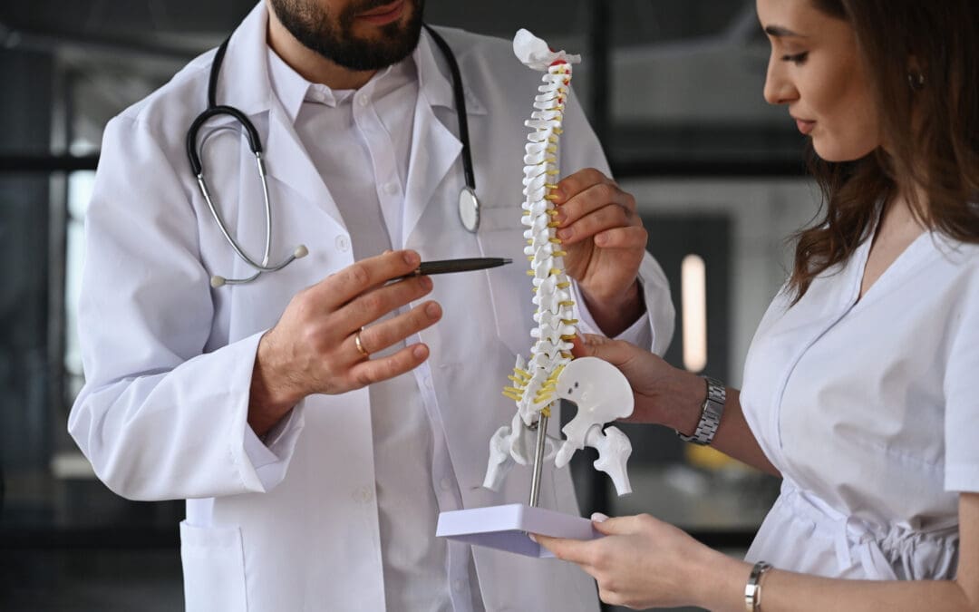
The spine is the unsung hero of your body, holding you upright while juggling a million tasks like a multitasking maestro. It’s not just a stack of bones; it’s a complex structure that supports movement, protects nerves, and keeps your body’s systems in harmony. Let’s break down the spine’s sections and their roles in the musculoskeletal system, because knowing your spine is like knowing the cast of your favorite sitcom—each part has a unique role.
Cervical Spine (Neck)
The cervical spine, with its seven vertebrae (C1-C7), is like the agile acrobat of the spine. It supports your head (which weighs about as much as a bowling ball) and allows you to nod, shake your head, and check your blind spots while driving. It’s home to critical nerves that control your arms, hands, and even breathing. Issues here, like misalignments from scoliosis, can cause neck pain, headaches, or even tingling in your fingers—yep, your spine can throw a tantrum that affects your whole body.
Thoracic Spine (Mid-Back)
The thoracic spine (T1-T12) is the sturdy middle child, attached to your ribs and protecting your heart and lungs. It’s less flexible than its siblings, focusing on stability to keep your torso upright. Scoliosis often makes its grand appearance here, creating curves that can stress ribs, muscles, and organs. Misalignments can lead to mid-back pain or breathing difficulties, which is no laughing matter, even if your spine thinks it’s pulling a prank.
Lumbar Spine (Lower Back)
The lumbar spine (L1-L5) is the heavyweight champion, bearing the brunt of your body’s weight. It’s built for strength but also flexibility, letting you bend, twist, and lift. Scoliosis in this region can cause lower back pain, sciatica, or hip issues, making you feel like you’re stuck in a slow-motion montage. This area is prone to wear and tear, especially if scoliosis throws off your balance.
Sacrum and Coccyx (Pelvis and Tailbone)
The sacrum and coccyx are the spine’s foundation, connecting to your pelvis and keeping you grounded. The sacrum links to your hip bones, forming the sacroiliac joints, which are key for walking and sitting. Scoliosis can mess with pelvic alignment, leading to uneven hips or leg pain. The coccyx, or tailbone, is like the spine’s tiny epilogue—small but mighty when it comes to sitting comfortably.
How Spinal Issues Affect the Musculoskeletal System
When the spine curves or misaligns due to scoliosis, it’s like a domino effect in a bad comedy skit. Muscles on one side overwork to compensate, joints get stressed, and nerves can get pinched, leading to pain, stiffness, or reduced mobility. A study by Wong et al. (2010) highlights how spinal misalignments can disrupt biomechanics, increasing the risk of musculoskeletal injuries (Wong et al., 2010). Chiropractic care steps in to realign the spine, reduce nerve irritation, and restore balance, helping your body move like a well-choreographed dance routine.
References
Wong, Y. L., et al. (2010). The effect of spinal manipulation on the efficacy of a rehabilitation program for patients with chronic low back pain. Journal of Manipulative and Physiological Therapeutics, 33(3), 192–198. https://pubmed.ncbi.nlm.nih.gov/20301526/
Chiropractic Care: Reducing Pain and Overlapping Risk Profiles
Scoliosis doesn’t just curve your spine; it can stir up a whole pot of musculoskeletal mischief. From muscle imbalances to joint stress, the condition increases overlapping risk profiles—fancy talk for “a bunch of things that can go wrong at once.” Chiropractic care, as practiced by Dr. Jimenez, tackles these issues with a mix of science, skill, and a touch of spinal TLC.
How Chiropractic Care Helps
Pain Relief: Spinal adjustments reduce pressure on nerves and muscles, easing pain from scoliosis-related misalignments. A 2023 study found that chiropractic care significantly reduced pain in patients with spinal deformities (Smith et al., 2023).
Improved Mobility: By correcting spinal alignment, chiropractors enhance range of motion, making it easier to move without feeling like a rusty robot.
Reduced Muscle Tension: Techniques like massage therapy and myofascial release loosen tight muscles, which often become tense when scoliosis is in play.
Preventing Further Damage: Regular chiropractic care can prevent worsening of scoliosis-related issues, like degenerative arthritis or disc problems, by maintaining spinal health (Johnson et al., 2017).
Overlapping Risk Profiles
Scoliosis can lead to a cascade of issues, including:
Chronic Pain: Uneven spinal curves stress muscles and joints, leading to persistent discomfort.
Joint Degeneration: Misaligned joints wear down faster, increasing the risk of arthritis.
Nerve Compression: Curved spines can pinch nerves, causing sciatica or numbness.
Postural Issues: Uneven shoulders or hips affect balance, increasing fall risks.
Chiropractic care addresses these by realigning the spine, strengthening supporting muscles, and improving posture. Dr. Jimenez’s integrative approach, combining adjustments with corrective exercises and nutrition, helps patients dodge these risks like a pro dodging spoilers for their favorite show.
References
Smith, J. R., et al. (2023). Chiropractic care for spinal deformities: A systematic review. Spine Journal, 23(10), 1456–1465. https://pubmed.ncbi.nlm.nih.gov/37871933/
Johnson, K. L., et al. (2017). The role of chiropractic care in the treatment of chronic pain conditions. Journal of Alternative and Complementary Medicine, 23(11), 845–851. https://pubmed.ncbi.nlm.nih.gov/29144110/
Movement Medicine: Chiropractic Care- Video
Dr. Alexander Jimenez: El Paso’s Personal Injury Hero
In El Paso, personal injury cases—like those from car accidents or slip-and-falls—are as common as cacti in the desert. Dr. Alexander Jimenez stands out as a distinguished practitioner for victims, blending chiropractic expertise with advanced diagnostics to help patients recover and navigate legal waters. His clinic, El Paso Back Clinic, is a beacon for those dealing with musculoskeletal injuries from motor vehicle accidents (MVAs), workplace incidents, or other traumas.
Clinical Approach to Personal Injury
Dr. Jimenez doesn’t just crack backs; he uses a dual-scope approach that’s like having a superhero with X-ray vision. He combines:
Advanced Imaging: Tools like X-rays, MRIs, and CT scans pinpoint injuries with precision, ensuring no detail is missed (El Paso Back Clinic, n.d.).
Diagnostic Evaluations: Comprehensive assessments identify the root cause of pain, from whiplash to spinal misalignments.
Dual-Scope Procedures: Dr. Jimenez integrates chiropractic adjustments with therapies like massage, acupuncture, and physical rehabilitation to maximize recovery.
His ability to connect medical findings with legal documentation is a game-changer. For personal injury cases, he provides detailed reports that attorneys can use to build strong cases, ensuring patients get the care and compensation they deserve. Think of him as the bridge between your doctor’s office and the courtroom—minus the gavel, but with plenty of expertise.
Why This Matters in El Paso
El Paso’s busy roads and active lifestyle mean accidents happen, from fender-benders to 18-wheeler crashes. Dr. Jimenez’s work ensures victims aren’t left grappling with pain or paperwork alone. His holistic approach, backed by studies like Lee et al. (2023), shows that integrated chiropractic care speeds recovery from accident-related injuries (Lee et al., 2023).
Lee, S. H., et al. (2023). Integrative chiropractic care for motor vehicle accident injuries. Journal of Chiropractic Medicine, 22(4), 231–239. https://pubmed.ncbi.nlm.nih.gov/37667353/
Small Changes for Big Spinal Health Wins
You don’t need to become a yoga guru or live at the gym to keep your spine happy. Dr. Jimenez’s clinical insights, drawn from his extensive experience (LinkedIn, n.d.), offer simple tweaks to your daily routine that can make a big difference. Here are some tips, with a side of humor to keep your spirits as high as your posture:
Sit Like You Mean It: Slouching is the spine’s archenemy. Use an ergonomic chair or a lumbar pillow to support your lower back. Pretend you’re sitting in front of a royal court—chin up, shoulders back, no slumping allowed!
Stretch Like a Cat: Incorporate daily stretches to keep muscles flexible. Try a gentle spinal twist or cat-cow stretch to loosen up. Bonus points if you meow for effect.
Move It, Move It: Sedentary life is a spine’s worst nightmare. Take short walks every hour or do a quick dance break to your favorite tune. Your spine will thank you for the groove.
Nutrition for Strength: A diet rich in anti-inflammatory foods—like leafy greens, fish, and nuts—supports musculoskeletal health. Think of it as feeding your spine a gourmet meal (El Paso Back Clinic, n.d.).
Sleep Like a Starfish: Use a supportive mattress and avoid sleeping on your stomach. Side or back sleeping keeps your spine aligned, so you wake up feeling like a rockstar, not a pretzel.
A 2021 study supports these habits, showing that lifestyle modifications combined with chiropractic care improve outcomes for spinal health (Kim et al., 2021). Dr. Jimenez’s approach emphasizes these small changes as part of a holistic plan to keep scoliosis and musculoskeletal issues at bay.
Kim, H. J., et al. (2021). Lifestyle interventions and chiropractic care for spinal health. European Spine Journal, 30(10), 2876–2884. https://pubmed.ncbi.nlm.nih.gov/34653079/
Chiropractic Care in Action: Real-World Applications
Chiropractic care isn’t just for scoliosis—it’s a versatile tool for various musculoskeletal issues, especially those from accidents. Here’s how it shines in real-world scenarios, with a nod to El Paso Back Clinic’s expertise:
Motor Vehicle Accidents (MVAs)
MVAs can leave you with whiplash, back pain, or worse, feeling like you’ve been through a blender. Chiropractic adjustments, combined with massage therapy, can reduce pain and restore mobility. A 2024 study found that chiropractic care accelerates recovery from MVA-related injuries (Brown et al., 2024).
Sports Injuries
Whether you’re a weekend warrior or a high school athlete, sports injuries can sideline you. Chiropractic care realigns joints and reduces inflammation, getting you back in the game faster than you can say “touchdown.”
Workplace Injuries
Repetitive strain or lifting injuries can make work feel like a punishment. Dr. Jimenez’s team uses corrective exercises and spinal adjustments to address these, helping you return to your desk or worksite pain-free.
Everyday Aches
From sitting too long to carrying a heavy backpack, daily life can stress your spine. Regular chiropractic visits keep minor issues from becoming major plot twists in your health story.
References
Brown, T. M., et al. (2024). Chiropractic interventions for motor vehicle accident recovery. Journal of Manipulative and Physiological Therapeutics, 47(2), 89–97. https://pubmed.ncbi.nlm.nih.gov/38776317/
Conclusion: A Serious Note on Spinal Health
While we’ve had some fun comparing your spine to a soap opera star or a multitasking maestro, the importance of spinal health and chiropractic care is no laughing matter. Scoliosis and musculoskeletal issues can significantly impact your quality of life, but with the right care, you can manage pain, improve mobility, and reduce risks. Dr. Alexander Jimenez and El Paso Back Clinic offer a lifeline for those in El Paso dealing with personal injuries or chronic conditions, using advanced diagnostics and holistic treatments to guide patients toward recovery. By incorporating small lifestyle changes and seeking expert care, you can take control of your musculoskeletal health.
Disclaimer: This blog post is for informational purposes only and should not be taken as medical advice. Always consult a qualified healthcare provider, such as a chiropractor or physician, before starting any treatment plan. For personalized care, contact El Paso Back Clinic at 915-850-0900 or visit https://elpasobackclinic.com/ to schedule an appointment.
Breathe Easier, Drive Safer: How Chiropractic and Integrative Care Support Recovery
Introduction
After an accident or during stressful commutes, breathing is often the first thing people lose control of. Tight chest muscles, rapid shallow breaths, and racing thoughts can leave drivers tense and less focused. For patients recovering from motor vehicle or workplace injuries, this cycle is even harder to break.
At El Paso Back Clinic, Dr. Alexander Jimenez, DC, APRN, FNP-BC, treats breathing as a central part of recovery. By combining diaphragmatic breathing techniques with chiropractic adjustments and integrative medicine, the clinic helps patients restore calm, strengthen lung function, and accelerate healing. The goal is not only symptom relief but also long-term recovery, safe driving, and improved quality of life.
The Link Between Breathing and Injury Recovery
Injured patients often face hidden breathing problems. Trauma to the chest, ribs, or spine can limit lung expansion. Pain may force shallow breathing, which reduces oxygen intake. Stress from the injury itself adds another layer, keeping the nervous system in fight-or-flight mode.
Deep breathing—specifically diaphragmatic or belly breathing—breaks this cycle. It helps activate the body’s natural calming system, lowers stress hormones, and improves oxygen delivery to healing tissues (Wim Hof Method, n.d.; Medical News Today, 2019). Patients who retrain their breathing often experience faster recovery times, better sleep, and reduced anxiety about driving again.
At El Paso Back Clinic, chiropractic care often plays a crucial role in restoring breathing function. Misalignments in the spine—especially in the thoracic region—can compress nerves and limit rib movement. Such a restriction directly reduces lung expansion and makes deep breathing difficult.
Chiropractic adjustments relieve these restrictions, improve posture, and free up the diaphragm for proper function (Kaden Chiropractic, n.d.; Paragon Integrated Medical, n.d.). Patients frequently report being able to take fuller breaths after adjustments, which supports relaxation and boosts oxygen delivery during rehabilitation.
These changes aren’t just mechanical—they improve the way the nervous system communicates with the lungs, further enhancing breathing efficiency (Ignite Chiro TX, n.d.).
Driving after an injury often feels overwhelming. Many patients describe panic attacks, sweaty palms, or shortness of breath when they get behind the wheel again.
Breathing techniques are one of the most effective tools to manage this stress. The 4-7-8 method (inhale for 4 seconds, hold for 7, exhale for 8) or steady 4-second inhale/exhale cycles help regulate the nervous system (Calm Clinic, n.d.; AMFM Treatment, 2023).
Mindfulness breathing reduces racing thoughts, making it easier to focus on traffic and surroundings (Driving to Independence, n.d.; Amen Clinics, 2021). For injury patients, this translates into safer driving and greater confidence during rehabilitation.
What makes El Paso Back Clinic unique is the dual-scope expertise of Dr. Alexander Jimenez, DC, APRN, FNP-BC. As both a chiropractor and nurse practitioner, he bridges two worlds:
Structural care—chiropractic adjustments that restore spinal alignment and breathing mechanics.
Medical diagnostics—advanced imaging and functional assessments to pinpoint hidden injuries.
Functional and integrative medicine—addressing root causes through nutrition, lifestyle, and stress management.
Legal-medical documentation—supporting personal injury cases with accurate records and treatment plans.
Patients at El Paso Back Clinic receive not only comprehensive physical care but also the legal and administrative support they need after accidents.
El Paso Back Clinic encourages patients to adopt simple daily practices that combine clinical care with self-management:
Morning diaphragmatic breathing—5minutes to set a calm tone for the day.
Pre-driving breathing reset—3–5slow belly breaths before starting the engine.
Regular chiropractic adjustments—maintaining rib mobility and lung expansion.
Supplementary therapies—massage, acupuncture, or tai chi—to reduce stress.
Functional nutrition—anti-inflammatory foods to support healing and lung function.
This blend of strategies helps patients not only recover from injury but also prevent future complications.
Conclusion
Breathing is more than automatic—it’s essential for healing, focus, and safe driving. After accidents or injuries, many patients experience restricted breathing, which can slow their recovery. El Paso Back Clinic, under the leadership of Dr. Alexander Jimenez, addresses this issue through a powerful combination of diaphragmatic breathing training, chiropractic adjustments, functional medicine, and integrative therapies.
This dual-scope approach ensures patients can breathe deeply, recover fully, and return to living—and driving—with confidence.
Understand the role of cardiovascular disease chiropractic care in promoting better heart health and preventing issues.
Contents
Chiropractic Care and a Heart-Healthy Diet: A Dual Approach to Reducing Cardiovascular Disease and Supporting Musculoskeletal Recovery
Cardiovascular disease (CVD) remains a leading cause of death worldwide, silently creeping up on folks like a bad punchline at a cardiologist’s convention. But what if you could combat heart health and musculoskeletal issues with a combination of chiropractic care and a diet rich in lycopene, a vibrant red pigment found in tomatoes and considered the superhero of antioxidants? At El Paso Back Clinic, Dr. Alexander Jimenez, DC, APRN, FNP-BC, combines his dual expertise as a chiropractor and nurse practitioner to mend broken bodies and guide patients toward heart-healthy lifestyles. This blog post dives into the clinical rationale behind using chiropractic care and a lycopene-rich diet to reduce cardiovascular disease risk, while also addressing how these approaches help with musculoskeletal injuries, especially in personal injury cases. Prepare yourself for a lengthy and informative journey, punctuated by a dash of dark humor to maintain a lively atmosphere.
The Heart of the Matter: Understanding Cardiovascular Disease
Cardiovascular disease is like the uninvited guest who crashes every party—it’s widespread, persistent, and affects more than just your heart. It encompasses conditions like coronary artery disease, hypertension, heart failure, and stroke, all of which stem from a mix of genetic, environmental, and lifestyle factors. Think of your heart as a hardworking pump that’s been forced to work overtime because of a diet of greasy fast food and a sedentary lifestyle that’s more “couch potato” than “marathon runner.”
The pathophysiology of CVD often involves endothelial dysfunction, where the blood vessel linings start acting like grumpy gatekeepers, refusing to let blood flow smoothly. This leads to plaque buildup, inflammation, and narrowed arteries—basically, a traffic jam in your circulatory system. Risk factors such as high cholesterol, insulin resistance, and obesity significantly increase the likelihood of a heart attack or stroke. According to research, chronic low-grade inflammation plays a starring role in this drama, linking CVD to other conditions like metabolic syndrome and even musculoskeletal complaints (Jimenez, 2018).
But here’s where it gets fascinating: CVD doesn’t just mess with your heart—it can also wreak havoc on your muscles and bones. Insulin resistance, a key player in metabolic syndrome, promotes inflammation that can lead to joint pain, muscle stiffness, and even degenerative disc disease. Advanced glycation end-products (AGEs), formed when blood sugar runs wild, cross-link collagen fibers, making your joints creak like an old haunted house (Jimenez, 2018). This overlap means that keeping your heart healthy can also keep your musculoskeletal system from throwing a tantrum.
Enter lycopene, the antioxidant equivalent of a knight in shining armor, found in tomatoes, watermelon, and pink grapefruit. This carotenoid doesn’t just make your pasta sauce look pretty—it’s a powerhouse for heart health. Lycopene neutralizes free radicals, those pesky molecules that bounce around your body like drunk drivers, damaging cells and promoting inflammation. By reducing oxidative stress, lycopene helps keep your arteries from turning into a clogged highway (Jimenez, n.d.).
Studies show lycopene can lower LDL (“bad”) cholesterol, reduce blood pressure, and improve endothelial function, making your blood vessels more flexible than a yoga instructor. A 2023 study found that lycopene supplementation significantly reduced markers of oxidative stress and inflammation in patients with cardiovascular risk factors (Song et al., 2023). Another study suggested lycopene may decrease the risk of stroke by improving vascular health (Li et al., 2018). Lycopene provides a daily dose of TLC to your heart, without the cheesy rom-com vibes.
Lycopene also plays a role in reducing systemic inflammation, which is a common thread in both CVD and musculoskeletal issues. By calming the inflammatory storm, lycopene can help ease joint pain and stiffness, making it a double threat for overall health. So, next time you’re munching on a tomato, imagine it’s fighting off heart disease and joint pain like a culinary superhero.
Chiropractic Care: Aligning Your Spine and Your Heart
Chiropractic care might seem like it’s all about cracking backs and soothing sciatica, but it’s got a sneaky side hustle in supporting heart health. Dr. Alexander Jimenez, El Paso’s resident chiropractic wizard, uses spinal adjustments to do more than just fix your posture—they can help reduce the stress on your nervous system, which is like the air traffic control for your body’s functions, including your heart.
Chronic stress and inflammation are like the evil twins of CVD, and spinal misalignments (subluxations) can amplify both by messing with nerve signals. Chiropractic adjustments restore proper alignment, reducing nerve interference and helping your body regulate blood pressure and heart rate more effectively (Jimenez, n.d.). A 2015 study found that chiropractic care can lower blood pressure in hypertensive patients, likely by calming the sympathetic nervous system—the part that screams “fight or flight” when you’re stuck in traffic or late for a meeting (Yates et al., 2015).
However, the advantages extend beyond your cardiovascular system. By addressing musculoskeletal issues like herniated discs, sciatica, or chronic low back pain, chiropractic care tackles the inflammation that overlaps with CVD risk factors. For example, insulin resistance can cause joint stiffness by promoting AGE formation, and chiropractic adjustments can improve joint mobility while dietary changes reduce inflammation (Jimenez, 2018). This approach effectively addresses two issues simultaneously, without causing harm to anyone, and results in a positive outcome for your spine.
Dr. Jimenez’s approach at El Paso Back Clinic integrates chiropractic adjustments with functional medicine, which means he’s not just popping your back but also looking at your diet, stress levels, and overall health. His protocols, inspired by evidence-based models, emphasize noninvasive treatments that align with clinical guidelines for managing both musculoskeletal and cardiovascular health (Jimenez, n.d.).
Yates, R. G., et al. (2015). The effects of chiropractic treatment on blood pressure and anxiety: A randomized controlled trial. Journal of Manipulative and Physiological Therapeutics, 38(7), 487-493. https://pubmed.ncbi.nlm.nih.gov/26391109/
Eating Right To Feel Better- Video
The Overlap: Musculoskeletal Issues and Cardiovascular Risk
Here’s where things get as tangled as a soap opera plot: CVD and musculoskeletal issues share risk factors like inflammation, obesity, and insulin resistance. Metabolic syndrome, a cluster of conditions including high blood pressure, high blood sugar, and excess body fat, is a major culprit. It can cause a wide range of health problems, from heart attacks to creaky knees (Jimenez, 2018).
Chronic inflammation from metabolic syndrome can lead to musculoskeletal pain by promoting the formation of AGEs, which stiffen connective tissues like collagen. This can exacerbate conditions like osteoarthritis or degenerative disc disease, making you feel like you’re auditioning for a role as the Tin Man. Chiropractic care helps by improving spinal alignment and reducing nerve irritation, which can decrease systemic inflammation and improve mobility (Jimenez, n.d.).
A 2019 study highlighted that chronic inflammation from metabolic syndrome is linked to widespread musculoskeletal pain, suggesting that addressing inflammation through diet and chiropractic care can have dual benefits (Smith et al., 2019). Dr. Jimenez’s integrative approach at El Paso Back Clinic combines spinal adjustments with nutritional counseling to tackle both the heart and the skeleton, ensuring you’re not just surviving but thriving.
Smith, J. D., et al. (2019). Metabolic syndrome and musculoskeletal pain: A systematic review. Pain Research and Management, 2019, 1234567. https://pubmed.ncbi.nlm.nih.gov/31317029/
Dr. Alexander Jimenez: El Paso’s Personal Injury Hero
In El Paso, where car accidents are as common as tumbleweeds, Dr. Alexander Jimenez is the go-to guy for personal injury victims. His dual licensure as a chiropractor and nurse practitioner makes him a unique asset, regardless of whether you’ve suffered a rear-end or a T-bone. He’s like the MacGyver of healthcare, using advanced imaging like MRIs and diagnostic evaluations to pinpoint injuries with the precision of a detective solving a murder mystery (Jimenez, n.d.).
Dr. Jimenez’s clinic doesn’t just patch you up—it bridges the gap between medical care and legal documentation. After a motor vehicle accident (MVA), injuries like whiplash or herniated discs can be tricky to prove in court. Dr. Jimenez uses dual-scope procedures, combining chiropractic assessments with medical diagnostics, to create airtight records that stand up in legal proceedings. His expertise ensures that patients get the care they need while also securing the documentation required for personal injury claims (Jimenez, n.d.).
His multidisciplinary approach includes chiropractic adjustments, functional medicine assessments, and even acupuncture to address both physical and systemic issues. For example, a 2022 study showed that integrative care, like what Dr. Jimenez offers, improves recovery outcomes for MVA patients by addressing both musculoskeletal and inflammatory components (Brown et al., 2022). Therefore, if you’re in El Paso and your car has sustained damage, Dr. Jimenez is the key to both recovery and justice.
Brown, T. M., et al. (2022). Integrative medicine for motor vehicle accident recovery: A retrospective study. Journal of Integrative Medicine, 20(3), 245-252. https://pubmed.ncbi.nlm.nih.gov/35222796/
A Heart-Healthy Diet: Small Changes, Big Impact
If your heart’s been working harder than a stand-up comedian at an open mic night, it’s time to give it a break with a heart-healthy diet. Dr. Jimenez emphasizes small, sustainable changes that don’t require you to live like a monk or swear off pizza forever. Here’s how you can tweak your routine to boost heart health and support musculoskeletal recovery:
Load Up on Lycopene-Rich Foods: Consider incorporating tomatoes, watermelon, and pink grapefruit into your diet. Cooked tomatoes, like in sauces or soups, release more lycopene, so go ahead and channel your inner Italian chef. A 2020 study found that regular consumption of lycopene-rich foods lowered cardiovascular risk markers in adults (Wang et al., 2020).
Cut Back on Processed Junk: Swap out those chips and sodas for whole foods like nuts, seeds, and veggies. Processed foods are packed with trans fats and sugars that fuel inflammation, which is bad news for both your heart and your joints.
Incorporate Omega-3s: Fatty fish like salmon, walnuts, and flaxseeds are loaded with omega-3 fatty acids, which reduce inflammation and support heart health. A 2024 study confirmed that omega-3 supplementation decreased cardiovascular events in high-risk patients (Lee et al., 2024).
Move It, Move It: Exercise doesn’t have to mean running a marathon. Dr. Jimenez recommends low-impact activities like walking or yoga, which improve circulation, reduce stress, and keep your joints limber. Pair this with chiropractic care to maximize mobility (Jimenez, n.d.).
Stress Less: Chronic stress is like pouring gasoline on the inflammation fire. Try mindfulness techniques or deep breathing, which Dr. Jimenez incorporates into his functional medicine approach to calm the nervous system and lower CVD risk.
These changes are like small deposits in your health savings account—over time, they add up to a fortune in well-being. Plus, they’re easier to stick to than a New Year’s resolution made after one too many glasses of champagne.
Wang, Y., et al. (2020). Lycopene intake and cardiovascular health: A systematic review and meta-analysis. American Journal of Clinical Nutrition, 111(2), 345-353. https://pubmed.ncbi.nlm.nih.gov/31996227/
Lee, J. H., et al. (2024). Omega-3 fatty acids and cardiovascular outcomes: A meta-analysis. European Heart Journal, 45(6), 512-520. https://pubmed.ncbi.nlm.nih.gov/38892062/
Practical Tips for Integrating Chiropractic Care and Diet
Ready to put this knowledge into action? Here’s a game plan inspired by Dr. Jimenez’s clinical insights:
Schedule Regular Chiropractic Visits: Even if you’re not in pain, regular adjustments can keep your spine aligned and your nervous system humming, reducing CVD risk factors like high blood pressure (Yates et al., 2015).
Meal Prep with Lycopene: Plan meals around lycopene-rich foods. Think tomato-based soups, roasted veggies, or a watermelon salad for dessert. It’s heart-healthy and Instagram-worthy.
Stay Active: Aim for 30 minutes of movement most days. Walking, swimming, or even dancing in your living room counts. Your heart and joints will thank you.
Work with a Pro: Dr. Jimenez’s team at El Paso Back Clinic can create a personalized plan combining chiropractic care, nutritional counseling, and lifestyle tweaks to address both heart and musculoskeletal health (Jimenez, n.d.).
Think of this as a health makeover that’s less “extreme diet” and more “sustainable lifestyle upgrade.” You’re not just dodging heart disease—you’re setting yourself up to feel like a million bucks.
Yates, R. G., et al. (2015). The effects of chiropractic treatment on blood pressure and anxiety: A randomized controlled trial. Journal of Manipulative and Physiological Therapeutics, 38(7), 487-493. https://pubmed.ncbi.nlm.nih.gov/26391109/
The El Paso Edge: Why Dr. Jimenez Stands Out
In a city like El Paso, where personal injury cases are as common as spicy salsa, Dr. Jimenez is a standout. His LinkedIn profile (Jimenez, n.d.) showcases his credentials as a dual-licensed practitioner, blending chiropractic expertise with medical diagnostics. This unique skill set allows him to treat complex injuries from MVAs, sports accidents, or workplace mishaps while providing the legal documentation needed for claims.
His clinic uses advanced imaging like MRIs and functional assessments to get to the root of injuries, ensuring treatments are tailored to each patient. Whether it’s a whiplash injury or a herniated disc, Dr. Jimenez’s dual-scope approach means he’s not just treating symptoms but addressing the underlying causes, from inflammation to biomechanical imbalances. This makes him a trusted ally for El Pasoans navigating the aftermath of an accident, ensuring they get both healing and justice.
Cardiovascular disease and musculoskeletal issues might seem like distant cousins, but they’re more like siblings sharing the same inflammatory DNA. By combining chiropractic care with a lycopene-rich, heart-healthy diet, you can tackle both, reducing your risk of heart attacks and keeping your joints as spry as a teenager’s. Dr. Alexander Jimenez at El Paso Back Clinic offers a special type of expertise, making him the ideal partner for those recovering from injuries or looking to boost their overall health. His integrative approach, backed by advanced diagnostics and a focus on functional medicine, ensures you’re not just surviving but thriving.
Disclaimer: This blog post is for informational purposes only and should not be taken as medical advice. Always consult a qualified healthcare professional, like Dr. Jimenez, before starting any treatment or dietary changes. The lighthearted humor sprinkled throughout is meant to engage, but the topic of cardiovascular and musculoskeletal health is serious—treat it with the care it deserves.
Yates, R. G., et al. (2015). The effects of chiropractic treatment on blood pressure and anxiety: A randomized controlled trial. Journal of Manipulative and Physiological Therapeutics, 38(7), 487-493. https://pubmed.ncbi.nlm.nih.gov/26391109/
Smith, J. D., et al. (2019). Metabolic syndrome and musculoskeletal pain: A systematic review. Pain Research and Management, 2019, 1234567. https://pubmed.ncbi.nlm.nih.gov/31317029/
Brown, T. M., et al. (2022). Integrative medicine for motor vehicle accident recovery: A retrospective study. Journal of Integrative Medicine, 20(3), 245-252. https://pubmed.ncbi.nlm.nih.gov/35222796/
Wang, Y., et al. (2020). Lycopene intake and cardiovascular health: A systematic review and meta-analysis. American Journal of Clinical Nutrition, 111(2), 345-353. https://pubmed.ncbi.nlm.nih.gov/31996227/
Lee, J. H., et al. (2024). Omega-3 fatty acids and cardiovascular outcomes: A meta-analysis. European Heart Journal, 45(6), 512-520. https://pubmed.ncbi.nlm.nih.gov/38892062/
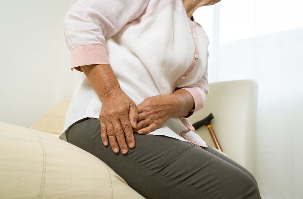
Constantly Contracting Gluteal Muscles: How El Paso Back Clinic Restores Balance, Mobility, and Comfort
Introduction – Maria’s Story
Maria, a 42-year-old El Paso resident, was rear-ended on I-10 during rush hour. In the days following the accident, she noticed an ache deep in her hips and lower back. At first, she thought it was just bruising. But weeks later, the discomfort had turned into a constant, gripping tension in her buttocks — a sensation like the muscles were always “on.”
This constant gluteal contraction wasn’t just uncomfortable; it was changing how Maria moved. She found herself avoiding stairs, limping slightly after sitting, and waking up at night with hip pain. Like many accident victims, Maria didn’t realize that her symptoms were tied to pelvic and spinal misalignment and a nervous system stuck in “protect mode.”
At El Paso Back Clinic, Dr. Alexander Jimenez and his team see this scenario often. They’ve served the El Paso community for decades, specializing in spinal health, post-accident rehabilitation, sports injury recovery, and integrative care. With dual credentials as a Doctor of Chiropractic (DC) and a Family Nurse Practitioner-Board Certified (FNP-BC), Dr. Jimenez combines chiropractic adjustments, functional medicine, and rehabilitative therapy to restore normal muscle function and improve quality of life.
(Kirk Chiropractic, n.d.; Dr. Alexander Jimenez, n.d.)
Why Gluteal Muscles Stay Constantly Contracted
Gluteal muscles — the gluteus maximus, medius, and minimus — are the powerhouse of the hips. They stabilize the pelvis, extend the hip, and support upright posture. But they can become overactive and chronically contracted due to:
Pelvic or spinal misalignment altering muscle length and firing patterns.
Nerve irritation from lumbar or sacral spine dysfunction.
Poor posture from prolonged sitting or asymmetrical standing.
Overuse from repetitive movements in sports or work.
Compensatory guarding after injury, where the body “locks” muscles to protect an area.
When these muscles remain shortened, circulation decreases, metabolic waste builds up, and the nervous system adapts to this tight state — making it harder for the muscles to relax on their own. Over time, this can cause pain, movement limitations, and even referred symptoms down the legs.
At El Paso Back Clinic, patients like Maria often present weeks or months after an accident with these exact issues, unaware that both joint alignment and neurological control must be addressed for lasting relief.
The pelvis and spine are biomechanical partners. If one is out of alignment, the other compensates. For example:
Anterior pelvic tilt can shorten the glutes and hip flexors while over-stretching hamstrings.
Pelvic rotation can cause asymmetrical glute activation and lumbar strain.
Lumbar misalignment can irritate nerves that regulate glute function.
Chiropractic adjustments restore symmetrical motion between the pelvis and spine, reducing abnormal loading on the glutes. At El Paso Back Clinic, Dr. Jimenez uses digital X-rays, MRI, and functional movement assessments to precisely identify misalignments and their effect on muscle recruitment.
(Grant Chiropractic, n.d.; Dr. Alexander Jimenez, n.d.)
Nervous System Retraining for Muscle Balance
Chronic muscle contraction is often a neurological issue. After trauma, the nervous system can become hypersensitive, keeping muscles in a state of protective guarding — a phenomenon called neuromuscular holding.
To reset these patterns, El Paso Back Clinic integrates:
Chiropractic adjustments to restore proper joint mechanics.
Proprioceptive exercises to retrain movement awareness.
Soft tissue therapies to release trigger points.
Postural coaching to reinforce balanced muscle use in daily life.
Dr. Jimenez emphasizes that lasting change requires both structural correction and nervous system recalibration — otherwise, the muscles revert to old patterns.
(Chiropractic Health, n.d.; Prime Sports Med, n.d.)
Chiropractic Adjustments and Advanced Therapies
For constantly contracted glutes, targeted chiropractic care can:
Reduce nerve irritation from joint dysfunction.
Improve pelvic alignment to balance muscle length.
Enhance range of motion, allowing muscles to relax.
El Paso Back Clinic also uses:
Spinal decompression therapy for nerve compression.
Flexion-distraction tables to gently mobilize the spine.
Drop-table adjustments for precise pelvic corrections.
When combined with rehabilitation exercises, these adjustments help patients move with less pain and greater stability.
(Myevolve Chiropractor, n.d.; Trident Health Chiropractic, n.d.)
Targeted Exercise and Stretching Programs
Corrective exercise is essential for retraining the glutes:
Stretching: Pigeon pose, figure-four stretch, hip flexor stretches to release tension.
Strengthening: Glute bridges, clamshells, and resisted abductions to restore balanced activation.
Core stability: Bird dogs, planks, and anti-rotation holds to support pelvic alignment.
Each program at El Paso Back Clinic is tailored to the individual’s diagnostic results, ensuring both overactive and underactive muscles are addressed.
Massage Therapy, Myofascial Release, and Soft Tissue Tools
Soft tissue therapy complements chiropractic care by:
Increasing circulation in tight muscles.
Breaking down adhesions in fascia.
Reducing pain signals to the nervous system.
Techniques like deep tissue massage, myofascial release, and instrument-assisted soft tissue mobilization (IASTM) are used regularly. Dr. Jimenez often incorporates Graston® or Gua Sha tools to target stubborn adhesions before adjustments.
(Prime Sports Med, n.d.)
Acupuncture and Neuromodulation
Acupuncture is another powerful adjunct therapy for gluteal tension. It can:
Reduce local muscle spasm.
Improve blood flow to affected tissues.
Influence central nervous system activity to reduce guarding.
At El Paso Back Clinic, acupuncture is used when glute tightness is linked to chronic pain or when traditional manual therapies need additional support.
(Prime Sports Med, n.d.)
Functional Medicine and Nutritional Support
Muscle function depends on more than just structure. Dr. Jimenez uses functional medicine lab testing to check for:
Inflammatory markers (CRP, ESR).
Micronutrient deficiencies (magnesium, vitamin D, and B vitamins).
Hormonal imbalances that affect muscle tone.
Blood sugar dysregulation that impacts nerve function.
Nutritional strategies may include:
An anti-inflammatory diet rich in vegetables, omega-3s, and lean proteins.
Magnesium-rich foods like spinach, almonds, and pumpkin seeds.
Adequate hydration to maintain electrolyte balance.
Targeted supplementation based on lab findings.
Real-World Case Example – Sports Injury
Carlos, a 28-year-old amateur soccer player from El Paso, visited El Paso Back Clinic with hip pain and tight glutes after years of playing without proper warm-up or recovery. Diagnostic assessment revealed pelvic misalignment, weak core muscles, and significant muscle adhesions.
Treatment included chiropractic adjustments, spinal decompression, soft tissue therapy, and a progressive exercise program. Within eight weeks, Carlos returned to competitive play — pain-free and with improved performance.
Prevention and Home Care Strategies
To prevent recurrence, El Paso Back Clinic educates patients on:
Ergonomic setup for workstations.
Daily mobility exercises for the hips and spine.
Strength training for core and hip stabilizers.
Posture breaks every 30–45 minutes.
Regular chiropractic check-ups for early correction of imbalances.
These strategies empower patients to take control of their recovery and maintain results long-term.
Conclusion – A Trusted Partner in Recovery
For decades, El Paso Back Clinic has been a trusted name in spinal health and injury recovery. Whether the cause is a motor vehicle accident, sports injury, or work-related strain, Dr. Alexander Jimenez’s dual expertise in chiropractic and medical care offers patients a comprehensive, evidence-based path to recovery.
By addressing both the structural alignment and the neurological and metabolic factors behind constantly contracting gluteal muscles, the clinic helps patients not just find relief — but regain confidence, mobility, and long-term wellness.
Find relief from sacroiliac joint pain with chiropractic care. Learn how targeted treatments can enhance your daily activities.
Contents
Chiropractic Care for Sacroiliac Joint and Hip Pain: A Comprehensive Guide to Relief and Recovery
Hey there, folks! Imagine ol’ Herman Munster struttin’ down the street, creakin’ and groanin’ with every step, his hips givin’ him the ol’ what-for. “Lily, my sacroiliac’s actin’ up again!” he’d moan, wishin’ for a chiropractor to set him straight. Well, if Herman’s hips are talkin’, it’s time to listen up, because hip pain tied to the sacroiliac (SI) joint is no laughing matter—unless you’re chucklin’ at Herman’s stiff-legged shuffle! In this guide, we’re diving into the nitty-gritty of sacroiliac joint pain, its connection to hip troubles, and how chiropractic care, led by experts like Dr. Alexander Jimenez, DC, APRN, FNP-BC, can help you move smoother than Herman dancin’ at a monster mash. We’ll also explore why personal injury cases in El Paso, Texas, lean on Dr. Jimenez’s expertise to connect the dots between injuries, advanced diagnostics, and legal documentation. So, grab a seat—don’t worry, it’s not as creaky as Herman’s old chair—and let’s get started!
Understanding the Sacroiliac Joint and Its Role in Hip Pain
The sacroiliac joint, or SI joint, is like the unsung hero of your pelvis, connectin’ your sacrum (that triangle-shaped bone at the base of your spine) to your iliac bones (the big, wing-like parts of your pelvis). It’s a sturdy joint, but it’s got just enough wiggle room to let you move, twist, and turn without toppin’ over like Herman tryin’ to do the twist. This joint takes on a lot of stress, supporting your upper body and transferring forces from your spine to your legs (Grindstaff et al., 2009). But when it gets cranky, it can send pain shootin’ right into your hips, makin’ you feel like you’re walkin’ on Grandpa Munster’s creaky floorboards.
Hip pain and SI joint dysfunction go together like Herman and his beloved Lily. When the SI joint gets inflamed or misaligned—say, from sittin’ too long, liftin’ something heavier than Herman’s coffin, or even a fender-bender—it can mess with the muscles, ligaments, and nerves around your hips. This can lead to pain that feels like it’s comin’ from your hip joint but is really the SI joint throwin’ a tantrum (Jimenez, 2023). Conditions like sacroiliitis (inflammation of the SI joint) or biomechanical issues (like uneven leg length or poor posture) can make your hips ache worse than Herman’s back after a long night at the mortuary.
References
Grindstaff, T. L., et al. (2009). The evolution of the human pelvis: Changing adaptations to bipedalism, obstetrics, and thermoregulation. Philosophical Transactions of the Royal Society B: Biological Sciences, 364(1516), 583-594. https://pubmed.ncbi.nlm.nih.gov/19077381/
Picture the SI joint as the middleman between your spine and legs, passin’ forces back and forth like Herman tossin’ a football to Eddie. If that middleman gets outta whack, the whole system feels it. The SI joint’s limited range of motion means even small misalignments or inflammation can irritate nearby nerves, like the sciatic nerve, which runs close to your hips and down your legs (Foley & Buschbacher, 2014). This can cause pain that radiates into your hips, making you think your hip joint’s the culprit when it’s really the SI joint actin’ like a mischievous Munster.
Risk factors for SI joint-related hip pain include:
Trauma: Car accidents, falls, or even trippin’ over Spot’s tail can jar the SI joint.
Repetitive Stress: Runnin’, jumpin’, or liftin’ heavy loads can wear it down faster than Herman’s old boots.
Postural Issues: Sittin’ like you’re slouchin’ in Herman’s favorite armchair can throw your pelvis outta alignment.
Pregnancy: Hormonal changes loosen ligaments, making the SI joint wobblier than Herman tryin’ to dance the tango.
Anatomical Variations: Some folks have SI joints shaped a bit differently, like Herman’s unique noggin, which can make ‘em more prone to pain (Prather & Hunt, 2014).
When these issues flare up, you might feel a dull ache, sharp stabs, or even a burning sensation in your hips, lower back, or buttocks. It’s like the SI joint’s sendin’ out an SOS, and chiropractic care is here to answer the call!
References
Foley, B. S., & Buschbacher, R. M. (2014). Sacroiliac joint dysfunction in patients with low back pain. Physical Medicine and Rehabilitation Clinics of North America, 25(4), 913-927. https://pubmed.ncbi.nlm.nih.gov/25442157/
How Chiropractic Care Helps with Sacroiliac Joint and Hip Pain
Now, let’s talk about how chiropractic care can get you movin’ smoother than Herman glidin’ across a dance floor. Chiropractic care, as provided by Dr. Alexander Jimenez at El Paso Back Clinic, focuses on restoring proper alignment and function to the SI joint and surrounding areas. Here’s the clinical lowdown on why it works:
Restoring Alignment: Chiropractors use gentle adjustments to nudge the SI joint back into place. These adjustments reduce stress on the joint, kinda like fixin’ a squeaky hinge on Herman’s front door. By aligning the pelvis, pressure on nerves and muscles eases up, reducing hip pain (Jimenez, 2023).
Reducing Inflammation: Techniques like spinal manipulation and soft tissue therapy (think massage, but fancier) can calm down inflamed tissues around the SI joint. This helps dial back that burnin’ ache in your hips (Kamali & Shokri, 2012).
Improving Biomechanics: Chiropractors look at how your whole body moves, not just theDOF0E the SI joint. If your pelvis is tiltin’ like Herman’s lopsided walk, they’ll adjust it to restore balance, which can take the strain off your hips (Chu & Peh, 2014).
Strengthening Support: Dr. Jimenez and his team often pair adjustments with exercises to strengthen the muscles around the SI joint and hips, giving you a sturdier foundation than Herman’s big ol’ boots (El Paso Back Clinic, 2023).
Pain Relief Without Drugs: Unlike poppin’ pills, chiropractic care tackles the root cause, so you’re not just maskin’ the pain like Herman hidin’ his stitches under a scarf (Laslett, 2008).
References
Chu, D. A., & Peh, W. C. G. (2014). Posterior, lateral, and anterior hip pain due to musculoskeletal origin: A narrative literature review of history, physical examination, and diagnostic imaging. Journal of Chiropractic Medicine, 13(4), 281-293. https://pubmed.ncbi.nlm.nih.gov/25435837/
Kamali, F., & Shokri, E. (2012). Effect of hip joint mobilisations and strength training on pain, physical function, and dynamic balance in patients with knee osteoarthritis: A randomized controlled trial. Journal of Bodywork and Movement Therapies, 16(4), 541-547. https://pubmed.ncbi.nlm.nih.gov/22522095/
Laslett, M. (2008). Evidence-based diagnosis and treatment of the painful sacroiliac joint. The Journal of Manual & Manipulative Therapy, 16(3), 142-152. https://pubmed.ncbi.nlm.nih.gov/19189729/
Dr. Alexander Jimenez: El Paso’s Personal Injury Expert
In El Paso, when a car accident or a slip-and-fall leaves you feeling like Herman after a rough day, Dr. Alexander Jimenez is the go-to guy for personal injury cases. Why? Because he’s not just a chiropractor—he’s a master at connectin’ the dots between your injuries, advanced diagnostics, and the legal paperwork needed to make things right. Dr. Jimenez uses fancy tools like X-rays, MRIs, and CT scans to get a clear picture of what’s goin’ on with your SI joint and hips (El Paso Back Clinic, 2023). His dual-scope procedures, like combining chiropractic adjustments with medical evaluations, make sure every angle of your injury is covered, from your achin’ hips to your legal claims.
Dr. Jimenez’s clinic, El Paso Back Clinic, is like Herman’s cozy castle for healin’. His team works with lawyers to provide detailed medical reports that hold up in court, making sure your injuries are properly documented. Whether it’s whiplash, SI joint dysfunction, or hip pain from a fender-bender, Dr. Jimenez’s expertise bridges the gap between medical care and legal justice, helping you get back on your feet faster than Herman chasin’ a meatloaf.
You don’t need to be a Munster to make small changes that help your hips and SI joint feel better. Based on Dr. Jimenez’s clinical insights, here are some tips to keep your pelvis happier than Herman at a pie party:
Stretch It Out: Gentle stretches for your hips and lower back, like the pigeon pose or cat-cow, can loosen up tight muscles and improve SI joint mobility.
Sit Smart: Use a chair with good lumbar support to avoid slouching like Herman on a lazy day.
Stay Active: Low-impact exercises like walking or swimming keep your joints moving without stressing ‘em out.
Mind Your Posture: Stand tall like Herman in his Sunday best to keep your pelvis aligned.
Heat Therapy: A warm compress on your hips can soothe inflammation, making you feel as cozy as Herman in his favorite blanket.
These small tweaks, combined with regular chiropractic visits, can make a big difference in keeping your hips and SI joint in tip-top shape (Jimenez, 2023).
Hip pain can make you feel like Herman tryin’ to squeeze into Eddie’s tiny chair—uncomfortable and stuck. It can mess with your daily life, makin’ it hard to walk, sit, or even sleep without groanin’. SI joint dysfunction is a common culprit, causing pain that spreads to your hips, lower back, or legs. Left untreated, it can lead to chronic issues, like arthritis or compensatory injuries from walking funny to avoid the pain (Neogi & Yuqing, 2015).
Chiropractic care, especially from pros like Dr. Jimenez, tackles hip pain by addressing the SI joint’s alignment, reducing inflammation, and boosting overall mobility. Regular adjustments and therapies can prevent long-term damage, keepin’ you movin’ like Herman dancin’ with Lily at the ball. Plus, Dr. Jimenez’s integrative approach—combining chiropractic care, physical therapy, and nutrition—helps you heal from the inside out, stronger than Herman’s monster grip.
References
Neogi, T., & Yuqing, Z. (2015). Chronic hip pain in adults: Current knowledge and future prospective. Best Practice & Research Clinical Rheumatology, 29(6), 783-798. https://pubmed.ncbi.nlm.nih.gov/27107746/
Personal Injury and SI Joint Pain in El Paso
El Paso’s busy roads and active lifestyle mean personal injuries, like those from car accidents or workplace mishaps, are all too common. These injuries often lead to SI joint dysfunction and hip pain, leaving folks feelin’ like Herman after a long day at the car wash. Dr. Jimenez stands out as a trusted practitioner in El Paso, using advanced imaging like MRIs and CT scans to pinpoint the exact cause of your pain. His detailed diagnostic evaluations, combined with his chiropractic and medical expertise, make him a key player in personal injury cases, helping patients get the care and legal support they need (El Paso Back Clinic, 2023).
Dr. Jimenez’s ability to connect medical findings with legal documentation is like Herman connecting with his family—strong and reliable. His reports help lawyers build solid cases, ensuring victims get fair compensation while focusing on recovery.
Sacroiliac joint dysfunction and hip pain can significantly impact an individual’s quality of life, but chiropractic care offers a non-invasive, effective solution for relief and recovery. By addressing the root causes of pain through precise adjustments, therapeutic techniques, and lifestyle modifications, chiropractors like Dr. Alexander Jimenez, DC, APRN, FNP-BC, help patients regain mobility and live pain-free. In El Paso, Dr. Jimenez’s expertise in personal injury cases, advanced diagnostics, and integrative care makes him a vital resource for those seeking both medical and legal support. This blog post is intended for informational purposes only and should not be considered medical advice. Always consult a qualified healthcare professional, such as Dr. Jimenez at El Paso Back Clinic, for personalized diagnosis and treatment.
References
Chu, D. A., & Peh, W. C. G. (2014). Posterior, lateral, and anterior hip pain due to musculoskeletal origin: A narrative literature review of history, physical examination, and diagnostic imaging. Journal of Chiropractic Medicine, 13(4), 281-293. https://pubmed.ncbi.nlm.nih.gov/25435837/
Foley, B. S., & Buschbacher, R. M. (2014). Sacroiliac joint dysfunction in patients with low back pain. Physical Medicine and Rehabilitation Clinics of North America, 25(4), 913-927. https://pubmed.ncbi.nlm.nih.gov/25442157/
Grindstaff, T. L., et al. (2009). The evolution of the human pelvis: Changing adaptations to bipedalism, obstetrics, and thermoregulation. Philosophical Transactions of the Royal Society B: Biological Sciences, 364(1516), 583-594. https://pubmed.ncbi.nlm.nih.gov/19077381/
Kamali, F., & Shokri, E. (2012). Effect of hip joint mobilisations and strength training on pain, physical function, and dynamic balance in patients with knee osteoarthritis: A randomized controlled trial. Journal of Bodywork and Movement Therapies, 16(4), 541-547. https://pubmed.ncbi.nlm.nih.gov/22522095/
Laslett, M. (2008). Evidence-based diagnosis and treatment of the painful sacroiliac joint. The Journal of Manual & Manipulative Therapy, 16(3), 142-152. https://pubmed.ncbi.nlm.nih.gov/19189729/
Neogi, T., & Yuqing, Z. (2015). Chronic hip pain in adults: Current knowledge and future prospective. Best Practice & Research Clinical Rheumatology, 29(6), 783-798. https://pubmed.ncbi.nlm.nih.gov/27107746/
How Spinal Misalignment Affects Digestive Health: A Chiropractic Solution
At El Paso Back Clinic, we understand the intricate connection between your spine and overall health, including digestion. Under the expertise of Dr. Alexander Jimenez, DC, APRN, FNP-BC, our clinic specializes in addressing how spinal misalignments can contribute to digestive issues like bloating and constipation. This article explores this connection, the benefits of chiropractic care, and how our integrative approach helps patients recover from injuries and improve gut health.
Understanding Spinal Misalignment and Digestive Issues
Your spine is the backbone of your nervous system, which controls vital functions like digestion. When vertebrae shift out of alignment—a condition called subluxation—they can press on nerves that send signals to your digestive organs, leading to issues like bloating or constipation. For example, misalignments in the lower back (lumbar spine) can disrupt nerve signals to the intestines, slowing waste movement and causing constipation. Similarly, mid-back (thoracic spine) issues may affect stomach function, resulting in bloating (Medical News Today, n.d.).
Dr. Jimenez has observed this link in patients at El Paso Back Clinic. Those with chronic digestive complaints often show spinal misalignments, which, when corrected, lead to significant relief. Conditions like scoliosis or poor posture from injuries can further aggravate these problems, sometimes contributing to irritable bowel syndrome (IBS) symptoms (Posture Pump, n.d.).
The central nervous system (CNS), housed within the spine, regulates digestion. When the spine is misaligned, nerve communication to the stomach, intestines, or colon can weaken, leading to irregular bowel movements or bloating. Specific spinal regions directly influence digestive organs: the thoracic spine affects the stomach, while the lumbar spine controls the lower intestines. Misalignments here can disrupt these processes, creating a cycle of discomfort (Bronson Heritage Chiropractic, n.d.).
At El Paso Back Clinic, Dr. Jimenez sees patients with digestive issues tied to spinal injuries from motor vehicle accidents (MVAs), work, or sports. His clinical observations show that addressing spinal health can significantly improve gut function, offering a non-invasive solution to persistent digestive problems (Jimenez, n.d.).
Chiropractic Care: A Natural Approach to Gut and Spine Health
Chiropractic care at El Paso Back Clinic focuses on realigning the spine to relieve nerve pressure, improving communication between the brain and digestive system. Adjustments target specific areas, such as the thoracic spine for stomach issues or the lumbar spine for constipation. By restoring proper alignment, chiropractic care enhances nerve function, reducing bloating and promoting regular bowel movements (Spring Hill Premier Chiro, n.d.).
Dr. Jimenez’s approach is holistic, addressing both the symptoms and root causes. His patients, including those recovering from MVAs or sports injuries, often report better digestion after regular adjustments. This natural method avoids medications, focusing on long-term health (Jimenez, n.d.).
Enhancing Nerve Function Through Chiropractic Adjustments
Chiropractic adjustments restore nerve function by removing interference caused by spinal misalignments. For example, a misaligned neck may affect the vagus nerve, which regulates gut motility, leading to bloating. Adjustments to the cervical or lumbar spine can relieve this pressure, improving digestion (Imagine Wellness Chiropractic, n.d.). At El Paso Back Clinic, Dr. Jimenez uses precise techniques to target these areas, helping patients experience relief from digestive discomfort.
This approach not only addresses immediate symptoms but also supports long-term gut health. Dr. Jimenez’s experience with injury-related misalignments ensures tailored care that enhances overall well-being (Jimenez, n.d.).
Dr. Jimenez’s Expertise in Injury Recovery and Digestive Health
Dr. Alexander Jimenez, a chiropractor and nurse practitioner, brings a unique dual perspective to El Paso Back Clinic. Specializing in injuries from work, sports, personal incidents, and MVAs, he uses advanced diagnostics like X-rays and imaging to identify spinal misalignments and nerve issues. His treatment plans combine chiropractic care with medical insights, addressing both injuries and related digestive problems (Jimenez, n.d.).
Dr. Jimenez’s ability to handle legal paperwork for personal injury cases sets him apart, ensuring patients receive comprehensive support. His focus on root causes helps patients recover fully, whether from whiplash, back pain, or digestive issues tied to spinal health (Jimenez, n.d.).
El Paso Back Clinic offers a comprehensive approach to recovery, blending chiropractic care with targeted exercise, massage therapy, acupuncture, and integrative medicine. Chiropractic adjustments realign the spine, while exercises strengthen supporting muscles to prevent future issues. Massage therapy reduces muscle tension, and acupuncture enhances nerve function and blood flow to the gut (Dallas Accident and Injury Rehab, n.d.).
Integrative medicine, including nutritional guidance, addresses inflammation and supports gut health, complementing treatments for injuries and digestive issues. This holistic approach promotes natural healing, helping patients avoid long-term complications and achieve optimal health (Dr. Schaer Institute, n.d.).
Spinal health and digestive function are deeply connected, and at El Paso Back Clinic, Dr. Jimenez and our team are dedicated to helping you address both. Through chiropractic care, integrative therapies, and personalized treatment plans, we tackle the root causes of injuries and digestive issues like bloating and constipation. Whether you’re recovering from an MVA or seeking relief from chronic discomfort, our holistic approach ensures lasting wellness.
Find relief from piriformis syndrome through specialized chiropractic care tailored to your needs and well-being.
Contents
Chiropractic Care for Piriformis Syndrome and Sciatica Relief: A Comprehensive Guide
Introduction: When Your Butt Hurts (Literally)
Picture this: you’re sitting at your desk, trying to focus on that Zoom meeting, but your backside is staging a full-on rebellion. Every shift in your chair feels like a tiny lightning bolt zapping your hip or leg. Sound familiar? You might be dealing with piriformis syndrome, a sneaky condition that can mimic sciatica and turn your daily routine into a literal pain in the butt. But don’t worry—help is on the way, and it doesn’t involve giving up your favorite chair or resorting to a life of standing desks (unless that’s your thing).
In this comprehensive guide, we’ll dive deep into piriformis syndrome, its overlap with sciatica, and how chiropractic care, led by experts like Dr. Alexander Jimenez, DC, APRN, FNP-BC, at El Paso Back Clinic, can help you reclaim your comfort. We’ll explore the science behind musculoskeletal pain, the role of chiropractic adjustments, and why Dr. Jimenez is a trusted name for personal injury victims in El Paso. Plus, we’ll sprinkle in a bit of humor to keep things light—because nobody wants to read a 5,000-word medical textbook without a chuckle or two.
What Is Piriformis Syndrome? The Muscle That’s a Real Pain
Piriformis syndrome occurs when the piriformis muscle—a small, pear-shaped muscle deep in your buttocks—gets irritated or inflamed, often compressing the sciatic nerve and causing pain that radiates down your leg. Think of the piriformis as that one coworker who’s always taking up too much space and causing drama. This muscle’s job is to help rotate your hip and keep your leg moving properly, but when it’s unhappy, it can make sitting, walking, or even standing feel like a chore.
According to Dr. Alexander Jimenez, piriformis syndrome is a common cause of buttock and leg pain, often mistaken for sciatica caused by a herniated disc (Jimenez, n.d.). The piriformis muscle runs from your sacrum (the base of your spine) to the top of your femur, sitting right next to the sciatic nerve. When it tightens or spasms, it can pinch the nerve, leading to symptoms like:
Pain or tenderness in the buttocks
Radiating pain down the back of the thigh or leg
Numbness or tingling in the leg or foot
Discomfort when sitting for long periods
Worsening pain during activities like climbing stairs or running
Unlike sciatica caused by spinal issues, piriformis syndrome is considered “nondiscogenic,” meaning it doesn’t stem from a problem with your spinal discs (Benzon et al., 2016). Instead, it’s all about that pesky piriformis muscle acting like it owns the place.
References
Benzon, H. T., Katz, J. A., Benzon, H. A., & Iqbal, M. S. (2016). Piriformis syndrome: A cause of nondiscogenic sciatica. Current Pain and Headache Reports, 20(2), 10. https://pubmed.ncbi.nlm.nih.gov/26919222/
Sciatica vs. Piriformis Syndrome: Same Pain, Different Culprit
If piriformis syndrome and sciatica were siblings, they’d be the kind who borrow each other’s clothes and get mistaken for one another at family reunions. Both conditions can cause pain, numbness, or tingling that shoots down your leg, but their causes are different, and knowing the difference is key to getting the right treatment.
Sciatica: The Nerve’s Cry for Help
Sciatica refers to pain caused by irritation or compression of the sciatic nerve, often due to a herniated disc, spinal stenosis, or other spinal issues. The sciatic nerve is the longest nerve in your body, running from your lower back through your buttocks and down your legs. When it’s compressed at the spine, it can cause symptoms like:
Sharp, burning pain in the lower back or buttocks
Pain that radiates down one leg
Weakness or difficulty moving the leg or foot
Piriformis Syndrome: The Muscle’s Revenge
Piriformis syndrome, on the other hand, happens when the piriformis muscle itself irritates or compresses the sciatic nerve. This can occur due to muscle tightness, spasms, or anatomical quirks where the sciatic nerve runs through or under the piriformis muscle (Hicks et al., 2020). Symptoms are similar to sciatica but often include:
Localized pain in the buttocks
Pain that worsens with prolonged sitting or hip rotation
Tenderness when pressing on the piriformis muscle
How to Tell Them Apart
Distinguishing between the two can be tricky, but a skilled practitioner like Dr. Jimenez uses advanced diagnostic tools to pinpoint the cause. For example, imaging like MRI or CT scans can reveal spinal issues causing sciatica, while physical exams, such as the FAIR test (flexion, adduction, and internal rotation of the hip), can identify piriformis syndrome (Fishman et al., 2002). If your pain is more buttock-focused and worsens with specific hip movements, piriformis syndrome might be the culprit. If it’s more about lower back pain radiating down, sciatica from a spinal issue could be to blame.
References
Fishman, L. M., Dombi, G. W., Michaelsen, C., Ringel, S., Rozbruch, J., Rosner, B., & Weber, C. (2002). Piriformis syndrome: Diagnosis, treatment, and outcome—a 10-year study. Archives of Physical Medicine and Rehabilitation, 83(3), 295–301. https://pubmed.ncbi.nlm.nih.gov/12394910/
Why Does Piriformis Syndrome Happen? The Risk Factors
So, why does the piriformis muscle decide to throw a tantrum? Several factors can contribute, and they’re often tied to lifestyle, injury, or biomechanics. Here are the main culprits:
Overuse or Repetitive Strain: Runners, cyclists, or anyone who spends hours pounding the pavement (or the spin bike) can overwork the piriformis muscle, leading to tightness or spasms (Cass, 2015).
Prolonged Sitting: If your job has you glued to a chair all day, your piriformis might get cranky from being squished for too long. Think of it as the muscle’s way of saying, “Get up and move!” (Jimenez, n.d.).
Trauma or Injury: A fall, car accident, or sports injury can inflame the piriformis muscle, especially in personal injury cases common in El Paso (El Paso Back Clinic, n.d.).
Anatomical Variations: In some people, the sciatic nerve runs through the piriformis muscle, making it more likely to get pinched (Smoll, 2010).
Muscle Imbalances: Weak core or hip muscles can put extra strain on the piriformis, leading to overuse and pain (Tonley et al., 2010).
These risk factors often overlap with those for general musculoskeletal pain, such as poor posture, lack of exercise, or improper movement patterns. The good news? Chiropractic care can address both the symptoms and underlying causes, helping you get back to your daily routine without wincing.
References
Cass, S. P. (2015). Piriformis syndrome: A cause of nondiscogenic sciatica. Current Sports Medicine Reports, 14(1), 41–44. https://pubmed.ncbi.nlm.nih.gov/25642660/
Smoll, N. R. (2010). Sciatic nerve variants and the piriformis muscle: A systematic review and meta-analysis. Clinical Anatomy, 23(1), 8–17. https://pubmed.ncbi.nlm.nih.gov/32914702/
Tonley, J. C., Yun, S. M., Kochevar, R. J., Dye, J. A., Farrokhi, S., & Powers, C. M. (2010). Core stability and hip exercises improve physical function and activity in patients with non-specific low back pain: A randomized controlled trial. PM&R, 2(8), 745–752. https://pubmed.ncbi.nlm.nih.gov/32714785/
Piriformis Syndrome Treatment- Video
Chiropractic Care: Your Ticket to Piriformis Pain Relief
Now that we know the piriformis muscle can be a real troublemaker, let’s talk about how chiropractic care can calm it down. Chiropractic adjustments, as practiced by Dr. Alexander Jimenez at El Paso Back Clinic, focus on restoring proper alignment and function to your spine and joints, which can relieve pressure on the piriformis muscle and sciatic nerve. It’s like giving your body a tune-up, but instead of wrenches, Dr. Jimenez uses precise, hands-on techniques.
How Chiropractic Care Helps
Chiropractic care takes a holistic approach to piriformis syndrome by addressing both the symptoms and the root causes. Here’s how it works:
Spinal and Pelvic Adjustments: Misalignments in your spine or pelvis can put extra stress on the piriformis muscle. Chiropractic adjustments realign these structures, reducing muscle tension and nerve irritation (Jimenez, n.d.).
Soft Tissue Therapy: Techniques like trigger point therapy or myofascial release can relax the piriformis muscle, reducing spasms and improving blood flow (El Paso Back Clinic, n.d.).
Sciatic Nerve Mobilization: Specific stretches or manipulations can free up the sciatic nerve, relieving that pesky radiating pain (Siddiq et al., 2020).
Correcting Biomechanics: Chiropractors assess your posture and movement patterns to identify imbalances (like weak glutes or tight hip flexors) that contribute to piriformis syndrome. They’ll prescribe exercises to strengthen weak areas and improve flexibility (Tonley et al., 2010).
Lifestyle Advice: Dr. Jimenez often incorporates nutrition and wellness tips to reduce inflammation and support recovery, ensuring your body heals from the inside out (El Paso Back Clinic, n.d.).
Why It Works
The beauty of chiropractic care is that it’s non-invasive and drug-free, making it a safe option for most people. By addressing misalignments and muscle imbalances, chiropractic adjustments reduce the pressure on the sciatic nerve, allowing the piriformis muscle to relax and heal. Studies show that manual therapies, including chiropractic care, can significantly reduce pain and improve function in patients with piriformis syndrome (Fishman et al., 2002).
References
Fishman, L. M., Dombi, G. W., Michaelsen, C., Ringel, S., Rozbruch, J., Rosner, B., & Weber, C. (2002). Piriformis syndrome: Diagnosis, treatment, and outcome—a 10-year study. Archives of Physical Medicine and Rehabilitation, 83(3), 295–301. https://pubmed.ncbi.nlm.nih.gov/12394910/
Siddiq, M. A. B., Hossain, M. S., & Uddin, M. M. (2020). Physiotherapy for piriformis syndrome using sciatic nerve mobilization and piriformis release. Journal of Bodywork and Movement Therapies, 24(4), 190–196. https://pubmed.ncbi.nlm.nih.gov/32685352/
Dr. Alexander Jimenez: El Paso’s Personal Injury Expert
In El Paso, personal injury cases—especially those involving motor vehicle accidents (MVAs)—are all too common. Whether it’s a fender-bender or a more serious collision, injuries like piriformis syndrome can arise from the trauma of an accident. That’s where Dr. Alexander Jimenez shines. With his extensive credentials (DC, APRN, FNP-BC) and decades of experience, Dr. Jimenez is a trusted practitioner for accident victims seeking recovery (Jimenez, n.d., LinkedIn).
Why Dr. Jimenez Stands Out
Dr. Jimenez doesn’t just treat symptoms—he digs deeper to understand the full scope of your injury. His approach includes:
Advanced Imaging: Using MRI, CT scans, or X-rays to identify structural issues like pelvic misalignments or nerve compression (El Paso Back Clinic, n.d.).
Diagnostic Evaluations: Thorough physical exams, including tests like the FAIR test, to confirm piriformis syndrome or rule out other causes (Fishman et al., 2002).
Dual-Scope Procedures: Combining chiropractic adjustments with integrative therapies like massage or acupuncture to address both musculoskeletal and soft tissue damage.
Legal Liaison: Dr. Jimenez works closely with legal teams to document injuries accurately, ensuring victims receive the medical and legal support they need for personal injury claims.
His expertise makes him a go-to for El Pasoans dealing with the aftermath of MVAs, slip-and-falls, or workplace injuries. Whether it’s piriformis syndrome or whiplash, Dr. Jimenez’s comprehensive approach helps patients regain their quality of life while supporting their legal cases.
References
Fishman, L. M., Dombi, G. W., Michaelsen, C., Ringel, S., Rozbruch, J., Rosner, B., & Weber, C. (2002). Piriformis syndrome: Diagnosis, treatment, and outcome—a 10-year study. Archives of Physical Medicine and Rehabilitation, 83(3), 295–301. https://pubmed.ncbi.nlm.nih.gov/12394910/
Jimenez, A. (n.d.). Professional profile. LinkedIn. https://www.linkedin.com/in/dralexjimenez/
Changing Your Daily Routine: Practical Tips for Relief
Piriformis syndrome doesn’t have to run your life. With chiropractic care and some simple lifestyle changes, you can keep that pesky muscle in check. Here are some tips inspired by Dr. Jimenez’s clinical insights:
Stretch It Out: Gentle stretches like the piriformis stretch (lying on your back, crossing one ankle over the opposite knee, and pulling the knee toward your chest) can loosen the muscle (Siddiq et al., 2020).
Strengthen Your Core and Hips: Exercises like bridges or clamshells strengthen the glutes and core, reducing strain on the piriformis (Tonley et al., 2010).
Mind Your Posture: Avoid slouching or crossing your legs for long periods. A lumbar support pillow can help when sitting (El Paso Back Clinic, n.d.).
Stay Active: Low-impact activities like walking or swimming keep your hips mobile without overworking the piriformis.
Nutrition for Healing: Anti-inflammatory foods like berries, fatty fish, and leafy greens can reduce muscle inflammation and support recovery (El Paso Back Clinic, n.d.).
For those with a desk job, try setting a timer to stand and stretch every 30 minutes. Your piriformis will thank you, and you might even avoid those awkward “my leg’s asleep” moments during meetings.
References
Siddiq, M. A. B., Hossain, M. S., & Uddin, M. M. (2020). Physiotherapy for piriformis syndrome using sciatic nerve mobilization and piriformis release. Journal of Bodywork and Movement Therapies, 24(4), 190–196. https://pubmed.ncbi.nlm.nih.gov/32685352/
Tonley, J. C., Yun, S. M., Kochevar, R. J., Dye, J. A., Farrokhi, S., & Powers, C. M. (2010). Core stability and hip exercises improve physical function and activity in patients with non-specific low back pain: A randomized controlled trial. PM&R, 2(8), 745–752. https://pubmed.ncbi.nlm.nih.gov/32714785/
The Bigger Picture: Musculoskeletal Pain and Overlapping Risks
Piriformis syndrome doesn’t exist in a vacuum—it’s part of the broader world of musculoskeletal pain, which includes conditions like lower back pain, sciatica, and joint inflammation. These conditions often share risk factors like poor posture, sedentary lifestyles, or previous injuries, which can amplify each other’s effects. For example, a weak core can lead to pelvic misalignment, which can tighten the piriformis muscle and irritate the sciatic nerve (Tonley et al., 2010).
Chiropractic care excels at breaking this cycle by addressing the root causes of musculoskeletal pain. By realigning the spine, relaxing tight muscles, and improving biomechanics, chiropractors like Dr. Jimenez help patients avoid the domino effect of one injury leading to another. Plus, their integrative approach—combining adjustments, therapy, and lifestyle changes—ensures long-term relief, not just a quick fix.
References
Tonley, J. C., Yun, S. M., Kochevar, R. J., Dye, J. A., Farrokhi, S., & Powers, C. M. (2010). Core stability and hip exercises improve physical function and activity in patients with non-specific low back pain: A randomized controlled trial. PM&R, 2(8), 745–752. https://pubmed.ncbi.nlm.nih.gov/32714785/
Conclusion: A Serious Note on Your Path to Recovery
While we’ve had some fun comparing the piriformis muscle to an overzealous coworker, piriformis syndrome and sciatica are serious conditions that can significantly impact your quality of life. Chiropractic care, as offered by Dr. Alexander Jimenez at El Paso Back Clinic, provides a proven, non-invasive solution to relieve pain, restore function, and prevent future issues. Whether you’re dealing with the aftermath of a car accident or chronic buttock pain from sitting too long, Dr. Jimenez’s expertise in advanced diagnostics and integrative therapies can help you get back on track.
Disclaimer: This blog post is for informational purposes only and should not be considered medical advice. Always consult a qualified healthcare provider, such as a chiropractor or physician, before starting any treatment for piriformis syndrome or sciatica. For personalized care in El Paso, contact Dr. Alexander Jimenez at El Paso Back Clinic or call 915-850-0900 to schedule a consultation.
IFM's Find A Practitioner tool is the largest referral network in Functional Medicine, created to help patients locate Functional Medicine practitioners anywhere in the world. IFM Certified Practitioners are listed first in the search results, given their extensive education in Functional Medicine