Are individuals with high foot arches or participating in sports involving repetitive ankle motion at risk for developing peroneal tendon injuries?
Peroneal Tendon Injuries
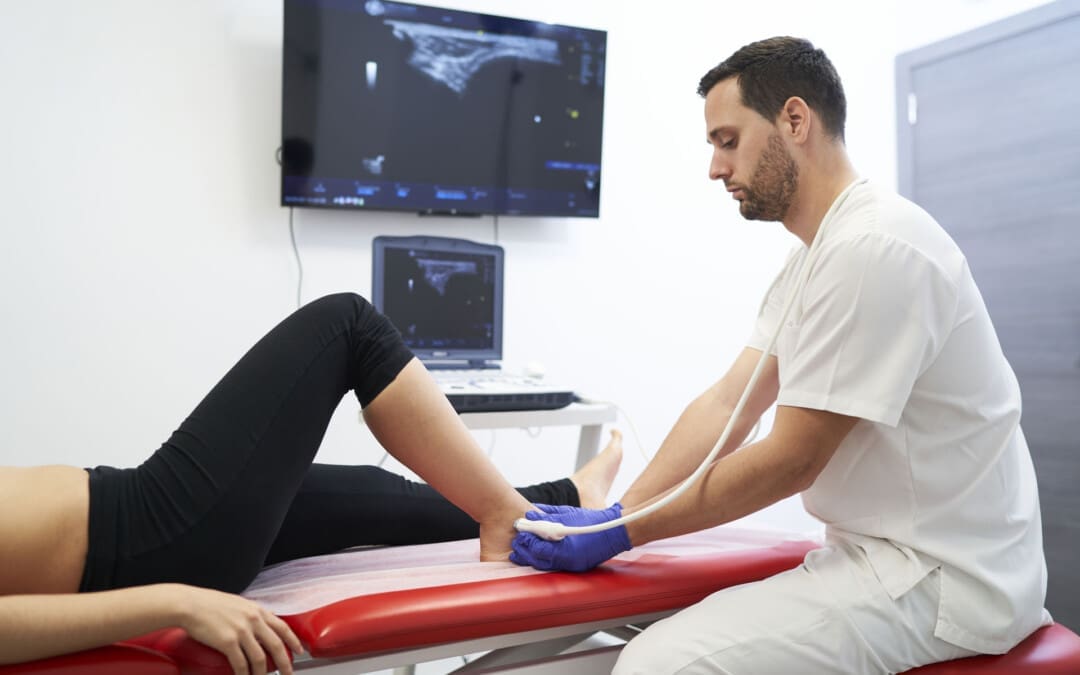
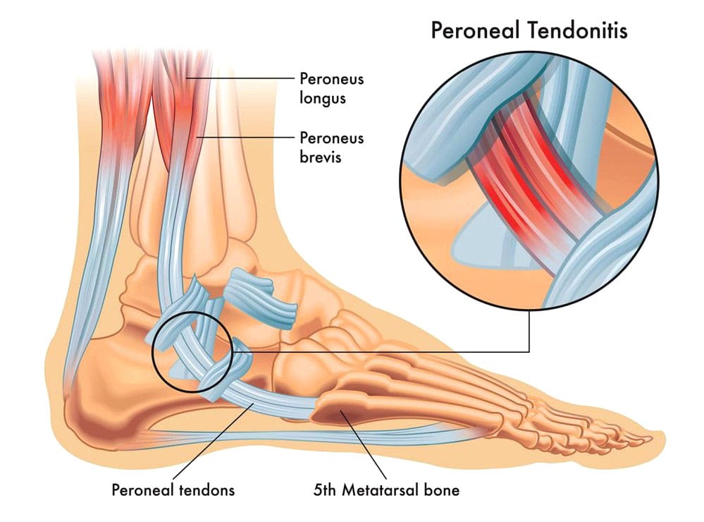
The peroneal tendons connect the muscles of the outer side of the lower leg to the foot. They may be acute—occurring suddenly—or chronic—developing over time. The basic types of peroneal tendon injuries are tendonitis, tears, and subluxation.
Anatomy and Function
The two major peroneal muscles (peroneus longus and peroneus brevis) are outside the lower leg, next to the calf muscles. The peroneal tendons run along the outer side of the ankle and attach to the foot, connecting these muscles to bone. They help stabilize the ankle joint, point the foot downward (plantarflexion), and turn the foot outward (eversion). In normal gait, the motion of the peroneal muscles is balanced by the muscles that invert the foot or rock the foot inward from the ankle. The two peroneal tendons sit one on top of the other right behind the fibula (the smaller lower leg bone). This closeness can contribute to problems with the peroneal tendons, as they rub together behind the ankle.
Tendonitis
The most common problem is inflammation or tendonitis. The tendons are usually inflamed just behind the fibula bone at the ankle joint. This part of the fibula is the bump on the outside of the ankle, and the peroneal tendons are located just behind that bony prominence. Tendonitis can either result from repetitive overuse or an acute injury. Common symptoms of tendonitis include:
Pain behind the ankle
Swelling over the peroneal tendons
Tenderness of the tendons
Pain usually worsens if the foot is pulled down and inwards, stretching the peroneal tendons.
Typical treatment of peroneal tendonitis is accomplished by:
Ice application
Applying ice to the area can help reduce swelling and control pain.
Rest
Resting is important to allow the tendon to heal.
A supportive device (walking boot or ankle brace) or crutches can help in severe cases.
Braces and boots provide support, reduce tendons’ stress, and allow rest and inflammation to subside.
Anti-inflammatory Medications
Motrin or Aleve are anti-inflammatory and can reduce the swelling around the tendon.
Physical Therapy
Physical therapy can help restore normal ankle joint mechanics, help with swelling and pain relief, and correct strength imbalances.
Cortisone Injections
Cortisone injections are low-risk if administered to the area around the tendon and not more often than every three months.
Ultrasound guidance can help ensure the medication is injected into the correct area. (Walt J. & Massey P. 2023)
Tendon Tears
Tears can occur and are more likely to happen in the peroneus brevis tendon. Tears are believed to be the result of two issues with the tendon. One is the blood supply. Tears of the peroneus brevis tendon almost always occur in the area where the blood supply and nutrition of the tendon are the poorest.
The second issue is the closeness between the two tendons, causing the peroneus brevis tendon to be wedged between the peroneus longus tendon and the bone. (Saxena A., & Bareither D. 2001) Tears of the peroneus brevis tendon are often treated with the same treatments for tendonitis. About half of the tears diagnosed by imaging are found to be asymptomatic. For individuals who don’t find lasting relief from symptoms, surgery may be necessary. Surgical options for peroneal tendon tears (Dombek M. F. et al., 2001)
Tendon Debridement and Repair
During a tendon debridement, the damaged tendon and the surrounding inflammatory tissue are removed.
The tear can be repaired, and the tendon can be tubularized to restore its normal shape.
Tenodesis
A tenodesis is a procedure where the damaged area of the tendon is sewn to the normal tendon.
In this case, the damaged segment of the peroneal tendon is removed, and the ends left behind are sewn to the adjacent remaining peroneal tendon.
Tenodesis is often recommended for tears involving more than 50% of the tendon. (Castilho R. S. et al., 2024)
Depending on the surgical procedure, Recovery after surgery can take several weeks of restricted weight-bearing and immobilization. Following immobilization, therapy can begin. Recovery is usually six to 12 weeks, depending on the surgery, but a full return to sports and activities may take several months. Risks of surgery include infection, stiffness, skin numbness near the incision, persistent swelling, and persistent pain.
Injury Medical Chiropractic and Functional Medicine Clinic
Injury Medical Chiropractic and Functional Medicine Clinic works with primary healthcare providers and specialists to build optimal health and wellness solutions. We focus on what works for you to relieve pain, restore function, prevent injury, and help mitigate issues through adjustments that help the body realign itself. They can also work with other medical professionals to integrate a treatment plan to resolve musculoskeletal problems.
The Difference of Using Custom Foot Orthotics
References
Walt, J., & Massey, P. (2025). Peroneal Tendon Syndromes. In StatPearls. https://www.ncbi.nlm.nih.gov/pubmed/31335074
Saxena, A., & Bareither, D. (2001). Magnetic resonance and cadaveric findings of the “watershed band” of the Achilles tendon. The Journal of foot and ankle surgery: official publication of the American College of Foot and Ankle Surgeons, 40(3), 132–136. https://doi.org/10.1016/s1067-2516(01)80078-8
Dombek, M. F., Orsini, R., Mendicino, R. W., & Saltrick, K. (2001). Peroneus brevis tendon tears. Clinics in podiatric medicine and surgery, 18(3), 409–427.
Castilho, R. S., Magalhães, J. M. B., Veríssimo, B. P. M., Perisano, C., Greco, T., & Zambelli, R. (2024). Minimally Invasive Peroneal Tenodesis Assisted by Peroneal Tendoscopy: Technique and Preliminary Results. Medicina (Kaunas, Lithuania), 60(1), 104. https://doi.org/10.3390/medicina60010104
For individuals dealing with newly formed or chronic lower back pain, can making daily walks a part of a weekly routine help relieve pain and discomfort symptoms and prevent strains and injuries?
Walking For Low Back Pain Relief
Walking is recommended to treat and prevent chronic or recurrent lower back pain. A study found that a personalized and progressive weekly walking program that builds up to 130 minutes of moderate intensity can significantly relieve severe lower back pain and prevent future flare-ups. (Pocovi N. C. et al., 2024) Walking is a cost-effective and easily accessible way to relieve lower back pain and prevent recurring or future injuries. It strengthens the back muscles, improves posture, and stabilizes the spine. (Suh JH, et al., 2019) Other benefits include improved overall physical health, posture, and circulation.
How Walking Helps
Walking for low back pain relief and general movement is better than not engaging in physical activities for individuals with recurrent lower back pain. Being sedentary can worsen back pain symptoms. (National Library of Medicine. 2019) Walking is second nature and is easy to incorporate into a weekly routine to help relieve back pain and improve overall health (Macquarie University, 2024)
Increases Spinal Flexibility
Walking and gentle movements increase the lower back’s functional range of motion, improve spinal flexibility, and reduce stiffness. (Smith J. A. et al., 2022)
Stabilizes Lumbar/Low Back Muscles
Walking builds muscle endurance and strength in the paraspinal muscles, increasing lumbar spinal stabilization. (Suh JH, et al., 2019)
Strengthens Core Muscles
Walking increases the body load and strengthens core muscles like the transversus abdominis, which lowers the risk of chronic lower back pain. (Lee J. S. and Kang S. J. 2016)
Improves Posture
Movement of the legs during walking enhances bodily awareness and helps correct posture.(Henry M. and Baudry S. 2019)
Increases Blood Circulation
Walking increases blood circulation to the muscles, supplying essential nutrients to spinal discs. It also reduces the frequency and severity of lower back muscle spasms. (Sitthipornvorakul E. et al., 2018)
Lubricates Spinal joints
Low-impact walking improves synovial fluid production and circulation, lubricating the lumbar spine’s facet joints and other joints that tend to get achy, such as the knees. (Zhang S. L. et al., 2013)
Relieves Inflammation
Walking helps reduce the presence of pro-inflammatory cytokines, like (IL-8 and TNF-alpha) associated with chronic lower back pain. (Slouma M. et al., 2023)
Promotes Weight Loss
Walking and a healthy diet can help individuals lose excess fat, which puts added strain on the lower back and correlates with lumbar intervertebral disc degeneration. (Wang M. et al., 2024)
Stress Relief
Regular walking can reduce mental stress associated with chronic lower back pain. (Choi S. et al., 2021)
Releases Endorphins
Moderate to vigorous physical activity, like walking at about 3 miles per hour for a half-hour daily, stimulates the release of endorphins, the body’s natural pain relievers. (Bruehl S. et al., 2020)
Walking Correctly
To get all the benefits of walking for low back pain relief, it is recommended to practice the following (Macquarie University, 2024)
Start slowly.
Gradually build intensity.
Stay consistent with the walking program.
Track progress to maintain motivation.
Healthcare Provider Consultation
Walking is a low-risk, low-impact activity well-tolerated by most individuals with nonspecific low back pain. (Pocovi N. C. et al., 2022) Because it doesn’t involve twisting or vigorous movements, it is considered a safe exercise for individuals with back pain symptoms (Gordon R. and Bloxham S. 2016). However, individuals experiencing severe lower back pain due to a traumatic injury or medical condition should consult a healthcare provider before starting a regular walking program.
Limit High Impact Activities
High-impact activities like running on hard surfaces or playing sports can exacerbate chronic lower back pain. If there is chronic lower back pain, it is recommended to limit activities that involve: (Al-Otaibi S. T. 2015)
Heavy lifting
Repetitive bending
Twisting motions
Injury Medical Chiropractic and Functional Medicine Clinic
Walking for low back pain relief. Injury Medical Chiropractic and Functional Medicine Clinic works with primary healthcare providers and specialists to develop an optimal health and wellness solution. We focus on what works for you to relieve pain, restore function, and prevent injury. Regarding musculoskeletal pain, specialists like chiropractors, acupuncturists, and massage therapists can help mitigate the pain through spinal adjustments that help the body realign itself. They can also work with other medical professionals to integrate a treatment plan to resolve musculoskeletal issues.
Movement as Medicine
References
Pocovi, N. C., Lin, C. C., French, S. D., Graham, P. L., van Dongen, J. M., Latimer, J., Merom, D., Tiedemann, A., Maher, C. G., Clavisi, O., Tong, S. Y. K., & Hancock, M. J. (2024). Effectiveness and cost-effectiveness of an individualised, progressive walking and education intervention for the prevention of low back pain recurrence in Australia (WalkBack): a randomised controlled trial. Lancet (London, England), 404(10448), 134–144. https://doi.org/10.1016/S0140-6736(24)00755-4
Suh, J. H., Kim, H., Jung, G. P., Ko, J. Y., & Ryu, J. S. (2019). The effect of lumbar stabilization and walking exercises on chronic low back pain: A randomized controlled trial. Medicine, 98(26), e16173. https://doi.org/10.1097/MD.0000000000016173
National Library of Medicine., & InformedHealth.org [Internet]. Cologne, G. I. f. Q. a. E. i. H. C. I. (2022). Low back pain: Learn More – Why movement is so important for back pain. https://www.ncbi.nlm.nih.gov/books/NBK284944/
Macquarie University. (2024). Macquarie University. Walking to combat back pain: world-first study shows dramatic improvement. https://lighthouse.mq.edu.au/article/june-2024/walking-away-from-pain-world-first-study-shows-dramatic-improvement-in-lower-back-trouble
Smith, J. A., Stabbert, H., Bagwell, J. J., Teng, H. L., Wade, V., & Lee, S. P. (2022). Do people with low back pain walk differently? A systematic review and meta-analysis. Journal of sport and health science, 11(4), 450–465. https://doi.org/10.1016/j.jshs.2022.02.001
Suh, J. H., Kim, H., Jung, G. P., Ko, J. Y., & Ryu, J. S. (2019). The effect of lumbar stabilization and walking exercises on chronic low back pain: A randomized controlled trial. Medicine, 98(26), e16173. https://doi.org/10.1097/MD.0000000000016173
Lee, J. S., & Kang, S. J. (2016). The effects of strength exercise and walking on lumbar function, pain level, and body composition in chronic back pain patients. Journal of exercise rehabilitation, 12(5), 463–470. https://doi.org/10.12965/jer.1632650.325
Henry, M., & Baudry, S. (2019). Age-related changes in leg proprioception: implications for postural control. Journal of neurophysiology, 122(2), 525–538. https://doi.org/10.1152/jn.00067.2019
Sitthipornvorakul, E., Klinsophon, T., Sihawong, R., & Janwantanakul, P. (2018). The effects of walking intervention in patients with chronic low back pain: A meta-analysis of randomized controlled trials. Musculoskeletal science & practice, 34, 38–46. https://doi.org/10.1016/j.msksp.2017.12.003
Zhang, S. L., Liu, H. Q., Xu, X. Z., Zhi, J., Geng, J. J., & Chen, J. (2013). Effects of exercise therapy on knee joint function and synovial fluid cytokine levels in patients with knee osteoarthritis. Molecular medicine reports, 7(1), 183–186. https://doi.org/10.3892/mmr.2012.1168
Slouma, M., Kharrat, L., Tezegdenti, A., Metoui, L., Ghazouani, E., Dhahri, R., Gharsallah, I., & Louzir, B. (2023). Pro-inflammatory cytokines in patients with low back pain: A comparative study. Reumatologia clinica, 19(5), 244–248. https://doi.org/10.1016/j.reumae.2022.07.002
Wang, M., Yuan, H., Lei, F., Zhang, S., Jiang, L., Yan, J., & Feng, D. (2024). Abdominal Fat is a Reliable Indicator of Lumbar Intervertebral Disc Degeneration than Body Mass Index. World neurosurgery, 182, e171–e177. https://doi.org/10.1016/j.wneu.2023.11.066
Choi, S., Nah, S., Jang, H. D., Moon, J. E., & Han, S. (2021). Association between chronic low back pain and degree of stress: a nationwide cross-sectional study. Scientific reports, 11(1), 14549. https://doi.org/10.1038/s41598-021-94001-1
Bruehl, S., Burns, J. W., Koltyn, K., Gupta, R., Buvanendran, A., Edwards, D., Chont, M., Wu, Y. H., Qu’d, D., & Stone, A. (2020). Are endogenous opioid mechanisms involved in the effects of aerobic exercise training on chronic low back pain? A randomized controlled trial. Pain, 161(12), 2887–2897. https://doi.org/10.1097/j.pain.0000000000001969
Pocovi, N. C., de Campos, T. F., Christine Lin, C. W., Merom, D., Tiedemann, A., & Hancock, M. J. (2022). Walking, Cycling, and Swimming for Nonspecific Low Back Pain: A Systematic Review With Meta-analysis. The Journal of orthopaedic and sports physical therapy, 52(2), 85–99. https://doi.org/10.2519/jospt.2022.10612
Gordon, R., & Bloxham, S. (2016). A Systematic Review of the Effects of Exercise and Physical Activity on Non-Specific Chronic Low Back Pain. Healthcare (Basel, Switzerland), 4(2), 22. https://doi.org/10.3390/healthcare4020022
Al-Otaibi S. T. (2015). Prevention of occupational Back Pain. Journal of family & community medicine, 22(2), 73–77. https://doi.org/10.4103/2230-8229.155370
Lower back leg pain depends on specific symptoms and their duration. Can having a better idea of symptoms help individuals inform their medical providers to develop an effective treatment plan?
Low Back Leg Pain
Lower back leg pain, sciatica, and weakness of the lower-extremity muscles are often diagnosed as a herniated (compressed or ruptured) disc. Nerves surrounding the spine are sensitive to irritation and pressure caused by a disc shifting out of position or physical damage to the disc and surrounding area, ranging from mild to severe. This is why it is important to be evaluated by a healthcare provider. Treatment depends on the type of spinal disc herniation and the severity, but getting an early medical evaluation helps ensure optimal outcomes. Non-surgical conservative treatments are often effective, but some cases may require more aggressive treatment, especially if the pain persists.
Spine and Nerves
Spinal discs are the shock absorbers between vertebrae. They consist of a tough outer layer, annulus fibrosis, covering a soft gel core, nucleus pulposus. When a disc is damaged, it can bulge and irritate surrounding nerves. In more severe cases, the annulus fibrosis can weaken and tear, allowing the material to leak and compress the spinal cord or nerves. As the nerves are not functioning properly, abnormal signals may be sent to and from the brain. The most common lower back herniations occur in the lumbar region, where five vertebrae near the base of the spine are classified from top to bottom as L1 through L5. (Dydyk A.M. et al., 2023) Pain resulting from an injury to this part of the spine can be debilitating because it may involve sciatic nerve irritation. Herniated disc causes are generally a combination of age-related degeneration, being overweight/obese, trauma, a sedentary lifestyle, and overloading of the spine. (Cleveland Clinic, 2021)
Symptoms
The most common symptoms include:
Back Pain
Caused by nerve irritation, muscle spasms, and inflammation.
Radiculopathy
Abnormal signaling of the nerves.
Electrical Shooting Pain
Nerve pressure can cause abnormal sensations, commonly experienced as electric shooting pains.
For low back herniations, the shocks go down one or both legs.
Tingling – Numbness
There are often abnormal sensations such as tingling, numbness, or pins and needles down one or both legs.
Muscle Weakness
Nerve signals may be interrupted, causing lower-body muscle weakness. (Dydyk A.M. et al., 2023)
Bowel – Bladder Symptoms
These symptoms may signal cauda equina syndrome, a rare condition resulting from a herniated disc between the L5 vertebrae and the first vertebrae of the sacrum.
Diagnosis
Diagnosing a herniated disc as the cause of low back leg pain involves testing sensation, muscle strength, and reflexes. MRI also aids this process (American Association of Neurological Surgeons, 2024). MRIs can often show herniated discs and other abnormalities, especially in older patients.
Treatment
A herniated disc treatment plan is based on patient symptoms, physical examination findings, and imaging results. Most herniated disc symptoms resolve themselves in four to six weeks. Lower back pain is generally treated conservatively through:
Topical pain ointments or creams for muscle spasms.
Non-surgical decompression relieves pressure, activates healing, and restores circulation and nutrients.
Chiropractic adjustments realign the spine and musculoskeletal system.
Massage loosens the muscles and maintains their relaxation.
Total rest is never recommended, even if movement is challenging,
Exercise and stretching help avoid muscle degeneration and strengthen the muscles.
Relaxation techniques and other natural pain therapies can help manage symptoms and restore overall health.
Pain-blocking injections which can include anesthetics or corticosteroids at the source (Cleveland Clinic, 2021)
Surgery is recommended only when conservative treatments are ineffective after six weeks, if there is significant muscle weakness from nerve damage, or if motor functions are compromised. (American Association of Neurological Surgeons, 2024)
Injury Medical Chiropractic and Functional Medicine Clinic
Chiropractic therapy is among the more conservative treatment options and may be tried first before proceeding with surgery. Injury Medical Chiropractic and Functional Medicine Clinic works with primary healthcare providers and specialists to develop an optimal health and wellness solution. We focus on what works for you to relieve pain, restore function, and prevent injury. Regarding musculoskeletal pain, specialists like chiropractors, acupuncturists, and massage therapists can help mitigate the pain through spinal adjustments that help the body realign itself. They can also work with other associated medical professionals to integrate a treatment plan to improve the body’s flexibility and mobility and resolve musculoskeletal issues.
Disc Herniation
References
Dydyk AM, Ngnitewe Massa R, Mesfin FB. Disc Herniation. [Updated 2023 Jan 16]. In: StatPearls [Internet]. Treasure Island (FL): StatPearls Publishing; 2024 Jan-. Available from: https://www.ncbi.nlm.nih.gov/books/NBK441822/
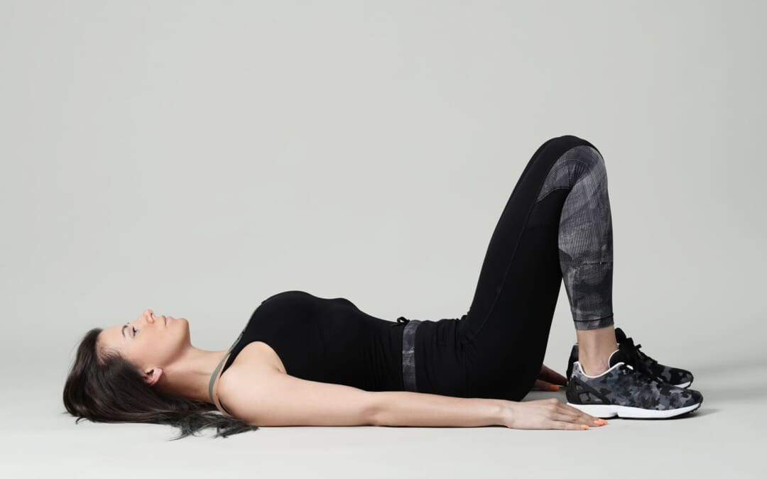
For individuals wanting to try Pilates for back pain and exercise, can learning how to find their neutral spine help improve flexibility and increase the range of motion in the joints?
Pilates Neutral Spine
Pilates is a functional exercise modality emphasizing core stability, which is fundamental to developing a balanced body. The exercises strengthen the muscles, improve flexibility, and increase the range of motion in the joints. (Kloubec J. 2011) It is considered a functional fitness method because its principles work to establish more graceful, efficient movements from everyday life, such as improving posture. Pilates has shown its effectiveness in that it is often used in physical therapy and rehabilitation settings. (Byrnes, K., Wu, P. J., and Whillier, S. 2018) However, knowing how to find the neutral spine is essential for performing various Pilates exercises correctly. (Barbosa, A. C. et al., 2018) This subtle adjustment during practice may help prevent injury and increase overall performance. A neutral spine is the natural position of the spine when all three curves:
Cervical (neck)
Thoracic (middle)
Lumbar (lower)
Are active and in healthy alignment.
This is the strongest position for the spine when standing or sitting, allowing the body to move more naturally.
Alignment
The following exercise can help find the Pilates neutral spine.
Basic Position
Lie on the back with knees bent and feet flat on the floor.
Ensure the legs are parallel to the hips, knees, heels, and toes.
Let the arms rest at your sides.
Relax
Relax the body, including the shoulders, neck, and jaw.
Allow the back to melt into the floor.
The rib cage will drop when the lower ribs are released to the floor.
Breathe Deep
Inhale all the way into the body, allowing it to move into the back and sides of the rib cage and all the way to the pelvis.
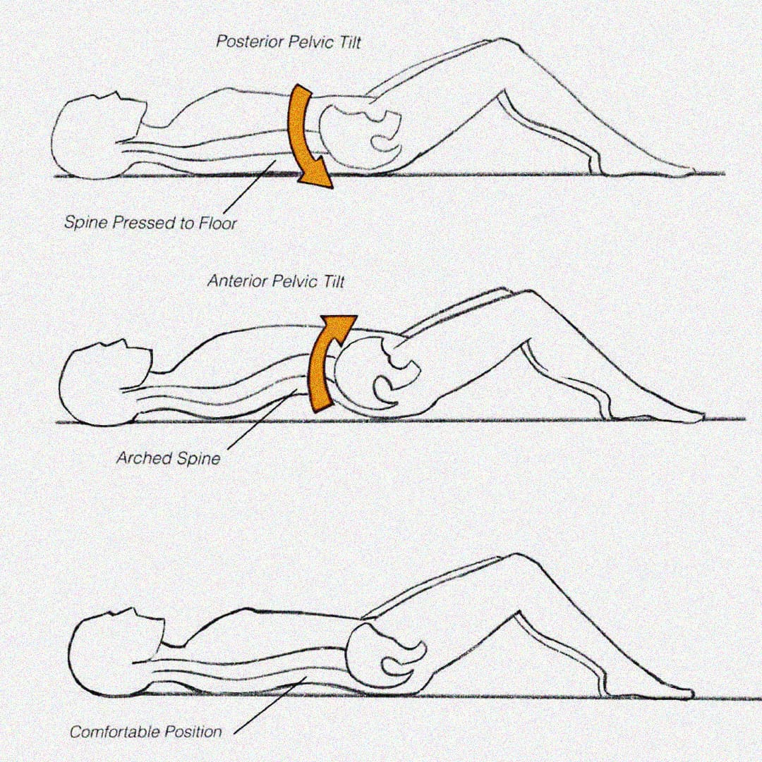
Pelvic Tilt
Exhale and use the abdominals to press the lower spine into the floor in a pelvic tuck. (Eickmeyer S. M. 2017)
Inhale to release.
Exhale and pull the lower spine off the floor, creating a pelvic tilt.
Inhale to release.
Pilates exercises don’t use excess energy or tension. Proper alignment and a neutral spine position can ensure that tension is released and excess energy is not exerted. (Byrnes, K., Wu, P. J., and Whillier, S. 2018) When performing the exercise, ensure that the shoulders, neck, and legs are relaxed and not involved in the movement.
When Exercising
Once a neutral spine is achieved, the goal is to maintain this spinal position during the exercises and when changing positions.
Start by lifting the right leg and placing it back down without letting the hips move.
Then, repeat the motion with the left leg.
Engage the abdominal muscles to help stabilize the pelvis, keeping it from moving and maintaining a neutral spine.
Repeat this process with each leg.
Once each leg can be lifted easily, test with both legs.
Exhale deeply and lift the legs while keeping the core and pelvis stable.
Then, lower them back down.
When performing this progression, there may be a want to release the abs and let the back arch.
This will cause a tuck and tilt position away from the neutral spine position.
If this progression is difficult, keep practicing until you can maintain a neutral spine.
Once this basic progression feels easy, try additional progressions and positioning.
Visualization Tips
Most people have their spines in one of two positions: tucked or tilted. A neutral spine requires individuals to be in between, with the lower abdominals flat and the lower spine’s natural curve slightly off the floor. The following visualization can help establish a neutral spine.
Balanced Pelvic Placement
Imagine a cup of water sitting on the lower abdomen, a couple of inches below the belly button.
Allow the abdominal muscles to drop toward the spine, flattening the belly.
You don’t want the water to spill, so the pelvis cannot be tipped forward or tucked under.
Body Scan Meditation
Once the body is relaxed with a balanced alignment on the floor.
Allow breathing to become deep and full and the abdominals to drop toward the floor.
The natural neck and lower spine curves should be off the floor.
Ensure the lower spine is not pressed into the floor, as this indicates a pelvic tilt.
If there is any discomfort or pain when working to increase endurance, seek advice from a healthcare professional. Injury Medical Chiropractic and Functional Medicine Clinic uses an integrated approach to treating injuries and chronic pain syndromes. It offers personalized care plans that improve ability through flexibility, mobility, and agility programs to relieve pain. Our providers use an integrated approach to create personalized care plans for each patient, including Functional Medicine, Acupuncture, Electro-Acupuncture, and Sports Medicine principles. Our goal is to relieve pain naturally by restoring health and function to the body. If other treatment is needed, Dr. Jimenez has teamed up with top surgeons, clinical specialists, medical researchers, and rehabilitation providers to provide the most effective treatments.
Is Motion Key to Healing?
References
Kloubec J. (2011). Pilates: how does it work and who needs it?. Muscles, ligaments and tendons journal, 1(2), 61–66.
Byrnes, K., Wu, P. J., & Whillier, S. (2018). Is Pilates an effective rehabilitation tool? A systematic review. Journal of bodywork and movement therapies, 22(1), 192–202. https://doi.org/10.1016/j.jbmt.2017.04.008
Barbosa, A. C., Vieira, E. R., Silva, A. F., Coelho, A. C., Martins, F. M., Fonseca, D. S., Barbosa, M. A., & Bordachar, D. (2018). Pilates experience vs. muscle activation during abdominal drawing-in maneuver. Journal of bodywork and movement therapies, 22(2), 467–470. https://doi.org/10.1016/j.jbmt.2017.05.002
Eickmeyer S. M. (2017). Anatomy and Physiology of the Pelvic Floor. Physical medicine and rehabilitation clinics of North America, 28(3), 455–460. https://doi.org/10.1016/j.pmr.2017.03.003
Can understanding the nucleus pulposus help in body positioning and prevention for individuals wanting to practice spinal hygiene and protect their discs from injury?
Nucleus Pulposus
The spinal discs are located between the spine’s vertebrae and are the body’s natural impact and shock absorbers. Within the disc is the nucleus pulposus, which plays a major role in providing the spine with shock absorption during movement. (Zhou Z. et al., 2014) The discs have a tough outer portion and a soft inner core. They are the:
It forms the tough circular exterior and comprises concentric sheets of collagen fibers or lamellae surrounding the inner core.
It has cartilaginous endplates that firmly attach to the vertebrae above and below.
Nucleus Pulposus
The nucleus pulposus is the inner core soft filling of the discs.
It contains a network of fibers suspended in a mucoprotein gel with a water base to maintain strength and pliability.
The near-liquid consistency makes it responsive to movement to handle the body’s axial load.
It helps maintain spinal suspension to prevent pressure on the bones and prevent bone-to-bone contact, reducing the potential for injuries and pain.
Shock Absorber
Each intervertebral disc is a shock-absorbing cushion, with the nucleus pulposus providing shock-absorbing properties (Zhou Z. et al., 2014). The intervertebral discs move as the body moves. For example, when arching the back, the disc moves forward slightly, and when twisting, the disc twists as well.
Spinal Action
The intervertebral disc supports spinal movements. When bending, twisting, arching, or tilting the spine, the nucleus pulposus swivels to accommodate these actions. These repeated spinal actions, which occur throughout the day and night, contribute to shifting positions while sitting, working, playing sports, carrying groceries, performing house chores, etc. An example is bending forward to pick something up. This action involves forward spinal flexion, which is bending the spine forward, flattening, or rounding. When bending using flexion, the spinal bones come closer together, pushing the nucleus pulposus toward the back.
Injuries
The disc can be pushed too far back with persistent or excessive spinal flexion. If the fibers of the annulus fibrosus become weak, they can tear, causing the nucleus pulposus to leak out and disc herniation. Generally, the nucleus pulposus will leak to the side and back; however, this corresponds to the location of the very sensitive nerve root/s with which it can come into contact, causing pain and other symptoms. The most common causes of disc herniation are degenerative wear and tear changes of the disc and trauma. Disc degeneration occurs as the body ages; it weakens the annulus fibers, allowing the nucleus pulposus to distend, bulge, or herniate.
Aging
Disc degeneration occurs with age but can also occur with injuries to the area. In young individuals, the nucleus pulposus is mostly water. For this age group, a herniation from trauma is more likely than in older individuals. (Ucar, D. et al., 2021) But as the body ages, the discs, especially the nucleus pulposus, begin to dry out. This dehydration leads to a significant loss of disc height. (UCLA Health, 2024) By age 60 or 70, the discs may be composed entirely of fiber, which can cause the shock absorption function not to work and disappear.
Chiropractic therapy is among the more conservative treatment options for a herniated disc and may be tried first before proceeding with more invasive treatments. Injury Medical Chiropractic and Functional Medicine Clinic works with primary healthcare providers and specialists to develop an optimal health and wellness solution that fully benefits the individual to get back to normal.
The Science of Functional Healing
References
Zhou, Z., Gao, M., Wei, F., Liang, J., Deng, W., Dai, X., Zhou, G., & Zou, X. (2014). Shock absorbing function study on denucleated intervertebral disc with or without hydrogel injection through static and dynamic biomechanical tests in vitro. BioMed research international, 2014, 461724. https://doi.org/10.1155/2014/461724
Nosikova, Y. S., Santerre, J. P., Grynpas, M., Gibson, G., & Kandel, R. A. (2012). Characterization of the annulus fibrosus-vertebral body interface: identification of new structural features. Journal of anatomy, 221(6), 577–589. https://doi.org/10.1111/j.1469-7580.2012.01537.x
Ucar, D., Duman, S., Bayram, Y., & Ucar, B. Y. (2021). Extruded disc herniations are experienced earlier by inactive young people in the high-tech gaming era. Journal of medicine and life, 14(3), 402–407. https://doi.org/10.25122/jml-2021-1059
Can kimchi benefit individuals trying to incorporate more fermented foods into their diet?
Kimchi
Kimchi is a flavorful and nutritious food packed with nutritious vegetables. It is high in vitamin C, vitamin A, and iron. It is made of salted, fermented vegetables and typically served as a side dish that starts with cabbage as the base. Other varieties use different vegetables, like radish, cucumber, and onion. It has minimal calories, a low carb count, zero fat, and health benefits like an abundance of probiotics from its fermentation process.
Nutrition
Kimchi is an excellent source of vitamin C and vitamin A. A typical half-cup of kimchi is 85 grams and provides the following. (U.S. Department of Agriculture. 2017)
Calories – 20
Fat – 0g
Sodium – 290 milligrams
Carbohydrates – 4 grams
Fiber – 1 grams
Sugars – 2 grams
Protein – 1 grams
Vitamin C – 18 milligrams
Iron – 1.08 milligrams
Vitamin A – 375 micrograms
Calcium – 40 milligrams
Calories
A half-cup serving provides 20 calories, about 53% of which are carbohydrates, 21% are protein, and 26% are fat.
Carbohydrates
Kimchi comprises 4 grams of carbohydrates per serving, with 1 being fiber.
However, many kimchi recipes add sweeteners, like honey or fruit juice, to balance the sourness.
More sweeteners means more carbohydrates.
Fats
Because it is primarily vegetables, it is naturally fat-free.
Protein
Kimchi isn’t exactly a protein-power player.
A half-cup serving provides just 1 gram of plant-based protein from veggies.
However, recipes that include seafood like shrimp or squid will contain higher amounts of this macronutrient.
Vitamins and Minerals
Vitamins and minerals vary depending on the vegetables used.
A Napa cabbage-based kimchi includes abundant vitamins C and K and smaller amounts of iron, calcium, copper, and potassium.
A recipe with carrots will contain significant vitamin A.
A recipe with radishes will supply folate, potassium, and riboflavin.
All varieties are made with salt, so sodium is a mineral to watch.
A half-cup serving may provide nearly 300 milligrams or 13% Daily Value of sodium.
Benefits
Kimchi is a versatile food that can provide health benefits.
Digestion
The lactic acid that ferments the cabbage also provides healthy gut bacteria.
Consuming probiotics through kimchi promotes healthy digestion and helps alleviate constipation problems. (Higashikawa, F. et al., 2010)
Compatible with Special Diets
With simple plant-based ingredients it can be suitable for specialized diets.
It suits vegan, vegetarian, low-carb, gluten-free, and dairy-free diets.
Immune System Support
The probiotics in fermented foods improve digestion and may help improve immune function.
Research has suggested that when individuals stop eating fermented foods, their immune response decreases. (Olivares, M. et al., 2006)
Researchers isolated a compound in kimchi called HDMPPA – 3-(4′-hydroxyl-3′,5′-dimethoxyphenyl) propionic acid –
and studied its interaction with inflammatory proteins.
They discovered that HDMPPA counteracted the proteins’ inflammatory effect.
It is not enough to conclude that kimchi readily reduces inflammation, but further research could help confirm its ability. (Jeong, J. W. et al., 2015)
Improve Asthma Symptoms
A study of Korean adults with asthma found that the more kimchi they consumed, the less likely they were to experience an asthma attack.
Further research is needed, but the results are promising. (Kim, H. et al., 2014)
Allergies
Commercial and home-prepared kimchi is often free of all top eight food allergens—but check ingredient labels to be sure.
Some preparations, for example, may contain fish sauce, shrimp, or shrimp paste, which are a no-go for those with a fish or shellfish allergy.
Adverse Effects
Kimchi may have adverse effects on some individuals depending on its preparation.
It could be high in sodium, which may not be recommended for individuals on a heart-healthy or sodium-restricted diet.
With high levels of probiotics, it could cause bloating or an upset stomach.
Individuals sensitive to strong flavors may not enjoy the taste.
Varieties
Traditionally, kimchi is made from cabbage, but a wide variety of vegetables can be substituted for or combined with recipes that use alternative vegetables, spices, or other additions. Some recipes include fish or meat to turn it inta a main dish. Water kimchi is a soup version served in broth. But what makes kimchi is its base of fermented vegetables.
Storage and Safety
Fermentation can be tricky when it comes to food safety. Store-bought or homemade kimchi properly canned in a sterilized jar can be kept at room temperature for up to a week after opening. Stored in the refrigerator, it will stay fresh for three to six months. The beneficial bacteria working and fermentation process is ongoing, making the taste increasingly sour and texture mushier over time. This does not mean the jar has gone bad as long as it has no odd smell or mold.
Preparation
The process is not that complex.
Select a recipe with vegetables like cabbage, radish, and carrots.
Slice the vegetables into chunks and rub with salt.
Leave the vegetables in salt; some recipes include water for several hours to allow fermentation.
Drain the excess water, then add flavoring ingredients like sweeteners and spices.
Serve as a side dish with fried rice or noodles, or make it a main course by adding fish, meat, or tofu.
Injury Medical Chiropractic and Functional Medicine Clinic focuses on and treats injuries and chronic pain syndromes through personalized care plans that improve ability through flexibility, mobility, and agility programs to relieve pain. Our providers use an integrated approach to create personalized care plans for each patient, to restore health and function to the body through Nutrition and Wellness, Functional Medicine, Acupuncture, Electro-Acupuncture, and Sports Medicine protocols. If the individual needs other treatment, they will be referred to a clinic or physician best suited for them, as Dr. Jimenez has teamed up with the top surgeons, clinical specialists, medical researchers, nutritionists, and health coaches to provide the most effective clinical treatments.
The Healing Diet
References
U.S. Department of Agriculture. FoodData Central. (2017). Kimchi. Retrieved from https://fdc.nal.usda.gov/fdc-app.html#/food-details/516912/nutrients
Higashikawa, F., Noda, M., Awaya, T., Nomura, K., Oku, H., & Sugiyama, M. (2010). Improvement of constipation and liver function by plant-derived lactic acid bacteria: a double-blind, randomized trial. Nutrition (Burbank, Los Angeles County, Calif.), 26(4), 367–374. https://doi.org/10.1016/j.nut.2009.05.008
Olivares, M., Paz Díaz-Ropero, M., Gómez, N., Sierra, S., Lara-Villoslada, F., Martín, R., Miguel Rodríguez, J., & Xaus, J. (2006). Dietary deprivation of fermented foods causes a fall in innate immune response. Lactic acid bacteria can counteract the immunological effect of this deprivation. The Journal of dairy research, 73(4), 492–498. https://doi.org/10.1017/S0022029906002068
National Institutes of Health Office of Dietary Supplements. (2021). Vitamin C: Fact sheet for health professionals. Retrieved from https://ods.od.nih.gov/factsheets/VitaminC-HealthProfessional/
Jeong, J. W., Choi, I. W., Jo, G. H., Kim, G. Y., Kim, J., Suh, H., Ryu, C. H., Kim, W. J., Park, K. Y., & Choi, Y. H. (2015). Anti-Inflammatory Effects of 3-(4′-Hydroxyl-3′,5′-Dimethoxyphenyl)Propionic Acid, an Active Component of Korean Cabbage Kimchi, in Lipopolysaccharide-Stimulated BV2 Microglia. Journal of medicinal food, 18(6), 677–684. https://doi.org/10.1089/jmf.2014.3275
Kim, H., Oh, S. Y., Kang, M. H., Kim, K. N., Kim, Y., & Chang, N. (2014). Association between kimchi intake and asthma in Korean adults: the fourth and fifth Korea National Health and Nutrition Examination Survey (2007-2011). Journal of medicinal food, 17(1), 172–178. https://doi.org/10.1089/jmf.2013.3013
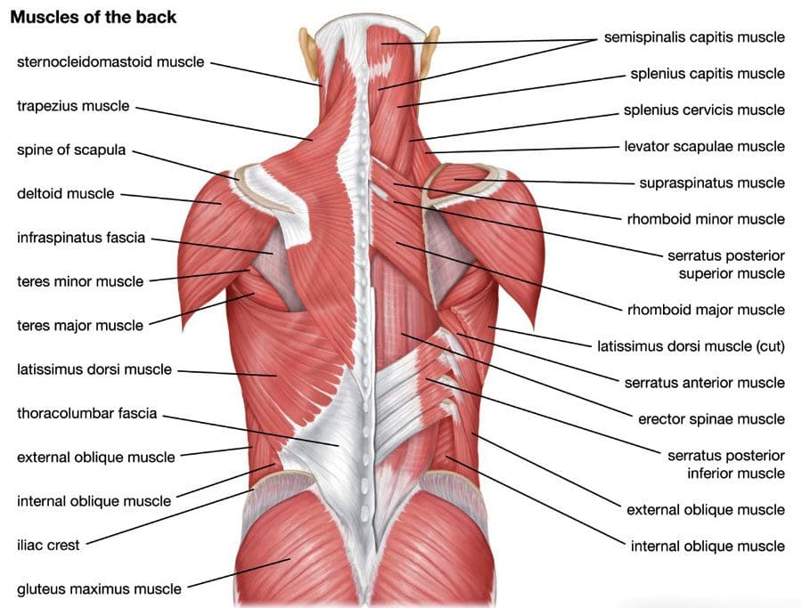
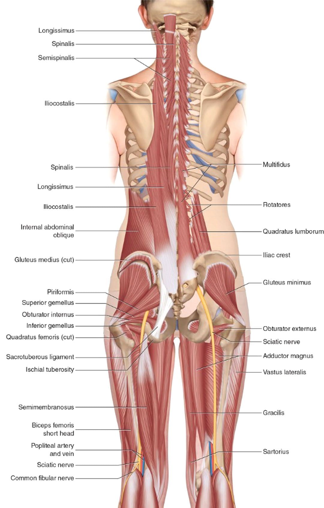
Can the thoracolumbar fascia cause or contribute to lower back pain and inflammation?
Thoracolumbar Fascia
Tissue behind the spinal column, positioned at both the lower back and mid-back levels, is connected to the thoracolumbar fascia, also called the lumbodorsal fascia or LF. The fascia is a thick connective tissue that covers and supports all the body’s muscles, bones, tendons, ligaments, and organs. The fascia also contains nociceptive nerve endings, also known as free nerve endings, that arise from the central nervous system, i.e., the brain and spinal cord, which may be responsible for some forms of back pain and stiffness caused by injury or inflammation.
Anatomy
The thoracolumbar fascia is divided into three layers:
Many of the back muscles attach to the thoracolumbar fascia. The erector spinae muscle group, known as the paraspinals, runs longitudinally down the spine. They are attached to the thoracolumbar fascia and the bony spine. The lumbar part of the posterior layer of the thoracolumbar fascia extends from the lowest rib to the top of the hip bone or the iliac crest. On the same path, it connects with the transverse abdominal muscle. The thoracolumbar fascia connections help bridge the back muscles to the abdominal wall muscles. The latissimus dorsi, a large back muscle that bears and moves the body’s weight with the arms and shoulders, is also connected to the thoracolumbar fascia, with the fibers extending outward from the fascia. The front part of the thoracolumbar fascia, or anterior layer, covers a muscle called the quadratus lumborum. This muscle bends the trunk to the side, helps maintain a healthy posture, and is often focused on muscle-related lower back pain.
What the Fascia Does
The thoracolumbar fascia, examined from the back of an anatomical drawing or diagram, is diamond-shaped. Its shape, large size, and central location uniquely position it to unify and synchronize the upper body’s movements with the lower body’s. The fascia’s fibers are very strong, enabling the tissue sheath to lend support (Willard, F. H. et al., 2012) . The tissue is also flexible, enabling it to help circulate forces of movement and contralateral movements as the back muscles contract and relax. An example is walking.
Back Pain
Scientists and doctors don’t know for sure, but it’s possible that the thoracolumbar fascia may contribute to lower back pain. A study found that the fascia may generate back pain based on: (Wilke, J. et al., 2017)
Sustaining micro-injuries and/or inflammation, which are often related, may cause signal changes in the free nerve endings in the fascia. Nerve endings acquire information from the outer areas of the body, like skin and other fascia, and relay it back to the central nervous system. The theory is that when the fascia close to the skin becomes injured, damaged, and/or backed up with inflammatory chemicals and substances, it is communicated as pain and other sensations back to the brain and spinal cord.
After a back injury, tissues tighten and stiffen. Some studies of patients with back pain noted alterations in their thoracolumbar fascia.
Injuries tend to stimulate nerves, which can lead to increased sensitivity.
Injury Medical Chiropractic and Functional Medicine Clinic focuses on and treats injuries and chronic pain syndromes through personalized care plans that improve ability through flexibility, mobility, and agility programs to relieve pain. Our providers use an integrated approach to create personalized care plans for each patient, including Functional Medicine, Acupuncture, Electro-Acupuncture, and Sports Medicine principles. Our goal is to relieve pain naturally by restoring health and function to the body. If other treatment is needed, Dr. Jimenez has teamed up with top surgeons, clinical specialists, medical researchers, and rehabilitation providers to provide the most effective treatments.
Sciatica, Causes, Symptoms, and Tips
References
Willard, F. H., Vleeming, A., Schuenke, M. D., Danneels, L., & Schleip, R. (2012). The thoracolumbar fascia: anatomy, function and clinical considerations. Journal of anatomy, 221(6), 507–536. https://doi.org/10.1111/j.1469-7580.2012.01511.x
Wilke, J., Schleip, R., Klingler, W., & Stecco, C. (2017). The Lumbodorsal Fascia as a Potential Source of Low Back Pain: A Narrative Review. BioMed research international, 2017, 5349620. https://doi.org/10.1155/2017/5349620
IFM's Find A Practitioner tool is the largest referral network in Functional Medicine, created to help patients locate Functional Medicine practitioners anywhere in the world. IFM Certified Practitioners are listed first in the search results, given their extensive education in Functional Medicine


 What the Fascia Does


