Clinical Walkthrough: Ultrasound-Guided Hydrodissection for Radial Tunnel Pain
Abstract
In this educational post, I present a clear, evidence-based walkthrough of ultrasound-guided hydrodissection for radial tunnel–related lateral elbow pain, grounded in modern musculoskeletal research and my clinical observations.
I explain why patients with forearm-burning pain aggravated by pronation and supination often benefit from targeted nerve hydrodissection, how we safely perform perineural injections under ultrasound guidance, and where integrative chiropractic care and rehabilitation accelerate functional recovery.
I also describe our multidisciplinary workflow at Injury Medical Clinic PA (Mission Plaza Injury Medical Clinic) in El Paso, Texas, where I collaborate with Dr. Maria Guadalupe Cardenas, MD, to combine medical oversight with integrative chiropractic, functional rehabilitation, and personal injury care.
My focus here is on chiropractic and physical therapy–driven strategies, using medications and hormones only as background support when necessary.
By the end, you will understand the physiologic rationale for hydrodissection, how it reduces nerve irritation and restores movement, and how a coordinated plan supports lasting outcomes.
Hydrodissection For Radial Tunnel–Related Lateral Elbow Pain: A First-Person Clinical Walkthrough
As Dr. Alex Jimenez, DC, APRN, FNP-BC, CFMP, IFMCP, ATN, CCST, I routinely evaluate persistent lateral elbow and dorsal forearm pain—especially when classic “tennis elbow” does not fit the picture. When a patient reports a dull, burning discomfort in the dorsum of the forearm, worsened by pronation and supination, and tenderness is distal to the lateral epicondyle, I consider involvement of the deep branch of the radial nerve (posterior interosseous nerve) near the radial tunnel and the arcade of Frohse over the supinator muscle.
In the case described below, the patient experienced more than six months of pain. A 1 mL lidocaine test injection delivered to the vicinity of the radial nerve temporarily improved symptoms, corroborating a nerve-entrapment pain generator. From there, we proceeded with ultrasound-guided hydrodissection—a perineural technique using carefully controlled fluid volumes to free and “float” the nerve away from surrounding fascial planes, adhesions, and compressive interfaces.
Why Hydrodissection? Physiologic Rationale And Evidence
Entrapment physiology:
The radial tunnel is a tight anatomical corridor where fascial thickening, muscle hypertonicity, or micro-adhesions can compress the deep branch of the radial nerve.
This compression causes ischemia, mechanical deformation, and neurogenic inflammation—manifesting as burning pain, weakness in wrist or finger extension, and symptom provocation with forearm rotation.
Perineural hydrodissection mechanism:
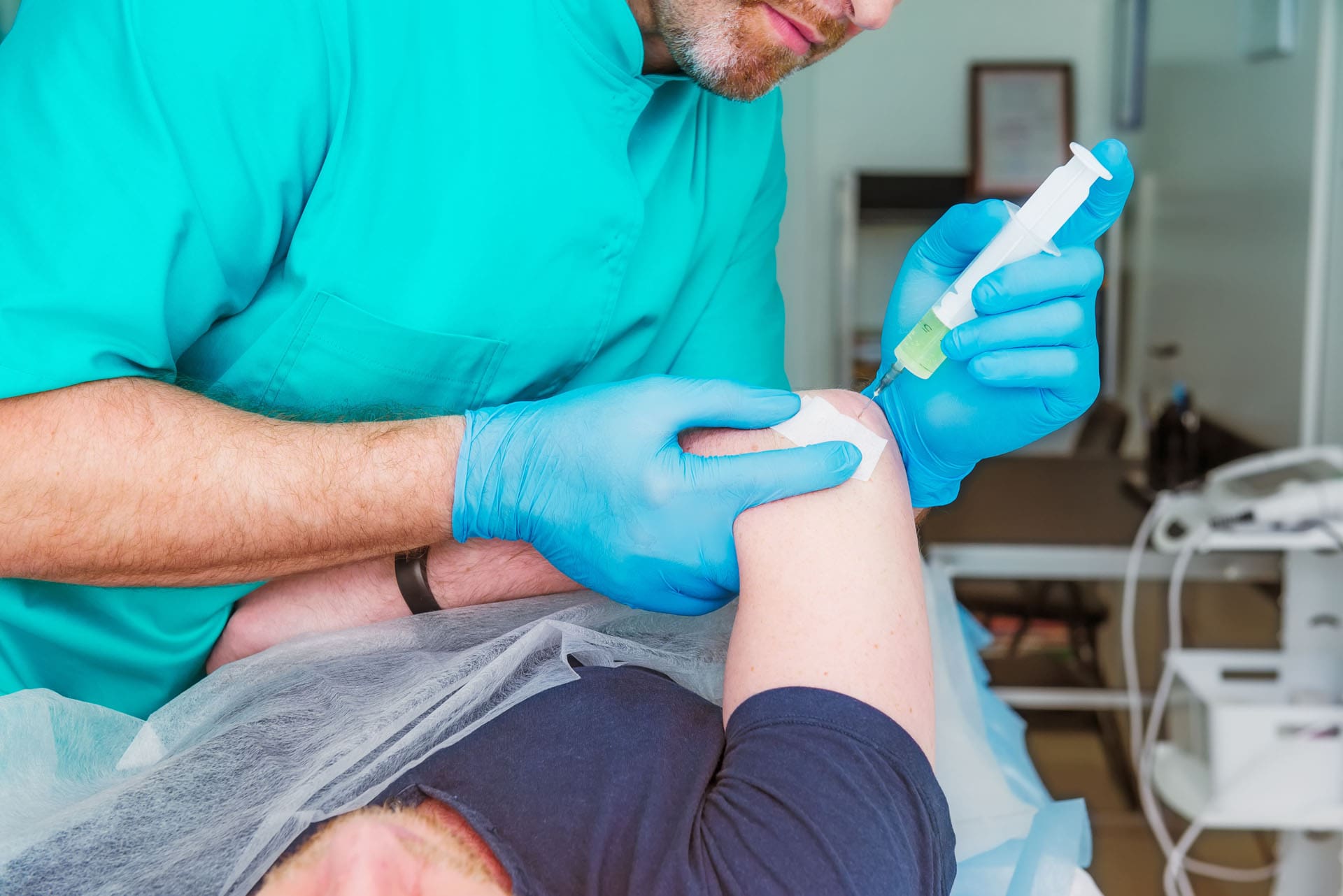
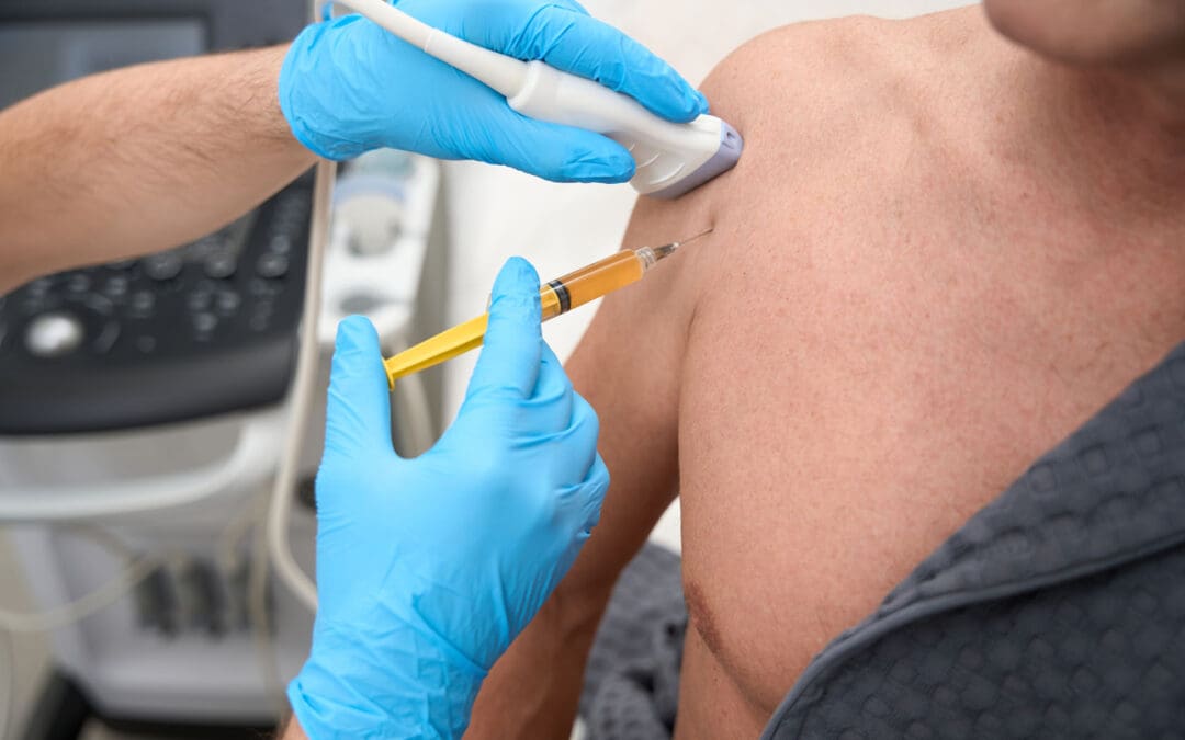
Using ultrasound guidance, a needle is advanced to the perineural plane—never intraneural—and small, pulsed injections of fluid are delivered circumferentially.
The fluid creates a “halo” effect around the nerve, gently separating it from tight fascial envelopes and adjacent tissues, reducing local mechanical stress and improving microvascular perfusion.
The result is decreased nociceptor activation, improved axoplasmic flow, and the potential restoration of normal neurodynamics during movement (Brown et al., 2022; Cass et al., 2021).
Evidence-based insight:
Ultrasound-guided perineural hydrodissection has been studied in peripheral nerve entrapments, showing favorable short- and mid-term outcomes, especially when combined with rehabilitation to address root causes and movement patterns (Wu et al., 2017; Lam et al., 2021).
The superiority of ultrasound guidance lies in precise needle visualization, reduced risk of intraneural placement, and real-time confirmation of fluid spread (Özçakar et al., 2018).
Clinical Orientation: What I See On Ultrasound
Landmarks:
Brachioradialis lies superficially and laterally; the supinator wraps around the proximal radius; the deep branch of the radial nerve courses between them, just proximal to the arcade of Frohse.
The neurovascular bundle is identified between the brachioradialis and supinator, with the nerve appearing as a honeycomb structure in short axis.
Approach:
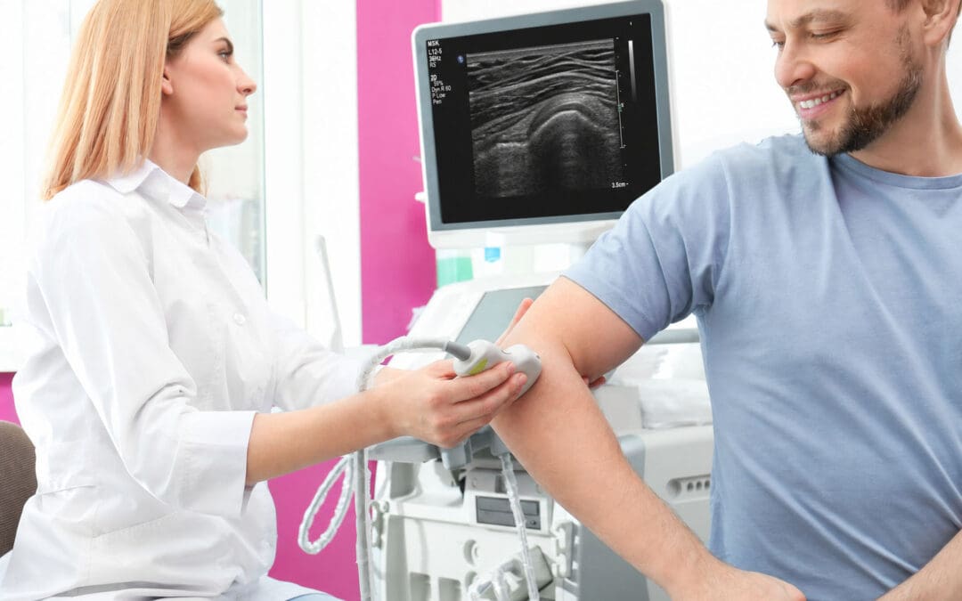
I position a linear ultrasound probe for a short-axis view of the deep branch of the radial nerve.
Using an in-plane technique, I introduce a fine needle (often a 25-gauge) under ultrasound visualization, advancing through the brachioradialis toward the perineural space around the nerve.
Confirmation:
I watch for muscle twitching when passing through contractile tissue—expected and typically benign—and constantly verify needle-tip location relative to the nerve.
The target is the perineural plane. We avoid the nerve fascicles themselves. The goal is a circumferential fluid halo that elevates and frees the nerve from adhered tissue.
Step-By-Step Hydrodissection: Perineural, Not Intraneural
Preparation and safety:
Anesthetize the skin with a cold spray or local infiltration.
Strict sterile technique with probe cover and sterile gel.
Real-time ultrasound ensures continuous visualization of the needle path and the nerve.
We check for prior response to diagnostic local anesthetic—useful for confirming the pain generator.
Needle movement and fluid delivery:
I make very small, subtle adjustments of the needle tip while keeping the ultrasound beam aligned.
I pulse small aliquots of fluid—often lidocaine with a minute dose of corticosteroid or buffered saline—to create gentle blunt dissection around the nerve.
I rotate the needle around the nerve’s perimeter (above, lateral, and below) to complete the halo, avoiding direct nerve penetration.
Why these choices?
Lidocaine provides immediate analgesia, facilitating comfortable movement post-procedure.
A small corticosteroid component may reduce perineural inflammation for several weeks, giving tissue time to remodel and patients time to implement corrective rehabilitation (Chang et al., 2019).
The hydrodissection itself is the primary mechanical solution—releasing entrapment and normalizing glide.
Integrative Chiropractic Care: How We Restore Function After Hydrodissection
Hydrodissection relieves the mechanical compression, but patients achieve durable results when we address biomechanics and neuromuscular control. At El Paso’s Injury Medical Clinic PA (Mission Plaza Injury Medical Clinic), I integrate chiropractic, functional rehabilitation, and physical therapy to correct movement patterns that feed radial tunnel stress.
Chiropractic neuro-biomechanical assessment:
I evaluate cervical and thoracic alignment, scapular mechanics, first rib mobility, and radial head mechanics.
Dysfunctions at the cervical spine (particularly C5–C7) can alter motor patterns through the radial nerve distribution; thoracic stiffness can disrupt scapulothoracic rhythm, increasing demand on forearm musculature.
Specific chiropractic techniques and reasoning:
Radial head mobilization: Restores proximal radio-ulnar mechanics, reducing supinator strain on the deep radial nerve during pronation-supination cycles.
Cervical and thoracic adjustments: Improve segmental mobility, reduce aberrant muscle guarding, and normalize descending motor control—beneficial for forearm extensor loading.
First rib and scapular mobilization: Optimizes scapular upward rotation and reduces compensatory forearm overuse.
Physical therapy integration:
Nerve gliding for the radial nerve: Gentle, pain-free sliders that encourage normal neural excursion and reduce post-procedure adhesiogenesis. We avoid aggressive tensioners early to protect the perineural interface (Shacklock, 2005).
Isometric extensor loading: Early-stage isometrics reduce pain via central modulation and prepare tissues for progressive strengthening.
Eccentric-concentric forearm training: Targets extensor carpi radialis brevis/longus and supinator-pronator balance, gradually building resilience.
Proprioceptive and motor control drills: Wrist stabilization, scapular setting, and kinetic chain integration to reduce forearm overload during work or sport.
Soft tissue and myofascial methods:
Instrument-assisted soft tissue mobilization and precise myofascial release around the supinator-brachioradialis interval can complement hydrodissection by promoting glide and lymphatic flow.
Dry needling may be considered for myofascial trigger points, performed conservatively to avoid nerve irritation.
Medical Oversight With Dr. Maria Guadalupe Cardenas, MD: Safety And Coordination
Our multidisciplinary clinic benefits from the seasoned medical direction of Dr. Maria Guadalupe Cardenas, MD (Board Certified in Internal Medicine, NPI #1164426749, Texas MD License #J2933), who has practiced internal medicine for over 40 years. She collaborates with me across personal injury, musculoskeletal pain, and functional recovery settings.
Role of medical oversight:
Ensures patient selection, contraindication screening (e.g., bleeding disorders, anticoagulant use, active infection), and medically appropriate use of local anesthetics and corticosteroids during hydrodissection.
Coordinates diagnostic frameworks and follow-up schedules, including consideration of imaging if atypical features emerge (e.g., profound weakness suggesting true posterior interosseous nerve palsy).
Provides backup strategies for complex cases where systemic factors—like diabetes or inflammatory conditions—increase neuropathic vulnerability.
Why this matters:
Interprofessional collaboration increases procedural safety and supports evidence-based care—from diagnosis to post-procedure rehabilitation—while keeping medication and hormonal therapy in the background unless clearly indicated for comorbidities.
Functional Medicine In Context: Keeping It Focused On Movement
Although my credentials include advanced functional medicine training, at El Paso Back Clinic we focus on movement-based solutions and reserve metabolic or hormonal interventions for select cases. Where appropriate:
We may recommend anti-inflammatory nutrition basics to support tissue healing.
We prioritize sleep optimization, stress modulation, and graded activity, all shown to affect pain perception and recovery trajectories (Clarke et al., 2023).
Supplements, if used, are adjuncts—not replacements—for chiropractic and physical therapy.
Personal Injury Care: Documentation And Recovery Pathways
In personal injury cases, clear causation and precise documentation are crucial:
High-resolution ultrasound documentation of nerve appearance, surrounding fascial tightness, and fluid spread during hydrodissection.
Objective measures: Grip strength, dynamometry, range-of-motion in pronation-supination, and functional tests tied to occupational demands.
Return-to-work plans: Staged exposure with ergonomic coaching and task modifications to prevent recurrence.
My Clinical Observations: What Patients Experience And Why
Drawing from years of care at El Paso Back Clinic and my clinical notes shared via our platforms, including our website and professional profiles:
Patients with dorsal forearm burning pain often report rapid reduction in symptoms after hydrodissection, particularly when the fluid halo is well established and the radial tunnel is decompressed.
Those who adhere to nerve glides, proximal kinetic chain correction, and graded strengthening experience more durable outcomes.
When cervical or scapular mechanics are neglected, symptoms tend to creep back during high-repetition tasks. Addressing the whole chain prevents over-reliance on the forearm extensors.
Safety Considerations And How We Minimize Risk
Perineural, not intraneural: We never inject directly into the nerve fascicles; ultrasound confirms placement.
Conservative volumes: Adequate to create separation without excessive tissue pressure.
Real-time visualization: The needle tip is visible throughout the procedure; we observe and respect tissue twitch.
Post-procedure monitoring: We encourage gentle movement the same day to promote neural glide and reduce stiffness. We advise patients on transient numbness from lidocaine and set expectations for gradual improvement over days to weeks.
When Hydrodissection Is Appropriate—And When It’s Not
Appropriate when:
Pain localizes distally in the dorsal forearm, worse with rotation, and examination indicates radial tunnel irritation.
The patient demonstrates temporary relief with a diagnostic local anesthetic block.
There is no frank motor deficit suggesting a complete posterior interosseous nerve palsy requiring surgical evaluation.
Consider alternatives or adjuncts when:
Pure lateral epicondylitis without nerve involvement is present—then tendinopathy protocols dominate the plan (eccentric loading, manual therapy).
Systemic neuropathies or inflammatory arthropathies are the primary drivers—then medical and rheumatologic collaboration guides care.
Putting It All Together: A Patient-Centered Care Pathway
Initial visit:
Thorough history, functional assessment, and targeted ultrasound of the radial tunnel.
Rule in nerve involvement with examination maneuvers and consider a small test lidocaine injection.
Procedure day:
Ultrasound-guided perineural hydrodissection using a fine needle and pulsed fluid delivery.
Immediate post-procedure instructions: gentle range of motion; avoid aggressive loading on day one.
Rehabilitation phase:
Begin radial nerve sliders, isometric extensor work, and scapular posture drills.
Progress to eccentric-concentric strengthening and functional task replication.
Maintenance:
Address workstation ergonomics, grip variation strategies, and sport technique refinements.
Periodic rechecks to ensure sustained neurodynamics and joint mechanics.
Summary
Today’s post provides a transparent, first-person account of how I identify and treat radial tunnel–related lateral elbow pain using ultrasound-guided hydrodissection, followed by integrative chiropractic and physical therapy. The core physiologic goals are to reduce perineural compression, normalize nerve glide, and retrain movement patterns across the kinetic chain. With medical oversight by Dr. Maria Guadalupe Cardenas, MD, we execute a safe, coordinated plan that emphasizes manual care, exercise therapy, and patient education—keeping medications and hormones in the background unless clinically necessary. This integrated approach delivers rapid symptom relief and durable function for forearm-burning pain aggravated by pronation and supination.
Key Takeaways
Hydrodissection creates a perineural fluid halo that frees the deep branch of the radial nerve in the radial tunnel.
Ultrasound guidance improves safety and precision and confirms fluid spread.
Integrative chiropractic and physical therapy restore biomechanics, ensuring durable results.
Medical oversight ensures appropriate selection, safety, and follow-up, especially in complex cases.
Unlocking the Body’s Healing Potential: Integrating PRP, Peptides, and Advanced Chiropractic Care
Abstract
Welcome to our educational series. As Dr. Alex Jimenez, I am dedicated to bringing you the latest in evidence-based, integrative healthcare. Today, we will explore the fascinating world of platelet-rich plasma (PRP) and peptide therapies, powerful tools in regenerative medicine. This post will take you on a journey through the science behind these treatments, from the meticulous laboratory process of preparing PRP to its application in treating joint pain and promoting tissue regeneration.
We will discuss the physiological mechanisms that make these therapies so effective, including how they stimulate the body’s natural healing cascade. Furthermore, we will explore how these advanced regenerative techniques are seamlessly integrated into our multidisciplinary practice at Injury Medical Clinic. Here, under the collaborative medical direction of Dr. Maria Guadalupe Cardenas, MD, we combine these innovative treatments with foundational care like integrative chiropractic, physical therapy, and functional medicine to create a holistic and personalized path to wellness for our patients in El Paso, Texas.
Our Collaborative Approach to Integrative Healthcare
At the Injury Medical Clinic, our philosophy is rooted in a comprehensive, patient-centered model. My journey as a doctor of chiropractic, advanced practice registered nurse, and certified functional medicine practitioner has shown me that the most effective healing occurs when we address the body as an interconnected system. This is why our practice is built on a multidisciplinary foundation.
I work in close collaboration with Dr. Maria Guadalupe Cardenas, MD, our esteemed medical director. With over 40 years of experience as a board-certified internist, Dr. Cardenas provides essential medical oversight, ensuring that our treatments are safe, effective, and grounded in sound medical principles. This partnership between a chiropractor (DC) and a medical doctor (MD) is a cornerstone of our integrative approach. It allows us to bridge the gap between different healthcare disciplines, offering our patients the best of both worlds.
Our team integrates a wide range of services:
Chiropractic Care: We focus on optimizing musculoskeletal function, correcting spinal misalignments, and relieving nerve pressure, which is often the foundational step in recovery.
Medical Oversight: Dr. Cardenas provides medical evaluations, diagnostics, and collaborative treatment planning, particularly for personal injury cases and complex chronic conditions.
Regenerative Medicine: We utilize advanced therapies like PRP and peptides to stimulate the body’s own healing mechanisms.
Physical Therapy & Rehabilitation: Our programs are designed to restore strength, mobility, and function, helping patients return to their daily activities without pain.
Functional Medicine: We investigate the root causes of illness, looking at genetics, lifestyle, and environmental factors to create personalized wellness plans.
This synergistic model ensures that every patient receives a tailored treatment protocol that addresses their unique needs, whether they are recovering from a personal injury or seeking solutions for chronic pain. Our primary focus at elpasobackclinic.com remains on physical medicine—chiropractic and rehabilitation—while leveraging these advanced biological treatments as powerful adjuncts to accelerate healing.
The Science and Art of Preparing Platelet-Rich Plasma (PRP)
One of the most exciting frontiers in regenerative medicine is the use of Platelet-Rich Plasma (PRP). PRP is a concentrated solution derived from a patient’s own blood, rich in platelets and growth factors that orchestrate the body’s natural healing and repair processes. Preparing PRP is a precise laboratory procedure that requires both technical skill and a deep understanding of the underlying biology.
The Centrifugation Process
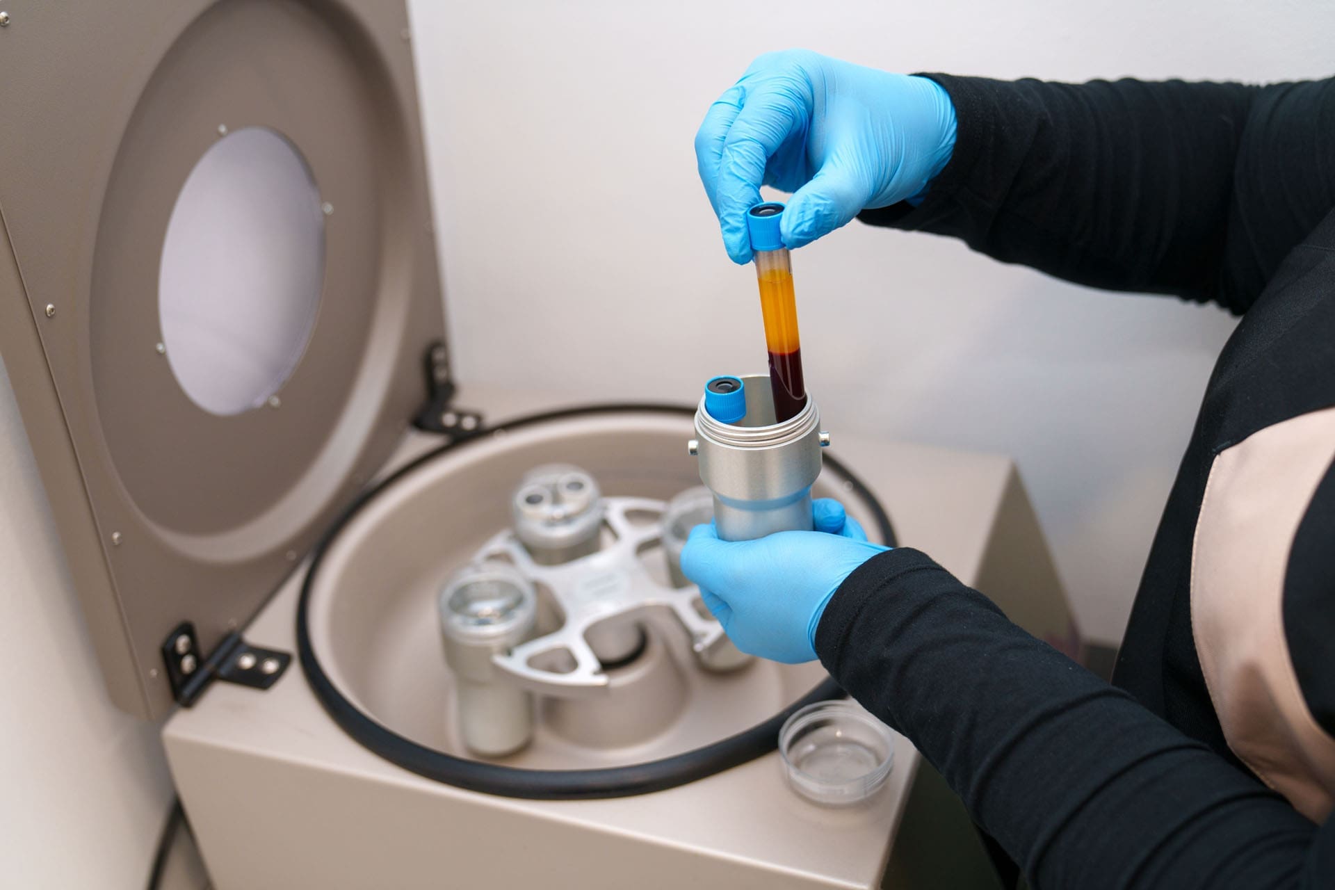
The journey begins with a simple blood draw, similar to what you would have for routine lab work. This whole blood is then placed in a specialized centrifuge, a machine that spins at high speeds to separate the blood into its different components based on their density.
Red Blood Cells: Being the densest, these cells settle at the very bottom of the tube.
Plasma: This is the liquid component of blood, which is the least dense and rises to the top.
The Buffy Coat: Between these two layers lies a thin, whitish layer known as the “buffy coat.” This is the treasure we are after. It contains a high concentration of platelets and white blood cells.
After the initial spin, the plasma layer, which now contains a high concentration of platelets, is carefully separated. This is what we call platelet-rich plasma.
The Importance of Technique: A Clinical Observation
During the extraction process, precision is paramount. The goal is to collect the maximum amount of PRP without disturbing the layers below it. We use a gel separator in our collection tubes, which forms a physical barrier between the red blood cells and the plasma after centrifugation.
A critical detail in this process is ensuring the tip of the collecting syringe does not touch or puncture this gel separator. If the gel is disturbed, it can cause the red blood cells to leak back into the plasma. This would contaminate the PRP, diminishing its purity and efficacy. Should this happen, the sample would need to be re-spun to separate the components again, a step we strive to avoid to maintain the highest quality of the preparation. This meticulous attention to detail is why I prefer a technique where I have maximum control, anchoring my hand and using a steady, precise motion to draw the plasma from just above the gel layer. It’s an art form guided by science.
The final product is a golden-hued liquid, densely packed with the healing potential of the patient’s own body. We can use this powerful biologic for various applications, from injecting it into arthritic joints to using it in aesthetic procedures like microneedling to rejuvenate the skin.
Applying PRP: From Joint Injections to Topical Creams
Once we have prepared the PRP, it’s ready to be deployed to the areas of the body that need it most. The versatility of PRP is one of its greatest strengths.
Joint Injections: For patients suffering from osteoarthritis or ligament and tendon injuries, injecting PRP directly into the affected joint can be transformative. We recently had a case where we prepared multiple tubes of PRP for a single patient to treat her ankle, knee, and hip. The concentrated growth factors in the PRP, such as Platelet-Derived Growth Factor (PDGF) and Transforming Growth Factor-Beta (TGF-β), signal local stem cells to migrate to the area of injury. They then initiate a cascade of repair processes, including the formation of new cartilage, ligaments, and blood vessels. This reduces inflammation, alleviates pain, and, most importantly, promotes true tissue regeneration rather than just masking symptoms. This approach aligns perfectly with the chiropractic goal of restoring function, as healthier joints move better and with less pain.
Topical Applications: Not all PRP is used for injections. Any leftover PRP can be repurposed for topical use. For instance, it can be mixed with a specialized carrier cream, like Pro-Cell, which is designed to stabilize the platelets and allow for transdermal absorption. This PRP-infused cream can then be taken home by the patient and applied to the skin, prolonging the regenerative effects of a procedure like microneedling or providing anti-inflammatory benefits to a sore joint.
Enhancing Healing with Peptides: The Next Level of Regenerative Therapy
While PRP harnesses the body’s existing healing components, we can further amplify this process by introducing peptides. Peptides are short chains of amino acids, which are the building blocks of proteins. They act as signaling molecules in the body, telling cells what to do. Certain peptides have powerful regenerative, anti-inflammatory, and healing properties.
In our practice, we often combine PRP treatments with the application of specific peptides to enhance the therapeutic outcome. For example, after performing a PRP treatment for hair restoration on the scalp, we can apply a specialized peptide solution to the area.
The PRP, which we’ve already injected, has created micro-channels in the skin and initiated the healing response. By massaging the peptide solution into the scalp immediately after, we leverage these open channels for enhanced absorption. The peptides can then penetrate deeper into the hair follicles, providing an additional layer of stimulation. For instance, a peptide like GHK-Cu (Copper Peptide) is known to stimulate hair growth by enlarging the follicle size and prolonging the growth phase of the hair cycle.
This combination therapy is synergistic. The PRP prepares the tissue and starts the healing process, while the peptides provide specific, targeted instructions to the cells to amplify that regeneration.
The Patient Experience: A Moment of Relief
Patients are often curious and a bit apprehensive about these procedures. A common question is, “What will it feel like?” When treating the scalp, for instance, patients may experience a temporary sensation of pressure or a feeling like a tension headache. As one patient described it, it felt like a “headache on the scalp,” not a deep, internal pain.
This sensation is a normal response to the injections and the volume of fluid being introduced. However, it is typically very brief. My clinical observation is that this feeling of “heaviness” or pressure resolves within minutes. The massaging of the peptides afterward is often described as the best part of the treatment—it feels soothing and helps to distribute the peptides evenly. This immediate transition from a strange pressure to a relaxing massage is reassuring for patients and is an integral part of the positive therapeutic experience we aim to create.
The Role of Integrative Chiropractic Care in Regenerative Medicine
So, where does chiropractic fit into this picture of high-tech regenerative therapies? It’s the foundation upon which everything else is built.
The central nervous system, which is housed and protected by the spine, is the master controller of every function in the body, including healing and regeneration. Spinal misalignments (subluxations) can create nerve interference, hindering the body’s ability to heal itself efficiently.
My role as a chiropractor is to identify and correct these misalignments through specific chiropractic adjustments. By restoring proper spinal alignment and motion, we achieve several critical goals:
Optimize Nerve Function: A well-aligned spine allows for clear communication between the brain and the rest of the body. This ensures that the healing signals initiated by treatments like PRP are transmitted effectively.
Improve Biomechanics: When we inject a joint like the knee or hip with PRP, we want that joint to function in a mechanically sound environment. If the patient has poor posture, an imbalanced gait, or spinal misalignments, these conditions place abnormal stress on the joint, which can counteract the benefits of the regenerative injection. Chiropractic care and physical therapy correct these biomechanical faults, creating an optimal environment for the repaired tissue to thrive.
Reduce Systemic Inflammation: Spinal misalignments can contribute to a state of chronic, low-grade inflammation. Chiropractic adjustments have been shown to help down-regulate inflammatory processes, creating a more favorable systemic environment for healing.
Therefore, we see chiropractic care not as a separate treatment but as an essential, integrated component of our regenerative protocols. We first ensure the body’s framework and communication systems are functioning optimally. Then, we introduce advanced therapies like PRP and peptides to target specific areas of injury and accelerate the healing that the body is now better equipped to perform. This holistic approach, combining hands-on structural care with cutting-edge biological treatments under medical supervision, is the future of musculoskeletal and personal injury medicine. It allows us to help our patients not just manage their pain but truly heal and regain their quality of life.
References
Everhart, J. S., Jayaram, P., & Dragoo, J. L. (2020). Autologous biologics in the treatment of knee osteoarthritis: A systematic review of the past 10 years of clinical evidence. The American Journal of Sports Medicine, 48(8), 2027–2040.https://journals.sagepub.com/doi/abs/10.1177/0363546519894762
Pickart, L., & Margolina, A. (2018). Regenerative and protective actions of the GHK-Cu peptide in the light of the new gene data. International Journal of Molecular Sciences, 19(7), 1987.https://www.mdpi.com/1422-0067/19/7/1987
Roy, R. A., Boucher, J. P., & Comtois, A. S. (2010). Inflammatory response following a short-term course of chiropractic treatment in subjects with and without chronic low back pain. Journal of Chiropractic Medicine, 9(3), 107–114.https://www.ncbi.nlm.nih.gov/pmc/articles/PMC3081333/
Integrative Peptide Science And Chiropractic Care For Regeneration, Repair, And Performance
As a doctor of chiropractic, advanced practice registered nurse, and board-certified family nurse practitioner with certifications in functional and integrative medicine, I, Dr. Alex Jimenez, am constantly exploring the forefront of evidence-based research to bring the most effective and innovative treatments to our patients. My journey is one of continuous learning, seeking the latest findings from leading researchers to integrate into our clinical practice. This commitment allows us to offer therapies that are not just cutting-edge but also grounded in a profound understanding of human physiology.
At Injury Medical Clinic, we pride ourselves on a multidisciplinary approach to health. Our team is a testament to this philosophy, bringing together diverse expertise to create comprehensive and personalized care plans. I am honored to work alongside Dr. Maria Guadalupe Cardenas, MD, a board-certified internist with over four decades of experience. As our Medical Director and Collaborative Physician, Dr. Cardenas provides essential medical oversight, ensuring our integrative model adheres to the highest standards of safety and efficacy.
Our unique clinical setup in El Paso, Texas, combines the strengths of chiropractic care, medical direction, functional medicine, personal injury care, and physical rehabilitation. This collaborative environment allows us to view each patient through multiple lenses, addressing not just their symptoms but the root causes of their health concerns. We believe in treating the whole person, and our integrated team is the cornerstone of that belief. Whether a patient is recovering from an injury, managing a chronic condition, or seeking to optimize their overall well-being, our coordinated efforts ensure they receive the most thorough and effective care possible.
Abstract
In this educational post, I walk you through how modern peptide science fits into integrative musculoskeletal care, rehabilitation, hair restoration, and weight management. I explain the difference between peptides and bioregulators, how these biological messengers support regeneration and homeostasis, and why delivery mechanisms matter. You will learn about commonly discussed compounds such as GHK-Cu (blue copper), BPC-157, and GLP-1-related strategies, alongside exercise-mimetic blends that target mitochondrial efficiency.
I also detail how our multidisciplinary team at Injury Medical Clinic PA (Mission Plaza Injury Medical Clinic) in El Paso, Texas, integrates chiropractic care, physical therapy-style rehabilitation, and functional medicine under the medical direction of Dr. Maria Guadalupe Cardenas, MD, with coordinated oversight for safe and evidence-informed use. Throughout, I share clinical observations from my practice and highlight evidence-based frameworks from leading researchers that shape our protocols. The emphasis here is on rehab-first, chiropractic-centric solutions, with peptides and other advanced therapies considered as adjuncts under appropriate medical guidance.
About Our Multidisciplinary Team in El Paso, Texas
I am Dr. Alexander Jimenez, DC, APRN, FNP-BC, CFMP, IFMCP, ATN, CCST. In our clinic—Injury Medical Clinic PA, also known as Mission Plaza Injury Medical Clinic—we operate within a collaborative, multidisciplinary model that is common in integrative injury-care environments. This structure allows us to prioritize chiropractic and active rehabilitation while maintaining medical oversight for safety, scope, and clinical appropriateness. Peptide-related strategies, when used, remain adjunctive—supporting tissue repair and metabolic function without overshadowing the foundation of spine-centric care and progressive loading.
Our team integrates:
Medical Director: Dr. Maria Guadalupe Cardenas, MD, Board Certified in Internal Medicine (NPI #1164426749, Texas MD License #J2933), with over 40 years of experience as an internist, serves as our collaborative physician and provides medical direction.
Chiropractic Care: I lead integrative chiropractic and functional neuromusculoskeletal care, focusing on biomechanical assessment, spinal and extremity adjustments, and manual therapies.
Rehabilitation: We include manual therapy, active care, therapeutic exercise, and movement corrections rooted in physical therapy-style protocols.
Functional Medicine: We incorporate nutrition, sleep optimization, stress modulation, and targeted biochemical support as appropriate to support the body’s innate healing capacity.
Personal Injury: We use evidence-based pathways for musculoskeletal trauma, ensuring documentation, objective measures, and standardized rehab progression for auto, work-related, and sports injuries.
Why Integrative Chiropractic First: The Physiology of Load and Adaptation
Chiropractic care and targeted rehab anchor our outcomes because they leverage mechanotransduction: the process by which cells convert mechanical stimuli into biochemical signals. This is the very foundation of how tissues heal and adapt.
Tendons and Ligaments: Eccentric loading, a cornerstone of our rehab programs, increases tenocyte activity and promotes collagen alignment. This enhances the tensile strength and stiffness of connective tissues, which is core to preventing re-injury (Kjaer, 2004).
Cartilage and Bone: Controlled loading stimulates chondrocyte metabolism in cartilage and osteogenesis (bone formation) via complex signaling pathways like Wnt/β-catenin and integrin signaling (Humphrey et al., 2019).
Neuromuscular Control: Chiropractic adjustments and corrective exercises reduce inhibitory reflexes that arise from pain, normalize motor unit recruitment patterns, and improve overall movement economy.
Biological messengers, such as peptides, complement this process by creating a more favorable internal environment for these mechanical inputs to work. They can help by:
Reducing local inflammatory mediators (e.g., IL-6, TNF-α).
Enhancing Extracellular Matrix (ECM) synthesis and angiogenesis (new blood vessel formation) for better nutrient delivery.
Supporting mitochondrial resilience to help patients tolerate higher training volumes during rehabilitation.
Peptides And Bioregulators: Clarifying The Biological Messenger Landscape
To understand how we support tissue repair, it’s essential to differentiate between two types of biological messengers.
Peptides: These are chains of amino acids—commonly 2 to 40 residues in the United States—that bind to receptors on cell membranes. This binding triggers downstream signaling cascades for regeneration, repair, and homeostasis. They act via a precise “lock-and-key” mechanism, creating selective effects with typically lower off-target risks compared to many small-molecule drugs (Fosgerau & Hoffmann, 2015).
Bioregulators: These are ultra-short peptides (often 2 to 4 residues) that are small enough to enter cells and their nuclei, where they modulate DNA transcription and epigenetic pathways. They directly influence the gene expression of structural proteins, antioxidant systems, and neurotrophic factors (Khavinson & Linkova, 2019).
The key concept is that both are biological messengers, not broad-spectrum synthetic drugs. Their role is to enhance existing signaling pathways rather than override normal physiology. In musculoskeletal care, tissues respond to mechanical loading via mechanotransduction. Peptides can potentiate this process, increasing ECM turnover and mitochondrial capacity, enabling better adaptation to chiropractic adjustments, soft-tissue work, and therapeutic exercise.
Endogenous Messengers: GHK-Cu And BPC-157 In Repair Biology
Two peptides frequently discussed in the context of tissue repair are GHK-Cu and BPC-157.
GHK-Cu (blue copper): This peptide is endogenously present in human saliva and plasma. It plays strong roles in musculoskeletal repair and has antimicrobial actions. Research shows it can upregulate collagen types I–III, decorin, elastin, and fibronectin, while also supporting angiogenesis (Pickart & Thaler, 2019). Clinically, I observe improved soft-tissue quality and skin elasticity in patients undergoing structured loading programs when topical or oral forms are used adjunctively.
BPC-157 (Body Protection Compound): Originally associated with gastric juice, this peptide has been studied for its effects on angiogenesis, fibroblast migration, tendon/ligament repair, and gut mucosal integrity (Sikiric et al., 2018). While it is frequently discussed, it is not extracted for medical use from natural sources; instead, it is synthesized. In our clinic, we keep discussions of such peptides under medical oversight and emphasize rehab-first protocols.
My clinical observation from our El Paso practice is that patients who maintain graded activity, receive precision chiropractic adjustments, and adhere to sleep and nutrition plans show superior outcomes. Adjunctive biological messengers can amplify results but do not replace mechanical stimulus, movement quality, or progressive strengthening.
A Comprehensive Approach to Hair Restoration
Hair loss, whether due to genetics, hormonal changes, or as a side effect of weight loss, can be distressing. Our hair restoration program is another example of our multi-faceted, integrative approach. We address the problem from multiple angles to achieve the best possible outcome.
Chiropractic Integration and Scalp Biomechanics: Restricted cervical and cranial soft tissues can impair local blood flow and lymphatic drainage. Gentle neck mobilization, soft tissue work around the occipital region and temporalis, and thoracic mobility can reduce tension, support autonomic balance, and improve scalp perfusion.
In-Office Regenerative Procedures: The foundation of our program involves stimulating the hair follicles directly. Under medical oversight, we use Platelet-Rich Plasma (PRP) and exosome injections into the scalp. PRP increases blood flow to dormant follicles and delivers a high concentration of growth factors, while exosomes provide signaling molecules that encourage follicles to shift from the resting (telogen) phase back into the active growth (anagen) phase.
Peptide Support (Oral and Topical): To support these in-office treatments, we use oral peptide capsules containing compounds like GHK-Cu. This peptide is crucial for building stronger, denser hair and has shown a remarkable ability to influence follicular pigmentation. I have seen this firsthand with my own patients and even my wife, who noticed that her graying hairs began growing back in their natural blonde color from the root. We also provide patients with a topical peptide spray for at-home use, delivering growth-promoting signals directly to the scalp daily.
Hormonal Modulation: For men experiencing androgenic alopecia (male pattern baldness), addressing the hormone dihydrotestosterone (DHT) is critical. Our program includes strategies to naturally modulate DHT levels, tackling one of the primary drivers of hair loss.
Success with hair restoration requires patience. It takes time for dormant follicles to re-enter the growth phase. We are transparent with our patients, letting them know it will likely be a month before they see the first new sprouts, and a full course of treatment may involve several sessions over six to nine months.
A Tiered, Evidence-Based Approach to Medical Weight Loss
Managing weight is one of the most significant challenges my patients face. For our readers at elpasobackclinic.com, we emphasize that movement correction, chiropractic care, and strength progression are the primary focus. However, when medically appropriate and under the direction of Dr. Cardenas, we have developed a tiered program that leverages the latest advancements in peptide therapy.
Good: Semaglutide Program: An excellent starting point for many patients.
Better: Tirzepatide Program: For those who need a more potent intervention, as it acts on two receptors (GLP-1 and GIP).
Best: Retatrutide Program: At the pinnacle of our program, this groundbreaking peptide targets three receptors (GLP-1, GIP, and glucagon) for comprehensive metabolic benefits.
A crucial component of our methodology is integrating lipotropic injections with each weekly GLP-1 agonist dose. These injections, containing compounds like L-carnitine and B vitamins, support the liver in metabolizing fat and improve cellular energy production.
Advanced Delivery Concepts: Getting Messengers Where They Need To Go
Delivery determines bioavailability and effectiveness. The route matters, whether we’re discussing supplements, topicals, or peptides.
Topical and Oral Routes: For compounds like GHK-Cu, these routes are more accessible and compliant with current regulations. Topical use supports local tissue (skin, superficial fascia), while oral forms may influence systemic availability. We emphasize non-invasive pathways to complement chiropractic care.
Advanced Oral Delivery: The gut often breaks down peptides like proteins. Modern formulations use enteric coatings to protect peptides from stomach acid and release them in the small intestine. Other strategies include trehalose lyophilized microcrystals to prevent denaturation and permeability enhancers to improve absorption without invasive methods (Maher et al., 2019).
Transdermal Delivery: For skin and aesthetics, the challenge is getting larger molecules past the stratum corneum. A properly formulated serum can facilitate passage to the dermis, allowing actives like PRP, exosomes, and peptides to exert local effects without injections. This is particularly useful after procedures like microneedling to reduce redness and speed recovery (Prausnitz & Langer, 2008).
Exercise-Mimetics and Mitochondrial Support: Why Energy Systems Matter
An exercise-mimetic approach aims to enhance cellular energy production without stimulants, which is critical for helping patients tolerate progressive rehab.
Mitochondrial Upregulation: Peptide strategies that increase ATP synthesis, support the electron transport chain, and reduce oxidative stress are invaluable. Enhanced mitochondrial efficiency reduces the burden of reactive oxygen species (ROS) during exercise, preserves muscle fiber integrity, and supports satellite cell activation.
NAD+ Support: As a central cofactor in redox reactions, NAD+ impacts sirtuin signaling, DNA repair, and mitochondrial function (Yoshino et al., 2018). When combined with mechanical loading, patients often report better recovery, less fatigue, and improved performance.
Structured Care Pathway: From Assessment To Return-To-Function
We translate this complex science into practical, actionable steps for our patients.
Comprehensive Evaluation:
History and Mechanism of Injury
Posture and Movement Screens
ROM, Strength, and Endurance Tests
Palpation and Regional Interdependence Assessment
Chiropractic and Manual Therapy:
Segmental Adjustments to optimize joint mechanics.
Myofascial Release and Instrument-Assisted Techniques.
Joint Mobilizations to restore motion.
Progressive Rehabilitation:
Isometrics for pain-modulated entry into exercise.
Eccentric Loading for tendon health.
Functional Integration for return-to-work or sport.
Lifestyle and Functional Medicine Supports:
Sleep Quality optimization.
Anti-inflammatory Nutrition.
Stress and Autonomic Modulation through breathwork.
Optional Adjuncts Under Medical Oversight:
Topical/Oral Biological Messengers.
Mitochondrial Supports.
PRP/Exosome therapies for hair and skin.
We use validated scales (pain, disability), performance metrics (strength, endurance), and functional milestones to measure progress and refine our protocols.
Real-World Observations From My El Paso Practice
From my clinical experience at elpasobackclinic.com, I have observed consistent themes:
Shoulder impingement with capsular tightness often resolves faster when we combine thoracic mobility, scapular retraction work, and rotator cuff eccentrics. Adjunct supports that foster collagen remodeling help patients reach end-range strength more quickly.
Chronic low back pain tied to hip flexor tightness and gluteal inhibition responds best to lumbar adjustments, psoas lengthening, and glute medius activation. Supporting tissue repair and energy metabolism helps sustain gains during work or sport.
Tendon recovery timelines shorten dramatically when patients adhere to sleep, nutrition, and graded loading. Adjunctive biological messengers may reduce soreness and enhance the quality of contraction during return-to-play phases.
Hair restoration patients often notice subtle vellus growth within 2–3 weeks when using copper peptide-based topicals and making lifestyle changes. More robust density changes typically require ongoing application with monthly photographic tracking.
Coordinated Care With Medical Oversight
Our clinic’s structure ensures safety, compliance, and data-informed decisions.
Safety: Dr. Cardenas provides medical direction, reviewing the appropriateness of all protocols and monitoring patient responses, especially for any adjuncts.
Scope and Compliance: We emphasize topical/oral approaches within regulatory guidelines and avoid overstepping our scope of practice.
Data-Informed Decisions: We use evidence-based protocols, reflect new research findings, and adjust care plans accordingly.
This disciplined approach keeps chiropractic and rehab at the forefront, using modern biological tools where they genuinely add value.
Practical Takeaways For Patients And Clinicians
Make movement and mechanical loading the foundation of any recovery plan. Biological messengers are adjuncts, not replacements.
Emphasize sleep, nutrition, and stress management to create the physiological environment needed for tissue adaptation.
Consider topical/oral supports where evidence and medical oversight align, especially for enhancing collagen synthesis, angiogenesis, and mitochondrial function.
Track outcomes and adjust care plans; what is measured can be improved.
Our mission in El Paso is simple: help patients move better, feel better, and live better—safely and consistently. With integrative chiropractic care at the forefront and medical oversight ensuring safety, we bring modern, evidence-based strategies to hair, skin, weight, and recovery, grounded in movement, tissue biology, and patient education.
Evidence-Based Men’s Health: Erectile Dysfunction, Vascular Markers, Shockwave Therapy, PRP, and Integrative Chiropractic Care in El Paso
Abstract
In this educational post, I share a practical, evidence-based roadmap for men’s health focused on erectile dysfunction (ED) as a vascular, neurologic, hormonal, and psychogenic condition. I explain why ED can be an early marker of cardiovascular disease, outline modern restorative therapies such as extracorporeal shockwave therapy (ESWT) and platelet-rich plasma (PRP), and discuss how to evaluate and optimize hormonal status—especially testosterone—while keeping medications and hormone therapy considerations in the background for this website’s focus on chiropractic and physical rehabilitation. I walk through the physiology of nitric oxide and endothelial function, the role of neuropathy and pelvic surgery, and how lifestyle, biomechanics, and integrative chiropractic care can support vascular health, pelvic floor function, and neurovascular signaling. I also demonstrate how our multidisciplinary team—under the medical direction of Dr. Maria Guadalupe Cardenas, MD (Internal Medicine), working collaboratively with me, Alex Jimenez, DC—coordinates diagnostics, personal injury care, and functional rehabilitation at Injury Medical Clinic PA (Mission Plaza Injury Medical Clinic) in El Paso, Texas. You will learn when and why we use shockwave therapy and PRP, how we monitor safety, and how we tailor care to each patient’s cardiovascular risk, musculoskeletal status, and functional goals.
Introduction: A Straightforward Path Through Men’s Health
I am Dr. Alex Jimenez, DC, APRN, FNP-BC, CFMP, IFMCP, ATN, CCST. In my clinical practice, I focus on musculoskeletal and functional rehabilitation and integrate modern, evidence-based approaches to men’s health. When a man says, “Doc, I’ve got ED—what can you do other than the blue pill?” I start by explaining that erectile dysfunction is not just a symptom; it can be a window into overall vascular health. If we take away one essential message, it’s this: evaluate for cardiovascular disease when ED is present. The latest research consistently associates ED with endothelial dysfunction, impaired nitric oxide signaling, and microvascular disease—mechanisms that precede overt cardiac events. By coupling chiropractic and physical therapy-based rehabilitation with restorative modalities like shockwave therapy and PRP, we aim to improve vascular dynamics, neuromuscular coordination, and pelvic biomechanics in a cohesive care plan.
Our Multidisciplinary Team in El Paso: Medical Oversight and Integrative Chiropractic
Medical Director and Collaborative Physician: Dr. Maria Guadalupe Cardenas, MD, Board Certified in Internal Medicine (NPI #1164426749, Texas MD License #J2933). With over 40 years of experience as an internist, Dr. Cardenas leads medical oversight in our clinic.
Chiropractic Care and Functional Rehabilitation: I, Dr. Alex Jimenez, DC, integrate spinal and pelvic alignment strategies, movement-based therapy, and targeted neuromuscular interventions.
Clinic: Injury Medical Clinic PA (Mission Plaza Injury Medical Clinic), El Paso, Texas.
How We Integrate Care
Medical Direction: Cardiovascular risk stratification, laboratory oversight (lipids, HbA1c, inflammatory markers), and guidance on when medication or specialty referral is necessary.
Chiropractic and Physical Therapy: Correction of pelvic misalignments, soft tissue interventions, and pelvic floor coordination to support neurovascular pathways critical for erection.
Functional Medicine: Lifestyle, sleep, stress, and metabolic support to optimize nitric oxide biology and endothelial health.
Personal Injury and Rehabilitation: Addressing lumbopelvic biomechanics, nerve entrapments, and scar tissue from prior surgeries or injuries that impair neurovascular signaling.
Restorative Modalities: Non-invasive extracorporeal shockwave therapy (ESWT) and platelet-rich plasma (PRP), coordinated with medical monitoring and functional rehab.
Why Erectile Dysfunction Is a Vascular and Neurofunctional Condition
ED is the inability to attain or maintain an erection sufficient for sexual performance. Clinically, it often reflects reduced arterial inflow, venous leak, impaired nitric oxide bioavailability, and autonomic or peripheral neuropathy. The endothelial lining of penile arteries—like coronary vessels—relies on nitric oxide (NO) generated by endothelial nitric oxide synthase (eNOS). Oxidative stress, atherosclerosis, and metabolic syndrome reduce NO, stiffen vessels, and limit smooth muscle relaxation in the corpus cavernosum. The result is compromised tumescence and maintenance of erection.
Key mechanisms:
Vascular Endothelium: Atherosclerosis narrows penile arteries, which are smaller-caliber vessels and may show dysfunction earlier than coronary arteries. Endothelial dysfunction reduces NO and cyclic GMP signaling in cavernosal smooth muscle, limiting vasodilation.
Hormonal Modulation: While low testosterone does not directly “cause” ED, it reduces libido and can diminish responsiveness to PDE5 inhibitors by affecting NO synthase expression and cavernosal smooth muscle integrity.
Psychogenic Components: Depression and anxiety suppress libido and sympathetic-parasympathetic balance, often heightening performance anxiety and decreasing erectile consistency.
Drug-Induced Effects: Antihypertensives, SSRIs, antipsychotics, and opioids may impair erectile function through vascular and neurochemical pathways.
Clinical Reasoning:
Because penile arteries manifest endothelial injury early, ED acts as a cardiovascular sentinel. Assess lipids, blood pressure, glycemic status, and inflammatory markers; consider calcium scoring or cardiology referral when risk is high.
Address biomechanics and neuromuscular integration. Pelvic tilt, sacroiliac dysfunction, and lumbar nerve irritation can degrade autonomic balance and perineal blood flow.
Chiropractic and Physical Therapy Foundations for Men’s Health
In our El Paso clinic, chiropractic care supports ED treatment by optimizing pelvic alignment, reducing neurogenic irritation, and enhancing blood flow dynamics.
What we focus on:
Pelvic Alignment and Sacroiliac Mechanics: Correcting anterior/posterior tilt and rotational dysfunction reduces strain on the pelvic floor and improves lumbosacral nerve signaling to the perineum.
Lumbar Spine Health: Addressing L4-S2 segments with manual therapy, mobilization, and stability exercises helps improve autonomic and somatic contributions to erectile reflexes.
Soft Tissue and Fascial Planes: Myofascial release of adductors, pelvic floor, and gluteal complexes improves venous return and arterial inflow by reducing fascial tension that restricts vascular dynamics.
Pelvic Floor Coordination: Biofeedback-informed exercises (relax-contract cycles) can reduce hypertonic guarding, improving arterial filling and reducing venous leak.
Breathing and Diaphragmatic Mechanics: Diaphragmatic breathing reduces sympathetic overdrive, supports NO production via improved endothelial shear stress during cardiovascular exercise, and enhances pelvic floor synergy.
Why These Techniques Help:
Neurovascular Integration: Better spinal mechanics decrease nociceptive input and sympathetic dominance while supporting parasympathetic pathways critical to erection.
Endothelial Shear and NO: Moderate aerobic exercise increases laminar shear stress, upregulating eNOS and NO, improving penile blood flow.
Venous Occlusion Mechanics: Coordinated pelvic floor activity enhances the veno-occlusive mechanism to maintain erection.
Extracorporeal Shockwave Therapy: Restoring Microvascular Health
Shockwave therapy (ESWT) uses low-intensity acoustic waves to create controlled microtrauma that stimulates repair biology.
Mechanisms:
Neovascularization: ESWT upregulates vascular endothelial growth factor (VEGF), fibroblast growth factor (FGF), and stromal cell-derived factors, driving angiogenesis and capillary density in penile tissue.
Endothelial Function: Microtrauma activates eNOS and enhances NO bioavailability, improving vasodilation and cavernosal smooth muscle relaxation.
Tissue Remodeling: ESWT promotes extracellular matrix turnover and reduces fibrosis that can impair tunica albuginea flexibility and veno-occlusive function.
Clinical Use:
Non-invasive and in-office, ESWT is scheduled over several sessions to progressively build vascular response.
Home-based handheld devices can extend benefits between sessions with proper medical guidance.
Why We Use ESWT:
It addresses root causes—poor microcirculation and endothelial dysfunction—rather than masking symptoms.
It pairs well with chiropractic-led movement and pelvic floor training that augment vascular and neuromuscular gains.
Platelet-Rich Plasma: Growth Factor-Driven Repair
PRP concentrates autologous platelets to deliver growth factors in penile tissue.
Mechanisms:
Angiogenesis: PDGF, VEGF, and TGF-β stimulate new vessel formation and improve perfusion.
Neurotrophic Effects: NGF and BDNF support nerve repair, particularly relevant in diabetic neuropathy or post-prostatectomy nerve injury.
Under sterile technique, PRP is injected into targeted penile structures by trained medical professionals. Post-procedure discomfort is typically minor.
Why We Use PRP:
It is minimally invasive and restorative, complementing ESWT to synergistically enhance vascular and neural repair.
It is particularly considered for men with diabetes or post-prostatectomy changes where neurovascular damage is profound.
ED as a Cardiovascular Marker: Practical Evaluation Steps
Lifestyle and Activity: Sedentary behavior worsens endothelial function; structured exercise improves NO signaling.
Sleep and Stress: Sleep apnea reduces nocturnal erections and worsens cardiometabolic risk; stress elevates sympathetic tone and impairs erection.
What We Do:
Coordinate with Dr. Cardenas for cardiovascular risk management and diagnostic workup.
Implement chiropractic-guided exercise prescriptions: brisk walking, cycling, and resistance training to upregulate eNOS and improve vascular compliance.
Provide pelvic floor rehabilitation and breathing protocols to recalibrate autonomic balance.
Hormone Considerations
In our clinic’s context, we emphasize musculoskeletal and rehabilitative strategies while acknowledging hormonal evaluation as part of comprehensive care.
Key points:
Low testosterone reduces libido and can blunt response to PDE5 inhibitors, but is not the primary cause of ED.
Evaluate morning total testosterone, consider free testosterone and sex hormone-binding globulin (SHBG) in obesity because low SHBG can mask free testosterone abnormalities.
Monitor clinical symptoms rather than treating numbers alone; labs must align with patient-reported issues.
Safety:
If testosterone therapy is considered under medical supervision, monitor hematocrit, PSA, and sleep apnea risk. Coordinate with cardiology for recent cardiac events.
Drug-Induced ED: What Patients Need to Know
Common culprits: antihypertensives, SSRIs, antipsychotics, and opioids.
Approach: Review the medication list, discuss alternatives with prescribing physicians when appropriate, and prioritize non-pharmacologic strategies like exercise and pelvic rehab that enhance NO signaling and autonomic balance.
Post-Prostatectomy and Radiation: Who Is a Candidate for Restorative Care?
After cancer treatment completion and appropriate medical clearance, ESWT and PRP can be considered to promote angiogenesis and nerve recovery.
Functional rehabilitation: pelvic floor and lumbopelvic mechanics are vital to reestablish neurovascular function and reduce scar-related restrictions.
Clinical Rationale:
Nerve-sparing surgeries still risk microvascular and neural disruption; restorative therapies aim to rebuild pathways rather than rely solely on symptomatic relief.
How We Structure Care: Step-by-Step
Intake and Assessment
Comprehensive history including cardiovascular risk, medications, sleep, activity, and psychosocial factors.
Physical examination focusing on lumbopelvic alignment, pelvic floor tone, fascial restrictions, and peripheral neuropathy screening.
Labs coordinated by Dr. Cardenas when indicated: lipids, HbA1c, inflammatory markers, and hormonal panel where appropriate.
Foundational Plan
Chiropractic adjustments for pelvic and lumbar segments to reduce neurogenic irritation and improve autonomic balance.
Targeted physical therapy: pelvic floor coordination, gluteal and adductor mobility work, and graded aerobic training.
Lifestyle coaching: nutrition for endothelial health (nitrate-rich vegetables), sleep hygiene, and stress management.
Restorative Modalities
ESWT cycle to drive neovascularization and endothelial repair.
PRP injections when indicated to enhance angiogenesis and nerve healing.
Monitoring and Progress
Functional endpoints: improved erectile quality, decreased reliance on PDE5 inhibitors, improved endurance and pelvic floor coordination.
Safety checks and adjustments: symptom tracking, cardiovascular monitoring, and careful pacing of exercise and ESWT sessions.
Clinical Observations from Our Practice
Many men improve erectile quality when lumbopelvic dysfunction is corrected and aerobic capacity increases—consistent with NO-mediated vasodilation and reduced sympathetic tone.
Pelvic floor hypertonicity is common; biofeedback-based relaxation before contraction training helps restore veno-occlusive competence and reduce performance anxiety.
Combining ESWT with structured rehab creates compounding gains: angiogenesis from shockwave meets improved hemodynamics from exercise and alignment.
Patients who reduce sedentary time and practice diaphragmatic breathing often report improved nocturnal erections and daytime vitality—reflecting autonomic recalibration.
Patient-Friendly Tools: Lowering Barriers to Care
Confidential questionnaires in the clinic help men communicate symptoms such as decreased libido, weaker erections, fatigue, and mood changes.
Clear explanations of physiology empower patients to engage in exercise, breathing work, and pelvic coordination with purpose.
Home-based shockwave devices can maintain momentum between sessions, with guidance to ensure safe and consistent usage.
Why We Prefer a Root-Cause Strategy
Symptomatic approaches alone may create tachyphylaxis and diminishing returns. Restorative care—ESWT, PRP, rehabilitation, and lifestyle—is built to improve microvascular perfusion, endothelial resilience, and neurovascular signaling.
Chiropractic and physical therapy interventions address the structural and functional systems that support penile hemodynamics and autonomic regulation.
Safety and Contraindications
ESWT and PRP: Generally well-tolerated; transient discomfort or bruising may occur.
Cardiovascular clearance: Essential for men with active or recent significant cardiac disease.
Professional oversight: Close coordination with Dr. Cardenas ensures appropriate screening and safeguards when additional medical factors are present.
The Takeaway: ED Is an Opportunity to Improve Whole-Body Health
Evaluate cardiovascular risk; ED can be a sentinel event.
Use chiropractic and physical therapy to correct pelvic mechanics and enhance neurovascular pathways.
Apply ESWT and PRP to build lasting microvascular and neural improvements.
Monitor progress, prioritize safety, and personalize care.
Conclusions: A Practical, Restorative Path Forward
Men’s health benefits from an integrative approach that starts with careful screening and proceeds to rehabilitative strategies designed to improve physiology, not just mask symptoms. By aligning chiropractic care, physical therapy, ESWT, and PRP under strong internal medicine oversight, we help men achieve durable improvements in erectile function and overall vitality. The combination of endothelial repair, neurovascular coordination, and optimized biomechanics creates a foundation for better performance and well-being.
Joint Pain Relief Through Regenerative Chiropractic
Abstract
In this educational post, I, Dr. Alexander Jimenez, DC, APRN, FNP-BC, CFMP, IFMCP, ATN, CCST, guide you through a practical, evidence-based approach to shoulder and knee care using integrative chiropractic methods, functional rehabilitation, ultrasound-guided procedures, and regenerative strategies. You will learn how we identify pain generators and biomechanical contributors, why we select specific manual therapies and corrective exercises, and how we safely use ultrasound to guide injections into targeted tissues. I also introduce our multidisciplinary team, led medically by Dr. Maria Guadalupe Cardenas, MD (Board Certified in Internal Medicine) (NPI #1164426749, Texas MD License #J2933), who serves as Medical Director and Collaborative Physician at Injury Medical Clinic PA (Mission Plaza Injury Medical Clinic) in El Paso, Texas. We show how chiropractic care, internal medicine oversight, functional medicine, personal injury care, rehab, and physical therapy combine to restore function and reduce pain, while keeping hormones and medications in the background for elpasobackclinic.com’s audience. Finally, I translate complex anatomy and physiology into clear, actionable steps and provide citations with linked references so you can explore the research behind each decision.
Chiropractic And Internal Medicine Collaboration In El Paso, Texas
At Injury Medical Clinic PA (Mission Plaza Injury Medical Clinic) in El Paso, Texas, our multidisciplinary model is designed for precision diagnostics, safe care, and sustainable outcomes.
Medical direction: Dr. Maria Guadalupe Cardenas, MD (Internal Medicine), brings over 40 years of clinical experience, ensuring medical safety, bi-directional care coordination, and evidence-based protocols across complex cases.
Chiropractic integration: I lead integrative chiropractic care, combining spinal biomechanics, regional joint assessment, soft-tissue methods, and functional rehabilitation targeted to the patient’s presentation.
Functional medicine lens: We prioritize nutrition, sleep, stress physiology, and metabolic health as supportive pillars for tissue healing, while minimizing reliance on hormones or medications unless medically indicated.
Physical therapy emphasis: Coordinated mobility, stability, motor control, and return-to-function plans are sequenced with chiropractic adjustments and soft-tissue care, including sports-specific and work-injury progressions.
Personal injury workflows: For PI cases, we document thoroughly, use validated outcome measures, and align care with imaging, guided procedures, and gradual load progressions to restore confidence and capacity.
Why This Integrative Model Matters
Safety first: Internal medicine oversight reduces procedural risk and guides comorbidity management.
Precision: Ultrasound-guided interventions and biomechanical assessments target the right tissue at the right dose.
Durability: Chiropractic care, physical therapy, and functional medicine together produce longer-lasting outcomes by addressing root causes.
Patient-centered: We build stepwise care pathways, educate patients, and align expectations to reduce fear and improve adherence.
Shoulder Pain: Anatomy, Biomechanics, And Why It Hurts
The shoulder is a dynamic, multi-planar joint system in which the glenohumeral joint, acromioclavicular (AC) joint, scapulothoracic articulation, and sternoclavicular joint must synchronize to ensure smooth function. The rotator cuff—supraspinatus, infraspinatus, teres minor, and subscapularis—stabilizes the humeral head to prevent excessive superior or anterior translation during elevation.
Key physiology driving pain:
Tendinopathy: Repetitive load and poor scapular control foster collagen disorganization, neovascularization, and nociceptive sensitization within cuff tendons, especially the supraspinatus footprint on the greater tuberosity.
Subacromial space mechanics: Limited thoracic extension or scapular upward rotation narrows the subacromial space, increasing bursal and tendinous stress.
AC joint degeneration: Microinstability and load transfer through the clavicle result in capsular irritation, osteophytes, and localized pain with cross-body movements.
Biceps-labral interface: The long head of the biceps traverses the bicipital groove and contributes to anterior shoulder pain when overloaded or in SLAP variants.
Neurovascular proximity: The neurovascular bundle in the anterior shoulder region requires meticulous mapping during procedures to avoid iatrogenic injury.
What I Look For During A Real Patient Encounter
Drawing from my clinical experience:
Visual and palpatory cues: I watch for asymmetry, protective guarding, and painful arcs. Palpation maps tenderness over the supraspinatus footprint, AC joint, subscapularis, and bicipital groove.
Functional patterns: I analyze bird-dog, superman, and scapular setting drills to identify deficits in anti-extension control and rotator cuff endurance. These tests help me see how trunk stability informs shoulder mechanics.
Ultrasound landmarks: I trace the humeral head, articular cartilage, supraspinatus footprint, subacromial bursa, AC joint, and biceps tendon sheath, maintaining a safe distance from neurovascular structures.
Load tolerance: I progress from low-load tasks to higher-load regions (e.g., triceps or deep cuff work), carefully managing patient expectations and discomfort.
Integrative Chiropractic Approach To Shoulder Care
Our shoulder pathway prioritizes chiropractic and physical therapy methods:
Thoracic mobility and rib mechanics
Why: Thoracic extension and rib mobility enable scapular upward rotation and posterior tilt, reducing impingement risk.
Methods: Thoracic spine manipulation and mobilization to improve segmental motion; breathing retraining for costovertebral rhythm.
Evidence: Manual therapy to the cervical-thoracic junction can reduce shoulder pain and improve function through regional interdependence (Domenech-Garcia et al., 2011).
Scapular motor control
Why: Proper serratus anterior and lower trapezius activation improves humeral head centering, decreasing superior migration under load.
Methods: Wall slides with lift-off, prone Y/T/W, serratus punches, anti-shrug carries to re-pattern scapular mechanics.
Evidence: Scapular-focused intervention enhances pain and function in shoulder disorders (Kibler et al., 2013).
Rotator cuff capacity building
Why: The cuff stabilizes micro-movements. Progressive isometrics and eccentrics remodel tendon integrity.
Methods: Isometric external rotation, eccentric abduction, side-lying ER, full-can holds; later closed-chain perturbations.
Evidence: Eccentric loading promotes tendon remodeling and reduces pain in tendinopathies (Rio et al., 2015).
Soft-tissue and fascia
Why: Myofascial restrictions elevate local shear and neural input.
Methods: Instrument-assisted soft-tissue mobilization, percussion, cupping, and nerve glides where appropriate.
Evidence: Soft-tissue approaches can modulate pain, improve ROM, and support exercise tolerance (Cheatham et al., 2015).
Patient education and pacing
Why: Expectation management reduces threat perception and enhances adherence.
Methods: Transparent planning, explaining why each step is chosen and how measurable progress is tracked.
Ultrasound-Guided Shoulder Procedures: What We Do And Why
When indicated, we use ultrasound to guide precise injections. While this post emphasizes chiropractic and physical therapy, understanding our interventional choices clarifies our iterative care model.
Subacromial bursa, supraspinatus footprint, and AC joint
Why: Pain may originate from bursitis, partial-thickness supraspinatus lesions, or AC joint capsular irritation. Ultrasound guidance ensures in-plane or out-of-plane needle control, keeping the needle away from neurovascular structures.
Technique: Identify bright cortical bone under the footprint; visualize bursal fluid and capsule integrity. Use small aliquots and reassess spread, avoiding intratendinous trauma unless intentionally performing a tendon fenestration or PRP in tendinopathic zones.
Evidence: Ultrasound-guided shoulder injections improve accuracy compared with landmark techniques and can more precisely target pathologic pain generators (Sibbitt et al., 2011).
Biceps tendon sheath
Why: Anterior shoulder pain often involves the long head of biceps. Sheath injection—distinct from intratendinous injection—reduces irritability and allows rehab to progress.
Technique: Map the groove, maintain longitudinal needle trajectory, and confirm spread along the sheath without tendon violation.
AC joint microvolume injection
Why: Small-volume injections can modulate capsular irritability. Cross-body adduction reproduction of pain is a clinical cue.
Technique: Orient to the joint cleft, avoid over-distention, and recheck cross-body ROM post-procedure.
Our Procedure Safety And Team Coordination
Pre-procedure planning: We plan labs, imaging, and rehab scheduling in advance. My nurse and lab tech process any biologics as needed, while I maintain room-side focus on mapping and safety.
Minimal staff burden: Our care flow allows other team members to handle follow-ups, therapy sessions, and patient education while I perform the procedure efficiently.
Internal medicine oversight: Dr. Cardenas reviews risk factors, comorbidities, contraindications, and post-procedural monitoring when warranted.
Rehabilitation Sequencing After Shoulder Interventions
We deliberately move from low-threat to higher-load tasks:
Start with what hurts least: Early sessions prioritize thoracic mobility, scapular setting, and isometric cuff work at angles that do not provoke pain.
Gradual load introduction: As irritability recedes, we add eccentrics, closed-chain stabilization, and overhead progressions using tempo, isometric holds, and pause reps.
Return-to-sport or work tasks: We simulate reach, lift, carry, and press patterns relevant to the patient’s goals, using pain-guided progression and rate of perceived exertion to keep tissues within safe adaptive ranges.
Knee Care: Integrative Chiropractic And Physical Therapy Emphasis
The knee often presents with MCL strain, medial meniscal involvement, and synovial irritability—themes echoed in the transcript. Our approach blends chiropractic, PT, and when appropriate, ultrasound guidance.
Knee Biomechanics And Physiology
Load transmission: The knee depends on hip control and ankle mobility for shock absorption and alignment. Poor hip abduction and external rotation strength elevate medial compartment stress.
Meniscal physiology: Menisci distribute load and contribute to joint stability. Intra-meniscal degeneration and synovial inflammation can perpetuate pain and mechanical symptoms.
MCL healing: The MCL typically responds to graded load and frontal-plane stability training. Excess valgus strain irritates healing tissue.
Chiropractic And PT Integration For The Knee
Pelvic and lumbar alignment
Why: Pelvic tilt and lumbar rotation alter femoral tracking and tibial alignment under dynamic load.
Methods: Lumbopelvic adjustments, hip mobilizations, and gluteal activation to normalize kinetic chain input.
Motor control and strength
Why: Stable knees require hip abductors, external rotators, hamstrings, and quadriceps working in harmony.
Methods: Side-steps with bands, split-squat isometrics, Spanish squats, hamstring bridges, and tempo squats to train tolerance and tissue remodeling.
Tendon and fascia support
Why: Tendinopathic tissues benefit from eccentric and isometric loading; fascia responds to improved glide and hydration.
Methods: Patellar tendon isometrics, eccentric decline squats as tolerated, and soft-tissue mobilization to quadriceps and adductors.
Progressive return to function
Why: Sequenced progressions reduce flare-ups and build confidence.
Methods: Low-impact conditioning, step-down drills, landings, and multi-directional gait under supervision.
Ultrasound-Guided Knee Procedures When Indicated
Intra-articular injections
Why: Targeted delivery to the joint space supports modulation of synovial irritation.
Technique: Short-axis or long-axis guidance to visualize needle entry and avoid neurovascular structures.
MCL and medial meniscus region
Why: Pain generators can localize to the MCL or posteromedial meniscus. High-precision mapping reduces the risk of non-target injections.
Technique: In-plane approach along the MCL with careful hydrodissection when necessary; avoid intrameniscal violation unless using a specialist technique aligned with current evidence.
Clinical Observations From Dr. Alex Jimenez
From practice patterns noted across my work at elpasobackclinic.com and shared on my LinkedIn profile, several themes consistently emerge:
Patients thrive when care is sequenced, explained, and measured. Clear progress markers—ROM, strength, pain thresholds—reduce anxiety and improve outcomes.
The shoulder and knee respond best when the spine and hip are addressed concurrently. Regional interdependence is not academic—it is observable daily in the clinic.
Education and expectation management are as therapeutic as manual care. When patients understand why a technique is used, adherence and results improve.
Small-aliquot injections with ultrasound guidance allow real-time adjustments based on tissue spread and patient feedback, enhancing comfort and safety.
We emphasize movement literacy, teaching patients how to maintain neutral positions, breathe, and move through ranges of motion without provoking symptoms.
How Our Team Coordinates Care
Intake and triage: Medical review by Dr. Cardenas for complex histories; chiropractic exam and movement analysis by me; imaging decisions based on need.
Plan creation: A written plan outlines manual therapy, exercise progression, imaging, procedural options, and follow-up cadence.
Execution: Therapy staff handles laser, shockwave, and exercise coaching; I manage manual and chiropractic care, as well as any ultrasound-guided procedures, as appropriate.
Reassessment: We use validated outcome scales, ROM, strength testing, and return-to-function checkpoints to iterate the plan.
Communication: Patients receive clear instructions on post-session expectations and a simple home exercise sequence.
Why We Prioritize Chiropractic and Physical Therapy for elpasobackclinic.com
For our web audience and community, practical hands-on care, exercise therapy, and movement education are the cornerstones of recovery. While medications and hormones are part of comprehensive medical practice, we keep them in the background here, emphasizing:
The power of adjustments to restore joint motion and relieve nociception.
The value of targeted strengthening and motor control to protect tissues.
The role of patient-guided progression to boost independence and long-term resilience.
Safety, Dosing, And Patient Comfort
Dosing matters: Whether we are adjusting, mobilizing, loading a tendon, or injecting, we dose according to irritability, stage of healing, and patient goals.
Comfort strategies: We start with low-pain tasks, use paced breathing, and deploy brief micro-breaks to maintain composure in procedures.
Monitoring: Signs of over-irritation (escalation of night pain, heat, swelling) prompt plan adjustments or a medical review.
Putting It All Together: An Easy-To-Follow Care Journey
Step 1: Assessment
Detailed history, movement analysis, palpation, and ultrasound mapping when indicated.
Step 2: Early Care
Thoracic and cervical-thoracic mobilization, scapular setting, isometric cuff work; knee lumbopelvic alignment, hip strength foundations.
Step 3: Load And Control
Eccentrics, closed-chain drills, perturbation training, and gait re-education.
Step 4: Targeted Procedures If Needed
Ultrasound-guided bursa, AC joint, or intra-articular knee injections based on clear indications, with medical oversight.
Step 5: Return To Function
Task-specific progressions, confidence building, and preventive strategies.
Evidence-Based References That Inform Our Practice
We continually incorporate high-quality research into decisions:
Ultrasound guidance improves injection accuracy and patient outcomes in shoulder pathology (Sibbitt et al., 2011).
Scapular-focused programs and regional interdependence considerations enhance the effectiveness of shoulder rehabilitation (Kibler et al., 2013).
Eccentric and isometric loading strategies reduce tendinopathy pain and remodel tissue (Rio et al., 2015).
Myofascial techniques can improve pain and functional outcomes, supporting active rehabilitation (Cheatham et al., 2015).
Practical Takeaways For Patients
Movement is medicine: Consistency beats intensity early on.
Pain-guided progression: Minor discomfort is normal; escalating night pain or swelling means you should check in with us.
Whole-system support: Sleep, nutrition, and stress management help tissues heal and adapt.
Team-based care: Chiropractic, physical therapy, and medical oversight ensure your pathway is safe, precise, and personalized.
How To Get Help
If you are in El Paso or nearby and dealing with shoulder or knee pain, our team can create a clear, step-by-step plan designed for your goals. We will explain why we select each technique, how it fits your stage of healing, and how we measure progress so you can return to life with confidence.
References
Domenech-Garcia, V., Palsson, T. S., Boudreau, S. A., & Arendt-Nielsen, L. (2011). Upper cervical and upper thoracic manipulation in patients with shoulder pain: A randomized clinical trial. Journal of Orthopaedic & Sports Physical Therapy. https://www.jospt.org/doi/10.2519/jospt.2011.3579
Kibler, W. B., Sciascia, A., & Wilkes, T. (2013). Scapular dyskinesis and its relation to shoulder pain. Journal of the American Academy of Orthopaedic Surgeons. https://journals.lww.com/jaaos/Abstract/2013/06000/Scapular_Dyskinesis_and_Its_Relation_to_Shoulder.3.aspx
Rio, E., Kidgell, D., Purdam, C., Gaida, J., Moseley, L. G., & Cook, J. (2015). Isometric exercise for pain relief in tendinopathy: Mechanisms and implications. British Journal of Sports Medicine. https://bjsm.bmj.com/content/49/10/645
Sibbitt, W. L., Band, P. A., Kettwich, S. C., et al. (2011). Does ultrasound-guided injection improve outcomes for shoulder pain? A randomized controlled trial. Journal of Rheumatology. https://www.jrheum.org/content/38/9/1917
Cheatham, S. W., Kolber, M. J., & Cain, M. (2015). Instrument-assisted soft tissue mobilization: A systematic review. Journal of the Canadian Chiropractic Association. https://www.ncbi.nlm.nih.gov/pmc/articles/PMC4566596/
Welcome to our exploration of photobiomodulation therapy (PBMT), a revolutionary approach that harnesses the power of light to stimulate cellular healing. In this educational post, I will guide you through the intricate biological processes that make PBMT so effective. We will explore how specific wavelengths of light can penetrate tissues to activate mitochondria, modulate the immune response, and accelerate recovery. This journey will cover the fundamental science behind PBMT, from its effects on ATP production and cytokine modulation to its role in promoting angiogenesis and neurogenesis. Furthermore, we will examine the synergistic potential of combining PBMT with orthobiologics, such as Platelet-Rich Plasma (PRP), and demonstrate how this integrated approach can enhance healing outcomes. Drawing on the latest research, including fascinating studies from the veterinary world and our laboratory findings on tenocyte proliferation, we’ll demonstrate why light is not just a modality but a cornerstone of modern regenerative medicine. At Injury Medical Clinic, we integrate these advanced therapies within a collaborative framework, combining my expertise in chiropractic and functional medicine with the medical oversight of our Medical Director, Dr. Maria Guadalupe Cardenas, MD, to provide comprehensive, evidence-based care for our patients.
About Our Integrated Practice: A Collaborative Approach to Wellness
I, Dr. Alex Jimenez, am honored to share my passion for integrative and regenerative medicine with you. With a diverse background as a Doctor of Chiropractic (DC), Advanced Practice Registered Nurse (APRN), board-certified Family Nurse Practitioner (FNP-BC), and certifications in Functional Medicine (CFMP, IFMCP), Applied Traumatology (ATN), and Cranial Spinal Integration (CCST), my goal has always been to find the most effective, evidence-based paths to healing.
Here at Injury Medical Clinic PA in El Paso, Texas, we have built a unique, multidisciplinary practice. We believe that the best patient outcomes are achieved through a collaborative team approach. I am privileged to work alongside Dr. Maria Guadalupe Cardenas, MD, who serves as our Medical Director and Collaborative Physician. Dr. Cardenas is a highly respected, board-certified Internist with over 40 years of experience (NPI #1164426749, Texas MD License #J2933). Her extensive medical knowledge provides invaluable oversight and complements our services, ensuring our patients receive safe, comprehensive, and well-rounded care.
Our clinic integrates:
Advanced Chiropractic Care: Focused on spinal health, biomechanics, and nervous system function.
Physical Therapy & Rehabilitation: Tailored programs to restore movement, strength, and function.
Medical Oversight: Guided by Dr. Cardenas to ensure clinical safety and efficacy.
Functional Medicine: Investigating the root causes of chronic conditions.
Personal Injury Care: Specialized treatment for injuries sustained in accidents.
This model allows us to address health from multiple angles. While our core focus at elpasobackclinic.com is chiropractic and physical rehabilitation, we incorporate advanced modalities such as photobiomodulation to enhance the body’s innate healing capabilities, with all treatments guided by a solid medical and scientific foundation.
The Awakening: From Skepticism to Cellular Biology
I have been on this journey for nearly a decade, and for the first five years, discussing “laser” therapy in medical circles often felt like an uphill battle. It was a path paved with skepticism, much like the initial reception many of you in the biologics field have likely experienced. But today, I am thrilled to see the conversation shifting as the science catches up with the clinical results.
My evolution as a clinician mirrors this shift. For the first two decades of my career, I was a “mechanic,” using established tools to address specific conditions. Over the last ten years, however, I have become a “biologist,” focused on understanding and facilitating the body’s own healing processes at a cellular level. This is why I am so excited to share the science of photobiomodulation (PBMT) with you. It represents a profound shift from treating symptoms to enabling cellular recovery.
Understanding Photobiomodulation: The Science of Light and Life
The concept is beautifully simple, rooted in a phenomenon we all accept: photosynthesis. The sun’s light fuels life on Earth, and as a species that has evolved under this light for hundreds of thousands of years, our cells have developed a deep, genetic sensitivity to it. We readily accept that sunlight is necessary for Vitamin D synthesis, yet a significant gap remains in medical education regarding the broader therapeutic applications of light.
Photobiomodulation breaks down as:
Photo: Light
Bio: Life
Modulation: To affect or change
Light is energy, delivered in units called photons. These photons can transfer their energy to our cells, triggering a cascade of biological responses. This is the essence of PBMT.
The Cellular Engine: How PBMT Activates Mitochondria
The primary target of photobiomodulation within the cell is the mitochondria, our cellular powerhouses. Specifically, an enzyme in the mitochondrial respiratory chain, cytochrome c oxidase, acts as a photoacceptor. This means it is designed to absorb photons of light.
Here is the cascade of events that follows:
Activation: When light photons of the correct wavelength strike cytochrome C oxidase, the enzyme becomes more active.
Increased ATP Production: This heightened activity accelerates the Krebs cycle, leading to more efficient production of adenosine triphosphate (ATP), the primary energy currency of the cell. More ATP means more energy available for cellular repair, replication, and function.
Signaling Cascade: This process also triggers the release of key signaling molecules, including nitric oxide and reactive oxygen species (ROS) in controlled, beneficial amounts.
Gene Transcription: These signaling molecules then travel to the cell’s nucleus, initiating gene transcription. This is where the cell is instructed to produce specific proteins, including cytokines, which orchestrate the healing process.
Modulating the Immune Response: From Inflammation to Repair
When an injury occurs, the body initiates an inflammatory response characterized by the production of pro-inflammatory cytokines. PBMT helps guide the body out of this chronic or stalled inflammatory phase and into a reparative one by modulating the cytokine profile.
Anti-Inflammatory Effects: Research has clearly shown that PBMT, when used at the right wavelengths, can increase the production of interleukin-10 (IL-10), a potent anti-inflammatory cytokine.
Pro-Inflammatory Reduction: It has also been shown to reduce levels of pro-inflammatory cytokines, such as interleukin-6 (IL-6).
This shift moves the cellular environment from a state of chronic inflammation—such as that seen in a thickened, bulbous Achilles tendon—toward active healing and regeneration.
Building the Foundation for Healing: Angiogenesis, Neurogenesis, and Muscle Recovery
The benefits of PBMT extend beyond simple control of inflammation. The cellular signaling it initiates promotes the foundational elements of tissue repair.
Enhanced Blood Flow (Angiogenesis): PBMT has been shown to promote angiogenesis by stimulating the production of cytokines such as galectin-1. This improved microcirculation is crucial for delivering oxygen and nutrients to injured tissue and removing waste products. For anyone focused on healing, whether through chiropractic adjustments or post-surgical recovery, enhanced blood flow is paramount.
Nerve Repair (Neurogenesis): We can also document the repair of nerve cells. PBMT stimulates the production of proteins that encourage axonal growth, helping to repair damaged neurons. This is particularly relevant in our practice for treating neuropathies and nerve entrapment syndromes like carpal tunnel.
Muscle and Tissue Recovery: Electron microscopy studies have provided clear evidence that PBMT improves muscle cell development and increases myoglobin production, which enhances oxygenation. It also activates fibroblasts, the cells responsible for producing collagen and building the structural framework for new tissue.
In essence, PBMT orchestrates a symphony of healing: it modulates the immune system, increases blood flow, repairs nerves, and rebuilds tissue.
The Therapeutic Window: Why Wavelength Matters
Not all light is created equal. The electromagnetic spectrum ranges from deadly short-wavelength gamma rays to long-wavelength radio waves that pass harmlessly through us. The therapeutic potential of light lies within a specific “therapeutic window,” approximately from 600 nanometers (red light) to 1200 nanometers (near-infrared light).
The primary challenge is getting the photons to the target tissue. Three main obstacles absorb light energy before it can penetrate deeply:
Skin (Melanin)
Blood (Hemoglobin)
Water
While red light is effective for superficial tissues (penetrating 3-4 millimeters), treating deeper musculoskeletal structures requires wavelengths in the near-infrared spectrum, which can penetrate more effectively.
In my practice, we leverage this science daily. For acute injuries, such as those our Division 1 athletes sustain, PBMT significantly reduces recovery time. Post-operatively, it minimizes swelling and bruising and improves incision healing. And for the chronic inflammatory conditions we see so often, it provides the cellular energy needed to break the cycle of pain and dysfunction.
Synergy in Action: Combining PBMT and Orthobiologics
This is where the conversation becomes truly exciting. We know that orthobiologics, such as Platelet-Rich Plasma (PRP), deliver a potent cocktail of growth factors and anti-inflammatory proteins. They are essentially sending a “message” to the cells, instructing them to heal.
Now, imagine providing the “fuel” for that message.
By combining PRP with PBMT, we are doing just that. The PRP provides the blueprint for repair, and the PBMT provides the cellular energy (ATP) needed to carry out those instructions. We turn on the mitochondrial engine, allowing the cells to fully utilize the growth factors and signaling proteins delivered by the biologic treatment. We are creating a synergistic effect where the whole is greater than the sum of its parts.
Evidence from Our Four-Legged Friends: A Canine Study
When exploring emerging therapies, I often look to veterinary medicine. Animals, particularly dogs, do not have confounding factors such as secondary gain or placebo effects associated with complex human emotions. A treatment either works or it does not.
An outstanding randomized controlled trial on canines with knee osteoarthritis provides compelling evidence for this synergy.
Study Design: Each dog served as its own control. The dogs first received PBMT alone. After a washout period, they received a PRP injection alone. Finally, after another washout period, they received a combination of PRP and PBMT.
Results: The outcomes, measured by owner-reported functional improvements (like climbing stairs or getting into a car), were significantly better with the combined therapy than with either treatment alone.
This study strongly suggests that combining light energy with biologics creates a more robust and effective healing response.
Our Own Research: Proving Cellular Proliferation
To further validate these concepts, we embarked on our own research. My son, Zachary, led a study at the Mass General Brigham Enable BioSkills Lab to investigate the direct effects of PBMT on human tendon cells.
We treated human tenocytes (tendon cells) with our laser therapy. The results were remarkable: we demonstrated a 20% dose-dependent increase in tenocyte proliferation with PBMT alone. We were able to literally watch the cells multiply under the influence of light.
We are now conducting additional qPCR and ELISA testing to analyze gene expression and protein levels, which will give us an even deeper understanding of the pathways being activated. This work confirms that PBMT is not a passive modality; it is an active biological stimulus that directly promotes cellular regeneration.
The Future of Medicine is Biology
We are moving away from an era of purely symptomatic treatments and toward a future of true disease modification. The goal is to intervene earlier and more effectively, harnessing the body’s innate biological wisdom to heal from within. Photobiomodulation is a cornerstone of this new paradigm. It has been validated by major health organizations, including its mention in the CDC’s revised opioid guidelines as a non-pharmacological option for pain.
I have seen the profound impact of this therapy in my clinic and in the research lab. It works. The synergy between photobiomodulation and other regenerative therapies, all within an integrated care model that prioritizes chiropractic and physical rehabilitation, represents the future of orthopedic and musculoskeletal health. It has been a pleasure to share this journey with you.
As a Doctor of Chiropractic, Advanced Practice Registered Nurse, and certified functional medicine practitioner, I am constantly exploring the leading edge of musculoskeletal health. In this educational post, I will share key insights from the forefront of orthobiologics, a revolutionary field that harnesses your body’s own substances to heal injuries and manage chronic conditions such as osteoarthritis (OA). We will delve into the nuances of Platelet-Rich Plasma (PRP), discussing the critical importance of understanding its cellular composition—specifically, the roles of platelets versus pro-inflammatory neutrophils. We will also explore advanced techniques, such as micro-fragmented adipose tissue (MFAT) and subchondral bone injections, and examine the latest research and clinical applications. Throughout this discussion, I will explain how our multidisciplinary practice integrates these advanced biological treatments with our foundational principles of integrative chiropractic care, physical rehabilitation, and functional medicine. Our goal is to provide a comprehensive, patient-centered approach that not only addresses symptoms but also corrects the underlying biomechanical and physiological imbalances that contribute to joint degeneration, all under the expert medical direction of Dr. Maria Guadalupe Cardenas, MD.
Our Collaborative Care Model: The Synergy of Chiropractic and Medicine
At Injury Medical Clinic, our strength lies in our multidisciplinary team approach. I, Dr. Alex Jimenez (DC, APRN, FNP-BC, CFMP), work in close collaboration with our Medical Director, Dr. Maria Guadalupe Cardenas, MD. Dr. Cardenas is a board-certified Internist with over 40 years of invaluable experience (NPI #1164426749, Texas MD License #J2933). This integrative model, common in advanced injury and wellness clinics, allows us to blend the best of different disciplines for superior patient outcomes.
Dr. Jimenez’s Role: I focus on the biomechanical, functional, and structural aspects of health. Through chiropractic adjustments, I address spinal and joint misalignments that create abnormal stress on the body. My expertise in functional medicine allows me to investigate and correct underlying metabolic and inflammatory issues. My role as a Family Nurse Practitioner enables me to bridge the gap between conservative care and medical interventions.
Dr. Cardenas’s Role: As the Medical Director, Dr. Cardenas provides essential medical oversight, ensuring all treatments are safe, appropriate, and aligned with the highest standards of medical care. Her deep knowledge of internal medicine is crucial for managing complex patient cases, especially those with comorbidities that could impact treatment outcomes. She collaborates on patient diagnoses, reviews treatment plans, and provides the necessary medical supervision for procedures that fall under the practice of medicine.
This partnership ensures that when we discuss and implement advanced therapies such as orthobiologics, we do so within a framework of comprehensive care. We can offer a spectrum of services from chiropractic adjustments and physical therapy to medically supervised regenerative procedures, all under one roof. This allows us to create truly personalized treatment plans that address the patient as a whole person, not just a symptom or a single joint.
The PRP Puzzle: Why Not All Platelet-Rich Plasma Is Created Equal
One of the most exciting and debated topics in orthobiologics is Platelet-Rich Plasma (PRP). The fundamental idea is simple: we concentrate the platelets from your blood and inject them into an injured area to stimulate healing. However, the details are crucial, and the clinical outcomes can vary dramatically based on the specific composition of the PRP.
It’s fascinating to look at the differences in preparation methods. For instance, European studies often describe manual preparation methods, which can yield a very different product from that of automated centrifuge systems commonly used in the United States. A key point of confusion in the literature and among practitioners concerns the white blood cell content of PRP, specifically the presence of neutrophils.
Leukocyte-Rich vs. Leukocyte-Poor PRP: The Neutrophil Question
When PRP was first being described, “leukocyte-rich” often implied it was rich in neutrophils. These are powerful immune cells that are excellent at fighting infection but are also highly pro-inflammatory. When injected into the sensitive, contained environment of a joint like the knee, a high concentration of neutrophils can trigger a significant inflammatory flare-up, leading to pain, swelling, and potentially even cartilage damage—an outcome we desperately want to avoid.
Many modern PRP systems in the U.S. are marketed as producing “leukocyte-poor” PRP. However, this term can be misleading. While these systems effectively reduce neutrophil counts, they often concentrate other white blood cell types, such as lymphocytes and monocytes. The total white blood cell count might remain the same or even increase, but the cell type has shifted.
My clinical takeaway for both patients and practitioners is this: Be meticulous.
Know Your System: If you are considering PRP, it’s crucial to understand what kind of preparation is being used. Ask the provider or the system manufacturer for data on the cellular composition. What is the typical platelet concentration? What are the final counts of neutrophils, lymphocytes, and monocytes?
The Differential is Key: The most important factor is the white blood cell differential. We generally want a preparation with a high concentration of platelets and monocytes (which can signal tissue repair) but a very low concentration of neutrophils. Injecting neutrophil-rich PRP into a joint with osteoarthritis is not a sound strategy and can lead to unhappy patients with increased pain and inflammation.
The future of this field may involve real-time analysis. I envision a time where we can aspirate fluid from a swollen knee, analyze its specific inflammatory profile in a lab, and then custom-tailor a biologic injection—be it a specific PRP formulation or another orthobiologic—to precisely counteract that patient’s unique inflammatory signature. Until then, diligence and a deep understanding of the product being used are paramount.
The Role of Integrative Chiropractic Care with PRP Therapy
When a patient receives PRP for a condition like knee osteoarthritis, the treatment doesn’t end with the injection. In our clinic, integrative chiropractic care is essential to maximizing the success of the biologic intervention.
Biomechanical Optimization: A degenerating knee is often the victim of poor biomechanics. There may be a pelvic tilt, a functional leg length discrepancy, or spinal misalignments that cause uneven weight distribution, placing excessive stress on one side of the joint. Through chiropractic adjustments, we can help restore proper alignment of the pelvis and spine, ensuring that forces are distributed more evenly through the lower extremities. This off-loading of the treated joint is critical; it creates a more favorable mechanical environment for the new tissue to regenerate and reduces the repetitive strain that caused the problem in the first place.
Neuromuscular Re-education: Our physical rehabilitation team works to strengthen weak muscles (such as the quadriceps and glutes) and release tight ones (such as the hamstrings). This corrects muscular imbalances that contribute to poor joint tracking and stability. Proper muscle function is vital for protecting the joint as it heals.
Reducing Systemic Inflammation: My functional medicine training enables me to address sources of systemic inflammation that can hinder healing. We may use dietary modifications, targeted nutritional supplements, and lifestyle coaching to lower the body’s overall inflammatory load, giving the PRP a better physiological environment in which to work its magic.
By combining the targeted regenerative power of PRP with a comprehensive plan to correct the underlying biomechanical and physiological dysfunctions, we give our patients the best possible chance for long-term success.
Micro-Fragmented Adipose Tissue (MFAT): A Powerful Second-Line Therapy
What happens when a patient has tried everything—physical therapy, bracing, cortisone shots, even PRP—and still suffers from persistent joint pain and swelling? For these individuals, who are often trying to delay or avoid a total knee replacement, we may consider a more advanced orthobiologic: micro-fragmented adipose tissue (MFAT), also known as a fat graft.
This procedure involves harvesting a small amount of fat, typically from the flank or abdomen, through a minimally invasive liposuction process. The fat is then specifically processed to create a micro-fragmented injectate rich in reparative cells, including mesenchymal stem cells (MSCs), which are contained within the fat tissue’s supportive structural matrix. This matrix, called the stromal vascular fraction (SVF), provides a natural scaffold and signaling environment for the cells.
Who Is a Candidate for MFAT?
We typically reserve MFAT as a second-line therapy for specific cases:
Patients with Osteoarthritis and Persistent Effusions (Swelling): These are individuals whose knees remain swollen and painful despite other treatments.
Post-Surgical Patients: Some patients elect to have an MFAT injection following an orthopedic surgery to provide a biologic boost to the healing process.
Patients Seeking to Avoid Joint Replacement: These are often individuals who have exhausted other non-surgical options and are seeking a more powerful intervention to preserve their native joint.
I have been pleasantly surprised by the number of patients who have responded favorably to MFAT after failing to respond to other biologics. This suggests that the cellular and structural components of adipose tissue confer a unique and potent capacity for healing. Does it work for everyone? No, just like any other medical procedure. But for the right patient, it can be a game-changing option.
The harvesting procedure itself is very well-tolerated. Interestingly, data from the plastic surgery field show that liposuction performed on an awake patient (using local anesthetic) is significantly safer than when performed under general anesthesia. We perform this procedure in a specialized treatment room in our clinic. We use a tumescent solution—a mixture of saline and local anesthetic—which is infused into the harvest area. A critical pearl of this process is time. We let the solution sit for 20-30 minutes. This not only numbs the area completely but also makes the fat tissue easier to harvest. It’s a comfortable and safe in-office procedure.
Decompressing the Bone: The Subchondral Injection Approach
For many years, the focus of osteoarthritis treatment has been on the cartilage. But we now understand that OA is a disease of the whole joint, including the subchondral bone—the layer of bone just beneath the cartilage. In advanced OA, this bone can become stressed, leading to bone marrow lesions (which appear as bruises on MRI), increased intraosseous pressure, and sclerotic changes. This “sick bone” is a major source of pain and contributes to the progression of cartilage breakdown.
This understanding has led to the development of subchondral bone injections. The procedure involves using fluoroscopic (X-ray) guidance to precisely place a needle into the area of diseased subchondral bone and decompress it. This act of creating a channel into the bone may itself be therapeutic by relieving the high pressure that causes pain.
What Do We Inject?
Once decompression is achieved, a biologic agent can be injected. Studies have explored using various substances, including:
Bone Marrow Aspirate Concentrate (BMAC): Rich in stem cells and growth factors to stimulate bone healing.
Calcium Phosphate Cement: A synthetic bone graft substitute that provides structural support.
A significant body of literature, including a notable French paper, has shown impressive results, with some studies reporting that up to 80-95% of patients avoided joint replacement for many years after the procedure. However, a consistent finding across most subchondral injection studies is a failure rate of about 20%. This tells us that while it is a powerful intervention for about 80% of patients, it’s not a silver bullet.
Maximizing Success: It’s All About the Environment
The key to improving that 80% success rate lies in what we do after the needle comes out. We must change the environment that made the bone sick in the first place.
This is where the principles of integrative and functional care are non-negotiable.
Offloading the Joint: From an orthopedic perspective, this might mean a surgical osteotomy to realign the bone. From a non-surgical and chiropractic perspective, it means using an offloading brace, correcting biomechanics through chiropractic adjustments, and, most importantly, weight loss. Every pound of body weight lost reduces the force on the knee by four pounds.
Treating the Biomechanics: As my surgical colleagues often point out, you can’t ignore the “roof collapsing on the foundation.” If a patient has poor core stability, weak quadriceps, and valgus collapse (knock-knees) during movement, they are constantly putting compressive stress on that joint. No biologic injection can fix that. This is why our physical rehabilitation programs are so vital. We must rebuild the functional foundation to protect the biological repair.
Patients who fail these advanced procedures are often those whose underlying biomechanical and metabolic issues are not addressed. The more variables we can modify—from spinal alignment and muscle function to body weight and systemic inflammation—the greater the patient’s chance of long-term success. It’s a testament to the fact that true healing is never about a single magic injection; it’s about a comprehensive, integrated strategy.
References
Hernigou, P., Auregan, J. C., Dubory, A., Flouzat-Lachaniette, C. H., Chevallier, N., & Rouard, H. (2018). Subchondral bone or intra-articular injection of bone marrow concentrate: what is the best treatment for knee osteoarthritis? International Orthopaedics, 42(10), 2265–2272. https://doi.org/10.1007/s00264-018-3926-5
Laudy, S., Boughedda, R., Musquer, N., & Verdot, F. (2020). Efficacy of autologous platelet-rich plasma to treat knee osteoarthritis: a systematic review. International Orthopaedics, 44(9), 1711–1725. https://doi.org/10.1007/s00264-020-04664-8
Pak, J., Lee, J. H., & Lee, S. H. (2013). A novel biological therapy for knee osteoarthritis: A combination of intra-articular and intraosseous injections of autologous adipose tissue-derived stromal cells. Journal of Medical and Biological Engineering, 33(5), 554-561. https://doi.org/10.5405/jmbe.1394
Sánchez, M., Delgado, D., Anitua, E., & Orive, G. (2019). The inflammatory paradox of platelet-rich plasma. Seminars in Thrombosis and Hemostasis, 45(6), 577-588. https://doi.org/10.1055/s-0039-1693444
IFM's Find A Practitioner tool is the largest referral network in Functional Medicine, created to help patients locate Functional Medicine practitioners anywhere in the world. IFM Certified Practitioners are listed first in the search results, given their extensive education in Functional Medicine