How can one effectively manage the early signs of bunions?
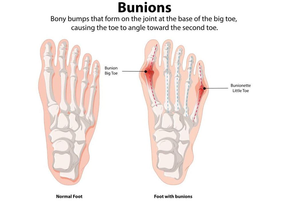
Bunions
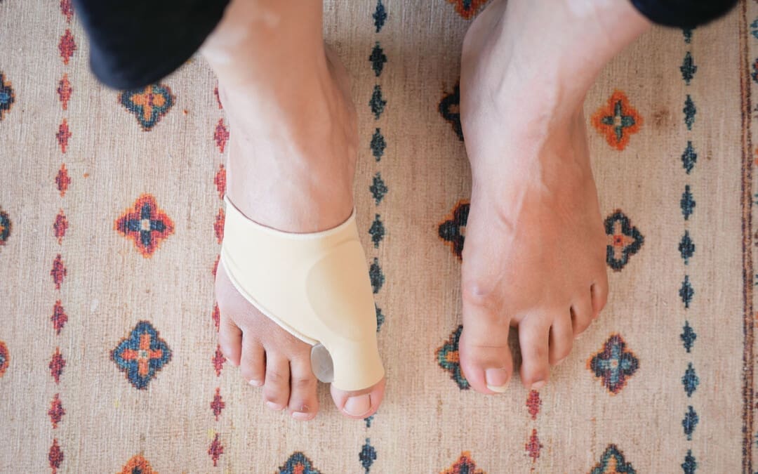
A bunion, also known as hallux valgus, is a bony, often painful protrusion on the side of the big toe. The most common cause of bunions is a misalignment of the big toe. Early symptoms of a bunion include redness, thicker skin, edema, and discomfort around the big toe joint. While you can’t stop a bunion from growing, you can manage the symptoms along the way. Early indicators of bunions include pain management measures and treatment choices. (MedlinePlus, 2024)
Early Visual Signs
Bunions are initially minor and don’t result in many noticeable issues. The following symptoms may be present in the early stages of a bunion around the metatarsophalangeal (MTP) joint, which joins the base of your big toe to the rest of your foot. (American Academy of Orthopaedic Surgeons, 2022)
Redness
Skin that is darker and swollen
Skin that has gotten thicker and harder
There is a bony bump
A bump where the big toe goes toward the second toe or even under it
Corns and calluses
Early Physical Signs
While bunions may not be visible immediately, they can cause discomfort even in the early stages. Here are some early physical indicators of a developing bunion. (MedlinePlus, 2024)
Pain in the foot and big toe
This pain is especially noticeable when walking or wearing tight, narrow-toed shoes.
Decreased movement of the big toe
Tenderness
Inflammation
Swelling
Stiffness
Heat
Stages
Bunions often worsen over time, a condition known as progressive. You could have problems if you don’t do anything to stop your bunions from getting bigger. Bunions in later stages might cause the following symptoms: (American Academy of Orthopaedic Surgeons, 2022)
Long-lasting, intense pain in and around the MTP joint and the sides and bottom of your foot
Bursitis causes a sac full of fluid to form at the bottom of your big toe.
The big toe bends toward and even crosses over the second toe.
Too much bone development on the side of your big toe
Not being able to wear your regular shoes
Hard to walk
Abnormalities known as “hammer toe” occur when your second, third, or fourth toes bend upward in the middle, resembling a hammer or claw.
Hallux rigidus is a form of arthritis affecting the big toe.
Manage the Progression
Bunions tend to remain in place once they start to develop. However, there are several steps you can take to prevent them from worsening or causing additional problems. These are some of them: (American Academy of Orthopaedic Surgeons, 2022)
Place spacers between your toes to prevent friction and chafing.
Use over-the-counter (OTC) pads made of felt, silicone, or foam to cover the bunion.
You should also stretch your calf muscles to improve joint alignment.
Managing Pain
Advil and Motrin (ibuprofen) are two examples of non-steroidal anti-inflammatory medicines (NSAIDs) that can aid with bunion pain. Studies also show that injecting Botox into the muscles in the forefoot can help with pain. (Hurn, S. E., et al., 2022)
Nonsurgical Early Stage Treatment
If your bunions continue to worsen despite using the self-care procedures listed above, you may need to consult a podiatrist (a foot expert) or another healthcare provider. A healthcare provider may recommend.
Orthotics, or foot orthoses
Foot orthoses, also known as orthotics, are customized inserts that help alleviate bunion pain and prevent chafing.
Splints
Bunion splints are orthotic devices that can help straighten out your toes. People typically use them at night. (Aebischer, A. S., & Duff, S. 2020)
Physical Therapy
A physical therapist can help you by giving you exercises that will help your feet and joints line up better. They might also do manual therapy to help with pain. (Hurn, S. E., et al., 2022)
Podiatrist
A podiatrist is a medical doctor (M.D.) who specializes in treating health problems that affect the feet, ankles, and lower legs. If you want help with your bunion issues, ask your doctor for a referral to a podiatrist. (American Podiatric Medical Association, 2025)
Chiropractic Treatment
Chiropractors and nurse practitioners (NPs) have distinct roles in managing bunions, which are bony bumps at the base of the big toe caused by joint misalignment, often resulting in pain, swelling, and restricted movement. Here’s how each can help:
Chiropractors:
Focus: Chiropractors primarily address musculoskeletal issues through manual adjustments and manipulations.
Bunion Support:
Foot Adjustments: They may perform adjustments to improve foot alignment and joint mobility, which can potentially reduce bunion-related discomfort.
Soft Tissue Therapy: Techniques such as massage or myofascial release can help alleviate tension in surrounding muscles and tissues.
Orthotics or Taping: Some chiropractors recommend custom orthotics or use taping to support proper foot mechanics.
Exercise Guidance: They may suggest stretches or exercises to strengthen foot muscles and improve alignment.
Limitations: Chiropractors don’t prescribe medications or perform surgeries, so severe cases requiring these interventions would need referral to a podiatrist or orthopedic specialist.
Evidence: While some patients report relief from chiropractic care for foot issues, evidence specifically for bunions is limited, and results vary.
Nurse Practitioners:
Focus: NPs are advanced practice registered nurses with broad medical training, able to diagnose, treat, and prescribe medications within their scope of practice.
Bunion Support:
Diagnosis and Assessment: NPs can evaluate bunion severity, often using physical examinations or ordering imaging, such as X-rays, to assess joint damage.
Pain Management: They may prescribe anti-inflammatory medications (e.g., ibuprofen) or corticosteroid injections for pain and swelling.
Conservative Treatments: NPs can recommend padding, splints, or orthotic devices to reduce pressure and improve alignment.
Lifestyle Advice: They provide guidance on footwear (such as wide-toed shoes) and weight management to reduce stress on the bunion.
Referrals: For severe cases, NPs can refer patients to podiatrists or orthopedic surgeons for surgical options, such as bunionectomies.
Scope: NPs offer a medical approach, bridging conservative care and coordination with specialists.
Key Differences:
Chiropractors focus on non-invasive, manual techniques and alignment, while NPs can incorporate medications and broader medical management.
NPs are more likely to coordinate with other healthcare providers for comprehensive care, whereas chiropractors tend to work more independently.
General Notes:
Both can assist in managing mild to moderate bunion symptoms, but neither can “cure” bunions, particularly if the structural deformity has progressed.
Consult a podiatrist or an orthopedic specialist for persistent or worsening symptoms, as severe cases may require surgical intervention.
Always verify the provider’s credentials and experience in treating bunions.
Injury Medical Chiropractic and Functional Medicine Clinic
Dr. Jimenez, a nurse practitioner, treats a wide range of conditions using a combination of medical knowledge and chiropractic care. The clinic offers personalized care plans that incorporate functional medicine, acupuncture, electroacupuncture, and sports medicine. The clinic treats chronic pain syndromes and injuries, focusing on strength, agility, and flexibility. Comprehensive care plans, when paired with in-person and virtual health coaching, offer personalized treatment and wellness outcomes for patients of all ages and abilities.
Enhance Your Performance with Functional Foot Orthotics
References
MedlinePlus (2024). Bunions. U.S. Department of Health and Human Services.
American Academy of Orthopaedic Surgeons. (2022). “Bunions.” OrthoInfo. from https://orthoinfo.aaos.org/en/diseases–conditions/bunions/.
Hurn, S. E., Matthews, B. G., Munteanu, S. E., & Menz, H. B. (2022). Effectiveness of Nonsurgical Interventions for Hallux Valgus: A Systematic Review and Meta-Analysis. Arthritis care & research, 74(10), 1676–1688. https://doi.org/10.1002/acr.24603
Aebischer, A. S., & Duff, S. (2020). Bunions: A review of management. Australian Journal of General Practice, 49(11), 720–723. https://doi.org/10.31128/AJGP-07-20-5541
American Podiatric Medical Association. (2025). “What is a podiatrist?” Advancing foot and ankle medicine and surgery. from https://www.apma.org/patients-and-the-public/what-is-a-podiatrist/.
Can older people who exercise regularly lessen their risk of dementia and enhance their overall health?
Exercise and the Prevention of Dementia
Dementia, a term used to cover several conditions that impact memory and cognition, is currently the seventh leading cause of mortality worldwide. (World Health Organization, 2025) More than 10 million new cases of dementia are found around the world each year. (J.H. Yoon et al., 2023) Research investigating the relationship between exercise and insulin in the brain suggests that regular exercise may improve brain function and decrease the prevalence of dementia. Scientists have discovered that variables, such as.
Muscles must be exercised and mobilized. People who do not engage in physical activity have rigid muscles, which impede the effectiveness of insulin. The body’s sensitivity to insulin is improved by the contraction and relaxation of the muscles during movement. Dementia can be prevented by understanding how to decrease the body’s insulin resistance.
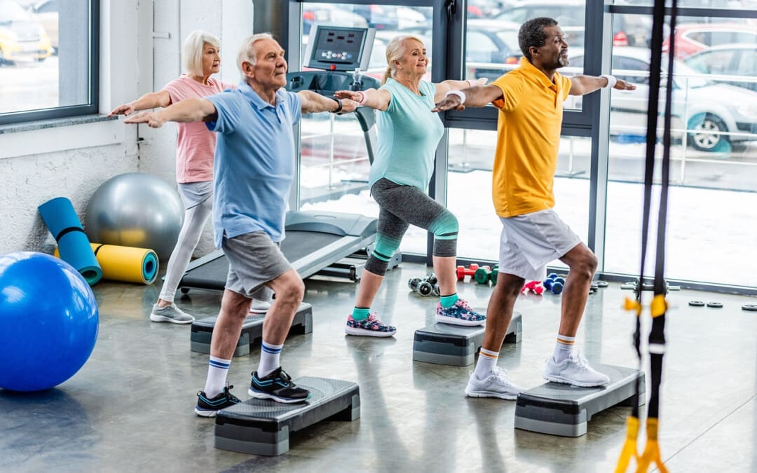
Physical Activity and Lower Risk
Over two weeks, researchers examined 21 older adults with prediabetes and found that exercise enhances cognitive performance by aiding the brain in insulin regulation. They conducted twelve supervised training sessions of moderate to extreme intensity. (Malin S. K. et al., 2025)
The results indicated that brain-derived extracellular vesicles containing insulin-related proteins had significantly increased.
These microscopic messengers are essential for brain maintenance and aid in neuronal communication.
Three serine/threonine-specific protein kinases known as AKT (protein kinase B, or PKB) are essential for several cellular functions, such as cell growth, survival, metabolism, and cell cycle control.
The protein affects the development and health of neuronal cells and is thought to play a significant role in insulin signaling.
The notion that exercise might help prevent dementia by potentially enhancing insulin signaling is important since poor insulin response can result in dementia.
Even while further research is needed, these findings prove that physical activity could be a cost-effective and easily accessible way to improve long-term brain health. To better understand how insulin affects brain activity, researchers will use MRIs and an insulin spray in the study’s next phase. To learn more about the effects of the insulin spray, they will compare the blood flow in the brain before and after it is administered.
Insulin and Exercise Are Essential for Brain Health
Insulin is a hormone that the pancreas makes. It controls blood sugar levels. But it’s just as vital for the health of the brain. Insulin binds to many receptors in the brain, which makes synaptic connections stronger and makes it easier for neurons to talk to each other. Both of these things help with memory and learning. (Gray, S. M., Meijer, R. I., & Barrett, E. J. 2014)
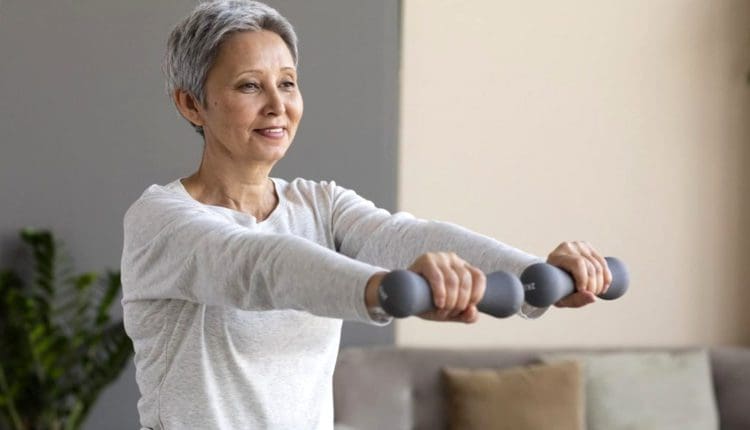
Type 2 diabetes, which impairs insulin transmission, increases the risk of cognitive impairment. (Yoon J.H. et al., 2023) Research has demonstrated that insulin resistance exacerbates tau tangles and amyloid-beta plaques, which are two critical characteristics of Alzheimer’s disease. (Hong, S., Han, K., & Park, C. Y. 2021) Increasing insulin sensitivity may slow down or even stop these changes in the brain. Working out, especially aerobic exercise, is good for the brain. According to earlier research, exercise can help people maintain or enhance their memory and brain function. (Rosenberg A. et al., 2020) Other ways to increase brain health include:
Maintaining social interaction
Challenging the mind
Getting a good night’s sleep
Controlling stress
A nutritious diet
Maintaining an active lifestyle
Controlling diabetes
Controlling blood pressure
To achieve the greatest possible effect, dementia prevention in the future will likely involve integrating lifestyle modifications, such as exercise, with medications. In individuals with diabetes or prediabetes, the risk of dementia is significantly reduced, and frequent exercise can assist in the preservation of optimal brain function.
Chiropractic and Functional Medicine Clinic
As a family nurse practitioner, Dr. Jimenez uses the latest medical expertise and chiropractic therapy to address many problems. Our clinic uses functional medicine, acupuncture, electro-acupuncture, and sports medicine to create individualized care plans that improve movement, encourage long-term health, and speed up the body’s natural healing process. We focus on strength, agility, and flexibility to help our patients thrive, regardless of age or health problems. At El Paso’s Chiropractic Rehabilitation Clinic & Integrated Medicine Center, we want to help people with chronic pain syndromes and injuries. We focus on improving flexibility, mobility, and agility through programs suitable for people of all ages and abilities. We ensure that each patient gets personalized care and reaches their health objectives through detailed care plans and health coaching in person and online.
Is Movement Essential to Recovery?
References
World Health Organization. (2025). “Dementia.” World Health Organization. from https://www.who.int/news-room/fact sheets/detail/dementia#:~:text=Alzheimer%20disease%20is%20the%20most,60%E2%80%9370%25%20of%20cases.
Yoon, J. H., Hwang, J., Son, S. U., Choi, J., You, S. W., Park, H., Cha, S. Y., & Maeng, S. (2023). How Can Insulin Resistance Cause Alzheimer’s Disease?. International Journal of Molecular Sciences, 24(4), 3506. https://doi.org/10.3390/ijms24043506
Malin, S. K., Battillo, D. J., Beeri, M. S., Mustapic, M., Delgado-Peraza, F., & Kapogiannis, D. (2025). Two weeks of exercise alters neuronal extracellular vesicle insulin signaling proteins and pro-BDNF in older adults with prediabetes. Aging cell, 24(1), e14369. https://doi.org/10.1111/acel.14369
Gray, S. M., Meijer, R. I., & Barrett, E. J. (2014). Insulin regulates brain function, but how does it get there?. Diabetes, 63(12), 3992–3997. https://doi.org/10.2337/db14-0340
Hong, S., Han, K., & Park, C. Y. (2021). The insulin resistance by triglyceride glucose index and risk for dementia: population-based study. Alzheimer’s research & therapy, 13(1), 9. https://doi.org/10.1186/s13195-020-00758-4
Rosenberg, A., Mangialasche, F., Ngandu, T., Solomon, A., & Kivipelto, M. (2020). Multidomain Interventions to Prevent Cognitive Impairment, Alzheimer’s Disease, and Dementia: From FINGER to World-Wide FINGERS. The Journal of Prevention of Alzheimer’s disease, 7(1), 29–36. https://doi.org/10.14283/jpad.2019.41
Can bone growth stimulators help promote bone healing in cases where fractures or fusions fail to heal properly?
Bone Growth Stimulator
Individuals who sustain broken bones typically heal the fracture with appropriate treatment, which may include casts, realignment, and surgery. This type of surgery is performed on the spine and joints throughout the body; typically, the bone heals without a problem. Bone healing is a natural process, as bones are constantly replaced with new ones, and after an injury, the body can heal the damage to the bone. However, bone healing sometimes does not happen correctly and/or completely. Bone healing can take a long time, which is known as a delayed union, or it may not occur at all, or a nonunion. This is when a healthcare provider could recommend bone growth stimulation.
How They Work
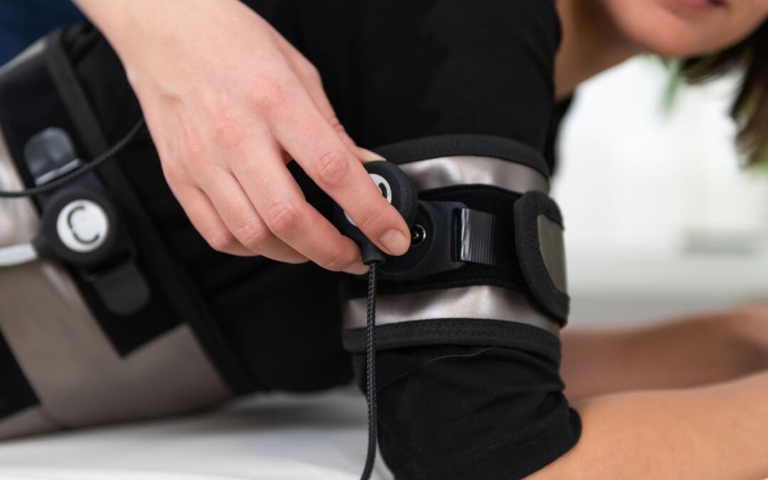
Bone growth stimulators apply external or implanted electrical or ultrasonic energy to the fracture or fusion site, stimulating bone growth. These devices are often used when a fracture doesn’t heal within the expected timeframe (a nonunion fracture) or when a spinal fusion has not successfully fused. (FDA, 2022)
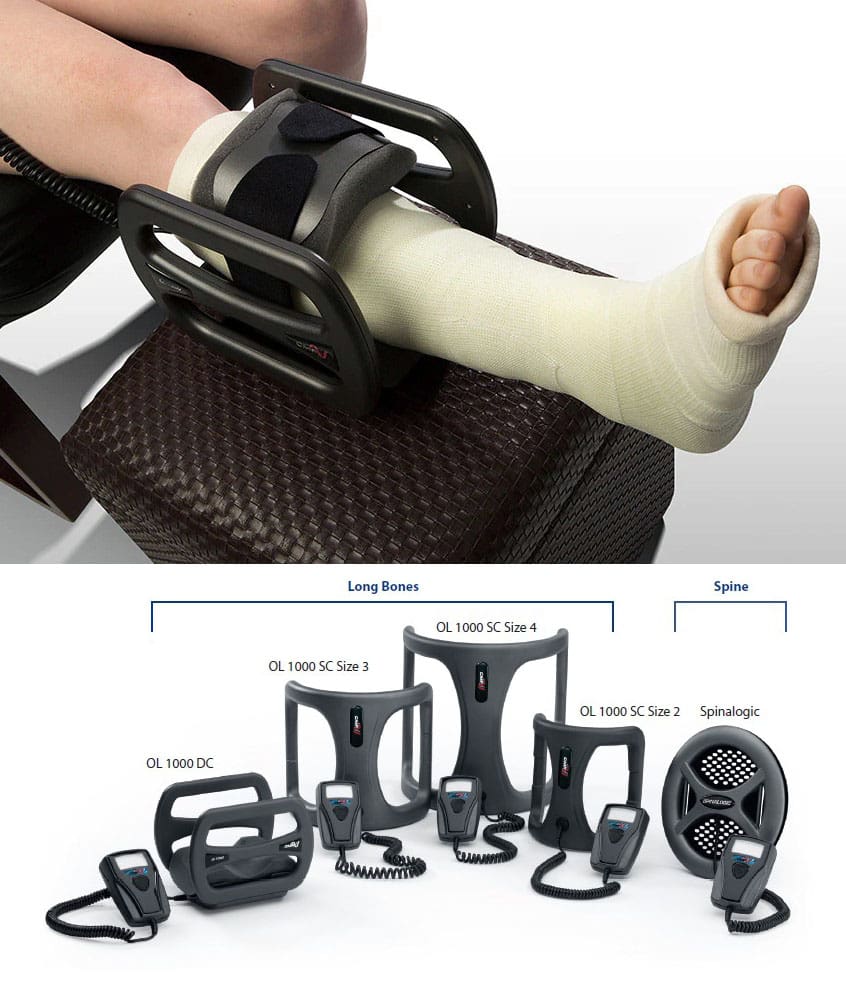
They come in various shapes, sizes, and technologies depending on the location of the fracture.
The most commonly used bone stimulators in orthopedics are electrical and ultrasound stimulators.
The stimulator emits a pulsed electromagnetic or ultrasonic impulse to the area where bone healing should occur.
Most modern bone stimulators are attached near the fracture site or fused with a small battery pack and worn for a period each day that may last minutes or hours.
Electrical Stimulation
Devices deliver low-level electrical pulses to the fracture site, which can stimulate bone cells to create new bone tissue.
Ultrasound Stimulation
They emit low-intensity pulsed ultrasound waves, which are absorbed by the bone and can promote bone healing.
Implantable vs. External
Some stimulators are surgically implanted, providing constant stimulation directly at the fracture site, while others are external and worn over the skin or cast.
The goal is to activate a series of receptors in the body to encourage a healing response. (Childs, S. G. 2003) The stimulator activates a pathway that releases chemicals within the body to promote fracture healing. This type of process in the body is called a cascade, and it happens when one signal stimulates another method, and so on until healing is complete. The bone stimulator ensures this cascade continues until the healing process is complete.
Effectiveness
Studies of bone stimulator effectiveness suggest two benefits:
Less pain is associated with the bone healing process.
Faster fracture healing.
However, these studies haven’t shown that these differences lead to improved patient functional outcomes. It would seem that if there is less pain and faster healing, then the patient should recover without complications. However, some researchers have suggested this is probably because the differences in pain and healing times are small and not necessarily noticeable. (Aleem, I. S. et al., 2016)
While bone growth stimulators can be effective, the results can vary, and their effectiveness is still under investigation.
Some studies have shown that stimulators can reduce pain and speed up healing time, while others have shown mixed results.
It’s important to discuss the benefits and risks of bone growth stimulation with a doctor to determine if it’s the right treatment option.
When Stimulation is Necessary
Bone stimulators are currently not used for routine fracture healing. It is certainly possible that bone healing stimulators will be used routinely in the future. They seem to show some benefit in non-healing fractures or fractures that are likely troublesome to heal. Some reasons individuals may have problems healing fractures are injuries to the blood supply to and around the fracture, injuries to specific bones, and overuse-related fractures. (Victoria, G. et al., 2009) These injuries may include (FDA, 2022)
Nonunion Fractures
When a fracture doesn’t heal within the expected timeframe.
Open fractures
Stress fractures
Scaphoid bone fractures
Talus fractures
Failed Fusions
When a spinal fusion hasn’t been successful.
High-Risk Patients
For individuals with factors that can hinder bone healing, such as smoking, diabetes, or certain medications
Bone healing typically proceeds without much problem. However, there are situations where people have issues healing after fractures or surgery. Bone stimulators are not used for routine bone healing but in situations where steps may be needed to help stimulate the body. While the improvement may be small, it may be critical if it is the difference between healing and nonhealing.
Injury Medical Chiropractic and Functional Medicine Clinic
As a Family Practice Nurse Practitioner, Dr. Jimenez combines advanced medical expertise with chiropractic care to address various conditions. Our clinic integrates Functional Medicine, Acupuncture, Electro-Acupuncture, and Sports Medicine to create customized care plans that promote natural healing, mobility, and long-term wellness. By focusing on flexibility, agility, and strength, we empower patients to thrive, regardless of age or health challenges. At El Paso’s Chiropractic Rehabilitation Clinic & Integrated Medicine Center, we passionately focus on treating patients after injuries and chronic pain syndromes. We focus on improving your ability through flexibility, mobility, and agility programs tailored for all age groups and disabilities. We use in-person and virtual health coaching and comprehensive care plans to ensure every patient’s personalized care and wellness outcomes.
From Injury to Recovery with Chiropractic Care
References
U.S. Food & Drug Administration. (2022). AccelStim Bone Growth Stimulator – P210035. Retrieved from https://www.fda.gov/medical-devices/recently-approved-devices/accelstim-bone-growth-stimulator-p210035#:~:text=What%20is%20it?,the%20transducer%20to%20the%20fracture
Childs, S. G. (2003). Stimulators of bone healing. Biologic and biomechanical. Orthopedic nursing, 22(6), 421–428. https://doi.org/10.1097/00006416-200311000-00010
Aleem, I. S., Aleem, I., Evaniew, N., Busse, J. W., Yaszemski, M., Agarwal, A., Einhorn, T., & Bhandari, M. (2016). Efficacy of Electrical Stimulators for Bone Healing: A Meta-Analysis of Randomized Sham-Controlled Trials. Scientific Reports, 6, 31724. https://doi.org/10.1038/srep31724
Victoria, G., Petrisor, B., Drew, B., & Dick, D. (2009). Bone stimulation for fracture healing: What’s all the fuss?. Indian Journal of Orthopaedics, 43(2), 117–120. https://doi.org/10.4103/0019-5413.50844
Can incorporating sauerkraut into one’s diet help promote healthy gut bacteria?
Sauerkraut
Sauerkraut, a fermented cabbage food, is a great source of probiotics and fiber, both of which can contribute to gut health. It’s a living food containing various microbial strains and can help improve digestion, reduce inflammation, and improve overall gut health. (Shahbazi R. et al., 2021)
Fermented foods like sauerkraut have live cultures that promote the growth of beneficial probiotics, bacteria that provide powerful health benefits. Probiotics also help make foods more digestible, increasing the gut’s ability to absorb the vitamins and minerals they contain. A study found that fermented cabbage may help protect intestinal cells from inflammatory damage more than raw cabbage. (Wei L., & Marco M. L., 2025)
Homemade sauerkraut likely contains more live cultures than store-bought since processing may destroy some of the healthy bacteria. (National Institutes of Health Office of Dietary Supplements, 2023) Depending on which supplement is chosen, sauerkraut may have a greater diversity of probiotic strains. This is because an oral supplement has a known quantity and strain of probiotics. Individuals who want to add probiotics to their diet for potential gut health benefits should take a food-first approach, which is generally recommended over oral supplements. (National Institutes of Health, 2025)
Look at the Benefits
Probiotics
Sauerkraut contains lactic acid bacteria, which are beneficial bacteria that can help support the immune system and reduce inflammation. (Healthline, 2023)
Fiber
Sauerkraut is a robust source of fiber, which aids in digestion and can help you feel full longer.
Digestion
The probiotics in sauerkraut can improve digestion by promoting a healthy gut microbiome. (Medical News Today, 2023)
Consult with a doctor before adding sauerkraut to your diet if you are taking monoamine oxidase inhibitors (MAOIs).
Start small and learn how you like to eat sauerkraut, which can be as a condiment, side dish, or sandwich ingredient.
Injury Medical Chiropractic and Functional Medicine Clinic
As a Family Practice Nurse Practitioner, Dr. Jimenez combines advanced medical expertise with chiropractic care to address various conditions. Injury Medical Chiropractic and Functional Medicine Clinic works with primary healthcare providers and specialists to develop highly effective treatment plans through an integrated approach for each patient and restore health and function to the body through nutrition and wellness, functional medicine, acupuncture, electroacupuncture, and integrated medicine protocols. We focus on what works for you to relieve pain, restore function, prevent injury, and mitigate issues through adjustments that help the body realign itself. The clinic can also work with other medical professionals to integrate a treatment plan to resolve musculoskeletal problems.
Body In Balance: Chiropractic, Fitness, and Nutrition
References
Shahbazi, R., Sharifzad, F., Bagheri, R., Alsadi, N., Yasavoli-Sharahi, H., & Matar, C. (2021). Anti-Inflammatory and Immunomodulatory Properties of Fermented Plant Foods. Nutrients, 13(5), 1516. https://doi.org/10.3390/nu13051516
Healthline. (2023). 8 Surprising Benefits of Sauerkraut (Plus How to Make It). https://www.healthline.com/nutrition/benefits-of-sauerkraut#nutrients
Medical News Today. (2023). What are the benefits of eating sauerkraut? https://www.medicalnewstoday.com/articles/health-benefits-of-sauerkraut
Wei, L., & Marco, M. L. (2025). The fermented cabbage metabolome and its protection against cytokine-induced intestinal barrier disruption of Caco-2 monolayers. Applied and environmental microbiology, e0223424. Advance online publication. https://doi.org/10.1128/aem.02234-24
National Institutes of Health Office of Dietary Supplements. (2023). Probiotics. Retrieved from https://ods.od.nih.gov/factsheets/Probiotics-Consumer/
National Institutes of Health. (2025). Probiotics. Retrieved from https://ods.od.nih.gov/factsheets/Probiotics-HealthProfessional/
U.S. Department of Agriculture. (2018). Sauerkraut, canned, solids and liquids. Retrieved from https://fdc.nal.usda.gov/food-details/169279/nutrients
Why do the muscles retighten days or weeks later for individuals who have received massage or chiropractic adjustments?
Muscles Retighten
Muscles can retighten again days or weeks after chiropractic treatment and/or a massage. A few factors may cause this.
Muscle memory: The body’s natural tendency to return to its previous state of tension.
This includes all the unhealthy postures/positioning
Delayed inflammatory response.
The body’s natural healing process
How muscles respond to manipulation
The need for ongoing maintenance of muscle health
After a massage, muscles can experience a temporary increase in soreness or tightness as the body heals from any minor damage or inflammation caused by the massage. Additionally, if the underlying issue causing muscle tightness is not addressed, the muscles may revert to their original state over time. (Cleveland Clinic, 2024)
Causes
Causes for muscles to retighten.
Underlying Issues
If the root cause of the muscle tightness is not addressed, such as unhealthy posture, injuries, repetitive motions, or muscle imbalances, it can lead to further complications.
In that case, the muscles may return to their original state after a few days or weeks.
Delayed Onset Muscle Soreness (DOMS)
For example, muscles feel sore after intense exercise, and individuals can also experience DOMS after a massage, especially if they are particularly tight or if it’s been a while since their last massage. (Healthline, 2020)
Delayed Inflammatory Response
Massage can sometimes cause microscopic tears or inflammation in muscle tissue, which triggers the body’s natural healing process.
This response can cause soreness and temporary tightness, as the body repairs the damaged tissue even after the initial massage. (Essential Chiropractic, 2025)
Muscle Memory
The body tends to revert to habitual patterns of muscle tension, especially if the underlying cause of the tightness isn’t addressed.
Muscles can retain how they were manipulated during the massage and may revert to their original tightness if they haven’t been consistently relaxed.
Muscle Guarding
The individual and body can subconsciously tense muscles to avoid pain or soreness, even after the massage.
This muscle guarding can counteract the benefits of the massage and lead to a renewed cycle of tension. (Bhimani R. H., & Soomar D. 2019)
Muscle Health Maintenance
Regular Maintenance
Muscles need consistent care to maintain their optimal state of health and function.
Regular massages and other lifestyle changes, such as stretching and strengthening exercises, can help maintain muscle flexibility and reduce the likelihood of muscle tightness returning.
Individual Variation
Factors like the type of massage received, the frequency of massages, and individual body responses can all influence how long a massage’s benefits last. (Cleveland Clinic, 2024)
Massage Treatment
Muscles can retighten after a massage due to the body’s response to the pressure and manipulation during the treatment. This can include individual sensitivity to massage, the type of massage, and hydration levels, which can also play a role.
Type of Massage
Deep tissue massage, which focuses on specific muscle groups and applies deeper pressure, may cause more soreness than other types of massage.
Some individuals may be more sensitive to massage and experience soreness, even with a gentle massage. (Cleveland Clinic, 2024)
Muscle Fiber Manipulation
Deep tissue massage can break down spasms or microtears in muscle fibers, leading to soreness.
This is similar to the microscopic tears that occur during exercise, which are necessary for muscle repair and growth.
Dehydration
Individuals who are not adequately hydrated may experience increased soreness after a massage. (Cleveland Clinic, 2024)
Lactic Acid Buildup
During an intense massage, particularly deep tissue, lactic acid can build up in the muscles, leading to soreness and tightness.
If the muscles were particularly tense or knotted before the massage, the manipulation could lead to soreness as the knots get worked out.
Injury Medical Chiropractic and Functional Medicine Clinic
As a Family Practice Nurse Practitioner, Dr. Jimenez combines advanced medical expertise with chiropractic care to address various conditions. Our clinic integrates Functional Medicine, Acupuncture, Electro-Acupuncture, and Sports Medicine to create customized care plans that promote natural healing, mobility, and long-term wellness. By focusing on flexibility, agility, and strength, we empower patients to thrive, regardless of age or health challenges. At El Paso’s Chiropractic Rehabilitation Clinic & Integrated Medicine Center, we passionately focus on treating patients after injuries and chronic pain syndromes. We focus on improving your ability through flexibility, mobility, and agility programs tailored for all age groups and disabilities. We use in-person and virtual health coaching and comprehensive care plans to ensure every patient’s personalized care and wellness outcomes.
Secrets of Optimal Wellness
References
Cleveland Clinic. (2024). How to Relieve Muscle Soreness After a Massage. https://health.clevelandclinic.org/why-does-my-body-feel-worse-after-a-massage
Healthline. (2020). How to Relieve Sore Muscles After a Massage. https://www.healthline.com/health/sore-after-massage#causes-of-soreness
Essential Chiropractic and Physiotherapy. (2025). Muscle Ache After Massage: Common Causes and Relief. https://essentialchiropractic.co.uk/muscle-ache-after-massage-common-causes-and-relief/#:~:text=Post%2Dmassage%20soreness%20is%20a,hours%20as%20the%20muscles%20recover.
Bhimani, R. H., & Soomar, D. (2019). Understanding Symptoms of Muscle Tightness, Weakness, and Rigidity From a Nursing Perspective. Rehabilitation Nursing: The Official Journal of the Association of Rehabilitation Nurses, 44(5), 271–281. https://doi.org/10.1097/rnj.0000000000000151
Can pace running help runners concentrate on other things, like breathing, form, or mental toughness?
Pace Running
Running is a sport many participate in and doesn’t require any equipment—only quality running shoes. Running in races comes with various physical and mental challenges. Some runners enjoy running in races and marathons but want to perfect their abilities and techniques, become more consistent, finish in a certain time, or pass a personal record. This is where pace running or working with a pace runner can help achieve those goals.
What is a Pace Runner?
A pace runner, or pacer, is an experienced runner who can run at a set pace for a long time. The pacer sets the speed so the runner can focus on running. Pacers run in races or marathons to help set the pace for a runner or runners. There are different reasons why professional and amateur runners work with a pace runner. Professional runners often use pace runners to work on techniques and help reach new levels in their running, while amateur runners can work on improving their overall running abilities. There are three typical types of pacers.
Race Pacer
Pacers wear or carry signs to make it easier for the runners and can often run split times.
A split time is the time it takes to run a certain distance.
Typically, these pacers will run at an even pace.
Many marathons will have pace groups that run at a specific speed or pace throughout the race.
The pace groups will have runners running a set time so other runners can key off these runners.
There are typically set paces for a 3-hour marathon time up to a 6-hour marathon time.
Runners can use pacers to help them reach their goals without relying on technology, such as a smartwatch or GPS, to determine if they maintain the correct speed throughout the race.
Distance Pacer
Professional and non-professional runners who run long distances or ultramarathons may also use a pace runner to set a specific tempo.
Ultramarathons are any race with a distance longer than the standard marathon of 26.2 miles.
Because some ultramarathons can be as long as 50 to 100 miles, runners who choose to have a pacer often have several pacers at different sections to motivate them and help set the rhythm.
Record Pacer or Rabbit
A record pacer, also known as a rabbit, is a pacer who helps a professional runner set a new record.
A rabbit often leads the race for a predetermined distance at a predetermined pace.
Some races have multiple pacers at various distances.
How Are Pacers Used?
There are different reasons for having a pacer, but they are based on the runner’s goals.
A runner being paced runs directly behind a pacer or pacers.
A pacer can run any pace requested, but typically, the pace groups run at an even pace or with a slight negative split.
A negative split is when a runner runs the race’s second half faster than the first half.
How Do They Help?
A pacer can be helpful because it takes more energy to lead a race than to sit back and follow another runner.
The pacer is responsible for timing and establishing the tempo so runners only have to focus on running.
A pacer does more work setting the pace, which allows the runner being paced to relax and not stress about hitting the pace.
A study focused on elite athletes who used pacers. It found that running together at a realistic speed helped optimize and achieve finishing time goals. (Casado A. et al., 2021) However, not all elite events allow pacers to participate. Some championship events, like the Olympics and NCAA Championships, do not allow pacer runners.
Benefits
Benefits of running with a pacer.
Prevent Running Too Fast
When a race begins, runners can take off and start too fast.
Starting too quickly can negatively impact the runners, like losing energy and stamina.
A pacer can help start the race with a steady pace, keeping them from going out too fast or expending too much energy.
Maintain an Even Pace
Maintaining an even pace can be difficult throughout a long race or a marathon.
Having a pacer can relieve some stress and pressure.
A pacer can keep the runners on an even pace or negative splits versus going out too fast and losing energy.
Focus on the Race
Pacers help the runners stay focused on the race without worrying about how they are running or maintaining the right speed.
A pacer can help keep runners relaxed.
Motivating
Pacers can help motivate runners.
The pace runners may encourage individuals along the way and help them stay motivated to keep working toward their goal, especially when the hard parts come.
Disadvantages
Running with a pacer can sometimes be more stressful than beneficial, especially for amateur runners. A pacer or pace group can be helpful, but it also can create more stress for some amateur runners.
Sometimes, individuals realize the pacer is running too quickly for what they can handle.
Anxiety can present as not being able to keep up.
Other times, runners underestimate their abilities and run more slowly than their bodies can handle.
However, runners don’t have to stay with a pace group just because they started with them.
Individuals can run ahead.
Or if the group is too fast, the runner can slow down and join the slower-paced group behind them.
The key is to listen to your body and do what feels right.
Becoming a Pacer
An experienced runner who wants to be a pacer for a race can contact a running organization about becoming a pace runner.
Individuals need to be able to have consistent racing times.
Be able to run at a set speed for a long duration.
Most pacers will contact the race organization to apply for a certain pace.
Some pacers enjoy the thrill of running in a race and helping other runners meet their goals.
The runner’s goals and the type of event also influence who the pacer is.
Injury Medical Chiropractic & Functional Medicine Clinic
Individuals can talk to local running experts or a running coach. Injury Medical Chiropractic and Functional Medicine Clinic works with primary healthcare providers and specialists to develop an optimal health and wellness solution. We focus on what works for you to relieve pain, restore function, and prevent injury. We can also work with other medical professionals to integrate a treatment plan to resolve musculoskeletal issues.
The Difference of Using Custom Foot Orthotics
References
Casado, A., Hanley, B., Jiménez-Reyes, P., & Renfree, A. (2021). Pacing profiles and tactical behaviors of elite runners. Journal of Sport and Health Science, 10(5), 537–549. https://doi.org/10.1016/j.jshs.2020.06.011
What is the body’s center of gravity to understand and maintain a healthy posture and balance?
Center of Gravity
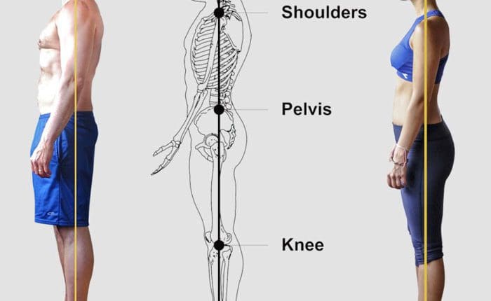
The human center of gravity, or COG, also known as the center of mass, with the two terms being interchangeable, is where the body’s weight appears to be concentrated. It’s a point in space where the entire body’s mass can be concentrated. When standing upright, the COG is generally located behind the navel and in front of the sacrum, around the level of the second vertebra. (Le Huec, J. 2011) The center of gravity is related to posture, including issues such as swayback, the design of posture exercise programs, and much more.
Gravity is a downward pull or force the Earth exerts on the body, creating weight. (NASA, 2013)
The center of gravity (COG) is where a body’s weight is equally balanced in all directions. (Physiopedia, 2025)
When the COG is defined, it is done from the reference of a static, standing position.
Because the body is in motion when we change positions, the COG is located in a new position with each new position.
The center of gravity is the point around which all the parts balance, which may be inside or outside the body.
Even slight changes in position can change where the COG is. (Physiopedia, 2025)
Key aspects of the COG
Location
When standing upright, the COG is typically found at the second vertebral level, behind the navel and in front of the sacrum. (Le Huec, J. 2011)
Shifting COG
The COG can shift depending on body position, posture, and movements like bending forward or backward. (Physiopedia, 2025)
Balance Impact
The relationship between the COG and the base of support (the area of the body in contact with the ground) is fundamental to maintaining balance.
Variations
There can be variations in the COG location based on factors like gender (men tend to have a slightly higher COG), body shape, and even conditions like obesity or chronic low back pain. (Physiopedia, 2025)
Biomechanics
Understanding the COG is crucial in biomechanics and movement analysis, as it’s an index of total body motion and how the body responds to external forces.
The Human Center
The center of gravity is the point at which the body’s mass is equally balanced. This point changes depending on one’s position:
Arms up/down
Leaning
Turning
With strength and flexibility training, the human body can change its center of gravity, as gymnasts and dancers do.
When standing, the center of gravity is normally located behind the navel and in front of the sacrum bone (made up of five vertebrae fused vertically) at about the second vertebra level. (Hasegawa K. et al., 2022)
Because the body has moving parts, its overall shape changes every time it moves. Carrying something like a suitcase or grocery bag or wearing a backpack adds weight to some areas but not others, changing the center of gravity as it does.
The center of gravity is a continually changing point inside or outside the body that represents where the weight or mass of the rest of the body is equally balanced in every direction.
This point can and does change based on what is being carried and how it is carried, as well as body position and movements.
Chronic Lower Back Pain
A study in the Journal of Back and Musculoskeletal Rehabilitation found that individuals with chronic lower back pain tend to have their center of gravity located excessively towards the back. (Kim D. H., Park J. K., & Jeong M. K. 2014)
In the study, the individuals had decreased low back strength upon extension and a reduced normal low back curve.
The researchers found that those with chronic lower back pain whose center of gravity was too far back may need physical therapy to retrain the body to overcome strength and balance challenges to re-establish and maintain a healthy posture.
Injury Medical Chiropractic and Functional Medicine Clinic
As a Family Practice Nurse Practitioner, Dr. Jimenez combines advanced medical expertise with chiropractic care to address various conditions. Our clinic integrates Functional Medicine, Acupuncture, Electro-Acupuncture, and Sports Medicine to create customized care plans that promote natural healing, mobility, and long-term wellness. By focusing on flexibility, agility, and strength, we empower patients to thrive, regardless of age or health challenges. At El Paso’s Chiropractic Rehabilitation Clinic & Integrated Medicine Center, we passionately focus on treating patients after injuries and chronic pain syndromes. We focus on improving your ability through flexibility, mobility, and agility programs tailored for all age groups and disabilities. We use in-person and virtual health coaching and comprehensive care plans to ensure every patient’s personalized care and wellness outcomes.
Enhance Your Lifestyle Today with Chiropractic Care
References
Le Huec, J. C., Saddiki, R., Franke, J., Rigal, J., & Aunoble, S. (2011). Equilibrium of the human body and the gravity line: the basics. European spine journal: official publication of the European Spine Society, the European Spinal Deformity Society, and the European Section of the Cervical Spine Research Society, 20 Suppl 5(Suppl 5), 558–563. https://doi.org/10.1007/s00586-011-1939-7
NASA. Jet Propulsion Laboratory, California Institute of Technology. (2013). What is gravity? Retrieved from https://gracefo.jpl.nasa.gov/news/5/what-is-gravity/
Physiopedia. (2025). Centre of gravity. https://www.physio-pedia.com/Centre_of_Gravity
Hasegawa, K., Amabile, C., Nesme, M., & Dubousset, J. (2022). Gravity center estimation for evaluation of standing whole body compensation using virtual barycentremetry based on biplanar slot-scanning stereoradiography – validation by simultaneous force plate measurement. BMC musculoskeletal disorders, 23(1), 22. https://doi.org/10.1186/s12891-021-04948-5
Kim, D. H., Park, J. K., & Jeong, M. K. (2014). Influences of posterior-located center of gravity on lumbar extension strength, balance, and lumbar lordosis in chronic low back pain. Journal of Back and Musculoskeletal Rehabilitation, 27(2), 231–237. https://doi.org/10.3233/BMR-130442
IFM's Find A Practitioner tool is the largest referral network in Functional Medicine, created to help patients locate Functional Medicine practitioners anywhere in the world. IFM Certified Practitioners are listed first in the search results, given their extensive education in Functional Medicine