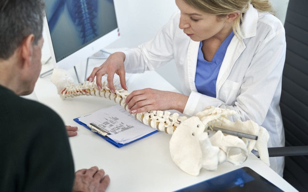
Spinal Stenosis Walking Issues:Stenosis means a narrowing. Spinal stenosis can happen in any spine region, but the neck and lower back are the most common locations. The spinal canal becomes narrower and can cause the nerves to become compressed, pinched, and irritated and can extend from the lumbar spine through the hips, buttocks, legs, and feet. Individuals with lumbar spinal stenosis may have difficulty walking caused by sensations of discomfort like numbness, electrical shocks, and pain, requiring the need to lean forward to relieve pressure and symptoms. Additionally, symptoms are likely to worsen the longer the walk. Chiropractic treatment can treat spinal stenosis because it corrects and re-aligns the spine, thus reducing pressure on the spinal cord, joints, and nerve roots.
Spinal Stenosis Walking Issues
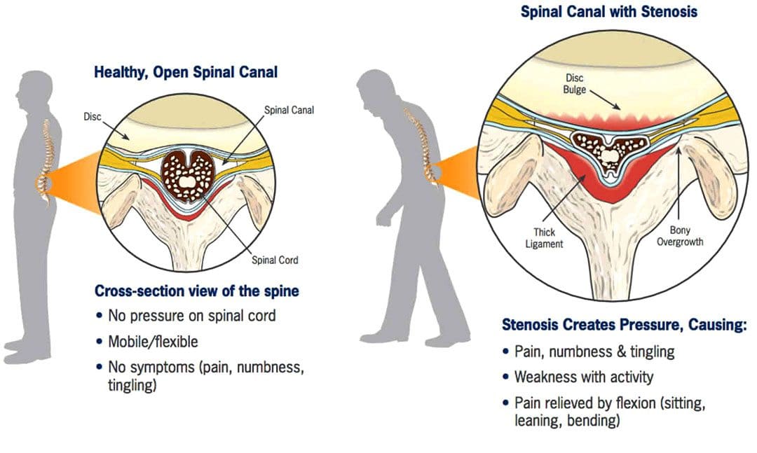
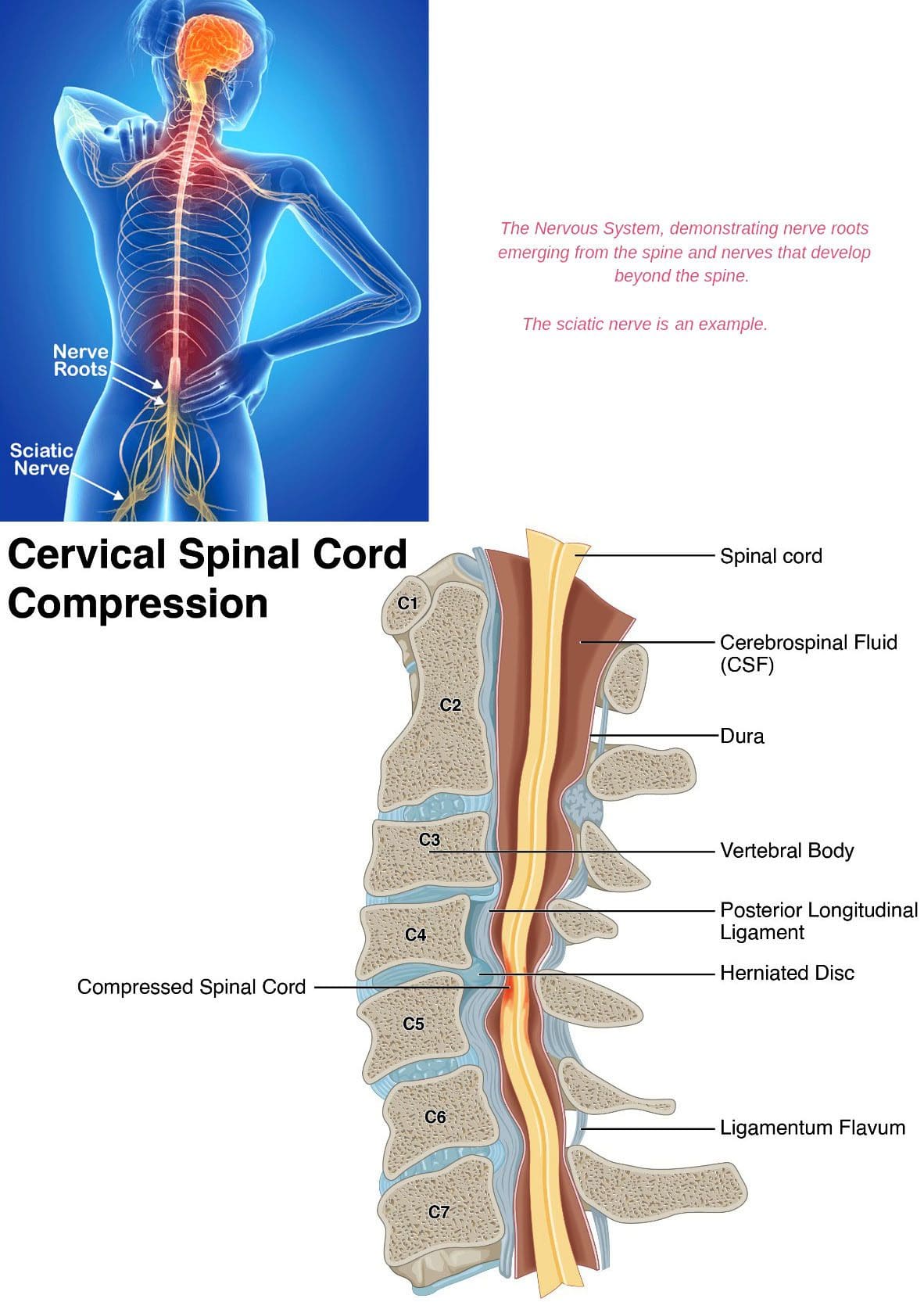
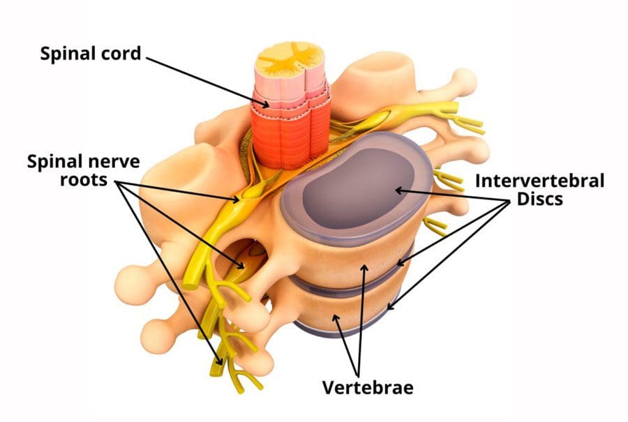
The spine is made up of interlocking vertebrae. The regions are cervical, thoracic, lumbar, and sacral bones with a foramen opening. These openings form the protective tunnel/spinal canal surrounding the spinal cord. The spinal cord is a group of nerves that run through the tunnel. The narrowing suffocates the nerves supplying the lower extremities that can influence walking activity.
Symptoms
There may be no symptoms with early lumbar spinal stenosis. Most individuals develop symptoms gradually and may begin to notice them while walking or standing. These can include:
Lower back pressure sensations when standing upright or walking.
Leg numbness, tingling, weakness, burning, and/or cramping.
Muscle weakness.
Persistent pain in the back, hips, buttocks, or legs while walking.
Difficulty lifting the top part of the foot – known as drop foot.
Loss of sensation in the feet.
A weak foot that drops/slaps down when walking.
Loss of sexual ability.
In more serious cases, severe numbness, bladder problems, and inability to stand.
Individuals begin to lean forward when symptoms start, bringing relief by reducing the pressure on the nerves. However, constantly leaning forward leads to other posture and health problems.
Diagnosis
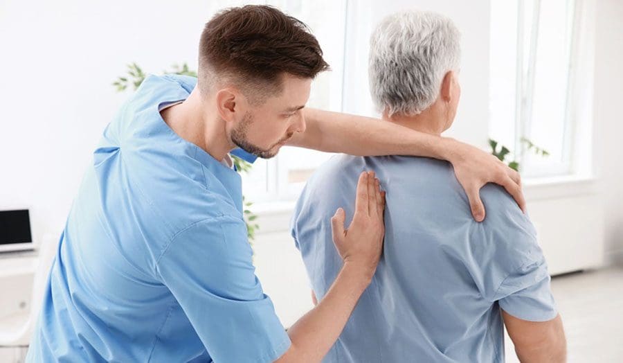
A doctor or chiropractor will ask questions about symptoms and medical history and perform a complete physical examination to diagnose lumbar spinal stenosis. During the physical examination, a healthcare provider will look for signs, such as loss of sensation, weakness, and abnormal reflexes.
Tests:
X-rays of the lumbar spine may show bone growths called spurs that push on spinal nerves and/or narrowing of the spinal canal.
Imaging tests – A CT or MRI scan can provide a detailed look at the spinal canal and nerve structures.
Other studies include – bone scans, myelogram, which is a CT scan that uses a color dye, and EMG, which is an electrical test of muscle activity.
Chiropractic Treatment
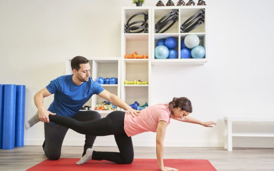
Chiropractic care combined with physical therapy is a tried-and-true treatment for spinal stenosis. A chiropractic treatment plan can include targeted and passive exercise programs. Targeted exercises involve strengthening the core and back muscles. Passive treatments include hot and cold therapy, massage, decompression, and electrical stimulation. The objective of chiropractic therapy is to:
Strengthen muscles in the core and legs
Correct posture and body mechanics.
Improve mobility.
Maintain ability to perform day-to-day activities.
Recommend stretches.
Educate on how to keep the spine and back muscles safe.
Train on using devices like a back brace, cane, or walker properly.
Advise about shoe inserts and splints.
Suggest work and home environment modifications, such as ergonomics and cushions.
Chiropractic Relief
References
Conway, Justin, et al. “Walking assessment in people with lumbar spinal stenosis: capacity, performance, and self-report measures.” The spine journal: official North American Spine Society journal vol. 11,9 (2011): 816-23. doi:10.1016/j.spinee.2010.10.019
Lurie, Jon, and Christy Tomkins-Lane. “Management of lumbar spinal stenosis.” BMJ (Clinical research ed.) vol. 352 h6234. 4 Jan. 2016, doi:10.1136/bmj.h6234
Macedo, Luciana Gazzi, et al. “Physical therapy interventions for degenerative lumbar spinal stenosis: a systematic review.” Physical therapy vol. 93,12 (2013): 1646-60. doi:10.2522/ptj.20120379
Tomkins-Lane, Christy C et al. “Predictors of walking performance and walking capacity in people with lumbar spinal stenosis, low back pain, and asymptomatic controls.” Archives of physical medicine and rehabilitation vol. 93,4 (2012): 647-53. doi:10.1016/j.apmr.2011.09.023
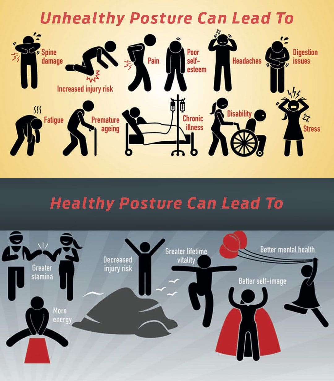
Postural dysfunction happens when unhealthy postures are practiced and maintained for prolonged periods. This can occur in any sitting, standing, or lying down position and is a major factor in musculoskeletal injuries. Injuries related to poor posture are normally caused by overuse that builds up over time. When the body starts to go out of alignment, the muscles must work harder to compensate, which further strains the body. This stress can lead to soft tissue injury and excess joint wear and tear. These injuries start as minor aches and pains in the short term. However, if left untreated, they can lead to chronic conditions. Injury Medical Chiropractic and Functional Medicine Clinic can rehabilitate the body to optimal function and provide postural training.
Postural Dysfunction
Posture is how the skeleton and muscles hold the body in a healthy position while standing or sitting, affecting breathing, muscle growth, and mobility. Practicing healthy posture means:
The bones are properly aligned.
The muscles, joints, and ligaments function correctly.
The organs, like the stomach, kidneys, and GI tract, are in the right position and can work efficiently.
The nervous system can operate at its full potential.
This allows the body to have:
More energy.
More room for the lungs to expand.
Experience less stress.
Alleviate muscle fatigue.
Achieve physical fitness.
Imbalance Causes
Unhealthy body positioning causes imbalances in muscle strength that pull the body out of alignment. This leads to muscles becoming tight/shortened and others becoming weak/lengthened, and it can also cause internal organ problems. For example, individuals that slump excessively cause the abdomen to compress, crowding the stomach and intestines, which leads to digestive issues. Postural dysfunction can be caused by the following:
Stress and strain from day-to-day activities.
Job responsibilities that involve sitting/standing for long periods and/or repetitive tasks like bending, lifting, reaching, twisting, etc.
Unhealthy driving position.
Non-supportive footwear.
Joint stiffness usually of the neck, upper and lower back, and hips.
Sedentary habits.
Lack of physical activity and exercise.
Muscle tightness.
Muscle weakness.
Weakened core stability.
Inadequate or failed post-surgical recovery.
Effects
Decreased blood circulation resulting in fatigue.
Overuse Injuries.
Breathing difficulties.
Balance issues.
Knee pain.
Joint misalignment.
Increased strain on the spine.
Compression of discs and joints.
Neck pain.
Lower back pain.
Less space for nerves to move due to compression.
Nerve problems.
Piriformis syndrome.
Shoulder impingement.
Chiropractic Rehabilitation
Chiropractic treatment for postural dysfunction provides adjustments, massage and decompression therapy, targeted stretching and exercises, retraining movement patterns, and nutritional and health coaching. Personalized treatment plans can include the following:
Targeted stretches and exercises to maintain posture correction.
Fix Posture
References
Korakakis, Vasileios, et al. “Physiotherapist perceptions of optimal sitting and standing posture.” Musculoskeletal science & practice vol. 39 (2019): 24-31. doi:10.1016/j.msksp.2018.11.004
Lee, Yongwoo, and Ki Bum Jung. “Effect of Physiotherapy to Correct Rounded Shoulder Posture in 30 Patients During the COVID-19 Pandemic in South Korea Using a Telerehabilitation Exercise Program to Improve Posture, Physical Function, and Reduced Pain, with Evaluation of Patient Satisfaction.” Medical science monitor: international medical journal of experimental and clinical research vol. 28 e938926. 27 Dec. 2022, doi:10.12659/MSM.938926
Shih, Hsu-Sheng, et al. “Effects of Kinesio taping and exercise on forward head posture.” Journal of back and musculoskeletal rehabilitation vol. 30,4 (2017): 725-733. doi:10.3233/BMR-150346
Snodgrass, Suzanne J et al. “Relationship between Posture and Non-Contact Lower Limb Injury in Young Male Amateur Football Players: A Prospective Cohort Study.” International journal of environmental research and public health vol. 18,12 6424. 14 Jun. 2021, doi:10.3390/ijerph18126424
Zhao, Mingming, et al. “Driver posture monitoring in highly automated vehicles using pressure measurement.” Traffic injury prevention vol. 22,4 (2021): 278-283. doi:10.1080/15389588.2021.1892087
Pinched Nerves and Muscle Spasms: A pinched or compressed nerve can occur in various body regions, from the wrist to the foot. When a nerve is compressed, a pins and needles feeling can present until the pressure is relieved, or there can be no sensation symptoms, but other symptoms like muscle spasms, especially in the arm or leg, can appear. Individuals will feel a repetitive fluttering or twitching when the arm or leg is not in motion. A pinched nerve could be the cause of spasms in the back or extremities. Injury Medical Chiropractic and Functional Medicine Clinic can help if symptoms are not stopping or worsening.
Pinched Nerves and Muscle Spasms
When multiple symptoms appear, individuals may not realize they are connected. Individuals may think aches, pains, and spasms are normal aging processes. Pinched nerves occur when there is an impingement on any one of the numerous nerves of the spine. Impingements can be caused by:
Repetitive motion injuries
Disc degeneration
Herniated/ruptured discs
Bone spurs
Arthritis
Trauma injury
The pain symptoms from the spasm can be quick, sharp, or pulsating and throbbing. The muscles respond by tightening or spasming as the nerve sends interrupted/incomplete signals. In addition to muscle spasms, a pinched nerve can contribute to other symptoms, including the following.
Tingling
Numbness
Pins and needles sensation
Reduced range of motion
Muscle weakness
Signs a Pinched Nerve Might Be Causing Spasms
Sudden shooting pain that radiates down the leg or arm.
Weak muscles
Muscle atrophy – shrinking or deteriorating.
Chronic tingling in the extremities.
A burning sensation in a specific area; this could but is not necessarily the source of the pinched nerve.
Electrical shock-type pain accompanies the spasms.
If a pinched nerve is left untreated and continues to generate symptoms, it can affect daily life and lead to uncomfortable long-term issues. Severe nerve compression combined with inflammation can cause damage to nearby soft tissues and muscles, leading to chronic conditions. When the nerves are damaged, it can be harder to control the muscles making certain motions uncomfortable or difficult to move certain body parts.
Chiropractic Care
Chiropractic care, massage, and decompression therapy will relieve pinched nerves and muscle spasms and restore neuromusculoskeletal system function. The body will be realigned, and patients will be trained on stretching exercises, muscle strengthening, posture training, and nutritional support to optimize the body’s natural healing abilities to repair the damaged nerves.
Low Back Pain
References
Bustamante, S, and P G Houlton. “Swelling of the leg, deep venous thrombosis, and the piriformis syndrome.” Pain research & management vol. 6,4 (2001): 200-3. doi:10.1155/2001/104091
Chu, Eric Chun-Pu, and Robert J Trager. “Thoracic Schwannoma as an Unusual Cause of Sciatic Pain in the Chiropractic Office: A Case Report.” The American journal of case reports vol. 23 e938448. 16 Nov. 2022, doi:10.12659/AJCR.938448
Coletti, Roger H. “The ischemic model of chronic muscle spasm and pain.” European journal of translational myology vol. 32,1 10323. 18 Jan. 2022, doi:10.4081/ejtm.2022.10323
Hirayama, Jiro, et al. “Relationship between low-back pain, muscle spasm and pressure pain thresholds in patients with lumbar disc herniation.” The European spine journal: official publication of the European Spine Society, the European Spinal Deformity Society, and the European Section of the Cervical Spine Research Society vol. 15,1 (2006): 41-7. doi:10.1007/s00586-004-0813-2
Kennedy, John G, and Donald E Baxter. “Nerve disorders in dancers.” Clinics in sports medicine vol. 27,2 (2008): 329-34. doi:10.1016/j.csm.2008.01.001
Waddell, Roger K. “Chiropractic care for a patient with spasmodic dysphonia associated with cervical spine trauma.” Journal of chiropractic medicine vol. 4,1 (2005): 19-24. doi:10.1016/S0899-3467(07)60108-6
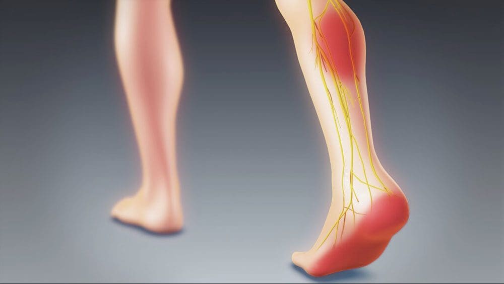
It didn’t happen at work, school, or exercise, and there haven’t been any trips and/or falls, but you can’t pinpoint what is causing foot and ankle discomfort and sensations. However, the cause could be originating in the lumbar spinal region. Sciatica is a set of symptoms that refer to pain, numbness, and tingling radiating down the leg from the lower back, affecting the legs, hips, buttocks, and feet. Injury Medical Chiropractic and Functional Medicine Clinic can release the compressed nerve, massage circulation back into the nerve, and restore mobility and function.
Sciatica Foot and Ankle
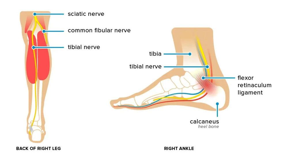
Sciatic nerve sensations can run down the back of the leg down into the foot.
Compression or irritation to any nerve roots can present with symptoms in the hip, thigh, calf, and foot.
Sciatica foot and ankle symptoms can accompany numbness and muscle weakness.
Sciatic nerve irritation mostly causes symptoms on the outside of the foot but can spread to other areas.
Nerve Roots
One or more of the lower spine’s sciatic nerve roots are being compressed or pinched. The foot symptoms location depends on which nerve root is affected.
If the S1 root is affected, symptoms will radiate to the sole and side of the foot.
If L5 is affected, symptoms will radiate to the top of the foot and the big toe.
If the L4 root is affected, symptoms can radiate to the medial or inside the ankle area.
Chiropractic Care and Relief
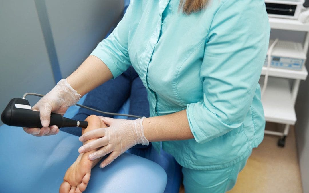
Foot Massage
A foot massage can be helpful.
A massage therapist finds points around the ankles that are tender.
Tenderness indicates a lymphatic blockage or muscle tension that needs to be worked out.
They will apply varying pressures to massage the muscles and get the circulation flowing.
The therapist will loosen the tarsal and metatarsal bones to loosen the muscles and nerves.
Moving the bones resupplies the joints, forces out inflammatory metabolic waste, opens the space for the nerves, and allows improved lymphatic drainage and blood flow to expedite healing.
A chiropractor will perform and train the individual on targeted stretches to the Achilles tendon and plantar fascia.
They will stretch, release, and open the ankle and sciatic nerve.
Injections
A cortisone injection where the nerve is affected can help in certain cases.
Injections of a corticosteroid, an anti-inflammatory medicine, can offer relief for up to three months and are given under local anesthesia.
The medicine reduces the inflammation and swelling around the nerve roots.
Foot Orthotics
Custom foot orthotics can help support a postural foot or ankle problem.
Overpronation is when the ankles collapse inward, which creates an imbalance of leg lengths that affects the hips, pelvis, and spine.
Orthotics can help provide symptom relief.
Nutrition
Part of a treatment plan will include an anti-inflammatory and antioxidant nutritional plan.
A professional nutritionist will make recommendations based on the individual’s case.
Magnesium-rich foods are generally recommended for sciatica as this nutrient aids the body in releasing muscle contractions.
99 percent of the body’s magnesium is stored in the bones, muscles, and soft tissues, with only 1 percent concentrated in the blood.
Foods rich in magnesium include:
Avocado
Bananas
Apricots
Dried pumpkin seeds
Dairy
Dark chocolate
Dried figs
Black beans
Brown rice
Fish
Spinach
Swiss chard
Yogurt
Benefits of Custom Foot Orthotics
References
Davis, David, et al. “Sciatica.” StatPearls, StatPearls Publishing, 6 May 2022.
Ge, Phillip S et al. “Iatrogenic pseudoaneurysm of the superior gluteal artery presenting as pelvic mass with foot drop and sciatica: case report and review of the literature.” Vascular and endovascular surgery vol. 44,1 (2010): 64-8. doi:10.1177/1538574409351990
Hughes, Michael S et al. “Post-traumatic catamenial sciatica.” Orthopedics vol. 31,4 (2008): 400. doi:10.3928/01477447-20080401-15
Mayo Clinic. “Sciatica.” https://www.mayoclinic.org/diseases-conditions/sciatica/symptoms-causes/syc-20377435?p=1
National Institutes of Health. “Sciatica.” https://medlineplus.gov/sciatica.html
Pan, Hung-Chuan, et al. “Magnesium supplement promotes sciatic nerve regeneration and down-regulates inflammatory response.” Magnesium research vol. 24,2 (2011): 54-70. doi:10.1684/mrh.2011.0280
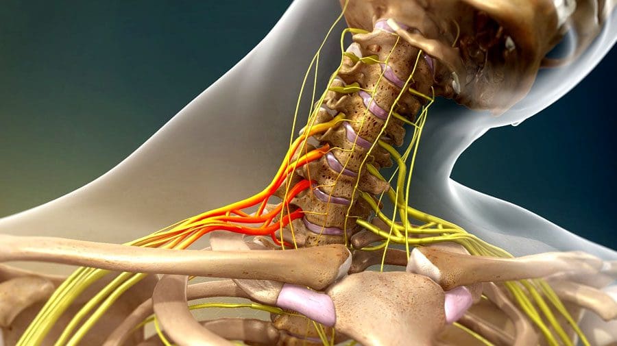
The body’s nerves are the communication system that carries messages between the brain and the rest of the body. Some nerves transmit messages from the brain to muscles to make the body move, while others relay pain, pressure, or temperature signals. Tiny fibers bundled inside each nerve carry the messages with an outer layer/sheathing that insulates and protects the nerves. The brachial plexus is a network of nerves that send signals from the spinal cord to the shoulders, arms, and hands. A brachial plexus nerve injury occurs when the nerves are over-stretched, compressed, torn, cut, or ripped from the spinal cord.
Brachial Plexus Nerve Injury
The injury involves the head or neck hitting or getting hit and shifting to one side while the shoulder is stretched/pulled in the opposite direction.
Minor brachial plexus injuries are commonly known as stingers or burners and are common in sports like football, wrestling, hockey, soccer, and basketball.
Severe brachial plexus injuries can cause arm paralysis and usually result from vehicle or motorcycle accidents.
Other conditions like inflammation or tumors can affect the brachial plexus.
Sometimes babies can sustain brachial plexus injuries during birth.
Pressure and stretching injuries do not physically sever the nerve but can disrupt communication.
Cutting injuries vary depending on the severity of the cut and because the nerves are in a protective canal that can also be fractured or broken. If the canal remains intact, the nerve fibers could grow back with time.
However, surgery is necessary to repair the damage if the canal is broken.
Signs and symptoms of a brachial plexus nerve injury can vary, depending on the severity and location of the injury. Usually, only one arm is affected.
Minor Injuries
Minor damage comes from over-stretching or mild compression.
An electric or burning sensation shoots down the arm.
Numbness and weakness in the arm.
Neck pain.
These symptoms usually last for a few seconds or minutes but can linger for days or longer.
Severe Injuries
More-severe symptoms result from injuries that impact, tear, or rupture the nerves.
The most severe injury occurs when the nerve root is torn from the spinal cord.
Symptoms include:
Intense pain.
Writhing neck pain.
Weakness or inability to use specific shoulder, arm, and/or hand muscles.
Complete lack of movement and feeling in the shoulder, arm, and/or hand.
Symptoms in both arms.
Complications
With time, most brachial plexus injuries in children and adults heal with minimal long-term damage. But some injuries can cause long-lasting problems that include:
Joint Stiffness
The joints can stiffen, making movement difficult.
Healthcare providers often recommend ongoing chiropractic and physical rehabilitation during recovery.
Atrophy
Nerves regrow slowly and can take some time to completely heal after the injury.
During that time, lack of use can cause the muscles to break down.
Chronic Pain
Nerve damage can cause pain signals to be constantly firing.
Numbness
It can occur in the arm or hand, increasing the risk of worsening the injury or causing new injuries.
Disability
Recovery from a severe brachial plexus injury depends on age, damage, location, and severity.
Even with surgery, individuals can experience long-term muscle weakness or paralysis.
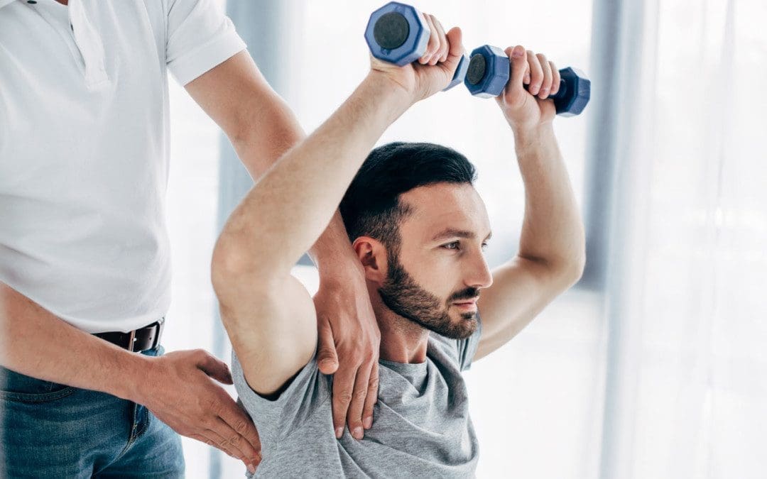
Chiropractic Treatment and Rehabilitation
Treatment depends on the severity of the damage. Chiropractic can help realign, rehabilitate, stretch, and strengthen the muscles, nerves, tendons, joints, and ligaments to expedite recovery. For less severe injuries:
Muscle strengthening and posture exercises help maintain motion.
Therapeutic massage will stimulate circulation and keep the muscles loose.
For severe injuries:
Surgery
Continued chiropractic and physical rehabilitation to maintain thorough circulation, range of motion, and relaxed muscles.
The Brachial Plexus
References
Brucker, J et al. “Brachial plexus birth injury.” The Journal of neuroscience nursing: Journal of the American Association of Neuroscience Nurses vol. 23,6 (1991): 374-80. doi:10.1097/01376517-199112000-00006
Gutkowska, Olga, et al. “Brachial plexus injury after shoulder dislocation: a literature review.” Neurosurgical review vol. 43,2 (2020): 407-423. doi:10.1007/s10143-018-1001-x
Joyner, Benny, et al. “Brachial plexus injury.” Pediatrics in review vol. 27,6 (2006): 238-9. doi:10.1542/pir.27-6-238
Noland, Shelley S et al. “Adult Traumatic Brachial Plexus Injuries.” The Journal of the American Academy of Orthopaedic Surgeons vol. 27,19 (2019): 705-716. doi:10.5435/JAAOS-D-18-00433
Spinal stress can affect nerve health. Neuropathy happens when disease or damage is sustained in the nerves that transmit messages from the brain through the spinal cord to the whole body. The source of the damage can be inside the spine, where a herniated disc could be squeezing the nerves, impeding or completely blocking blood circulation until deterioration begins to disease or damage nerve receptors. Removing the pressure from the spine and reversing the stress on the nerves can be done through manual or motorized spinal decompression.
Spinal Stress and the Nerves
The peripheral nervous system is comprised of three types of nerves that are directly influenced by the central nervous system, each with a distinct function which is why there is a wide range of symptoms associated with neuropathy. The types of nerves include:
Sensory nerves receive sensations from the skin like heat, cold, pleasure, and pain.
Spinal nerves contain sensory and motor fibers giving them sensory and motor functions. The spinal nerves receive sensory messages from the skin, internal organs, and bones. Any disruption from a bent, crushed, or entangled nerve group will not allow proper blood circulation and message transmission, causing delayed responses, tingling, numbness, and pain. If left untreated, it could cause permanent damage that can lead to chronic pain. Decompression therapy accelerates healing as it floods the spine with blood, oxygen, and nutrients.
Peripheral nerves originate from the spinal cord and extend a network of lines throughout the body called dermatomes. Injury to one dermatome can radiate/spread out to other dermatomes and the peripheral areas like the hands and feet. Once communication with the brain is compromised, results can lead to sensations like numbness and severe pain. Several factors can result in peripheral neuropathy, including:
Gordon, Tessa. “Peripheral Nerve Regeneration and Muscle Reinnervation.” International journal of molecular sciences vol. 21,22 8652. 17 Nov. 2020, doi:10.3390/ijms21228652
Menorca, Ron M G et al. “Nerve physiology: mechanisms of injury and recovery.” Hand clinics vol. 29,3 (2013): 317-30. doi:10.1016/j.hcl.2013.04.002
Wang, Mark L et al. “Peripheral nerve injury, scarring, and recovery.” Connective tissue research vol. 60,1 (2019): 3-9. doi:10.1080/03008207.2018.1489381
Tremors are extremely rare, but they can result from spinal compression and not necessarily a brain condition like Parkinson’s disease. Tremors are abnormal, involuntary body movements with various causes, most of which are connected to the brain and not the spine. A study reports that more than 75% of individuals with Parkinson’s experienced a resting tremor, and about 60% experience tremors while moving. Sometimes the spine is the contributor caused by compression of the spinal cord.
Spinal Compression Study
A 90-year-old man was hospitalized after having tremors, with Parkinson’s being the initial diagnosis. The tremors progressed to the point where the man could not feed himself or walk without support. The case became the focus of a medical report published by physicians in the Department of Orthopaedic Surgery, Division of the Spine, Singapore Tan Tock Seng Hospital. Along with the tremors, symptoms progressed to:
Difficulty with fine motor skills like buttoning a shirt.
However, it was ruled out because the patient was not presenting with other Parkinson’s symptoms.
For individuals with cervical spondylotic myelopathy tremors, surgery can be used to help the condition. However, with cervical myelopathy, there is often some permanent damage. Individuals have shown that post-surgery and decompression, symptoms still present, maybe not as much, but there will be a need for a symptom management plan.
Prevention
The best way to prevent tremors associated with cervical spondylotic myelopathy is to minimize the strain on the spine that can lead to herniated discs and/or other spinal injuries. The discs in the spine degenerate, dry out and start cracking with age, increasing the risk of rupture. If a tremor develops, contact a doctor, spine specialist, or chiropractor to help diagnose the condition. These doctors can perform physical and neurological tests to determine the cause and treatment options.
Body Composition
Aging Health
Steady weight gain throughout life can lead to adult-onset diabetes. This is partly caused by having more body fat and progressive muscle loss. Loss of skeletal muscle mass is linked to insulin resistance that involves:
The less muscle is available, the less insulin sensitive the body becomes.
As insulin sensitivity decreases, the body becomes more resistant, increasing risk factors for type II diabetes.
This can lead to osteoporosis, where the old bone is reabsorbed more and less new bone is created.
Both men and women can experience decreased muscle mass that can lead to:
Thinner bones
Weaker bones
Increased risk of osteoporosis and severe injury from falls.
To help prevent these issues, it is recommended to:
It is recommended to space out protein intake across meals rather than consuming it all at once. This helps to ensure the proper amount is acquired.
Monitoring body composition regularly can help minimize muscle mass loss and fat mass gain as the body ages.
A regular strength training routine will help strengthen bones muscles and maintain optimal circulation.
References
Heusinkveld, Lauren E et al. “Impact of Tremor on Patients With Early Stage Parkinson’s Disease.” Frontiers in neurology vol. 9 628. 3 Aug. 2018, doi:10.3389/fneur.2018.00628
Jancso, Z et al. “Differences in weight gain in hypertensive and diabetic elderly patients primary care study.” The Journal of nutrition, health & aging vol. 16,6 (2012): 592-6. doi:10.1007/s12603-011-0360-6
Srikanthan, Preethi, and Arun S Karlamangla. “Relative muscle mass is inversely associated with insulin resistance and prediabetes. Findings from the third National Health and Nutrition Examination Survey.” The Journal of clinical endocrinology and metabolism vol. 96,9 (2011): 2898-903. doi:10.1210/jc.2011-0435
Tapia Perez, Jorge Humberto et al. “Treatment of Spinal Myoclonus Due to Degenerative Compression Myelopathy with Cervical Spinal Cord Stimulation: A Report of 2 Cases.” World neurosurgery vol. 136 (2020): 44-48. doi:10.1016/j.wneu.2019.12.170
IFM's Find A Practitioner tool is the largest referral network in Functional Medicine, created to help patients locate Functional Medicine practitioners anywhere in the world. IFM Certified Practitioners are listed first in the search results, given their extensive education in Functional Medicine