As kids play sports like soccer and football with more frequency and force, many are damaging their knees, a new study finds.
A common knee injury — an anterior cruciate ligament (ACL) tear — has steadily increased among 6- to 18-year-olds in the United States, rising more than 2 percent a year over the last two decades, researchers report. These injuries peak in high school, said lead researcher Dr. Nicholas Beck. Girls have a higher rate of ACL injuries, added Beck, an orthopedic surgery resident at the University of Minnesota.
Sports that involve cutting or pivoting — such as soccer and basketball — are the riskiest for ACL tears. And contact sports like football can further increase the risk. But ACL tears can occur in tennis and volleyball, too, the researchers noted.
Rising ACL Tears, According to Researchers
Study co-author Dr. Marc Tompkins said the researchers didn’t look at why ACL tears are on the rise. But, he said, “one potential cause is the year-round sports specialization that is occurring in kids at an earlier age.” Tompkins is an assistant professor of orthopedic surgery at the University of Minnesota. Instead of getting cross-training from multiple sports and therefore using different muscle groups, this means the kids do the same thing over and over. This can lead to fatigue and an increased potential for injury, including ACL injury, Tompkins explained.
“Another potential cause is that children as athletes play with more intensity and force than 20 years ago, which may put the body at increased risk of injury,” he added.
More girls are playing sports, which could affect injury rates, the study authors said. And it’s also possible that rates are up “because we are getting better as a medical community at diagnosing ACL injury,” Tompkins suggested. Beck hopes this study will increase awareness of ACL tears in young athletes and promote interest in prevention programs or developing athletic participation guidelines.
The anterior cruciate ligament sits in the center of the front of the knee. It’s one of the ligaments that holds the knee bones together. When it tears, the ligament splits into two, causing knee instability, according to the American Academy of Orthopaedic Surgeons. When a tear occurs, you might hear a popping sound and your knee may give out from under you. Depending on the severity of the injury, treatment can range from physical therapy to surgery.
“ACL injuries are serious in the short term because they generally require six months’ to a year’s worth of hard recovery work before going back to sports. And even then it often takes longer to get back to pre-injury function,” Tompkins said. “ACL injuries are serious in the long term, too, because we know that even if they recover well with or without surgery, the risk of developing arthritis in the injured knee is higher than before the injury,” he added.
Dr. Stephen Swirsky is an orthopedic surgeon at Nicklaus Children’s Hospital in Miami. He said one of the best ways to reduce injuries is to teach good running techniques, which will improve function and agility.
“We have developed an injury prevention program, and we try to reduce the rates of ACL injuries,” Swirsky said. “In addition, kids need to be on a flexibility and stretching program,” he advised. “The more flexible they are, the less likely they are to have an injury.”
When ACL tears do happen, Swirsky said, he recommends a comprehensive rehab program after surgery. This is accompanied by advice for reducing the risk of injury when young patients return to play. To study the trends in ACL tears among U.S. children and teens, the study authors used insurance billing data for patients aged 6 to 18 from 1994 to 2013.
The researchers found that girls of all ages experienced a significant increase in the incidence of ACL tears over 20 years. In boys, however, only those aged 15 to 16 showed such an increase.
SOURCES: Nicholas Beck, M.D., resident, department of orthopaedic surgery, University of Minnesota, Minneapolis; Marc Tompkins, M.D., assistant professor, department of orthopaedic surgery, University of Minnesota, Minneapolis; Stephen Swirsky, D.O., orthopedic surgeon, Nicklaus Children’s Hospital, Miami; Feb. 22, 2017, Pediatrics, online
For more information, please feel free to ask Dr. Jimenez or contact us at 915-850-0900 .
Preventing Sports Injuries
Many athletes largely depend on chiropractic care to enhance their physical performance. New research studies have determined that aside from maintaining overall health and wellness, chiropractic can also help prevent sports injuries. Chiropractic is an alternative treatment option utilized by athletes to improve their strength, mobility and flexibility. Spinal adjustments and manual manipulations performed by a chiropractor can also help correct spinal issues, speeding up an athlete’s recovery process to help them return-to-play as soon as possible.
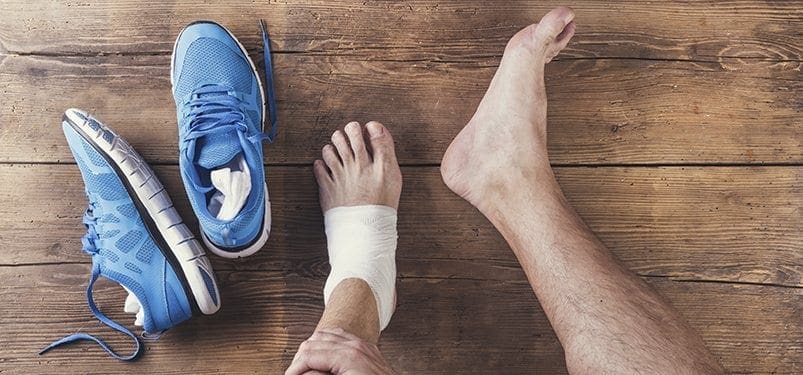
Sometimes you need to see a doctor for help in diagnosis and treatment. For strains or sprains, the pain can increase in the first one to two days, as the spasm surrounding the injury sets in.
When should you see a doctor for a sprain or strain?
If after trying RICE (an acronym for “rest, ice, compression, and elevation” of the injured limb) and over-the-counter medications the pain is not controlled or if the injury is thought to be more severe than initially believed, then a visit to a doctor is wise. A doctor’s visit also is important if swelling gradually develops over a large joint, such as a hip, knee, elbow, or wrist.
Sometimes you need the help of hospital equipment and specialists. Seek care immediately in any of the following cases:
If you are concerned that a bone is broken or a joint is dislocated
If you have numbness or tingling associated with the injury (This may signify damage to a nerve.)
If the injured part of the body is cold and discolored (This may be associated with damaged blood vessels and loss of circulation.)
Children present a special situation. Due to growing bones, muscles, and tendons, these structures can react differently to stress. Parents can be rightly concerned about possible broken bones. Remember, even if you can walk on an injured limb or move it, you may still have a broken bone. It just means that the muscles and tendons are working across the joint.
What tests do physicians use to diagnose a sprain or strain?
When visiting the doctor, expect many questions about the accident. The mechanism of injury can give clues as to what stresses were put on the body part and what injuries likely happened. The doctor will perform a thorough physical examination of the injured area. The physician will want to examine the joint above and the joint below an injury to make sure no hidden injuries are missed.
The doctor may need to take X-rays or perform other tests. X-rays only show bones and not the soft tissues, such as the muscles, tendons, and ligaments. The physician determines when it is appropriate to order X-rays. Injuries of knees, ankles, and the low back, are often unlikely to warrant X-rays to rule out any broken bones. The physician should discuss the reasons for or against taking X-rays.
Reference:
Young, Craig C. “Ankle Sprain.” Medscape.com. Dec. 16, 2014. <http://emedicine.medscape.com/article/1907229-overview>.
For more information, please feel free to ask Dr. Jimenez or contact us at 915-850-0900 .
Preventing Sports Injuries
Many athletes largely depend on chiropractic care to enhance their physical performance. New research studies have determined that aside from maintaining overall health and wellness, chiropractic can also help prevent sports injuries. Chiropractic is an alternative treatment option utilized by athletes to improve their strength, mobility and flexibility. Spinal adjustments and manual manipulations performed by a chiropractor can also help correct spinal issues, speeding up an athlete’s recovery process to help them return-to-play as soon as possible.
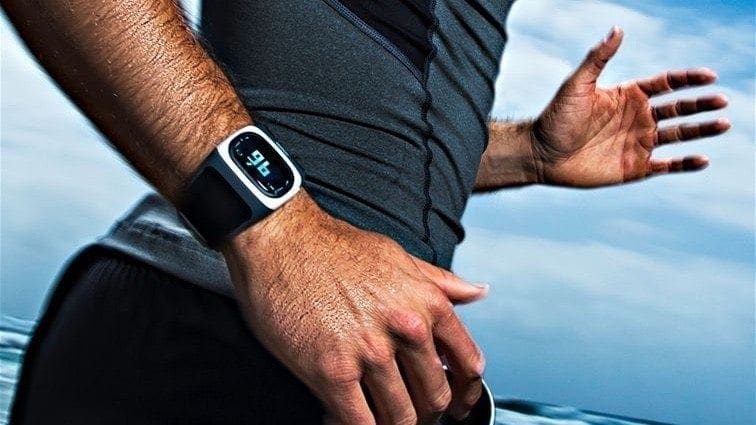
Fitbits and other wrist-worn fitness devices promise to keep track of your heart rate, but new research suggests they are less accurate than thought during certain exercises.
“If you need to know your heart rate with accuracy when exercising — either because you are training for a marathon or have safe heart rate limits set by your doctor, perhaps due to coronary artery disease, heart failure or other heart conditions — wrist-worn monitors are less accurate than the standard chest strap,” study author Dr. Marc Gillinov said in an American College of Cardiology news release.
The heart rates on the wrist-worn devices were compared to those from a continuous 4-lead electrocardiogram (EKG) and a chest strap monitor. Like an EKG, the chest strap measures electrical activity of the heart.
Functioning Errors in Heart Rate Trackers
Depending on the type of activity, the wrist devices were up to 34 beats a minute off. The wrist trackers could either overestimate or underestimate heart rate, Gillinov said. He’s a heart valve research, thoracic and cardiovascular surgery expert at the Cleveland Clinic.
The study included 50 volunteers. Their average age was 38. They tested popular wrist-worn fitness trackers, including the Apple Watch, Fitbit Blaze, Garmin Forerunner 235, and TomTom Spark Cardio.
The volunteers’ heart rates were recorded at rest and after light, moderate and vigorous exercise on a treadmill, stationary bike and elliptical trainer. All of them exercised for 18 minutes.
The chest strap monitor closely matched the readings from the EKG, which is the gold standard for measuring heart rate. And the wrist-worn devices were fairly accurate when a person was at rest. Most wrist devices gave acceptable readings during treadmill activity, but were fairly inaccurate while bicycling or using the elliptical, the study revealed.
Fitbit’s maker took issue with the findings.
“We stand behind our heart-tracking technology. Fitbit trackers are not intended to be medical devices,” Fitbit said in a statement. “Unlike chest straps, wrist-based trackers fit conveniently and comfortably into everyday life, providing continuous heart rate for up to several days without recharging [the device’s batteries].”
The San Francisco-based company added that internal studies involving 60 volunteers showed the device has an average error of 6 percent or less for measuring a person’s heart beat. And the Fitbit was tested against devices like the chest strap during walking, running, biking, using the elliptical and more, the company added.
Of all the wrist devices tested, the Apple watch seemed to fare the best. It performed well during bicycling and on elliptical machines without arm levels. The Apple watch’s heart rate monitor was only noticeably inaccurate compared to the chest strap when used on an elliptical machine with arm levers, the researchers said.
Why Are There Heart Rate Tracker Inaccuracies?
Wrist-worn devices use optical sensing, or light, to measure blood flow, the researchers said.
“It’s not measuring what the heart does, but rather blood flow — basically the volume of blood in the tissue,” Gillinov explained.
Wrist-worn devices also introduce many more variables that can result in incorrect readings, including insufficient contact with the skin due to sweating, poor fit or skin color, he said.
“Even though all these wrist-worn monitors work by the same general principles, there is considerable variation among them,” he said. “Overall, they were most accurate when someone was using the treadmill at low intensity and worst when exercising on the elliptical at high intensity,” Gillinov added.
The study is to be presented at the upcoming annual meeting of the American College of Cardiology, in Washington, D.C. Findings presented at meetings are typically viewed as preliminary until they’ve been published in a peer-reviewed journal.
SOURCE: American College of Cardiology, news release, March 8, 2017
For more information, please feel free to ask Dr. Jimenez or contact us at 915-850-0900 .
Preventing Sports Injuries
Many athletes largely depend on chiropractic care to enhance their physical performance. New research studies have determined that aside from maintaining overall health and wellness, chiropractic can also help prevent sports injuries. Chiropractic is an alternative treatment option utilized by athletes to improve their strength, mobility and flexibility. Spinal adjustments and manual manipulations performed by a chiropractor can also help correct spinal issues, speeding up an athlete’s recovery process to help them return-to-play as soon as possible.
What home remedies are effective for sprains and strains?
Initial treatment for sprains and strains should occur as soon as possible. Remember RICE!
Rest the injured part. Pain is the body’s signal to not move an injury.
Ice the injury. This will limit the swelling and help with the spasm.
Compress the injured area. This again, limits the swelling. Be careful not to apply a wrap so tightly that it might act as a tourniquet and cut off the blood supply.
Elevate the injured part. This lets gravity help reduce the swelling by allowing fluid and blood to drain downhill to the heart.
Over-the-counter pain medication is an option. Acetaminophen (Tylenol) is helpful for pain, but ibuprofen (Motrin, Advil) or naproxen (Aleve) might be better because these medications relieve both pain and inflammation. Remember to follow the guidelines on the bottle for appropriate dose of the medicine, especially for children and teens. Underlying medical conditions or use of other prescription medicines may limit the use of over the counter pain medications.
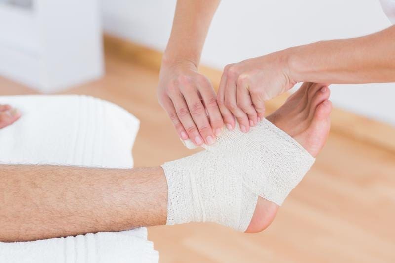
What is the treatment for sprains and strains?
Sprains and strains can usually be treated with home therapy using the RICE interventions. However, if the injury is more severe, your care provider may suggest splinting or casting to rest the injured joint. In some cases, operations are required to fix complete tears of muscles or tendons to allow complete return of function and to allow those muscles to do their job of moving the body. Significant tears of ligaments that stabilize joints also may need repair, but again, most are treated with short-term immobilization and early return to activity. Sometimes, resting the injury requires some help. Slings for arm injuries or crutches for leg injuries can be used, in addition to a variety of removable splints to protect the injured area from further damage and movement. Resting also helps relieve some of the muscle spasm associated with the injury.
Occasionally, if the injury is especially severe, the physician may want to use a nonremovable splint made of plaster or fiberglass. Although the splint may look like a cast, it doesn’t have plaster or fiberglass completely encircling the injured area. Instead, by only going partially around an injury, there is some room to allow for swelling that may occur during the next few days.
For more information, please feel free to ask Dr. Jimenez or contact us at 915-850-0900 .
Preventing Sports Injuries
Many athletes largely depend on chiropractic care to enhance their physical performance. New research studies have determined that aside from maintaining overall health and wellness, chiropractic can also help prevent sports injuries. Chiropractic is an alternative treatment option utilized by athletes to improve their strength, mobility and flexibility. Spinal adjustments and manual manipulations performed by a chiropractor can also help correct spinal issues, speeding up an athlete’s recovery process to help them return-to-play as soon as possible.
The body is meant to move. Muscles allow that movement to happen by contracting and making joints flex, extend and rotate. Muscles attach on each side of the joint to bone by thick bands of fibrous tissue called tendons. When a muscle contracts, it shortens and pulls on the tendon, which allows the joint to go through a range of motion.
A strain occurs when the muscle tendon unit is stretched or torn. The most common reason is the overuse and stretching of the muscle. The damage may occur in three areas:
The muscle itself may tear.
The area where the muscle and tendon blend can tear.
The tendon may tear partially or completely (rupture).
Joints are stabilized by thick bands of tissue called ligaments which surround them. These ligaments allow the joint to move only in specific directions. Some joints move in multiple planes; therefore, they need more than one group of ligaments to hold the joint in proper alignment. The ligaments are anchored to bone on each side of the joint. If a ligament is stretched or torn, the injury is called a sprain.
Causes of Sprains and Strains
Sprains and strains occur when the body is put under stress. In these situations, muscles and joints are forced to perform movements for which they are not prepared or designed to perform. An injury can occur from a single stressful incident, or it may gradually arise after many repetitions of a motion.
Signs and Symptoms of Sprains and Strains
The first symptom of a sprain or strain injury is pain. Other symptoms, such as swelling and spasm, can take time (from minutes to hours) to develop.
Pain is always a symptom that indicates that there is something wrong with the body. It is the message to the brain that warns that a muscle or joint should be protected from further harm. In work, exercise, or sport, the pain may come on after a specific incident or it may gradually progress after many repetitions of a motion.
Swelling almost always occurs with injury, but it may take from minutes to hours to be noticed. Any time fibers of a ligament, muscle, or tendon are damaged, some bleeding occurs. The bleeding (such as bruising on the surface of the skin) may take time to be noticed.
Because of pain and swelling, the body starts to favor the injured part. This may cause the muscles that surround the injured area to go into spasm. Hard knots of muscle might be felt near the site of the injury.
The combination of pain, swelling, and spasm causes the body to further protect the injured part, which results in difficulty with use. Limping is a good example of the body trying to protect an injured leg.
REFERENCE:
Young, Craig C. “Ankle Sprain.” Medscape.com. Dec. 16, 2014. <http://emedicine.medscape.com/article/1907229-overview>.
For more information, please feel free to ask Dr. Jimenez or contact us at 915-850-0900 .
Preventing Sports Injuries
Many athletes largely depend on chiropractic care to enhance their physical performance. New research studies have determined that aside from maintaining overall health and wellness, chiropractic can also help prevent sports injuries. Chiropractic is an alternative treatment option utilized by athletes to improve their strength, mobility and flexibility. Spinal adjustments and manual manipulations performed by a chiropractor can also help correct spinal issues, speeding up an athlete�s recovery process to help them return-to-play as soon as possible.
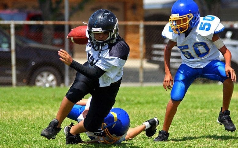
Female soccer players suffer the highest rate of concussions among all high school athletes in the United States, a new study finds.
“While American football has been both scientifically and colloquially associated with the highest concussion rates, our study found that girls, and especially those who play soccer, may face a higher risk,” said study author Dr. Wellington Hsu. He is a professor of orthopaedics at Northwestern University in Chicago.
“The new knowledge presented in this study can lead to policy and prevention measures to potentially halt these trends,” Hsu said in a news release from the American Academy of Orthopaedic Surgeons.
The researchers analyzed data on nearly 41,000 injuries suffered by high school athletes in nine sports between 2005 and 2015. The injuries included nearly 6,400 concussions. The sports studied included football, soccer, basketball, wrestling and baseball for boys; and soccer, basketball, volleyball and softball for girls. During the study period, participation in the sports rose 1.04-fold, but the number of diagnosed concussions increased 2.2-fold.
In sports played by both girls and boys, girls had much higher concussion rates than boys, Hsu’s team found. Between 2010 and 2015, the concussion rate was higher in girls’ soccer than in boys’ football, the findings showed. During the 2014-2015 school year, concussions were more common in girls’ soccer than in any other sport in the study.
Girls may be at greater risk of concussion while playing soccer due to “heading” the ball, a lack of protective gear, and an emphasis on contact during the game, the researchers suggested.
Each year, about 300,000 U.S. teens suffer concussions or mild traumatic brain injuries while participating in high school sports, the study authors said.
The findings were presented Tuesday at the American Academy of Orthopaedic Surgeons meeting in San Diego. Research presented at meetings should be considered preliminary until published in a peer-reviewed journal.
SOURCE: American Academy of Orthopaedic Surgeons, news release, March 14, 2017
For more information, please feel free to ask Dr. Jimenez or contact us at 915-850-0900 .
Additional Topics: Headache and Auto Injury
Whiplash is a common type of automobile accident injury. Characterized by symptoms of neck pain, whiplash is caused when the complex structures and tissues of the neck are stretched beyond their limit as a result of an abrupt back-and-forth motion of the head. While neck pain is the most common symptom associated with the auto injury, headaches can also occur due to complications along the cervical spine.
Focusing too much on playing one favorite sport probably isn’t a good idea for kids under 12, researchers report. That’s because specializing in a single sport seems to increase a child’s risk of injury, researchers say.
“Young athletes should participate in one competitive sport per season, and take at least three months off (non-consecutive) from competition per year,” said the study’s leader, Dr. Neeru Jayanthi. He’s a physician with Emory Sports Medicine and an associate professor of orthopaedics and family medicine at Emory University in Atlanta.
For the study, Jayanthi’s team assessed the risk of sports-related injuries among nearly 1,200 young athletes. After tracking their training schedules over the course of three years, the investigators found that nearly 40 percent of the athletes suffered an injury during the study period. The findings also showed that injured athletes began specializing in one sport at an average age younger than 12 years. In addition, nearly two-thirds of these athletes in highly specialized sports sustained a repeat injury. Athletes who didn’t sustain injuries began to focus on one sport when they were older than 12, on average, according to the report.
“While different for each sport, determining a possible age of specialization, as well as other training factors, may help guide young athletes in reducing risk,” Jayanthi said in an Emory news release.
Young athletes who had sports-related injuries during the study period tended to play more year-round sports, played more organized sports each week and were more specialized in specific sports than those who didn’t have an injury, the researchers found. The study authors advise young athletes to play more than one sport. In addition, they said, younger children shouldn’t train more hours than their age each week.
The study was published online March 16 in the British Journal of Sports Medicine. The findings were also presented Thursday at the International Olympic Committee World Conference on Prevention of Injury and Illness in Sport, in Monaco.
SOURCE: Emory University School of Medicine, news release, March 13, 2017
For more information, please feel free to ask Dr. Jimenez or contact us at 915-850-0900 .
Preventing Sports Injuries
Many athletes largely depend on chiropractic care to enhance their physical performance. New research studies have determined that aside from maintaining overall health and wellness, chiropractic can also help prevent sports injuries. Chiropractic is an alternative treatment option utilized by athletes to improve their strength, mobility and flexibility. Spinal adjustments and manual manipulations performed by a chiropractor can also help correct spinal issues, speeding up an athlete’s recovery process to help them return-to-play as soon as possible.
Low vitamin D levels are common among football players and may put them at increased risk for injuries, a new study suggests.
“Vitamin D has been shown to play a role in muscle function and strength,” said senior study author Dr. Scott Rodeo, co-chief emeritus of the sports medicine and shoulder service at the Hospital for Special Surgery in New York City.
“While most prior studies have focused on the aging population as the group most likely to experience the harmful effects of inadequate vitamin D, few reports have looked at the impact on muscle injury and function in the high-performance athlete,” he said in a hospital news release.
In the study, Rodeo’s team assessed 214 college football players, average age 22. The investigators found that nearly 60 percent had low levels of vitamin D, including 10 percent with a severe deficiency.
Those players with low vitamin D levels had higher rates of lower extremity muscle strain and core muscle injury than those with normal levels. Of the 14 players who missed at least one game due to a strain injury, 86 percent had low vitamin D levels.
LOW VITAMIN D CHANGES TISSUE COMPOSITION
Low levels may cause changes in muscle composition that increase the risk of injury, according to the researchers. But the study only found an association, rather than a cause-and-effect link, between levels and injury.
“Awareness of the potential for vitamin D inadequacy could lead to early recognition of the problem in certain athletes. This could allow for supplementation to bring levels up to normal and potentially prevent future injury,” Rodeo said.
“Although our study looked at high-performance athletes, it’s probably a good idea for anyone engaging in athletic activities to give some thought.” Rodeo said.
Adequate vitamin D is essential for musculoskeletal structure, function and strength, Rodeo explained. However, more than 40 percent of the U.S. population is vitamin D-deficient, he said.
Often called the “sunshine vitamin,” it is produced by the skin when exposed to sunlight. Milk and fortified foods, including orange juice and some cereals, can also provide nutrients. Supplements are usually prescribed for�deficiency, the researchers said.
The study was to be presented Thursday at the annual meeting of the American Academy of Orthopaedic Surgeons, in San Diego. Research presented at meetings is viewed as preliminary until published in a peer-reviewed journal.
SOURCE: Hospital for Special Surgery, news release, March 16, 2017
�Get Rich NOW! Lose 50 pounds in 5 Days AND Make $50! Eat What You Want and Still Lose Weight � GUARANTEED! We�ve heard �em all. And yes, we�d all like to be fitter (and wealthier). Are you sick and tired of reading false promises like these? Frankly, I am.
We all know that it takes hard work and dedication to become lean and fit. While both goals are attainable, they require commitment and a good amount of time to achieve the desired results. Okay, sure we may have faltered here and there, taking a few missteps on our health journey, but that�s why I�m here: to cut through the bull and tell you what REALLY works and what doesn�t.
Watch the video: 7 Fat-Burning Foods That Boost Metabolism
LIE #1: Cut carbs, lose weight
Why it’s not true: To burn fat, you must fuel your body with the calories it needs to achieve high-intensity ranges of exercise. Without that fuel (i.e. carbs), your tank will be on empty and you�ll ultimately be running on fumes. You�ll feel as though you�re working hard, but your workouts won�t be as long or effective as they would if you had fueled your body correctly.
LIE #2: Extended moderate exercise burns more fat than high-intensity exercise
Why it’s not true: While you will burn more fat than carbohydrates during a moderate exercise session, the total calorie burn depends on the duration of the workout. But there is not much post-exercise elevation in metabolic rate after this type of exercise. High-intensity exercise, however, causes a more intense �after-burn� that can last a day or more after working out. That after-burn is fueled mostly by fat, and that is when the body actually changes shape.
LIE #3: Lose a pound a week by cutting 500 calories a day from food
Why it’s not true: Human beings are built to survive and thus when calories are severely restricted the human body goes into survival mode, slowing down the metabolic rate and holding on to every calorie. Consuming fewer calories per day propels the body into conserving fuel. However, if you cut 300 calories from your daily diet you will lose more weight than if you lowered your calorie intake by 500 calories. Eating more calories will allow you to train harder and keep your metabolic rate up.
LIE #4: Lose weight by putting the fork down after 6pm
Why it’s not true: All food contains calories regardless what time you eat it. The simple truth is that eating too many calories will cause you to gain weight. A 2012 study compared overweight people who ate carbs throughout the day and those who ate them at dinner. The nighttime carb eaters lost more weight and body fat and experienced less hunger during the day�researchers noted that the evening group had better levels of hormones that regulate satiety and hunger. The explanation may also lie in the body�s production of Growth Hormone (GH). GH is a powerful hormone that controls how much fat your body burns and how much muscle it builds. At night, your GH peaks while you sleep, ultimately shutting off the moment you eat your first meal.
�If genius really is 1% inspiration and 99% perspiration, then some of us must be a lot smarter than others. While sweat is a normal human function, a lucky few�seem to produce higher-than-normal amounts�especially in the hot summer months.�But before you hole yourself up in air conditioning all season, there are a few things you should know about sweat. Here�s the basics on what it is, why it happens (to certain folks�more than others), and what you can do if you�re concerned about it.
There are three types of sweat
All sweat is not created equal, says Laure Ritti�, PhD, research assistant professor in the department of dermatology at the University of Michigan. And everyone sweats differently: Some people may have problems with all three types of perspiration, while others may really only ever notice one or two.
First, there�s body sweat�the odorless type that pours off you during a workout or when you stand out in the hot sun. This type of sweat exists to help cool the skin and keep the body�s internal temperature as close to 98.6 degrees as possible. You�ll notice it pretty much everywhere, but especially along the forehead and the spine.
Then, there�s perspiration on the palms and soles of the feet. This type of sweat helps increase adherence and grip, says Ritti�, and, evolutionarily, it�s the body�s response to a perceived threat. (That�s why some people notice it when they�re feeling anxious.) �When you want to hold onto something, you�ll do better with wet fingers,� Ritti� explains. �In the beginning, we didn�t wear shoes, so sweaty feet helped us run or climb when we needed to.�
Finally, there�s sweat that�s emitted from the armpits and the genital area. This is the type that produces so-called body odor, thanks to bacteria living in these places. �We�re not completely sure what the function here is, but we think there�s some pheromone-type of communication going on,� Ritti� says. �If one individual in a herd senses danger and starts to emit those strong smells, it could alert others around them.�
The body begins producing body sweat when it starts to heat up�either externally, from high temperatures, or internally, from muscle exertion (like when exercising). So if you push yourself harder than your body is used to, your body is more likely to kick on its internal air conditioning; that�s why a highly trained athlete may be able to run a 10-minute mile without breaking a sweat, while the same workout may�leave�a less conditioned person red-faced and drenched.
But the more you train your body, and the more time you spend in hot, humid climates, the more efficient you become at sweating. �The body will adjust and react a little earlier before you get too hot,� says Ritti�, �so your sweating will be more spread out over time and across your whole body�rather than building up and releasing all at once, and leaving one big spot on your shirt.�
Yes, that may translate into more sweating overall�just look at any NBA basketball game and you�ll see that even highly-trained athletes sweat a ton�but it�s ultimately a good thing. It means the body is better able to respond to the demands of heat and exercise, and stays cooler as a result.
How much you sweat is largely determined before age 2
Whether you sweat buckets or stay fresh as a daisy on hot days also has a lot to do with genetics. �If one or both of your parents were heavy sweaters, then there is a good chance that you will be too,� says exercise physiologist Michael Bergeron, PhD, President of Youth Sports of the Americas. Men also tend to sweat more than women, he says, although that�s not always the case.
Body composition matters, too: Larger people generally sweat more, because they work harder to carry a heavier load. �But many comparatively small people can sweat tremendously,� says Bergeron. And because muscle generates heat, he adds, people with more muscle mass also tend to sweat more than their leaner peers.
But actually, a lot of how much a person sweats has to do with the first two years of life. That�s when sweat glands are first activated, says Ritti�; if they don�t get fully turned on during this period, they likely never will. In other words, a super-active toddler who runs around in the heat will likely develop greater sweating ability (again, a good thing) than one who�s not very active.
Heavy sweaters should hydrate more
�The more you sweat, the more deliberate you need to be about replacing the water your body�s lost,� says Bergeron. When exercising, most adults can comfortably and safely take in about 1.5 liters (a little more than 50 ounces) of water an hour. If you�re sweating more than that amount�you can weigh yourself before and after a workout to find out�you should make up for it by drinking extra water before and after you work out.
Sweat also contains important electrolytes, like sodium, that the body needs to function properly. Most people get enough salt in their diets that they don�t need to worry about this, but if you�re exercising for longer than an hour and really sweating a lot, an electrolyte-enriched sports drink can help replace what�s lost.
Ritti� also points out that acclimating your body to exercise or to heat�so that you ultimately sweat less�won�t work if you don�t drink enough water on a regular basis. �Staying hydrated before and during exercise will help train your body to fight the heat,� she says. Cold water is best, she adds, �because it helps to cool down your internal organs.�
Besides staying in shape, wearing sweat-wicking clothing, and spending two to three weeks acclimating to the heat, there�s not much people can do about heavy sweating during exercise. (Putting antiperspirant on your hairline or on other body parts may help curb localized perspiration�but, Ritti� warns, when you block sweat glands in one part of the body, others will compensate by working even harder.)
People with excessive day-to-day sweating, however�a condition known as hyperhydrosis�may have more options. If you�re already using an over-the-counter aluminum-based antiperspirant and it just isn�t cutting it, your doctor may recommend a prescription-strength solution with aluminum chloride. These work best when applied before bed, but can cause skin and eye irritation for some people.
Botox injections and certain types of medications have been shown to block the nerves that� trigger sweat production, and may be useful for people who sweat excessively under their arms or on their hands and feet. (If those don�t work, electrical stimulation or even surgery may be considered.) Friction also contributes to sweaty feet, says Ritti�, so changing up your shoes and socks�and being sure you aren�t sliding around in them�may help, as well.
If sweating is truly causing a problem in your daily life, talk to your primary doctor or dermatologist about potential solutions. Otherwise, grab your water bottle and get outside. Embrace your sweaty self, and try to be thankful your body is doing its job.
IFM's Find A Practitioner tool is the largest referral network in Functional Medicine, created to help patients locate Functional Medicine practitioners anywhere in the world. IFM Certified Practitioners are listed first in the search results, given their extensive education in Functional Medicine