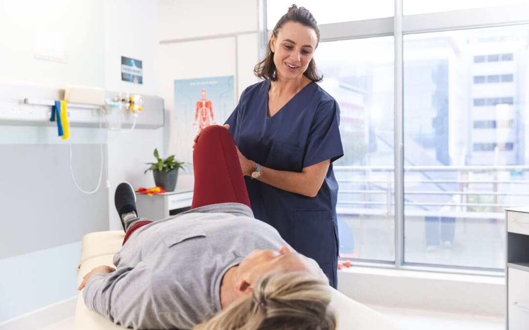
Individuals experiencing pain, numbness, tingling, or a burning sensation in the front and outer thigh could have meralgia paresthetica, a nerve entrapment. Can understanding the condition help healthcare providers develop an effective treatment plan?
Meralgia Paresthetica
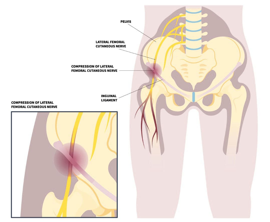
Meralgia paresthetica, or MP, is also known as Bernhardt-Roth syndrome, lateral femoral cutaneous nerve syndrome, or lateral femoral cutaneous neuralgia. It occurs when the lateral femoral cutaneous nerve, a sensory nerve that passes over the brim of the pelvis and down the front of the thigh, becomes compressed. The nerve supplies information about sensations over the front and outside of the thigh. This can happen for several reasons, including:
Recent hip injuries, such as from a motor vehicle collision/accident.
Repetitive hip activities, like cycling.
Pregnancy
Weight gain
Wearing tight clothing.
The nerve entrapment condition causes tingling, numbness, and burning pain in the front and/or outer thigh.
Causes
There can be several different causes of this condition, but it is frequently seen in pregnancy, sudden weight gain, wearing tight clothing or belts, and other conditions. (Ivins G. K. 2000) Sometimes, meralgia paresthetica can be caused by medical procedures. For example, the condition can present after an individual has surgery and is in an unusual position for a long period of time, where there is direct external pressure on the nerve. Also, the nerve can become damaged during a surgical procedure. (Cheatham S. W. et al., 2013) This can occur when a bone graft is obtained from the pelvis or anterior hip replacement surgery.
Sensitivity to lightly touching the outside of the thigh.
Worsening of symptoms with certain positions.
Increased symptoms when wearing belts, work belts, or tight-waist clothes.
The symptoms may come and go or be persistent. Some individuals are hardly noticeable and do not impact their lives or activities, while others can be very bothersome and cause significant pain. (Scholz C. et al., 2023)
Treatment
Treatment depends on how long the injury has been present and the frequency and severity of the condition.
Clothing Modifications
If the cause is due to tight clothing, belts, or work belts, then garment modification should alleviate symptoms.
If recent weight gain is thought to contribute to the condition, then a weight loss program may be recommended.
Cortisone Injections
If simple steps do not relieve symptoms, a cortisone injection around the nerve area may be recommended. The goal is to reduce inflammation that contributes to nerve pressure (Houle S. 2012) . Cortisone injections may be a definitive treatment or a temporary treatment.
Chiropractic
Chiropractic care can be an effective, natural, and safe treatment. Adjustments can help relieve pressure on the lateral femoral cutaneous nerve (LFCN) by realigning the spine and restoring nerve function. Chiropractors may also use soft tissue therapies, such as massage, to relieve muscle tension and support the body’s healing process. Other chiropractic techniques that may be used include:
A chiropractic treatment program may include 10–15 treatments over 6–8 weeks, but the number of treatments needed will vary from person to person. If there’s no noticeable progress after 3–4 weeks, it may be time to consult a specialist or surgeon.
Surgery
Surgery is rarely necessary. However, a surgical procedure may be considered when all conservative treatments fail to provide relief. (Schwaiger K. et al., 2018) A surgeon dissects and identifies the nerve, looks for compression locations, and tries to free the nerve from any areas where it may be pinched. Alternatively, some surgeons transect/cut the nerve so it no longer causes problems. If the transection procedure is performed, there will be a permanent area of numbness over the front of the thigh.
Injury Medical Chiropractic and Functional Medicine Clinic works with primary healthcare providers and specialists to develop a customized treatment plan to relieve pain, treat injuries, improve flexibility, mobility, and agility, and help individuals return to optimal function. If other treatments are needed, Dr. Jimenez has teamed up with top surgeons, clinical specialists, medical researchers, and rehabilitation providers to provide the most effective treatments.
Chiropractic Care for Leg Instability
References
Ivins G. K. (2000). Meralgia paresthetica, the elusive diagnosis: clinical experience with 14 adult patients. Annals of surgery, 232(2), 281–286. https://doi.org/10.1097/00000658-200008000-00019
Cheatham, S. W., Kolber, M. J., & Salamh, P. A. (2013). Meralgia paresthetica: a review of the literature. International journal of sports physical therapy, 8(6), 883–893.
Chung, K. H., Lee, J. Y., Ko, T. K., Park, C. H., Chun, D. H., Yang, H. J., Gill, H. J., & Kim, M. K. (2010). Meralgia paresthetica affecting parturient women who underwent cesarean section -A case report-. Korean journal of anesthesiology, 59 Suppl(Suppl), S86–S89. https://doi.org/10.4097/kjae.2010.59.S.S86
Scholz, C., Hohenhaus, M., Pedro, M. T., Uerschels, A. K., & Dengler, N. F. (2023). Meralgia Paresthetica: Relevance, Diagnosis, and Treatment. Deutsches Arzteblatt international, 120(39), 655–661. https://doi.org/10.3238/arztebl.m2023.0170
Hosley, C. M., & McCullough, L. D. (2011). Acute neurological issues in pregnancy and the peripartum. The Neurohospitalist, 1(2), 104–116. https://doi.org/10.1177/1941875211399126
Houle S. (2012). Chiropractic management of chronic idiopathic meralgia paresthetica: a case study. Journal of chiropractic medicine, 11(1), 36–41. https://doi.org/10.1016/j.jcm.2011.06.008
Schwaiger, K., Panzenbeck, P., Purschke, M., Russe, E., Kaplan, R., Heinrich, K., Mandal, P., & Wechselberger, G. (2018). Surgical decompression of the lateral femoral cutaneous nerve (LFCN) for Meralgia paresthetica treatment: Experimental or state of the art? A single-center outcome analysis. Medicine, 97(33), e11914. https://doi.org/10.1097/MD.0000000000011914
When muscle pains and aches present from health conditions, work, exercise, housework, etc., many individuals turn to topical sprays, creams, ointments, and gels to bring relief. Can magnesium spray be beneficial in the fight against neuromusculoskeletal pain?
Magnesium Spray
Magnesium spray is a liquid form of magnesium applied externally to the skin that has been marketed to promote muscle relaxation, improve sleep, and manage migraines. However, studies of its effectiveness have had mixed results. Some studies have shown that topical use can:
Improve chronic muscle and joint pain. Example: fibromyalgia.
Decrease the frequency and severity of nerve pain symptoms. Example: peripheral neuropathy.
Reduce the incidence and severity of an intubation-related sore throat after surgery.
Further studies of various groups are necessary to clarify the optimal dose for each condition and to determine how topical magnesium affects magnesium blood levels.
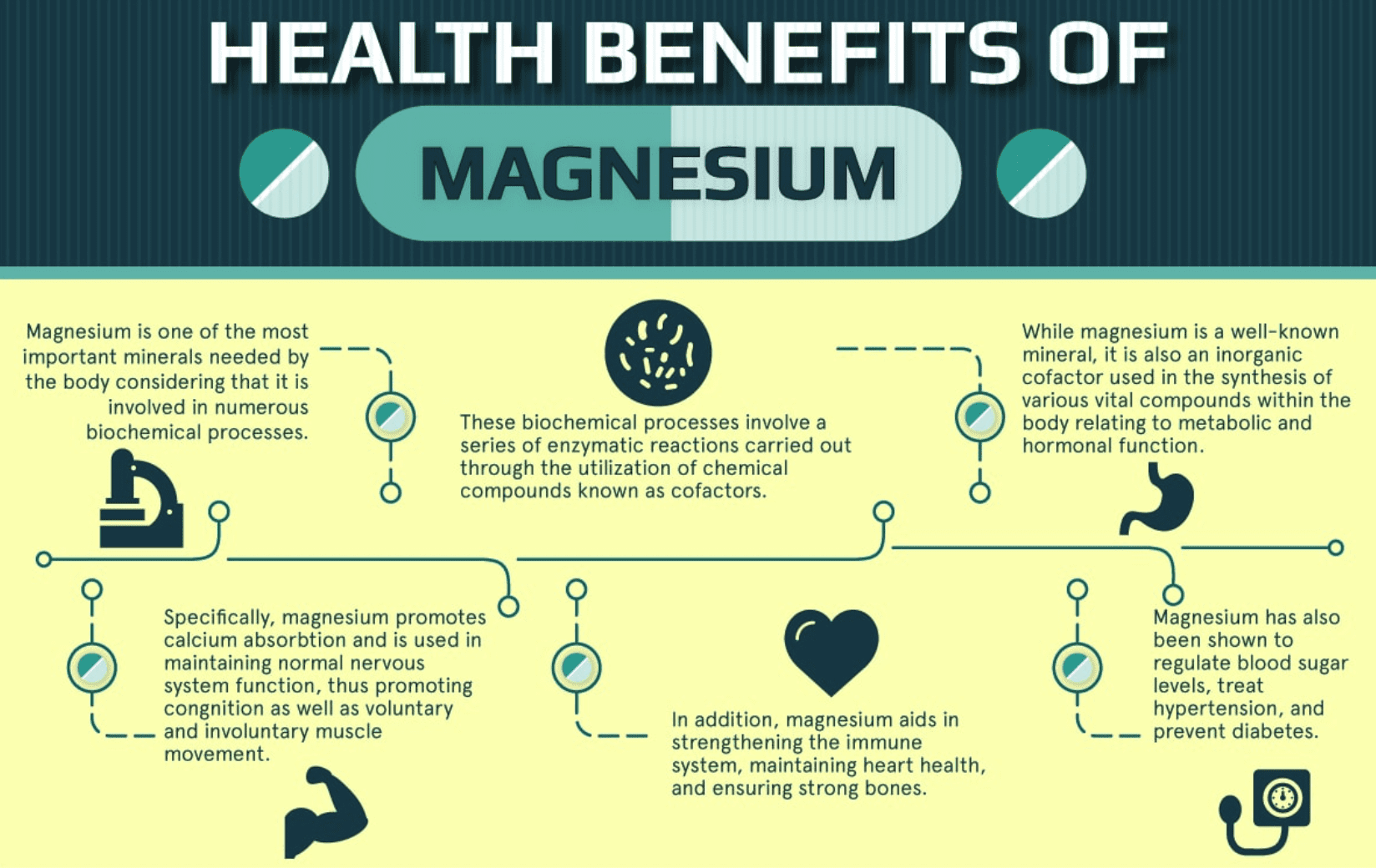
What is It?
Magnesium is a mineral that has an important role in many of the body’s processes and is essential for the following (Gröber U. et al., 2017)
Nerve transmission
Muscle contraction
Blood pressure regulation
Blood sugar regulation
Protein production
DNA and RNA production
Currently, there is no recommended dosage for topical magnesium use. However, some major health institutions have established a recommended daily amount taken by mouth. Listed are the recommended daily magnesium intake based on age and other factors. (National Institutes of Health Office of Dietary Supplements, 2022)
14 to 18 years old: 410 mg for males, 360 mg for females and when lactating, and 400 mg when pregnant.
19 to 30 years old: 400 mg for males, 310 mg for females and when lactating, and 350 mg when pregnant.
31 to 50 years old: 420 mg for males, 320 mg for females and when lactating, and 360 mg when pregnant.
51 years old and above: 420 mg for males and 320 mg for females.
Although self-care is appropriate for minor injuries or exercise, individuals are encouraged to see their healthcare provider for severe musculoskeletal pain symptoms.
Benefits
Though taking oral magnesium supplements is common, there is limited research on using magnesium on the skin to improve magnesium levels. Studies comparing the absorption of magnesium taken by mouth with the spray applied to the skin require further research. However, some studies look at the localized effect of magnesium spray on improving a sore throat after surgery and nerve, muscle, and joint pain.
Intubation-Related Sore Throat
Topical magnesium reduced the severity of sore throat after surgery in individuals undergoing tracheal intubation compared to a placebo. (Kuriyama, A. et al., 2019) However, further studies are necessary to clarify the optimal dose.
Nerve Pain
Peripheral neuropathy is nerve damage that causes a tingling and numbing sensation in the arms or legs. In a study of individuals with chronic kidney disease, the daily application of magnesium sprays to limbs affected by peripheral neuropathy for twelve weeks decreased the frequency and severity of nerve pain symptoms. However, one limitation was that it was performed mostly in females. (Athavale, A. et al., 2023)
Chronic Muscle and Joint Pain
A small study assessed whether applying magnesium to the skin could improve the quality of life of female participants with fibromyalgia – a chronic condition that causes muscle and joint pain, fatigue, and other symptoms. The study found that four sprays of magnesium chloride applied twice daily to the upper and lower limbs for four weeks could benefit those with fibromyalgia. However, further research with larger studies is needed to confirm the results. (Engen D. J. et al., 2015)
Does The Spray Increase Overall Magnesium Levels?
Magnesium is transported into cells through magnesium transporters. The outer layer of the skin does not contain these transporters, so absorption occurs in the small areas of the sweat glands and hair follicles. (Gröber U. et al., 2017) One study suggested that applying magnesium to the skin can help with magnesium deficiency within four to six weeks, compared to four to 12 months in the case of oral magnesium supplementation. However, there is minimal research on topical magnesium and its impact on magnesium levels. Another study suggested that 56 mg of magnesium cream applied daily on the skin for 14 days had no statistically significant effect on magnesium blood levels. Although the results were statistically insignificant, a clinically relevant increase in magnesium blood levels was observed. (Kass, L. et al., 2017) Because it remains unclear if magnesium absorption via the skin is more effective than by mouth, further studies are necessary to confirm the amount of magnesium absorbed into the skin.
Using The Spray
In one study, a magnesium chloride solution was poured into a spray bottle and applied as follows (Engen D. J. et al., 2015)
The solution was sprayed into the palm and applied evenly on the affected area.
There is a four-hour wait time between spray dose applications.
Individuals should wait at least one hour after application before showering or washing the product off.
Leave the product on the skin throughout the day and wash it off before bed.
Rinse the solution off with water if the skin becomes irritated.
Avoid applying to open wounds.
Precautions
Avoid magnesium chloride sprays if you are allergic to them or their components. If you have a severe allergic reaction, such as itching, hives, or shortness of breath, seek immediate medical attention. Topically applied magnesium solution has no known side effects other than skin irritation. (Engen D. J. et al., 2015)
Injury Medical Chiropractic and Functional Medicine Clinic works with primary healthcare providers and specialists to develop a personalized treatment plan through an integrated approach to treating injuries and chronic pain syndromes, improving flexibility, mobility, and agility programs to relieve pain and help individuals return to optimal function. If other treatments are needed, Dr. Jimenez has teamed up with top surgeons, clinical specialists, medical researchers, and rehabilitation providers to provide the most effective treatments.
Why Choose Chiropractic?
References
Gröber, U., Werner, T., Vormann, J., & Kisters, K. (2017). Myth or Reality-Transdermal Magnesium?. Nutrients, 9(8), 813. https://doi.org/10.3390/nu9080813
National Institutes of Health Office of Dietary Supplements. (2022). Magnesium. Retrieved from https://ods.od.nih.gov/factsheets/Magnesium-HealthProfessional/#h2
Kuriyama, A., Maeda, H., & Sun, R. (2019). Topical application of magnesium to prevent intubation-related sore throat in adult surgical patients: a systematic review and meta-analysis. Application topique de magnésium pour prévenir les maux de gorge liés à l’intubation chez les patients chirurgicaux adultes: revue systématique et méta-analyse. Canadian journal of anaesthesia = Journal canadien d’anesthesie, 66(9), 1082–1094. https://doi.org/10.1007/s12630-019-01396-7
Athavale, A., Miles, N., Pais, R., Snelling, P., & Chadban, S. J. (2023). Transdermal Magnesium for the Treatment of Peripheral Neuropathy in Chronic Kidney Disease: A Single-Arm, Open-Label Pilot Study. Journal of palliative medicine, 26(12), 1654–1661. https://doi.org/10.1089/jpm.2023.0229
Engen, D. J., McAllister, S. J., Whipple, M. O., Cha, S. S., Dion, L. J., Vincent, A., Bauer, B. A., & Wahner-Roedler, D. L. (2015). Effects of transdermal magnesium chloride on quality of life for patients with fibromyalgia: a feasibility study. Journal of integrative medicine, 13(5), 306–313. https://doi.org/10.1016/S2095-4964(15)60195-9
Kass, L., Rosanoff, A., Tanner, A., Sullivan, K., McAuley, W., & Plesset, M. (2017). Effect of transdermal magnesium cream on serum and urinary magnesium levels in humans: A pilot study. PloS one, 12(4), e0174817. https://doi.org/10.1371/journal.pone.0174817
Can chiropractic treatment alleviate pain and correct swayback posture, a postural deformity that can cause lower back pain and mobility issues, for individuals experiencing it?
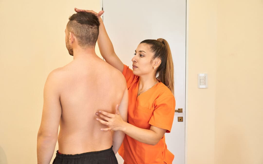
Swayback Posture
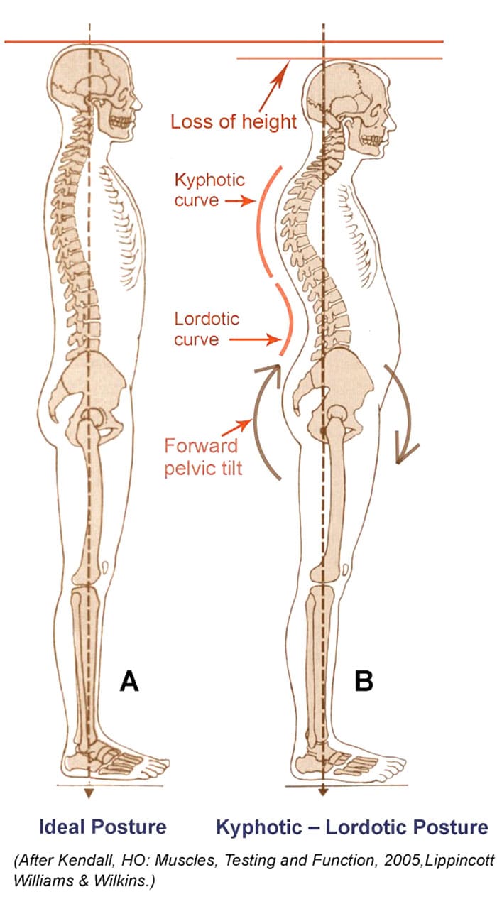
Swayback posture is a common dysfunction involving the pelvis and hip joints tilted forward in front. This causes the pelvis to shift forward, which exaggerates the curves in the lower and upper back, known as lordosis and kyphosis. The pelvis may tilt backward relative to the upper half, causing the buttocks to tuck under. The pelvis coordinates the movements of the head, shoulders, and trunk with those of the feet, legs, and thighs. A neutral pelvis, the ideal position, generally supports a mild curve/normal lordosis in the lower back. The small arch helps the body balance the skeletal parts as they work together to support and move the body’s weight. When a postural deformity occurs, one or more bones may shift from their ideal position to compensate for any pain or loss of balance caused by the original deviation. This deviation can lead to muscle strain, ligament sprain, and/or pain. (Czaprowski, D. et al., 2018)
Postural Deviations
Swayback posture causes the thoracic spine to move backward and round over into kyphosis. At the same time, the pelvis is tilted forward, resulting in an exaggeration of the normal lumbar lordosis. (Czaprowski, D. et al., 2018)
Healthcare providers, chiropractors, and physical therapists use exact measurements to define and treat postural deformities.
A neutral pelvis is a position of balance the entire body uses to help it stay upright, move, and be pain-free.
The ideal or neutral pelvic tilt is a 30-degree angle between the vertical and the plane that passes through the top of the sacrum and the axis of the hip joint socket in the front.
Swayback posture causes the pelvis to tilt forward another 10 degrees.
As a result, the spine compensates, exaggerating the curves in the lower back/lordotic curve and in the mid and upper back/kyphotic curve.
When viewed from the side, individuals can see a backward movement of the thoracic spine.
In front, the chest tends to sink in.
Muscle Group Imbalances
Healthcare providers look at different contributors or causes of postural deviations. Swayback can sometimes be associated with strength imbalances between muscle groups that move the hips, spine, and pelvis and hold the body upright. This includes:
Weakened hip flexors and overly strong or tense hip extensors/the hamstrings.
Tight upper abdominals, weak lower abdominals, and weak mid-back muscles may also contribute.
A corrective exercise program after seeing a physical therapist will help address some or all underlying muscle imbalances.
Risk Factors
Because weight in the abdominal region pulls the pelvis forward, pregnant women and obese individuals can have an increased risk of developing a swayback posture. (Vismara, L. et al., 2010)
Symptoms
The symptoms of swayback posture often include:
Severe lower back pain
Difficulty sitting or standing for long periods
Difficulty performing certain physical activities.
Tightness in the hamstrings and hip flexors
Tightness in the upper back muscles
Headaches or migraines
Chiropractic Treatment
Chiropractic adjustments are a common treatment used to correct swayback posture and can be corrected through various treatments. These include:
Spinal adjustments: The doctor applies pressure to specific spine areas to realign them and help restore proper spinal function.
Non-surgical decompression
Massage therapies
Muscle Energy Technique, or MET, improves muscle strength, flexibility, and function.
Acupuncture
Exercises to strengthen and stabilize the core muscles
Lifestyle adjustments to help reduce stress on the spine
Posture exercises
Biomechanics training
Injury Medical Chiropractic and Functional Medicine Clinic works with primary healthcare providers and specialists to develop a personalized care plan for each patient through an integrated approach to treating injuries and chronic pain syndromes, improving flexibility, mobility, and agility programs to relieve pain and help individuals return to optimal function. If other treatments are needed, Dr. Jimenez has teamed up with top surgeons, clinical specialists, medical researchers, and rehabilitation providers to provide the most effective treatments.
How I Gained My Mobility Back With Chiropractic Care
References
Czaprowski, D., Stoliński, Ł., Tyrakowski, M., Kozinoga, M., & Kotwicki, T. (2018). Non-structural misalignments of body posture in the sagittal plane. Scoliosis and spinal disorders, 13, 6. https://doi.org/10.1186/s13013-018-0151-5
Vismara, L., Menegoni, F., Zaina, F., Galli, M., Negrini, S., & Capodaglio, P. (2010). Effect of obesity and low back pain on spinal mobility: a cross sectional study in women. Journal of neuroengineering and rehabilitation, 7, 3. https://doi.org/10.1186/1743-0003-7-3
Do individuals with muscle pain know the difference between heat stroke and heat exhaustion and can find ways to stay cool?
Introduction
As the temperature rises worldwide, many individuals are enjoying their time outside and getting more sun in their lives. However, rising temperatures also mean the rise of heat-related illnesses. The two most common heat-related illnesses are heat stroke and heat exhaustion, which can impact an individual’s musculoskeletal system and have different symptoms in terms of severity. Today’s article focuses on the differences between these two heat-related illnesses, how they affect the musculoskeletal system and treatments to stay cool while reducing muscle pain. We discuss with certified associated medical providers who consolidate our patients’ information to assess heat-related illnesses associated with muscle pain. We also inform and guide patients while asking their associated medical provider intricate questions to integrate treatments and ways to stay cool when temperatures rise and reduce muscle pain. Dr. Jimenez, D.C., includes this information as an academic service. Disclaimer.
Heat Exhaustion VS Heat Stroke
By understanding the differences between heat stroke and heat exhaustion is crucial. Do you often feel overheated after simple activities? Have you experienced muscle pain or cramps? Or do you struggle to cool down? These are all signs of heat-related illnesses. Heat-related illnesses often occur when the body cannot dissipate heat, leading to dysfunctional thermoregulation. (Gauer & Meyers, 2019) The two most common types are heat exhaustion and heat stroke. While they share similar causes, they differ significantly in terms of severity, symptoms, and treatment. (Prevention, 2022)
Heat exhaustion is a mild condition that often occurs when the human body loses excessive water and salt from profusely sweating. This causes the external temperatures to be more moderate when associated with intense physical activity. (Leiva & Church, 2024) Additionally, when a person is dealing with heat exhaustion, some of the symptoms that they will experience include:
Heavy sweating
Fatigue
Headaches
Muscle cramps
Pale, cool, moist skin
Fast, weak pulse
Even though heat exhaustion is a mild heat-related condition, it can develop into severe heat-related conditions like heat stroke if not treated immediately. Heat stroke is a severe heat-related illness that is not only life-threatening buthas two forms that can affect a person’s body temperature: classic and exertional. Classic heat stroke often affects elderly individuals who have chronic medical conditions, while exertional heat stroke affects healthy individuals who are doing strenuous physical activities. (Morris & Patel, 2024) Some of the symptoms associated with heat stroke include:
High body temperature (104°F or higher)
Hot, red, dry skin
Rapid, strong pulse
Confusion
Seizures
Loss of consciousness
How Do Both Conditions Affect The Muscles?
Both heat-related illnesses can have a significant effect on the musculoskeletal system and cause muscle pain to not only the extremities but also the entire body system. The issue affects the musculoskeletal system and can lead to painful muscle cramps, involuntary muscle contractions, and muscle pain. Since muscle pain is a multi-factorial condition, heat-related illnesses like heat stroke and exhaustion can influence a person’s lifestyle and comorbid health factors. (Caneiro et al., 2021) When that happens, many individuals can seek treatments to stay cool from heat exhaustion and heat stroke and reduce muscle pain.
Secrets Of Optimal Wellness-Video
Treatments For Staying Cool & Reduce Muscle Pain
While it is important to understand the difference between heat stroke and heat exhaustion due to the crucial timing and effective interventions, finding various treatments to reduce muscle pain and find ways to stay cool is important. Many individuals can wear technology to monitor the person’s physiological status actively and prevent injuries while providing early detection for heat-related illnesses. (Dolson et al., 2022) This can reduce the chances of muscle pain and help regulate body temperature. For individuals dealing with heat exhaustion, they can:
Move to a cooler environment
Be well-hydrated with water and electrolyte-rich drinks
Rest
Wear cool clothes to lower body temperature
For individuals dealing with heat stroke, they can:
Call emergency services immediately
Apply cool clothes or ice packs to the body
Monitor vital signs
Both treatments can ensure positive results in preventing life-threatening situations that can affect the musculoskeletal system.
Conclusion
Given the significant impact both heat stroke and heat exhaustion can have on the musculoskeletal system, it’s essential to take proactive measures. Proper hydration, cooling, and rest can help manage and alleviate muscle pain associated with these heat-related illnesses. By staying informed, maintaining hydration, and taking proactive steps to protect yourself from excessive heat, you can significantly reduce the chances of these heat-related illnesses affecting your outdoor activities.
References
Caneiro, J. P., Bunzli, S., & O’Sullivan, P. (2021). Beliefs about the body and pain: the critical role in musculoskeletal pain management. Braz J Phys Ther, 25(1), 17-29. https://doi.org/10.1016/j.bjpt.2020.06.003
Dolson, C. M., Harlow, E. R., Phelan, D. M., Gabbett, T. J., Gaal, B., McMellen, C., Geletka, B. J., Calcei, J. G., Voos, J. E., & Seshadri, D. R. (2022). Wearable Sensor Technology to Predict Core Body Temperature: A Systematic Review. Sensors (Basel), 22(19). https://doi.org/10.3390/s22197639
Can fruit help with a sweet craving for individuals trying to limit sugar?
Fruits Low In Sugar
Fruits and their natural sugars: Whether following a low-carbohydrate diet or having diabetes and watching your A1C, many have heard that fruit is either bad or okay because of its natural sugars. Sugars in fruit are natural. How they affect blood sugar depends on various factors, like which foods they’re paired with and if diabetes is a factor. Counting carbs or noting the glycemic index or glycemic load of foods being eaten, understanding low-sugar fruits can help make choices that best fit your dietary needs. Certain fruits are considered lower in sugar because they contain fewer carbohydrates and sugar, allowing you to consume a larger portion.
One serving of fruit has about 15 grams of carbohydrates.
A serving is one small apple, half a medium-sized banana, or a cup of berries.
Fruits like berries can be eaten in more significant portions for the same amount of carbohydrates but less sugar.
Fruits
Low-sugar fruits include:
Lemons and Limes
Rhubarb
Apricots
Cranberries
Guava
Raspberries
Blackberries
Kiwi
Figs
Tangerines
Grapefruit
Natural Sugar
How much fruit an individual eats may differ if they follow a specific low-carb meal plan or are counting or modifying their carbohydrate intake because of diabetes. Adults should consume two cups of fruit or juice or a half-cup of dried fruit daily. (U.S. Department of Health and Human Services and U.S. Department of Agriculture, 2015) Most fruits have a low glycemic index/GI because of the amount of fiber they contain and because the sugar is mostly fructose. However, dried fruits like raisins, dates, sweetened cranberries, melons, and pineapples have a medium glycemic index. Sweetened dried fruits have an even higher glycemic index.
Fruits from Lowest to Highest Content
Fruits are a healthy way to satisfy a sweet craving. The fruits listed are ranked from lowest to highest sugar content, providing a quick way to assess sugar content. The fruits lowest in sugar have some of the highest nutritional values, plus antioxidants and other phytonutrients.
Limes and Lemons
Limes contain:
1.1 grams of sugar
7 grams of carbs
1.9 grams of fiber per fruit
Lemons contain:
1.5 grams of sugar
5.4 grams of carbs
1.6 grams of fiber per fruit
Rhubarb
Rhubarb contains:
1.3 grams of sugar
5.5 grams of carbs
2.2 grams of fiber per cup
Apricots
Apricots contain:
3.2 grams of sugar
3.8 grams of carbs
0.7 grams of fiber per small apricot
Apricots are available fresh in spring and early summer. They can be eaten whole, skin and all. However, watch portions of dried apricots as they shrink when dried.
Cranberries
Cranberries contain:
3.8 grams of sugar
12 grams of carbs
3.6 grams of fiber per cup when fresh.
While they’re low in sugar, be aware that they are usually sweetened when dried or used in a recipe.
Guavas
Guava contains:
4.9 grams of sugar
7.9 grams of carbs
3 grams of fiber per fruit
They can be sliced or dipped in salty sauce, including the rind.
Berries
These fruits generally have the lowest sugar content and are among the highest in fiber, antioxidants, and other nutrients. Berries, lemon, and lime can be added to flavor water.
Raspberries
Raspberries contain:
5.4 grams of sugar
14.7 grams of carbs
8 grams of fiber per cup
Eat a handful, or use them as a topping or ingredient. Fresh in summer or frozen year-round.
Blackberries
Blackberries contain:
7 grams of sugar
13.8 grams of carbs
7.6 grams of fiber per cup
Strawberries contain:
7.4 grams of sugar
11.7 grams of carbs
3 grams of fiber per cup
Berries are excellent choices for a snack, a fruit salad, or an ingredient in a smoothie, sauce, or dessert.
Blueberries
Blueberries contain:
15 grams of sugar
21 grams of carbs
3.6 grams of fiber per cup
While blueberries are higher in sugar than other berries, they’re packed with powerful antioxidants.
Kiwis
Kiwis contain:
6.2 grams of sugar
10.1 grams of carbs
2.1 grams of fiber per kiwi
Kiwis have a mild flavor, and the seeds and skin can be eaten.
Figs
Figs contain:
6.5 grams of sugar
7.7 grams of carbs
1.2 grams of fiber per small fig
These figures are for fresh figs, and it may be harder to estimate for dried figs of different varieties, which can have 5 to 12 grams of sugar per fig.
Tangerines
Tangerines contain:
8 grams of sugar
10.1 grams of carbs
1.3 grams of fiber per medium fruit
These low-sugar citrus fruits have less sugar than oranges and are great for salads. They are also portable, making them healthy additions to packed lunches and snacks.
Grapefruit
Grapefruit contains:
8.5 grams of sugar
13 grams of carbs
2 grams of fiber per half fresh grapefruit
Individuals can enjoy fresh grapefruit in a fruit salad or by itself, adjusting the amount of sugar or sweetener.
Low-Carb Diets
Individuals following a low-carb eating plan should remember that while some popular diet plans factor in the glycemic index or glycemic load of foods, others only factor in the number of carbohydrates.
20 Grams of Carbohydrates or Less
Individuals will likely not consume fruit or rarely substitute it for other food items with less than 20 grams of carbohydrates daily.
Nutrients are obtained from vegetables.
Some diets don’t even allow low-sugar fruits in the first phase.
20-50 Grams of Carbohydrates
These eating plans allow 20 to 50 grams of carbs daily, allowing room for one daily fruit serving.
50-100 Grams of Carbohydrates
If the eating plan allows 50 to 100 grams of carbs per day, individuals may be able to follow the FDA guidelines for two fruit servings a day, as long as other resources of carbohydrates are limited.
Other popular plans, like the Paleo diet and Whole30, don’t place a limit on fruit.
Although not necessarily a low-carb diet, Weight Watchers also allows fruit.
In general, individuals following a low-carb diet are recommended to try to eat fruits low in sugar.
Diabetes
Fruit choices when managing diabetes will depend on the type of diet being followed. For example, when counting carbohydrates, individuals should know that 1/2 cup of frozen or canned fruit has about 15 grams of carbohydrates.
Enjoy 3/4 to 1 cup of fresh berries, melon, or 17 grapes for the same carbs.
If using the plate method, add a small piece of whole fruit or 1/2 cup of fruit salad to the plate.
When using the glycemic index to guide food choices, remember that most fruits have a low GI and are encouraged.
Melons, pineapples, and dried fruits have medium GI index values, so watch portion size.
Individuals with diabetes may want to consult their primary doctor or a registered dietitian to help design an eating plan that incorporates fruit appropriately.
Body In Balance: Chiropractic, Fitness, and Nutrition
References
U.S. Department of Health and Human Services and U.S. Department of Agriculture. 2015–2020 Dietary Guidelines for Americans. 8th Edition. December 2015. Available at http://health.gov/dietaryguidelines/2015/guidelines/
Can individuals with osteoarthritis can incorporate cycling to reduce joint pain and regain their joint mobility?
Introduction
The joints in the musculoskeletal system allow the individual to be mobile while allowing the extremities to do their jobs. Just like the muscles and ligaments of the body, the joints can also wear and tear through repetitive motions, leading to joint pain in the extremities. Over time, the wear and tear from the joints can lead to the potential development of osteoarthritis, which then can affect joint mobility and lead to a life of pain and misery for individuals. However, numerous ways exist to reduce osteoarthritis’s pain-like symptoms and help restore joint mobility through cycling. Today’s article looks at how osteoarthritis affects the joints, how cycling is incorporated for osteoarthritis, and how it can reduce joint pain. We discuss with certified associated medical providers who consolidate our patients’ information to assess osteoarthritis and its associated pain symptoms affecting the joints in the extremities. We also inform and guide patients while asking their associated medical provider intricate questions to integrate cycling into their personalized treatment plan to manage the pain correlated with osteoarthritis affecting their joints. Dr. Jimenez, D.C., includes this information as an academic service. Disclaimer.
Osteoarthritis Affecting Joint Mobility
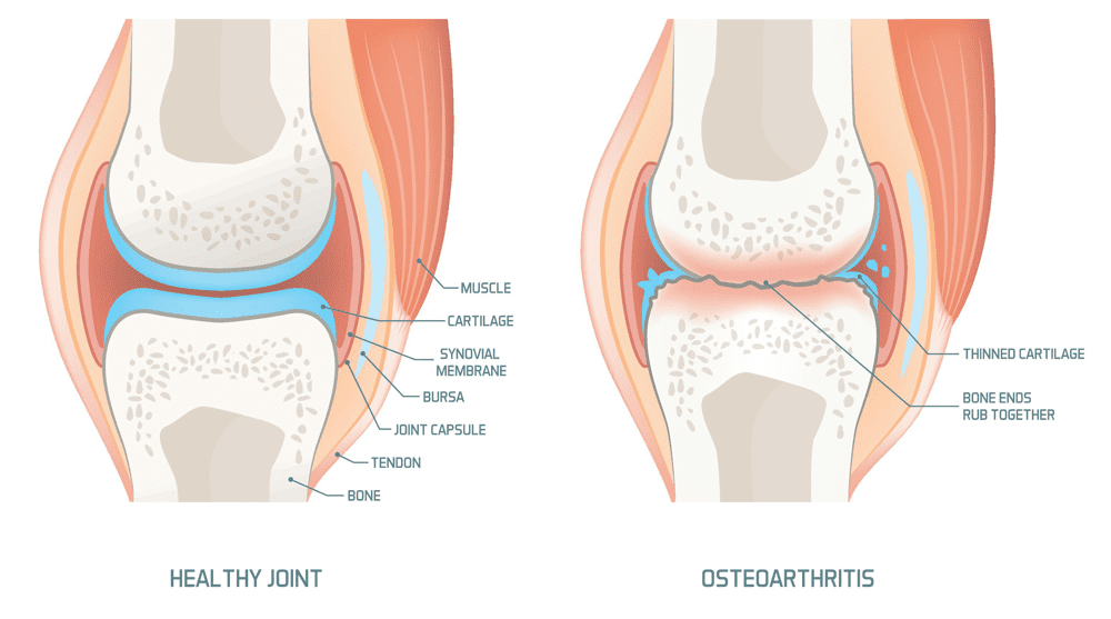
Do you feel pain and stiffness every morning in your joints only for it to feel better throughout the day? Do you experience pain in your knees, hips, and hands? Or have you noticed that your range of motion has decreased drastically? Many individuals, both young and old, can be affected by these pain-like issues and could be at risk of developing osteoarthritis in their joints. Osteoarthritis is the largest and most common musculoskeletal condition that causes a disturbance of the inflammatory cytokine balance, damaging the cartilage and other intra-articular structures surrounding the joints. (Molnar et al., 2021) This is because osteoarthritis develops over time, causing the cartilage to wear away and causing the connecting bones to rub against each other. This, in turn, can affect the extremity’s joint mobility, causing symptoms of stiffness, pain, swelling, and reduced range of motion to the joints.
Additionally, osteoarthritis is multifactorial as it can cause an imbalance in the joints due to genetics, environmental, metabolic, and traumatic factors that can contribute to its development. (Noriega-Gonzalez et al., 2023) This is because repetitive motions and environmental factors can impact the body and cause overlapping risk profiles to correlate with osteoarthritis. Some overlapping risk profiles associated with osteoarthritis are pathological changes in the joint structure that cause abnormal loading on the joints, which causes joint malalignment and muscle weakness. (Nedunchezhiyan et al., 2022) This causes many people to be in constant pain and trying to find relief from joint pain associated with osteoarthritis.
Chiropractic Solutions For Osteoarthritis-Video
Cycling For Osteoarthritis
Engaging in physical activities may seem daunting when managing osteoarthritis symptoms, but it can help restore joint mobility while reducing the pain associated with osteoarthritis. One of the physical activities that has little impact and does not impact the joints is cycling. Cycling for osteoarthritis has many beneficial properties as it can:
Strengthen surrounding muscles
Retain joint mobility
Improve range of motion
Weight management
Enhancing cardiovascular health
Cycling can help the individual focus on strengthening the lower extremity muscles surrounding the joints, which can help improve pain and functionality. (Katz et al., 2021) This, in turn, helps provide better support and stability to the joints, thus reducing overload on the body while minimizing the risk of injuries. Additionally, cycling can help improve many individuals looking for a healthier change and increase bone mineral density in the joints, thus decreasing the risk of fractures. (Chavarrias et al., 2019)
Cycling Reducing Joint Pain
Cycling is a safe and effective exercise for anyone, whether they’re just starting or haven’t been active for a while. The key to optimal recovery and joint functionality is to consult a doctor. This ensures that cycling is a safe option for you, helps you choose the right bike, and provides guidance on how to start slowly, warm up and stretch, maintain proper form, and stay consistent with the cycling sessions. This professional guidance is crucial, as it allows many individuals with joint pain to achieve complete functional recovery to their joints. (Papalia et al., 2020) Cycling is an excellent way to manage osteoarthritis and its associated symptoms. For many individuals with osteoarthritis, this low-impact exercise can be a game-changer, promoting muscle strengthening, improving joint range of motion, and helping alleviate osteoarthritis symptoms.
References
Chavarrias, M., Carlos-Vivas, J., Collado-Mateo, D., & Perez-Gomez, J. (2019). Health Benefits of Indoor Cycling: A Systematic Review. Medicina (Kaunas, Lithuania), 55(8). https://doi.org/10.3390/medicina55080452
Katz, J. N., Arant, K. R., & Loeser, R. F. (2021). Diagnosis and Treatment of Hip and Knee Osteoarthritis: A Review. JAMA, 325(6), 568-578. https://doi.org/10.1001/jama.2020.22171
Molnar, V., Matisic, V., Kodvanj, I., Bjelica, R., Jelec, Z., Hudetz, D., Rod, E., Cukelj, F., Vrdoljak, T., Vidovic, D., Staresinic, M., Sabalic, S., Dobricic, B., Petrovic, T., Anticevic, D., Boric, I., Kosir, R., Zmrzljak, U. P., & Primorac, D. (2021). Cytokines and Chemokines Involved in Osteoarthritis Pathogenesis. Int J Mol Sci, 22(17). https://doi.org/10.3390/ijms22179208
Nedunchezhiyan, U., Varughese, I., Sun, A. R., Wu, X., Crawford, R., & Prasadam, I. (2022). Obesity, Inflammation, and Immune System in Osteoarthritis. Front Immunol, 13, 907750. https://doi.org/10.3389/fimmu.2022.907750
Noriega-Gonzalez, D., Caballero-Garcia, A., Roche, E., Alvarez-Mon, M., & Cordova, A. (2023). Inflammatory Process on Knee Osteoarthritis in Cyclists. J Clin Med, 12(11). https://doi.org/10.3390/jcm12113703
Papalia, R., Campi, S., Vorini, F., Zampogna, B., Vasta, S., Papalia, G., Fossati, C., Torre, G., & Denaro, V. (2020). The Role of Physical Activity and Rehabilitation Following Hip and Knee Arthroplasty in the Elderly. J Clin Med, 9(5). https://doi.org/10.3390/jcm9051401
For individuals who don’t have time for a full workout, could incorporating sprint exercise training be an option to improve their cardiovascular and overall health?
Sprint Exercise Training
Most think of running when they hear the word sprinting. However, sprinting can be performed in any aerobic activity, whether swimming, cycling, rollerblading, or exercising on an elliptical machine. Sprint exercise training means varying the intensity levels of the activity. It is also known as sprint interval training or speed drills. It targets cardiovascular endurance and is suitable for all fitness levels, from beginners to advanced. This type of training is demanding and requires high motivation, but it can lead to significant improvements and help achieve fitness goals faster.
Sprint workouts are a time saver. Many exercise guidelines recommend up to 60 minutes of moderate exercise 3 times a week; however, many people don’t have the time. Studies have shown that short, high-intensity sprint exercise training improves aerobic capacity and endurance in half the time of traditional endurance exercise. Sprint exercise training burns calories, improves cardiovascular health, builds muscle, and increases speed and power. Sprint workouts are great for individuals who lack time for traditional steady endurance exercise but want to improve cardiovascular health. (Vollaard, N. B. J., and Metcalfe, R. S. 2017) Adding them to a workout routine can take training to a new level.
Training
The key to sprint training is performing an activity at a certain percentage of all-out effort to increase heart rate. Sprint exercise training is recommended three times a week, with at least one to two days of rest or other easy exercises between sprint workouts. How to do.
Warm-up
Warm up with easy exercise for five to 10 minutes.
Slowly perform the exercise that will be done for the sprints to prepare the body for the intense sprint.
Do the First Sprint
Perform the first sprint at around 60% intensity.
Slow down and continue warming up if there is muscle tightness or joint pain.
Rest
Recover for four minutes by slowing to a comfortable pace, but continue moving.
Do the Second Sprint
Perform the next sprint at 80% max intensity.
Rest
Rest for four minutes.
Do the Third Sprint
Perform the remainder of the sprints at 100% intensity or all-out efforts for 30 seconds.
Push to the maximum for each exercise.
Rest
Recover for four minutes after each sprint to slow down breathing and heart rate, and can hold a conversation without gasping.
Repeat
Repeat the sprint/recovery routine four to eight times, depending on fitness level and ability.
For the first workout, stop at four sprints.
Gradually build up to eight.
Benefits
Sprint exercise training enhances endurance performance and can be effectively used by athletes, fitness enthusiasts, and individuals who want to improve their fitness and health. (Litleskare, S. et al., 2020) In one study, participants who completed eight weeks of sprint training saw improvements in maximal oxygen uptake or VO2 max. The test is one way to measure cardiovascular fitness. (Litleskare, S. et al., 2020) These short bursts of intense exercise improve muscle health and performance comparable to several weeks of traditional training. (Gunnarsson, T. P. et al., 2013) Other studies have found that short, high-intensity exercise burns more calories than the same amount of moderate-level cardiovascular exercise. (Vollaard, N. B. J., and Metcalfe, R. S. 2017)
Variations
There are different ways to structure a sprinting routine, and different fitness goals will determine the intensity, duration, and number of sprints that should be performed.
Beginners
Those new to sprinting should start slow, as overdoing it can lead to injury. Work on building up a base level of fitness before introducing sprinting into an exercise routine. Start with one set of four sprint/rest cycles when trying sprints. As fitness goals are achieved, add more sprints to each set or different sprints.
Intermediate
Once a sprinting exercise routine is begun, it may only be a few weeks before one is ready to advance to an intermediate level. Try increasing the number of sprints at different intensity levels. However, avoid sprint exercises too often weekly as the body needs adequate rest.
Advanced
Advanced athletes can intensify the routine by increasing intensity and adding reps. One way is by adding resistance. For example, for those running or cycling, try sprinting hills, or if rollerblading, try wearing wrist and ankle weights to increase the load. Swimmers can use strength-building techniques to focus on specific body areas or add resistance. The intensity of any sprinting activity can be intensified by wearing a weighted vest.
Beginner Errors
A few common starting mistakes include going too hard, advancing too quickly, and doing too many for too long. Sprints are not meant to replace moderate-intensity exercise. The goal is to modulate the intensity of aerobic activities. A study showed that not getting enough rest between sprints led to an inability to perform as well during sprinting. (Selmi, M. A. et al., 2016)
Safety
Sprint workouts can be done with running, swimming, cycling, or other aerobic cardiovascular exercises. The following precautions should be considered before adding sprint training to a workout schedule:
Safety
Because sprinting is a high-intensity exercise, it is recommended that individuals consult with a healthcare professional and review the physical activity readiness questionnaire (PAR-Q) before beginning a sprint training workout.
Base Fitness
A strong fitness base in the sprint activity is also important.
To build a fitness base, follow the 10% rule and gradually increase training volume.
Frequency
Because of the intensity, sprint workouts should not be done more than three times a week.
Muscle Soreness
Launching into a sprint program can cause delayed-onset muscle soreness.
Experts recommend having about three to four weeks of base fitness before beginning.
Injuries are more likely if the body isn’t properly prepared.
The goal is to do a sprint workout six times in two weeks, then only perform 2 times a week for maintenance for six to eight weeks before changing the workout. On the days following a sprint workout, aim for 20–30 minutes of the same aerobic activity at an easier pace to help recover but maintain results. If pleased with the results, continue with the routine longer, but it is recommended to vary the workouts every few months and throughout the year. Modify the routine to find what works best.
Military Training and Chiropractic Care: Maximizing Performance
References
Vollaard, N. B. J., & Metcalfe, R. S. (2017). Research into the Health Benefits of Sprint Interval Training Should Focus on Protocols with Fewer and Shorter Sprints. Sports medicine (Auckland, N.Z.), 47(12), 2443–2451. https://doi.org/10.1007/s40279-017-0727-x
Litleskare, S., Enoksen, E., Sandvei, M., Støen, L., Stensrud, T., Johansen, E., & Jensen, J. (2020). Sprint Interval Running and Continuous Running Produce Training Specific Adaptations, Despite a Similar Improvement of Aerobic Endurance Capacity-A Randomized Trial of Healthy Adults. International journal of environmental research and public health, 17(11), 3865. https://doi.org/10.3390/ijerph17113865
Gunnarsson, T. P., Christensen, P. M., Thomassen, M., Nielsen, L. R., & Bangsbo, J. (2013). Effect of intensified training on muscle ion kinetics, fatigue development, and repeated short-term performance in endurance-trained cyclists. American journal of physiology. Regulatory, integrative and comparative physiology, 305(7), R811–R821. https://doi.org/10.1152/ajpregu.00467.2012
Selmi, M. A., Haj, S. R., Haj, Y. M., Moalla, W., & Elloumi, M. (2016). Effect of between-set recovery durations on repeated sprint ability in young soccer players. Biology of sport, 33(2), 165–172. https://doi.org/10.5604/20831862.1198636
IFM's Find A Practitioner tool is the largest referral network in Functional Medicine, created to help patients locate Functional Medicine practitioners anywhere in the world. IFM Certified Practitioners are listed first in the search results, given their extensive education in Functional Medicine