Can correcting body misalignments and the elements of unhealthy posture help achieve a healthy posture?
Healthy Posture
Maintaining a healthy posture is more important than ever, as individuals from all walks of life realize how practicing awkward and unhealthy postures can wreak havoc on their bodies and quality of life. Unhealthy postures include rounding the upper and lower back, slouching, and forward head posture. Over time, these postures make daily activities more difficult or painful. Mobility, stability, and strengthening exercises can address unhealthy posture problems and issues, along with practicing correct sitting, standing, and resting postural habits to reinforce proper alignment daily. A chiropractic and physical therapy team can treat and train individuals to restore correct and healthy posture.
Body Alignment
Ideal posture involves correct body alignment or how the structural parts, such as the head, trunk, hips, knees, etc., relate to an individual’s form. Whether standing, sitting, lying down, or moving, body parts need to be balanced in relationship to each other to avoid unnecessary stress on the spine and musculoskeletal system. (Bone Health & Osteoporosis Foundation, 2024)
Posture Types
Posture is considered static when sitting or standing still and dynamic when moving. Both are categorized as active postures as they require the activation of stabilizing muscles to deal with gravity and maintain alignment. Lying down and remaining still is considered an inactive posture, as muscle involvement is minimal. However, both have the potential to be healthy or unhealthy.
Proper Body Alignment
An easy way to check proper alignment while standing is to stand against a wall with the base of the head, shoulder blades, and buttocks flush against the wall, with enough space for a hand wide enough to slide in between the wall and the small of the back. This exercise correctly lines up the head, shoulders, and hips to reduce or eliminate undue stress on the spine. Body balance is the foundation for active and inactive postures concerning workstations, industrial ergonomics, daily activities, and sports. (Mayo Clinic, 2023) Healthy alignment is a standard position in which all body joints are centered and balanced and the most mechanically efficient position for static or dynamic activities. Biomechanical efficiency (the ability to use the body’s muscles and joints to perform movements while minimizing energy use and maximizing output) enables the muscles surrounding the joints to work in balance and efficiently activate, which, in turn, helps reduce strain, tension, and injury. Balanced muscles also conserve energy, leading to better daily stamina.
Development of Imbalances
Individuals develop position and movement habits in joint positioning that have led to imbalanced muscles. When this is the case, some muscles can be chronically stretched, and others become chronically tight, all to hold the body up or move around, which can lead to postural conditions like upper crossed (Physiopedia, 2024) or lower crossed syndrome (Physiopedia, 2024) which often leads to pain and/or mobility issues.
Posture Assessment
The recommended way to determine if one’s posture is healthy or poor is by conducting a posture assessment. The examination looks at joint positions and gathers visual information about the planes into which parts of each joint move and the axes around which those movements occur (Singla D. and Veqar Z., 2014) (Debra Coglianese et al., 2006). In a posture assessment, body alignment is compared with the ideal standard, a plumb line, usually a string with a small weight attached to the bottom to help maintain straightness. The other end of the string is affixed to the ceiling to be used as an accurate reference for correct alignment. (Singla D. and Veqar Z. 2014) During a posture assessment, the patient stands next to the plumb line while the doctor or therapist compares the relative positions of the following areas:
Ears
Shoulder joint
Spine
Hip joint
Knee joint
Ankle joint
Feet
Any areas that don’t match the reference can indicate misalignments in one or several regions.
Making Corrections
Chiropractic care can help correct unhealthy posture by realigning the spine and strengthening the musculoskeletal system:
Spinal Adjustments
Chiropractors use their hands or instruments to apply controlled force to the spine to realign the vertebrae. This can help relieve pressure on muscles, ligaments, and nerves, which can improve posture.
Corrective Exercises
Chiropractors can create custom exercises to strengthen postural muscles and maintain proper alignment.
Massage Therapy
Chiropractors can use massage therapy to work on strained ligaments and soft tissue.
Guidance and Training
Chiropractors can teach patients how to move to encourage a neutral spine and provide strategies for maintaining healthy posture.
A posture corrector or brace can also help teach and engage the correct muscles to achieve proper alignment. However, it should not be relied on long-term because promoting and activating one’s stabilizing muscles is important rather than relying on a brace for prolonged periods.
Injury Medical Chiropractic and Functional Medicine Clinic
Achieving and maintaining proper posture requires consistent work and development. Retraining the body and maintaining its optimal health requires daily efforts through exercise, conscious position corrections, and ergonomics. Injury Medical Chiropractic and Functional Medicine Clinic works with primary healthcare providers and specialists to develop an optimal health and wellness solution. We focus on what works for you to relieve pain, restore function, and prevent injury. Regarding musculoskeletal pain, specialists like chiropractors, acupuncturists, and massage therapists can help mitigate the pain through spinal adjustments that help the body realign itself. They can also work with other medical professionals to integrate a treatment plan to resolve musculoskeletal issues.
Upper Cross Syndrome
References
Bone Health & Osteoporosis Foundation. (2024). Proper body alignment. https://www.bonehealthandosteoporosis.org/patients/treatment/exercisesafe-movement/proper-body-alignment/
Mayo Clinic. (2023). Mayo Clinic Q and A: Proper posture and body alignment. https://newsnetwork.mayoclinic.org/discussion/mayo-clinic-q-and-a-proper-posture-and-body-alignment/
Singla, D., & Veqar, Z. (2014). Methods of postural assessment used for sports persons. Journal of clinical and diagnostic research: JCDR, 8(4), LE01–LE4. https://doi.org/10.7860/JCDR/2014/6836.4266
Coglianese, D. (2006). Muscles: Testing and Function With Posture and Pain, ed 5 (with Primal Anatomy CD-ROM). Physical Therapy, 86(2), 304-305. https://doi.org/https://doi.org/10.1093/ptj/86.2.304
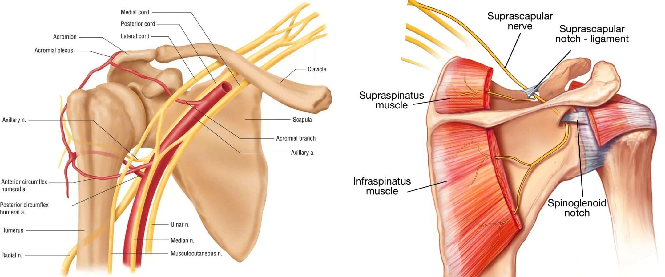
An axillary nerve injury can cause pain, weakness, and shoulder mobility loss. Can physical therapy help restore and maintain shoulder joint flexibility?
Axillary Nerve
The axillary nerve, or the circumflex nerve, is a peripheral nerve that runs through the shoulder and supports movement and sensation in the upper limbs. It originates in the neck at the brachial plexus, a network of nerves that extends from the neck and upper torso to the shoulders and arms. Its primary purpose is to supply nerve function to the shoulder joint and three muscles in the arm and also innervates some skin in the region.
Anatomy
Except for the cranial nerves, all the body’s nerves branch off from the spinal cord, emerge from between vertebrae and continue to branch off as they travel to various muscles and other structures. The axillary nerve is named after the axilla, the medical name for the armpit. Individuals have two, one on each side. After leaving the spinal column, the axillary nerve runs behind the axillary artery and continues to the shoulder blade’s lower edge of the subscapularis muscle. It winds back and travels down the arm along the posterior humeral circumflex artery, which then passes through the quadrangular space (a small area of the shoulder blade just above the armpit where there is a gap in the muscles that allows nerves and blood vessels to pass through to the arm before it divides into terminal branches, which are:
Anterior Division
Supplies motor innervation to the deltoid’s anterior and middle heads, allowing the arm to abduct or move away from the body.
It winds around the neck of the humerus/funny bone, goes beneath the deltoid muscle, and then connects to the forward edge of the deltoid.
A few small cutaneous branches serve the skin in that area.
Posterior Division
Innervates the teres minor muscles and the lower part of the deltoid.
It enters the deep fascia and becomes the superior lateral cutaneous nerve.
It then wraps around the lower edge of the deltoid, connects to the skin over the lower two-thirds of the muscle, and covers the long head of the triceps brachii.
Articular Branch
Comes from the trunk of the axillary nerve and enters the glenohumeral joint, which is in the shoulder, below the subscapularis muscle.
Anatomical Variations
In a case report, healthcare providers noted an incidence of the nerve branching directly off the upper trunk of the brachial plexus rather than the posterior cord. (Subasinghe S. K. and Goonewardene S. 2016) In this case, it innervated the subscapularis muscle, latissimus dorsi, and the deltoid and teres minor muscles and also had a communicating branch to the posterior cord. Another case documented multiple abnormalities in the course of the axillary nerve in an individual with pain and severely limited shoulder mobility. (Pizzo R. A. et al., 2019) During reverse shoulder arthroplasty, the surgeon discovered that the axillary nerve ran beside the coracoid process instead of underneath and stayed close to the subscapularis muscle instead of traveling through the quadrangular space. The case noted earlier reports of axillary nerves not running through the quadrangular space. In those cases, the nerve pierced the subscapularis muscle or split into branches before reaching the quadrangular space.
Function
The axillary nerve functions as a motor nerve that controls movement and a sensory nerve that controls sensations like touch or temperature.
Motor
As a motor nerve, the axillary nerve innervates three muscles in the arm and includes:
Deltoid
Allows flexing of the shoulder joint and rotating the shoulder inward.
Long Head of the Triceps
It runs down the back of the outer arm, allowing straightening, pulling the upper arm toward the body, or extending it backward.
The radial nerve can also innervate this muscle.
Teres Minor
One of the rotator cuff muscles starts outside the shoulder and runs diagonally along the bottom edge of the shoulder blade.
It works with other muscles to allow for the external rotation of the shoulder joint.
Sensory
In its sensory role, the nerve carries information to the brain from the following:
Glenohumeral joint or the ball-and-socket joint in the shoulder.
The skin on the lower two-thirds of the deltoid muscle through the superior lateral cutaneous branch.
Injuries and Conditions
Problems with the axillary nerve can be caused by injuries anywhere along the arm and shoulder and by disease. Common injuries include:
Dislocations
Of the shoulder joint, which can cause axillary nerve palsy.
Fracture
Of the surgical neck of the humerus.
Compression
This stems from walking with crutches, also known as crutch palsy.
Direct Trauma
This can be from an impact sports, work, automobile accident, collision, or laceration.
Added Pressure
This can be from wearing a cast or splint.
Surgical Accidental Injury
An injury or damage can come from shoulder surgery, especially arthroscopic surgery on the inferior glenoid and capsule.
Quadrangular Space Syndrome
This is where the axillary nerve is compressed where it passes through that space, which is most common in athletes who perform frequent overhead motions)
Nerve Root Damage
Between the fifth and sixth cervical vertebrae, where the nerve emerges from the spinal cord, which can be caused by traction, compression, spinal disc prolapse, or a bulging disc.
Systemic Neurological Disorders
Example – multiple sclerosis
Erb’s Palsy
A condition often is the result of a birth injury called shoulder dystocia, in which a baby’s shoulder/s becomes stuck during childbirth.
Axillary Nerve Palsy
Damage can result in a type of peripheral neuropathy that can cause weakness in the deltoid and teres minor muscles.
This can result in losing the ability to lift the arm away from the body and weakness in various shoulder movements.
If the damage is severe enough, it can cause paralysis of the deltoid and other minor muscles, resulting in flat shoulder deformity, in which individuals cannot lay their shoulders flat when lying down.
Axillary nerve damage also can lead to a change, reduction, or loss of sensation in a small part of the arm just below the shoulder.
Nerve Injury Statistics
Three times more common in men than women.
It may be present in as many as 65% of shoulder injuries.
The risk of injury due to dislocation is significantly increased after age 50.
Tests
If a healthcare provider suspects a problem with axillary nerve function, they’ll test the shoulder’s range of motion and skin sensitivity. A difference in the range of motion between the shoulders can indicate a nerve injury. Individuals may be sent for electromyography and a nerve conduction study to verify nerve palsy. In some cases, an MRI and/or X-rays may be ordered, especially if the cause of possible nerve damage is unknown.
Rehabilitation
Depending on the severity and cause of the injury, non-surgical treatments may be recommended, with surgery as a last resort. Non-surgical treatment can include some combination of immobilization, rest, ice, physical therapy, and anti-inflammatory meds. Physical treatment typically lasts about six weeks and focuses on strengthening and stimulating the muscles to prevent joint stiffness, which can impair long-term function.
Surgery
If conservative treatments don’t work, surgery may be recommended, especially if several months have passed without improvement. Surgical outcomes are generally better if surgery is performed within six months of the injury, and regardless of the time frame, the prognosis is considered positive in about 90% of cases. Surgical procedures performed for axillary nerve dysfunction or injury include:
Neurolysis
This procedure involves targeted damage/degeneration of nerve fibers, interrupts the nerve signals, and eliminates pain while the damaged area heals.
Neurorrhaphy
This procedure stitches a severed nerve back together.
Nerve Grafting
Grafting involves transplanting a portion of another nerve, usually the sural nerve, to reconnect severed nerves.
This helps, especially when the damaged portion is too large to be repaired by neurorrhaphy.
It allows a pathway for signals and encourages the regrowth of nerve axons.
Neurotization or Nerve Transfer
Similar to grafting but used when the nerve is too damaged to heal.
This procedure involves transplanting a healthy but less important nerve, or a portion of a nerve, to replace the damaged one and restore function.
Injury Medical Chiropractic and Functional Medicine Clinic
Injury Medical Chiropractic and Functional Medicine Clinic works with primary healthcare providers and specialists to develop an optimal health and wellness solution. We focus on what works for you to relieve pain, restore function, and prevent injury. Regarding musculoskeletal pain, specialists like chiropractors, acupuncturists, and massage therapists can help mitigate the pain through spinal adjustments that help the body realign itself. They can also work with other medical professionals to integrate a treatment plan to resolve musculoskeletal issues.
Shoulder Pain Chiropractic Treatment
References
Subasinghe, S. K., & Goonewardene, S. (2016). A Rare Variation of the Axillary Nerve Formed as Direct Branch of the Upper Trunk. Journal of clinical and diagnostic research : JCDR, 10(8), ND01–ND2. https://doi.org/10.7860/JCDR/2016/20048.8255
Pizzo, R. A., Lynch, J., Adams, D. M., Yoon, R. S., & Liporace, F. A. (2019). Unusual anatomic variant of the axillary nerve challenging the deltopectoral approach to the shoulder: a case report. Patient safety in surgery, 13, 9. https://doi.org/10.1186/s13037-019-0189-1
The vastus lateralis is a muscle on the outside part of the thigh. Injuries to the muscle include strains, tendinitis, femoral nerve compression, and others. Can rehabilitation like heat and ice, massage, and strength and mobility exercises help individuals return to normal activities and function?
Vastus Lateralis Muscle
The vastus lateralis is the largest of the four quadriceps muscles on the thigh’s outer portion. The vastus lateralis helps extend the knee joint and maintain the knee position when walking or running. The vastus lateralis functions to work with the other quad muscles to help extend the knee joint.
Anatomy
Most muscles are attached to bone points of attachment, called the origin and insertion points. The vastus lateralis origin and insertion points are as follows (Vieira, EPL. 2017)
Origin
The origin is on the upper inter-trochanteric line of the femur or thigh bone.
It also arises from the base of the greater trochanter and the linea aspera, the supracondylar ridge, and the lateral intermuscular septum.
Insertion
From its origin, the muscle courses down the lateral thigh and inserts as part of the lateral quadriceps tendon on the tibial tubercle, an elevated portion of the upper shin.
The muscle is a large, flat structure with different attachments and a flat aponeurosis or sheath of connective tissue on the outer thigh.
The femoral nerve from lower back levels two, three, and four controls or innervates the muscle.
Blood supply to the muscle goes through the lateral circumflex femoral artery of the upper thigh.
Function
The muscle works with the other quadriceps muscles to extend or straighten the knee. The quads are responsible for functional activities like walking, running, climbing stairs, and getting up from a seated position. The vastus lateralis and the iliotibial band, which courses down the lateral thigh next to this muscle, form the lateral wall of the thigh. The vastus lateralis is on the opposite side of the vastus medialis muscle on the inner portion of the thigh. These muscles work together to maintain the appropriate position of the patella/kneecap in the femoral groove of the thigh bone. Malfunctioning these muscles properly can lead to knee pain from patellofemoral stress syndrome. (American Academy of Orthopaedic Surgeons, 2024)
Conditions
Many different injuries and conditions can affect the vastus lateralis and quad muscles, especially in athletes or active individuals. These injuries can cause vastus lateralis pain and other problems. (Timothy J. Von Fange, 2024) Some of the injuries and conditions include:
Patellofemoral Stress Syndrome – PFSS
This occurs when the kneecap tracks improperly in the femoral groove of the knee joint.
This leads to pain and difficulty when walking and running.
Vastus Lateralis Strain
A sudden force on the thigh can cause the quad muscle to be strained.
If the vastus lateralis suffers a pull injury, individuals may have pain, muscle swelling, thigh bruising, and walking difficulties.
Patellar Tendinitis
Irritation of the quad tendon that courses over the kneecap can cause patellar tendinitis.
Femoral Nerve Compression Weakness
The femoral nerve may become pinched or irritated from a herniated disc, lumbar stenosis, or arthritis.
Pain, numbness, tingling, or weakness in the thigh may result.
Iliotibial Band Friction Syndrome
Tight or weak muscles can irritate the IT band, and the vastus lateralis muscle can be affected.
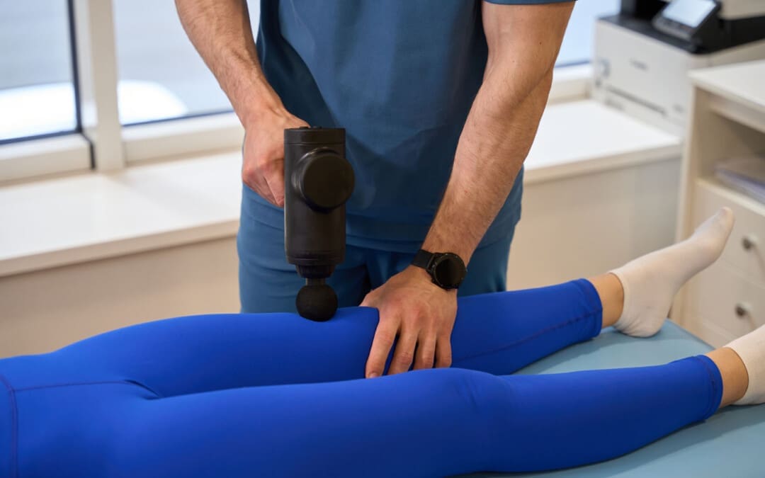
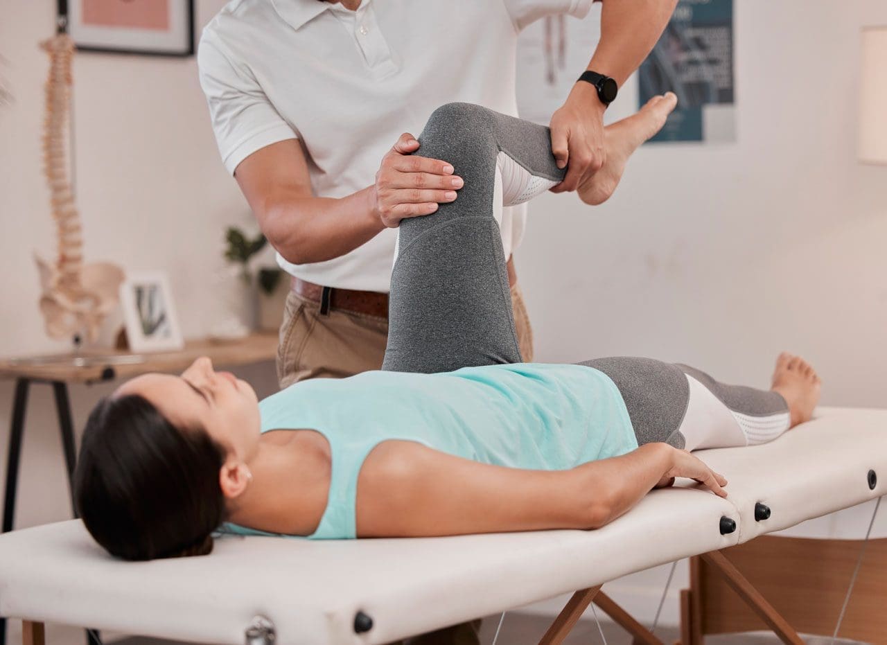
Injury Rehabilitation
Injury to the vastus lateralis or quad muscles can cause pain, swelling of the thigh, or limited walking ability. Various treatments are available to help expedite recovery. A primary healthcare provider may recommend working with a physical therapy team. Self-care techniques can include:
Heat and Ice
Ice may be applied to the lateral thigh the first few days after injury to control pain and decrease swelling and inflammation.
Ice should be applied for 10 to 15 minutes.
Individuals may switch to heat two to three days after to promote circulation and improve tissue mobility.
Heat should be applied for 10 to 15 minutes.
Massage
Massage can help decrease pain and promote circulation.
Massage techniques can improve tissue mobility before stretching to help improve quadriceps motion.
Exercises and Stretching
A physical therapy team will prescribe certain exercises and stretches to help regain strength and range of motion. After an injury, quad stretching can improve the mobility and function of the muscle group.
Prone Towel Quad Stretch
Lie on your stomach and place a towel or strap around the ankle.
Bend the knee up, and gently pull on the towel to bend the knee fully.
A pulling sensation should be felt in the front of the thigh.
Hold the stretch for 30 seconds and release.
Repeat three times.
Half-kneeling Quad and Hip Flexor Stretch
Kneel on one knee.
Slowly move forward until a stretch is felt in the front of the hip and thigh.
Hold this position for 30 seconds.
Relax back to the starting position.
Repeat three times.
Back Exercises
If femoral nerve irritation coming from the lower back is causing thigh pain or weakness, exercises to release the nerve may be helpful and can include:
Prone press-ups
Supine lumbar flexion
Lumbar side glides
The exercises are designed to relieve pressure on the lumbar nerve, and postural correction exercises may be performed to maintain decompression.
Strengthening
Weakness to the vastus laterals and quads may be causing injury, and strengthening exercises may be prescribed during rehabilitation and can include:
Hip-strengthening exercises
Straight leg raises
Leg extension exercises
Lunges
Squats
Strengthening exercises should be done two to four times weekly with appropriate rest between sessions.
Balance exercises and sport-specific plyometric training may be recommended to ensure the quad functions normally.
Most quadriceps and vastus lateralis muscle injuries heal within six to eight weeks.
Recovery may be shorter or longer depending on the nature of the injury.
Injury Medical Chiropractic and Functional Medicine Clinic
By understanding the anatomy and function of the vastus lateralis muscle, a healthcare provider can help individuals understand their specific injury and develop a treatment program to rehabilitate the muscle properly. At Injury Medical Chiropractic and Functional Medicine Clinic, we focus on what works for you and strive to develop fitness and better the body through research methods and total wellness programs. These natural programs use the body’s ability to achieve improvement goals, and athletes can condition themselves to excel in their sport through proper fitness and nutrition. Our providers use an integrated approach to create personalized programs, often including Functional Medicine, Acupuncture, Electro-Acupuncture, and Sports Medicine principles.
Knee Injury Chiropractor
References
Vieira EPL. (2017). Anatomic study of the portions long and oblique of the vastus lateralis and vastus medialis muscles. J Morphol Sci., 28(4), 0-. http://www.jms.periodikos.com.br/article/587cb49f7f8c9d0d058b47a1/pdf/jms-28-4-587cb49f7f8c9d0d058b47a1.pdf
American Academy of Orthopaedic Surgeons. (2024). Patellofemoral pain syndrome. https://orthoinfo.aaos.org/en/diseases–conditions/patellofemoral-pain-syndrome/
Timothy J Von Fange. (2024). Quadriceps muscle and tendon injuries. UpToDate. https://www.uptodate.com/contents/quadriceps-muscle-and-tendon-injuries/print
Ramírez-delaCruz, M., Bravo-Sánchez, A., Esteban-García, P., Jiménez, F., & Abián-Vicén, J. (2022). Effects of Plyometric Training on Lower Body Muscle Architecture, Tendon Structure, Stiffness, and Physical Performance: A Systematic Review and Meta-analysis. Sports medicine – open, 8(1), 40. https://doi.org/10.1186/s40798-022-00431-0
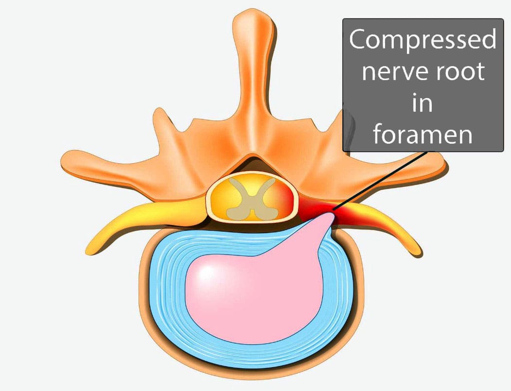
Individuals experiencing persistent pain, weakness, numbness, and tingling in the back could be suffering from nerve root encroachment. Could surgery ease nerve compression and improve symptoms for persistent and severe cases?
Surgical Decompression
The pain, weakness, numbness, and tingling associated with nerve root encroachment are usually first treated with non-surgical therapies that include:
Anti-inflammatory medications
Physical therapy
This can be enough to address the irritation of the spinal nerve root. But when cases become severe, surgical decompression may be recommended and necessary. It can be done in a couple of different ways.
Causes and Symptoms
Vertebrae are bones in the spine. Small openings called foramina allow a spinal nerve root to pass through on each side of the vertebra. When nerve root encroachment is present, the spinal nerve root gets compressed, pinched, and trapped, which can cause peripheral symptoms such as numbness, tingling, pain, or weakness to develop. Nerve root encroachment is typically caused by normal aging degenerative wear and tear changes in the vertebrae. (Choi Y. K. 2019) These degenerative changes can include:
Facet joint hypertrophy
Ligament and bone hypertrophy
Disc disorders
Formation of bone spurs or osteophytes.
If these degenerative changes progress, they can encroach and compress a nerve root, leading to peripheral symptoms. (Choi Y. K. 2019)
When Surgery Is Recommended
When symptoms occur, initial treatment will involve:
Physical therapy
Chiropractic realignment
Massage therapies
Rest
Lifestyle adjustments
Nonsteroidal anti-inflammatories – NSAIDs
Corticosteroid injections into the spine
If conservative therapies don’t fully heal or improve symptoms or there are neurological problems like difficulty with balance or walking, then surgery may be recommended. Severe pain that limits normal function is an indication for surgery, and rapidly progressive weakness of the arms and/or legs or signs of cauda equina syndrome are indications for emergency surgery.
Surgery Options
Different types of spinal surgery may be performed. A neurosurgeon will decide the best procedure for each patient based on their case, age, medical conditions, and other factors. Specific spinal surgical decompression depends on what is causing the nerve compression. In most cases, it involves removing bone or tissue to relieve nerve pressure or provide support to stabilize the joint. The most common types of surgical decompression include: (Mayo Clinic Health System, 2022)
Maintain the stability and alignment of the spine.
Improve the stability and alignment of the spine.
Anterior Surgery
The anterior approach to surgery means that the spine is accessed through the anterior/front of the spine. In this surgery, one or more discs and bone spurs may be removed through an incision in the front of the neck. (American Association of Neurological Surgeons, 2024) For example, an anterior cervical discectomy may alleviate pressure on one or more nerve roots in the neck. With an anterior lumbar interbody fusion, a surgeon removes a degenerative disc in the lower spinal area by going through a patient’s lower abdomen. (American Association of Neurological Surgeons, 2024) After the disc is removed, a structural device, usually made of bone, fills the space where it once was. This device encourages bone healing and helps the vertebrae’s bodies fuse.
Posterior Surgery
Posterior surgery means the spine is accessed through the posterior/back of the spine. An example is removing a thickened ligament, bone spur, or disc material in the neck. To do this, a small incision in the back of the neck may be made to remove part of the back of the vertebrae called the lamina. This is called a posterior cervical laminectomy. (American Association of Neurological Surgeons, 2024) A posterior lumbar interbody fusion removes a degenerative disc by going through the back. (American Association of Neurological Surgeons, 2024) Like the anterior approach, a structural device often contains bone to fill the space where the disc once was to fuse the bones.
Potential Risks
As with any surgery, it’s important that the individual and their healthcare provider carefully discuss the benefits and risks. Spinal surgical decompression includes: (Proietti L. et al., 2013)
Bleeding
Blood clots
Surgical site infection
Urinary tract infection
Lung infection
Intestinal blockage
There are also specific risks to the area of the spine being operated on and how it is surgically approached. For example, a cervical anterior procedure may injure the esophagus, trachea, or carotid artery. Likewise, damage to the C5 nerve root/C5 palsy can occur from cervical spinal decompressive surgery. This complication causes weakness, numbness, and pain in the shoulders. (Thompson S. E. et al., 2017) The spinal cord may also be injured during surgery and result in paralysis, although this is rare. (American Association of Neurological Surgeons, 2024)
Injury Medical Chiropractic and Functional Medicine Clinic
Injury Medical Chiropractic and Functional Medicine Clinic works with primary healthcare providers and specialists to develop an optimal health and wellness solution. We focus on what works for you to relieve pain, restore function, and prevent injury. Regarding musculoskeletal pain, specialists like chiropractors, acupuncturists, and massage therapists can help mitigate the pain through spinal adjustments that help the body realign itself. They can also work with other medical professionals to integrate a treatment plan to resolve musculoskeletal issues.
The Non-Surgical Solution
References
Choi Y. K. (2019). Lumbar foraminal neuropathy: an update on non-surgical management. The Korean journal of pain, 32(3), 147–159. https://doi.org/10.3344/kjp.2019.32.3.147
Mayo Clinic Health System. (2022). Decompress and stabilize: understanding types of back surgery. Speaking of Health. https://www.mayoclinichealthsystem.org/hometown-health/speaking-of-health/understanding-types-of-back-surgery
American Association of Neurological Surgeons. (2024). Cervical spine. https://www.aans.org/patients/conditions-treatments/cervical-spine/
American Association of Neurological Surgeons. (2024). Lumbar spinal stenosis. https://www.aans.org/patients/conditions-treatments/lumbar-spinal-stenosis/
Proietti, L., Scaramuzzo, L., Schiro’, G. R., Sessa, S., & Logroscino, C. A. (2013). Complications in lumbar spine surgery: A retrospective analysis. Indian journal of orthopaedics, 47(4), 340–345. https://doi.org/10.4103/0019-5413.114909
Thompson, S. E., Smith, Z. A., Hsu, W. K., Nassr, A., Mroz, T. E., Fish, D. E., Wang, J. C., Fehlings, M. G., Tannoury, C. A., Tannoury, T., Tortolani, P. J., Traynelis, V. C., Gokaslan, Z., Hilibrand, A. S., Isaacs, R. E., Mummaneni, P. V., Chou, D., Qureshi, S. A., Cho, S. K., Baird, E. O., … Riew, K. D. (2017). C5 Palsy After Cervical Spine Surgery: A Multicenter Retrospective Review of 59 Cases. Global spine journal, 7(1 Suppl), 64S–70S. https://doi.org/10.1177/2192568216688189
What type of concussion tests are there to help establish the extent of head injuries and help assess improvement during recovery?
Concussion Tests
A concussion is a temporary change in brain function that occurs from a traumatic brain injury or TBI. It can cause problems with thinking and mood and can take weeks to years to heal. Concussion tests are done after a suspected head injury and are also used after diagnosis to assess healing progress. They are noninvasive tests that measure brain functions. Several tests vary in how they are given and what they measure.
Tests
A mild or moderate traumatic brain injury can cause damage to the brain that is not detectable with brain imaging tests. However, the damage can cause serious symptoms, including headaches, emotional changes, difficulty concentrating, and memory problems. (Haider M. N. et al., 2021) The effects of a concussion can be hard to describe, but concussion testing can help identify and quantify these changes. For individuals who don’t have time to heal or experience further brain injuries while recovering, the effects can be prolonged and worsen. This is one reason why concussion testing is vital to get a diagnosis and follow medical recommendations to avoid further injury to the brain. Diagnosis can help set goals, adjust, and assess how the effects improve over time. With improvement, individuals can participate in rehabilitation and follow their doctor’s instructions for gradually returning to work, school, and other activities.
Measurements
Concussion tests can measure subtle aspects of brain function, like visual or auditory perception and response speed (Joyce A. S. et al., 2015). The damage sustained can impair these abilities, like slow decision-making. A traumatic brain injury can be associated with serious injuries, like a skull fracture, swelling, bruise, or bleeding in the brain. These injuries can be detected with imaging tests and often require surgery or other interventions. Brain damage from bleeding or swelling would cause focal neurological symptoms and signs, including partial vision loss, numbness, and weakness. Individuals can have a concussion along with detectable brain injuries or in the absence of detectable brain injuries.
Types of Tests
There are several types of concussion tests. Individuals may have one or more of these, depending on the standard test that is used in their school, sports league, or by their doctor. These can include:
Online Checklists
Several different online checklists are available for concussion screening.
These tests may include questions about symptoms and are often used as self-tests but are not intended to replace an evaluation by a medical professional.
Baseline and Post-Injury Tests
Many schools and sports leagues conduct preseason skill measurements, including memory tests or tests of speed and accuracy, either in an interview form or with computer testing.
Individuals might be asked to retake the test that is used as a comparison if they have experienced a traumatic brain injury.
Standardized Assessment of Concussion – SAC
This five-minute test can be done on the sidelines after a sports injury or later.
It evaluates orientation, immediate memory, neurologic function, concentration, and delayed recall. (Kaufman M. W. et al., 2021)
King-Devick Concussion Test
This two-minute test can be performed on the sidelines after a sports injury or later to assess language, eye movement, and attention. (Krause D. A. et al., 2022)
Post-Concussion Symptom Scale
This test includes 22 questions involving neurocognitive factors, including difficulty concentrating or remembering, physical symptoms like headaches and dizziness, and emotional symptoms like sadness or irritability. (Langevin P. et al., 2022)
Sport Concussion Assessment Tool – SCAT
This test includes an on-field assessment noting concussion symptoms, memory assessment using Maddocks questions (a short list of specific questions), Glasgow Coma Scale (GCS), and cervical spine assessment.
An off-field assessment involves the evaluation of cognitive, neurological, balance, and delayed recall. (Kaufman M. W. et al., 2021)
Buffalo Concussion Physical Examination – BCPE
A modified physical examination that assesses neck tenderness and range of motion, head, jaw, and face abnormalities, eye movements examination, and coordination. (Haider M. N. et al., 2021)
After a concussion, individuals will also have a physical examination, including a full neurological examination, in a doctor’s office.
Results
A doctor will diagnose based on symptoms, physical examination, and concussion test results. For example, for individuals who have broken several bones and are taking powerful pain medications, concussion test results can be abnormal even if they did not experience a concussion. The results of concussion testing can be compared with results before the head injury. Often, baseline testing is required for participation in certain sports leagues at professional and amateur levels. A low score can indicate that head injury has impaired brain function. Sometimes, testing can be done within a few hours of the head trauma and then again a few days later. Responses of individuals who did not have measurements taken before a head injury can be compared with the average results of people their age.
Injury Medical Chiropractic and Functional Medicine Clinic
Injury Medical Chiropractic and Functional Medicine Clinic works with primary healthcare providers and specialists to develop an optimal health and wellness solution. We focus on what works for you to relieve pain, restore function, and prevent injury. Regarding musculoskeletal pain, specialists like chiropractors, acupuncturists, and massage therapists can help mitigate the pain through spinal adjustments that help the body realign itself. They can also work with other medical professionals to integrate a treatment plan to resolve musculoskeletal issues.
Lumbar Spine Injuries in Sports: Chiropractic Healing
References
Haider, M. N., Cunningham, A., Darling, S., Suffoletto, H. N., Freitas, M. S., Jain, R. K., Willer, B., & Leddy, J. J. (2021). Derivation of the Buffalo Concussion Physical Examination risk of delayed recovery (RDR) score to identify children at risk for persistent postconcussive symptoms. British journal of sports medicine, 55(24), 1427–1433. https://doi.org/10.1136/bjsports-2020-103690
Joyce, A. S., Labella, C. R., Carl, R. L., Lai, J. S., & Zelko, F. A. (2015). The Postconcussion Symptom Scale: utility of a three-factor structure. Medicine and science in sports and exercise, 47(6), 1119–1123. https://doi.org/10.1249/MSS.0000000000000534
Kaufman, M. W., Su, C. A., Trivedi, N. N., Lee, M. K., Nelson, G. B., Cupp, S. A., & Voos, J. E. (2021). The Current Status of Concussion Assessment Scales: A Critical Analysis Review. JBJS reviews, 9(6), e20.00108. https://doi.org/10.2106/JBJS.RVW.20.00108
Krause, D. A., Hollman, J. H., Breuer, L. T., & Stuart, M. J. (2022). Validity Indices of the King-Devick Concussion Test in Hockey Players. Clinical journal of sport medicine: official journal of the Canadian Academy of Sport Medicine, 32(3), e313–e315. https://doi.org/10.1097/JSM.0000000000000938
Langevin, P., Frémont, P., Fait, P., & Roy, J. S. (2022). Responsiveness of the Post-Concussion Symptom Scale to Monitor Clinical Recovery After Concussion or Mild Traumatic Brain Injury. Orthopaedic journal of sports medicine, 10(10), 23259671221127049. https://doi.org/10.1177/23259671221127049
Elbow pain from lifting is a common symptom among individuals who lift weights, heavy objects, children, grocery bags, etc. Depending on the underlying cause, can conservative treatments relieve and heal elbow pain?
Elbow Pain Caused By Lifting
Elbow pain from lifting can result from weight training, repetitive daily tasks, or job duties like lifting small children or heavy objects. Pain can manifest at the sides or the front of the elbow. Most minor injury cases can be treated with ice, rest, and medications at home. However, pain after lifting can also be a sign of a serious injury, such as a tendon rupture/tear.
Minor Pain From Lifting
Lifting puts pressure on the tendons connecting the wrist and upper arm to the bones in the elbow joint. Minor elbow pain can occur from temporary inflammation in any of these structures after lifting an object. Tendonitis occurs when a tendon becomes inflamed, often from overuse or lifting something too heavy, and ranges from mild to severe. Mild tendonitis typically causes pain during the activity and improves with rest. (American Academy of Orthopaedic Surgeons, 2020) Common forms of tendonitis include:
Tennis elbow – tendonitis on the outside of the elbow
Golfer’s elbow – tendonitis on the inside of the elbow.
Add ice to the affected area for up to 20 minutes daily to decrease elbow pain.
Rest
Avoid lifting heavy objects as much as possible when pain is present.
Wearing A Brace
If the pain is at the tendons on the inside or outside of your elbow, try wearing a wrist brace to limit the use of your wrist muscles that connect to this area.
Stretching
Gently stretching the wrist flexors and extensors can help reduce elbow pain after lifting. Stretches can be performed several times daily, even after symptoms have resolved. (American Academy of Orthopaedic Surgeons, 2024)
Hold the arm out in front with the palm down. Keep the elbow straight.
Bend the wrist down so that the fingers are pointing toward the ground.
With the other hand, gently pull the wrist further down until a stretch is felt along the back of the forearm.
Hold this position for 15 seconds.
Repeat five times.
Next, bend the wrist upward so the fingers point toward the ceiling.
Using the other hand, gently pull the hand backward until the stretch is felt along the front of the forearm.
Mild cases can improve after a few days of self-care, whereas more pronounced elbow symptoms can take several weeks, months, or even a year. (Kheiran A. Pandey, A. & Pandey R. 2021) If self-care doesn’t work, physical therapy may be recommended. A physical therapy team can use various modalities and treatments to help reduce pain and inflammation from elbow injuries. The therapy can include targeted exercises to strengthen weak muscles and stretch tight muscles that might contribute to the condition. In addition, the therapy team will help individuals modify their lifting technique to help prevent further injury.
A biceps tendon rupture is a rare but serious injury usually caused from lifting. In addition to other visible signs of the injury, there will be a bulge at the top of the upper arm because the muscle bunches up as it is no longer attached to the elbow. (American Academy of Orthopaedic Surgeons, 2022) Individuals may hear an audible popping sound if an elbow ligament or tendon gets torn while lifting. (Johns Hopkins Medicine, 2024)
Treatment
Treatment depends on the severity of the injury, but most cases resolve on their own with rest and, if necessary, physical therapy. Conditions that cause severe pain require orthopedic surgeon expertise. These physicians specialize in treating musculoskeletal system injuries. Imaging such as X-rays, MRIs, or CT scans are often used to determine the extent of damage. Individuals with tendon or ligament tears in the elbow may need surgery to regain full range of motion and strength in their arm. After surgery, physical therapy will help restore function.
Injury Medical Chiropractic and Functional Medicine Clinic
Injury Medical Chiropractic and Functional Medicine Clinic works with primary healthcare providers and specialists to develop an optimal health and wellness solution. We focus on what works for you to relieve pain, restore function, and prevent injury. Regarding musculoskeletal pain, specialists like chiropractors, acupuncturists, and massage therapists can help mitigate the pain through spinal adjustments that help the body realign itself. They can also work with other associated medical professionals to integrate a treatment plan to improve the body’s flexibility and mobility and resolve musculoskeletal issues.
Shoulder Pain Chiropractic Treatment
References
American Academy of Orthopaedic Surgeons. (2020). Sprains, strains, and other soft-tissue injuries. https://orthoinfo.aaos.org/en/diseases–conditions/sprains-strains-and-other-soft-tissue-injuries/
Kheiran, A., Pandey, A., & Pandey, R. (2021). Common tendinopathies around the elbow; what does current evidence say?. Journal of clinical orthopaedics and trauma, 19, 216–223. https://doi.org/10.1016/j.jcot.2021.05.021
American Academy of Orthopaedic Surgeons. (2024). Therapeutic exercise program for epicondylitis (tennis elbow/golfer’s elbow). https://orthoinfo.aaos.org/globalassets/pdfs/2024-therapeutic-exercise-program-for-lateral-and-medial-epicondylitis.pdf
American Academy of Orthopaedic Surgeons. (2023). What are NSAIDs? https://orthoinfo.aaos.org/en/treatment/what-are-nsaids/
American Academy of Orthopaedic Surgeons. (2022). Biceps tendon tear at the elbow. https://orthoinfo.aaos.org/en/diseases–conditions/biceps-tendon-tear-at-the-elbow
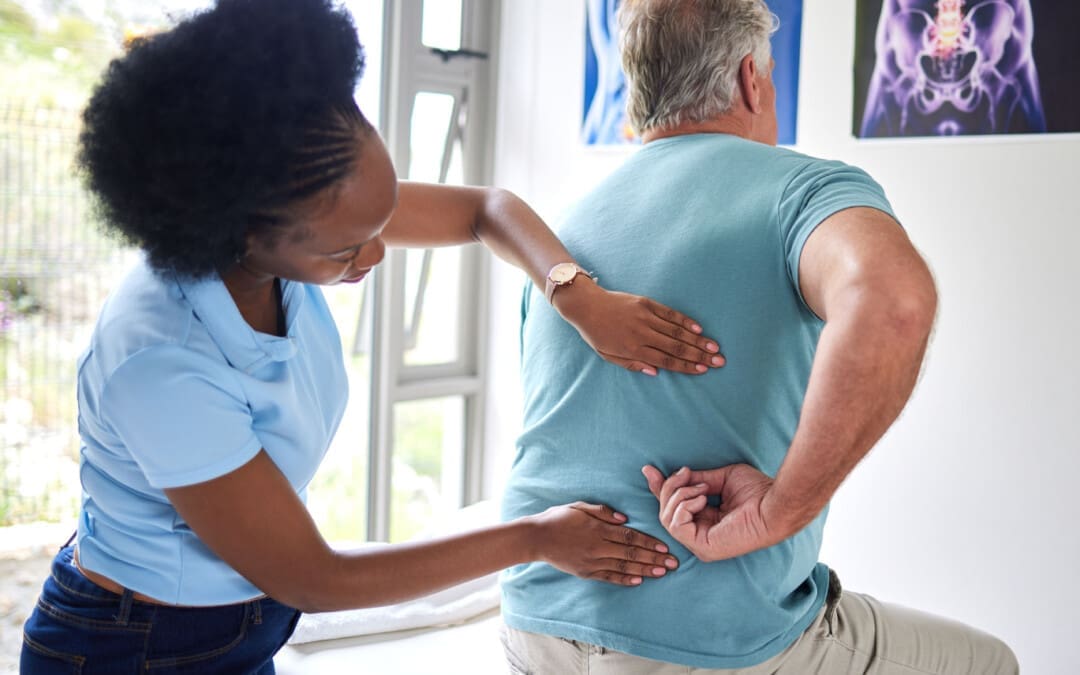
Lower back leg pain depends on specific symptoms and their duration. Can having a better idea of symptoms help individuals inform their medical providers to develop an effective treatment plan?
Low Back Leg Pain
Lower back leg pain, sciatica, and weakness of the lower-extremity muscles are often diagnosed as a herniated (compressed or ruptured) disc. Nerves surrounding the spine are sensitive to irritation and pressure caused by a disc shifting out of position or physical damage to the disc and surrounding area, ranging from mild to severe. This is why it is important to be evaluated by a healthcare provider. Treatment depends on the type of spinal disc herniation and the severity, but getting an early medical evaluation helps ensure optimal outcomes. Non-surgical conservative treatments are often effective, but some cases may require more aggressive treatment, especially if the pain persists.
Spine and Nerves
Spinal discs are the shock absorbers between vertebrae. They consist of a tough outer layer, annulus fibrosis, covering a soft gel core, nucleus pulposus. When a disc is damaged, it can bulge and irritate surrounding nerves. In more severe cases, the annulus fibrosis can weaken and tear, allowing the material to leak and compress the spinal cord or nerves. As the nerves are not functioning properly, abnormal signals may be sent to and from the brain. The most common lower back herniations occur in the lumbar region, where five vertebrae near the base of the spine are classified from top to bottom as L1 through L5. (Dydyk A.M. et al., 2023) Pain resulting from an injury to this part of the spine can be debilitating because it may involve sciatic nerve irritation. Herniated disc causes are generally a combination of age-related degeneration, being overweight/obese, trauma, a sedentary lifestyle, and overloading of the spine. (Cleveland Clinic, 2021)
Symptoms
The most common symptoms include:
Back Pain
Caused by nerve irritation, muscle spasms, and inflammation.
Radiculopathy
Abnormal signaling of the nerves.
Electrical Shooting Pain
Nerve pressure can cause abnormal sensations, commonly experienced as electric shooting pains.
For low back herniations, the shocks go down one or both legs.
Tingling – Numbness
There are often abnormal sensations such as tingling, numbness, or pins and needles down one or both legs.
Muscle Weakness
Nerve signals may be interrupted, causing lower-body muscle weakness. (Dydyk A.M. et al., 2023)
Bowel – Bladder Symptoms
These symptoms may signal cauda equina syndrome, a rare condition resulting from a herniated disc between the L5 vertebrae and the first vertebrae of the sacrum.
Diagnosis
Diagnosing a herniated disc as the cause of low back leg pain involves testing sensation, muscle strength, and reflexes. MRI also aids this process (American Association of Neurological Surgeons, 2024). MRIs can often show herniated discs and other abnormalities, especially in older patients.
Treatment
A herniated disc treatment plan is based on patient symptoms, physical examination findings, and imaging results. Most herniated disc symptoms resolve themselves in four to six weeks. Lower back pain is generally treated conservatively through:
Topical pain ointments or creams for muscle spasms.
Non-surgical decompression relieves pressure, activates healing, and restores circulation and nutrients.
Chiropractic adjustments realign the spine and musculoskeletal system.
Massage loosens the muscles and maintains their relaxation.
Total rest is never recommended, even if movement is challenging,
Exercise and stretching help avoid muscle degeneration and strengthen the muscles.
Relaxation techniques and other natural pain therapies can help manage symptoms and restore overall health.
Pain-blocking injections which can include anesthetics or corticosteroids at the source (Cleveland Clinic, 2021)
Surgery is recommended only when conservative treatments are ineffective after six weeks, if there is significant muscle weakness from nerve damage, or if motor functions are compromised. (American Association of Neurological Surgeons, 2024)
Injury Medical Chiropractic and Functional Medicine Clinic
Chiropractic therapy is among the more conservative treatment options and may be tried first before proceeding with surgery. Injury Medical Chiropractic and Functional Medicine Clinic works with primary healthcare providers and specialists to develop an optimal health and wellness solution. We focus on what works for you to relieve pain, restore function, and prevent injury. Regarding musculoskeletal pain, specialists like chiropractors, acupuncturists, and massage therapists can help mitigate the pain through spinal adjustments that help the body realign itself. They can also work with other associated medical professionals to integrate a treatment plan to improve the body’s flexibility and mobility and resolve musculoskeletal issues.
Disc Herniation
References
Dydyk AM, Ngnitewe Massa R, Mesfin FB. Disc Herniation. [Updated 2023 Jan 16]. In: StatPearls [Internet]. Treasure Island (FL): StatPearls Publishing; 2024 Jan-. Available from: https://www.ncbi.nlm.nih.gov/books/NBK441822/
IFM's Find A Practitioner tool is the largest referral network in Functional Medicine, created to help patients locate Functional Medicine practitioners anywhere in the world. IFM Certified Practitioners are listed first in the search results, given their extensive education in Functional Medicine