Can understanding how nociceptors function and their role in processing pain signals help individuals who are managing injuries and/or living with chronic pain conditions?
Nociceptors
Nociceptors are nerve endings that detect harmful stimuli, such as extreme temperatures, pressure, and chemicals, and signal pain. They are the body’s first defense against potentially damaging environmental inputs.
Nociceptors are in the skin, muscles, joints, bones, internal organs, deep tissues, and cornea.
They detect harmful stimuli and convert them into electrical signals.
These signals are sent to the brain’s higher centers.
The brain interprets the signals as pain, which prompts the body to avoid the harmful stimulus.
Nociceptors, often called pain receptors, are free nerve endings all over the body. They play a pivotal role in how the body feels and reacts to pain. The main purpose of a nociceptor is to respond to damage to the body by transmitting signals to the spinal cord and brain. (Purves D, Augustine GJ, Fitzpatrick D, et al., editors. 2001) If you bang your foot, the nociceptors on the skin are activated, sending a signal to the brain via the peripheral nerves to the spinal cord. Pain resulting from any cause is transmitted this way. Pain signals are complex, carrying information about the stimuli’s location and intensity. This causes the brain to fully process the pain and send communication back to block further pain signals.
Thermal nociceptors respond to extreme hot or cold temperatures.
For instance, when touching a hot stove, the nociceptors, which signal pain, are activated immediately, sometimes before you know what you’ve done.
Mechanical
Mechanical nociceptors respond to intense stretching or strain, such as pulling a hamstring or straining a tendon.
The muscles or tendons are stretched beyond their ability, stimulating nociceptors and sending pain signals to the brain.
Chemical
Chemical nociceptors respond to chemicals released from tissue damage.
For example, prostaglandins and substance P or external chemicals like topical capsaicin pain creams.
Silent
Silent nociceptors must be first activated by tissue inflammation before responding to a mechanical, thermal, or chemical stimulus.
Most visceral nociceptors are located on organs in the body.
Polymodal
Polymodal nociceptors respond to mechanical, thermal, and chemical stimuli.
Mechano-thermal
Mechano-thermal nociceptors respond to mechanical and thermal stimuli.
Pain Transmission
Nociceptors are also classified by how fast they transmit pain signals. Transmission speed is determined by the type of nerve fiber known as an axon a nociceptor has. There are two main types.
The first type is A fiber axon, fibers surrounded by a fatty, protective sheath called myelin.
Myelin allows nerve signals/action potentials to travel rapidly.
Because of the difference in transmission speed, the pain signals from the A fibers reach the spinal cord first. As a result, after an acute injury, an individual experiences pain in two phases, one from the A fibers and one from the C fibers. (Ngassapa D. N. 1996)
Pain Perception Phases
When an injury occurs, the stimulated nociceptors activate the A fibers, causing a person to experience sharp, prickling pain.
This is the first phase of pain, known as fast pain, because it is not especially intense but comes right after the stimulus.
During the second phase of pain, the C fibers are activated, causing an intense, burning pain that persists even after the stimulus has stopped.
The fact that the C fibers carry burning pain explains why there is a short delay before feeling the sensation.
The C fibers also carry aching, sore pain caused by organs within the body, such as a sore muscle or stomachache. (Ngassapa D. N. 1996)
Injury Medical Chiropractic and Functional Medicine Clinic
Injury Medical Chiropractic and Functional Medicine Clinic works with primary healthcare providers and specialists to build optimal health and wellness solutions. We focus on what works for you to relieve pain, restore function, prevent injury, and help mitigate issues through adjustments that help the body realign itself. They can also work with other medical professionals to integrate a treatment plan to resolve musculoskeletal problems.
From Injury To Recovery With Chiropractic Care
References
Purves D, A. G., Fitzpatrick D, et al., editors. (2001). Nociceptors. In Neuroscience. 2nd edition. (2nd ed.). Sunderland (MA): Sinauer Associates. https://www.ncbi.nlm.nih.gov/books/NBK10965/
University of Texas McGovern Medical School. (2020). Chapter 6: Pain Principles. https://nba.uth.tmc.edu/neuroscience/m/s2/chapter06.html
Ngassapa D. N. (1996). Comparison of functional characteristics of intradental A- and C-nerve fibres in dental pain. East African medical journal, 73(3), 207–209.
Can individuals utilize chiropractic care to reduce sciatica pain and help restore mobility to their lower back and legs?
Introduction
Many individuals are always on the move and utilize the lower half of the body to go from one location to another. From the lower back to the feet, many individuals do repetitive movement that causes the lower back muscles to be constantly stretched. This, in turn, can lead to overlapping risk profiles that can affect the lower back and a long nerve connected to the lower back, known as the sciatic nerve. When numerous issues affect the sciatic nerve, many individuals experience pain and discomfort over time, which can develop into something chronic without treatment. Luckily, when individuals begin to think about their health and wellness, it reduces the chances of pain and discomfort from returning. Today’s article looks at the causes of sciatica and how non-surgical treatments like chiropractic care can help relieve sciatica pain and restore mobility to the lower back and legs. We discuss with certified medical providers who inform our patients how sciatica pain correlates with the lower back. While asking informed questions to our associated medical providers, we advise patients to incorporate non-surgical treatments like chiropractic care to reduce sciatica pain and help realign the body. Dr. Alex Jimenez, D.C., encompasses this information as an academic service. Disclaimer.
What Causes Sciatica?
Do you feel numbness or tingling sensations in your legs, making walking unbearable? Do you experience pain or discomfort in your lower back after lifting a heavy object? Or how long have you stretched your body after sitting down or standing excessively at work? Many of these scenarios are often correlated with low back pain. However, low back pain is always connected with sciatica and can impact the body. While the sciatic nerve plays a huge role in direct motor function to the hamstrings and lower extremities, sciatica is when environmental factors impact or compress the sciatic nerve, causing pain-like symptoms to the lower extremities. (Davis et al., 2024) Since the sciatic nerve is large and travels down to the feet, it allows mobility to the lower extremities. When many individuals experience low back pain, they will also experience sciatica.
Sciatica also means that pain will always be transmitted along the sciatic nerve root, leading to various impairments and socioeconomic consequences and affecting a person’s quality of life. (Siddiq et al., 2020) Many of the causes of sciatica pain can vary as they are all correlated with low back pain. Some of the various environmental factors that can cause sciatica include:
Physical inactivity
Excessive sitting or standing
Poor posture
Musculoskeletal conditions
Another environmental factor that leads to the development of sciatica is disc degeneration. This can cause the nerves to be irritated due to herniation and cause asymptomatic overlapping risk profiles (Zhou et al., 2021). When people are dealing with the overlapping risk profiles of sciatica, many often seek treatment to reduce the pain and restore mobility to the lower extremities.
Sciatica: Causes, Symptoms, & Tips- Video
How Chiropractic Care Reduces Sciatica
Since sciatica is commonly experienced as a frequent symptom cause of low back and leg pain, the symptoms can range from a mild tingling sensation to a burning sensation, and treatment is needed. Many individuals will seek out non-surgical treatments to reduce the pain caused by sciatica. Non-surgical treatments are non-invasive, affordable, and customizable to the individual. One of the non-surgical treatments is chiropractic care, which can help many individuals dealing with sciatica. A chiropractor can diagnose a person’s sciatica and assess the root cause of the symptoms to provide the most appropriate form of treatment. The main goal for chiropractic care is to restore the natural mobility of the spine and provide progressive relief from pain and stiffness in the lower extremities while improving a person’s daily living. (Kruse et al., 2019)
Chiropractic Care Restores Lower Back & Leg Mobility
Chiropractors utilize manual and mechanical spinal manipulation to increase lumbar spinal mobility and flexibility and improve the legs biomechanically. (Siciliano et al., 2024) Chiropractic treatments can also use various methods like neural mobilization intervention to decrease the pain and restore nerve function in the lower extremities and lower back. (Peacock et al., 2023) This allows individuals to have a reduced disability from sciatica associated with low back pain, and it is possible to prevent sciatica from occurring in the future. As many people begin to become more mindful of what is causing their sciatica, they can find effective ways to improve their spine’s well-being and prevent sciatica symptoms from affecting the lower extremities.
Kruse, R. A., White, B. A., & Gudavalli, S. (2019). Management of Lumbar Radiculopathy Associated With an Extruded L4-L5 Spondylolytic Spondylolisthesis Using Flexion-Distraction Manipulation: A Case Study. J Chiropr Med, 18(4), 311-316. https://doi.org/10.1016/j.jcm.2019.02.001
Peacock, M., Douglas, S., & Nair, P. (2023). Neural mobilization in low back and radicular pain: a systematic review. J Man Manip Ther, 31(1), 4-12. https://doi.org/10.1080/10669817.2022.2065599
Siciliano, T. B., Gudavalli, M. R., & Kruse, R. (2024). Spinal manipulation and mobilization forces delivered treating sciatica: a case report. Front Integr Neurosci, 18, 1356564. https://doi.org/10.3389/fnint.2024.1356564
Siddiq, M. A. B., Clegg, D., Hasan, S. A., & Rasker, J. J. (2020). Extra-spinal sciatica and sciatica mimics: a scoping review. Korean J Pain, 33(4), 305-317. https://doi.org/10.3344/kjp.2020.33.4.305
Zhou, J., Mi, J., Peng, Y., Han, H., & Liu, Z. (2021). Causal Associations of Obesity With the Intervertebral Degeneration, Low Back Pain, and Sciatica: A Two-Sample Mendelian Randomization Study. Front Endocrinol (Lausanne), 12, 740200. https://doi.org/10.3389/fendo.2021.740200
An axillary nerve injury can cause pain, weakness, and shoulder mobility loss. Can physical therapy help restore and maintain shoulder joint flexibility?
Axillary Nerve
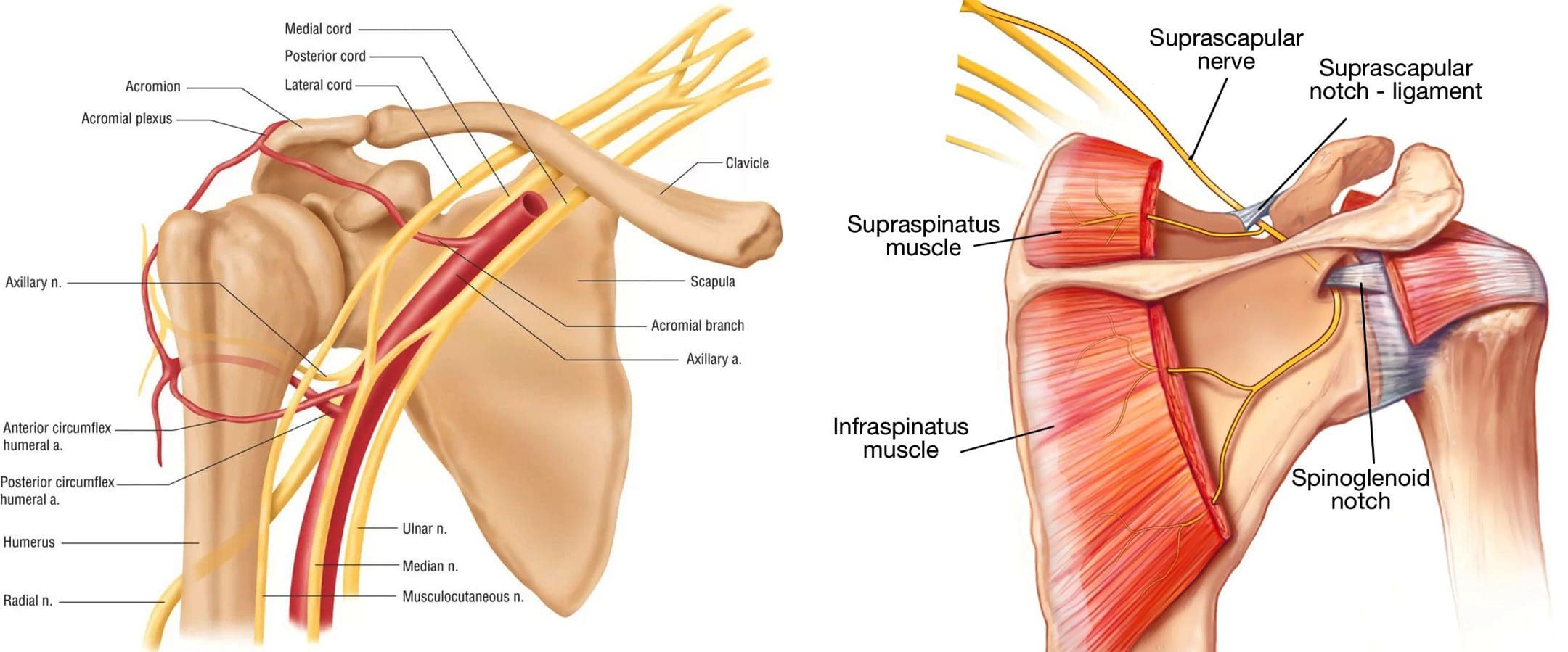
The axillary nerve, or the circumflex nerve, is a peripheral nerve that runs through the shoulder and supports movement and sensation in the upper limbs. It originates in the neck at the brachial plexus, a network of nerves that extends from the neck and upper torso to the shoulders and arms. Its primary purpose is to supply nerve function to the shoulder joint and three muscles in the arm and also innervates some skin in the region.
Anatomy
Except for the cranial nerves, all the body’s nerves branch off from the spinal cord, emerge from between vertebrae and continue to branch off as they travel to various muscles and other structures. The axillary nerve is named after the axilla, the medical name for the armpit. Individuals have two, one on each side. After leaving the spinal column, the axillary nerve runs behind the axillary artery and continues to the shoulder blade’s lower edge of the subscapularis muscle. It winds back and travels down the arm along the posterior humeral circumflex artery, which then passes through the quadrangular space (a small area of the shoulder blade just above the armpit where there is a gap in the muscles that allows nerves and blood vessels to pass through to the arm before it divides into terminal branches, which are:
Anterior Division
Supplies motor innervation to the deltoid’s anterior and middle heads, allowing the arm to abduct or move away from the body.
It winds around the neck of the humerus/funny bone, goes beneath the deltoid muscle, and then connects to the forward edge of the deltoid.
A few small cutaneous branches serve the skin in that area.
Posterior Division
Innervates the teres minor muscles and the lower part of the deltoid.
It enters the deep fascia and becomes the superior lateral cutaneous nerve.
It then wraps around the lower edge of the deltoid, connects to the skin over the lower two-thirds of the muscle, and covers the long head of the triceps brachii.
Articular Branch
Comes from the trunk of the axillary nerve and enters the glenohumeral joint, which is in the shoulder, below the subscapularis muscle.
Anatomical Variations
In a case report, healthcare providers noted an incidence of the nerve branching directly off the upper trunk of the brachial plexus rather than the posterior cord. (Subasinghe S. K. and Goonewardene S. 2016) In this case, it innervated the subscapularis muscle, latissimus dorsi, and the deltoid and teres minor muscles and also had a communicating branch to the posterior cord. Another case documented multiple abnormalities in the course of the axillary nerve in an individual with pain and severely limited shoulder mobility. (Pizzo R. A. et al., 2019) During reverse shoulder arthroplasty, the surgeon discovered that the axillary nerve ran beside the coracoid process instead of underneath and stayed close to the subscapularis muscle instead of traveling through the quadrangular space. The case noted earlier reports of axillary nerves not running through the quadrangular space. In those cases, the nerve pierced the subscapularis muscle or split into branches before reaching the quadrangular space.
Function
The axillary nerve functions as a motor nerve that controls movement and a sensory nerve that controls sensations like touch or temperature.
Motor
As a motor nerve, the axillary nerve innervates three muscles in the arm and includes:
Deltoid
Allows flexing of the shoulder joint and rotating the shoulder inward.
Long Head of the Triceps
It runs down the back of the outer arm, allowing straightening, pulling the upper arm toward the body, or extending it backward.
The radial nerve can also innervate this muscle.
Teres Minor
One of the rotator cuff muscles starts outside the shoulder and runs diagonally along the bottom edge of the shoulder blade.
It works with other muscles to allow for the external rotation of the shoulder joint.
Sensory
In its sensory role, the nerve carries information to the brain from the following:
Glenohumeral joint or the ball-and-socket joint in the shoulder.
The skin on the lower two-thirds of the deltoid muscle through the superior lateral cutaneous branch.
Injuries and Conditions
Problems with the axillary nerve can be caused by injuries anywhere along the arm and shoulder and by disease. Common injuries include:
Dislocations
Of the shoulder joint, which can cause axillary nerve palsy.
Fracture
Of the surgical neck of the humerus.
Compression
This stems from walking with crutches, also known as crutch palsy.
Direct Trauma
This can be from an impact sports, work, automobile accident, collision, or laceration.
Added Pressure
This can be from wearing a cast or splint.
Surgical Accidental Injury
An injury or damage can come from shoulder surgery, especially arthroscopic surgery on the inferior glenoid and capsule.
Quadrangular Space Syndrome
This is where the axillary nerve is compressed where it passes through that space, which is most common in athletes who perform frequent overhead motions)
Nerve Root Damage
Between the fifth and sixth cervical vertebrae, where the nerve emerges from the spinal cord, which can be caused by traction, compression, spinal disc prolapse, or a bulging disc.
Systemic Neurological Disorders
Example – multiple sclerosis
Erb’s Palsy
A condition often is the result of a birth injury called shoulder dystocia, in which a baby’s shoulder/s becomes stuck during childbirth.
Axillary Nerve Palsy
Damage can result in a type of peripheral neuropathy that can cause weakness in the deltoid and teres minor muscles.
This can result in losing the ability to lift the arm away from the body and weakness in various shoulder movements.
If the damage is severe enough, it can cause paralysis of the deltoid and other minor muscles, resulting in flat shoulder deformity, in which individuals cannot lay their shoulders flat when lying down.
Axillary nerve damage also can lead to a change, reduction, or loss of sensation in a small part of the arm just below the shoulder.
Nerve Injury Statistics
Three times more common in men than women.
It may be present in as many as 65% of shoulder injuries.
The risk of injury due to dislocation is significantly increased after age 50.
Tests
If a healthcare provider suspects a problem with axillary nerve function, they’ll test the shoulder’s range of motion and skin sensitivity. A difference in the range of motion between the shoulders can indicate a nerve injury. Individuals may be sent for electromyography and a nerve conduction study to verify nerve palsy. In some cases, an MRI and/or X-rays may be ordered, especially if the cause of possible nerve damage is unknown.
Rehabilitation
Depending on the severity and cause of the injury, non-surgical treatments may be recommended, with surgery as a last resort. Non-surgical treatment can include some combination of immobilization, rest, ice, physical therapy, and anti-inflammatory meds. Physical treatment typically lasts about six weeks and focuses on strengthening and stimulating the muscles to prevent joint stiffness, which can impair long-term function.
Surgery
If conservative treatments don’t work, surgery may be recommended, especially if several months have passed without improvement. Surgical outcomes are generally better if surgery is performed within six months of the injury, and regardless of the time frame, the prognosis is considered positive in about 90% of cases. Surgical procedures performed for axillary nerve dysfunction or injury include:
Neurolysis
This procedure involves targeted damage/degeneration of nerve fibers, interrupts the nerve signals, and eliminates pain while the damaged area heals.
Neurorrhaphy
This procedure stitches a severed nerve back together.
Nerve Grafting
Grafting involves transplanting a portion of another nerve, usually the sural nerve, to reconnect severed nerves.
This helps, especially when the damaged portion is too large to be repaired by neurorrhaphy.
It allows a pathway for signals and encourages the regrowth of nerve axons.
Neurotization or Nerve Transfer
Similar to grafting but used when the nerve is too damaged to heal.
This procedure involves transplanting a healthy but less important nerve, or a portion of a nerve, to replace the damaged one and restore function.
Injury Medical Chiropractic and Functional Medicine Clinic
Injury Medical Chiropractic and Functional Medicine Clinic works with primary healthcare providers and specialists to develop an optimal health and wellness solution. We focus on what works for you to relieve pain, restore function, and prevent injury. Regarding musculoskeletal pain, specialists like chiropractors, acupuncturists, and massage therapists can help mitigate the pain through spinal adjustments that help the body realign itself. They can also work with other medical professionals to integrate a treatment plan to resolve musculoskeletal issues.
Shoulder Pain Chiropractic Treatment
References
Subasinghe, S. K., & Goonewardene, S. (2016). A Rare Variation of the Axillary Nerve Formed as Direct Branch of the Upper Trunk. Journal of clinical and diagnostic research : JCDR, 10(8), ND01–ND2. https://doi.org/10.7860/JCDR/2016/20048.8255
Pizzo, R. A., Lynch, J., Adams, D. M., Yoon, R. S., & Liporace, F. A. (2019). Unusual anatomic variant of the axillary nerve challenging the deltopectoral approach to the shoulder: a case report. Patient safety in surgery, 13, 9. https://doi.org/10.1186/s13037-019-0189-1
Individuals experiencing persistent pain, weakness, numbness, and tingling in the back could be suffering from nerve root encroachment. Could surgery ease nerve compression and improve symptoms for persistent and severe cases?
Surgical Decompression
The pain, weakness, numbness, and tingling associated with nerve root encroachment are usually first treated with non-surgical therapies that include:
Anti-inflammatory medications
Physical therapy
This can be enough to address the irritation of the spinal nerve root. But when cases become severe, surgical decompression may be recommended and necessary. It can be done in a couple of different ways.
Causes and Symptoms
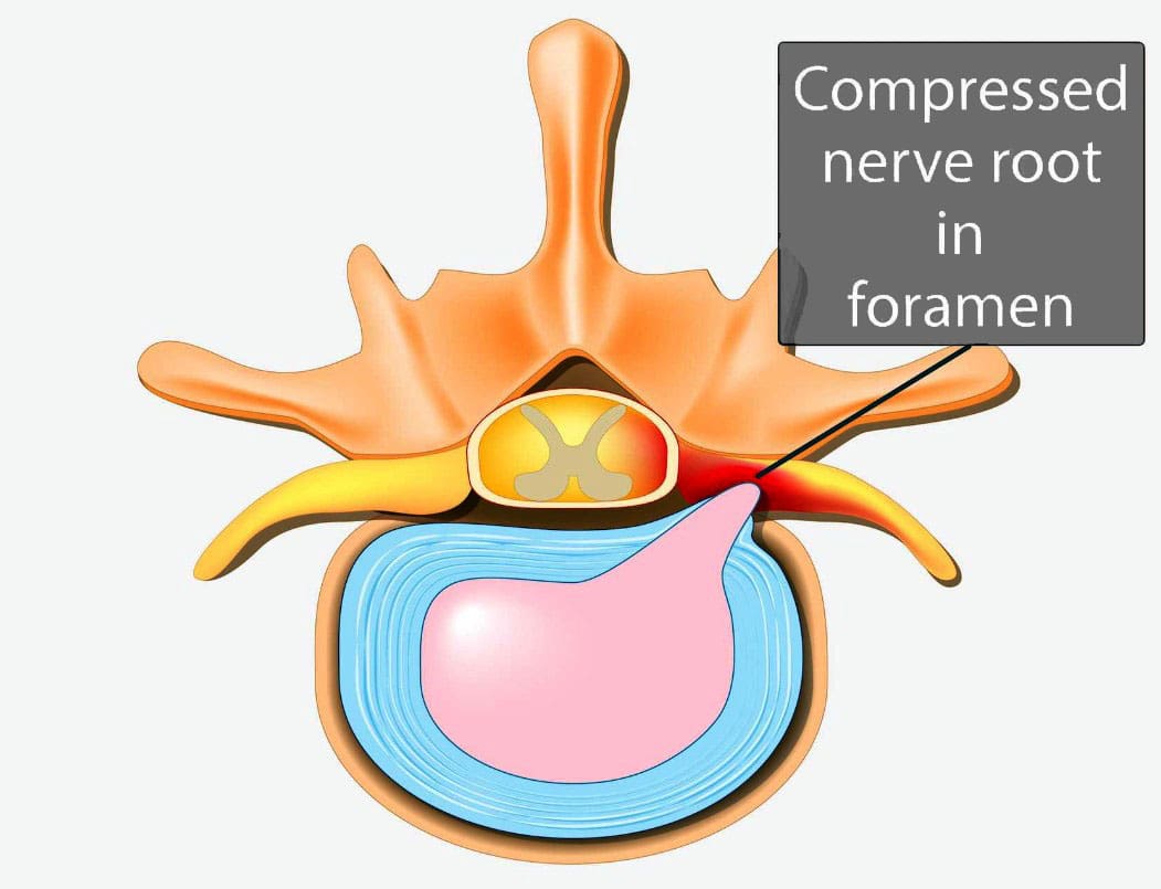
Vertebrae are bones in the spine. Small openings called foramina allow a spinal nerve root to pass through on each side of the vertebra. When nerve root encroachment is present, the spinal nerve root gets compressed, pinched, and trapped, which can cause peripheral symptoms such as numbness, tingling, pain, or weakness to develop. Nerve root encroachment is typically caused by normal aging degenerative wear and tear changes in the vertebrae. (Choi Y. K. 2019) These degenerative changes can include:
Facet joint hypertrophy
Ligament and bone hypertrophy
Disc disorders
Formation of bone spurs or osteophytes.
If these degenerative changes progress, they can encroach and compress a nerve root, leading to peripheral symptoms. (Choi Y. K. 2019)
When Surgery Is Recommended
When symptoms occur, initial treatment will involve:
Physical therapy
Chiropractic realignment
Massage therapies
Rest
Lifestyle adjustments
Nonsteroidal anti-inflammatories – NSAIDs
Corticosteroid injections into the spine
If conservative therapies don’t fully heal or improve symptoms or there are neurological problems like difficulty with balance or walking, then surgery may be recommended. Severe pain that limits normal function is an indication for surgery, and rapidly progressive weakness of the arms and/or legs or signs of cauda equina syndrome are indications for emergency surgery.
Surgery Options
Different types of spinal surgery may be performed. A neurosurgeon will decide the best procedure for each patient based on their case, age, medical conditions, and other factors. Specific spinal surgical decompression depends on what is causing the nerve compression. In most cases, it involves removing bone or tissue to relieve nerve pressure or provide support to stabilize the joint. The most common types of surgical decompression include: (Mayo Clinic Health System, 2022)
Maintain the stability and alignment of the spine.
Improve the stability and alignment of the spine.
Anterior Surgery
The anterior approach to surgery means that the spine is accessed through the anterior/front of the spine. In this surgery, one or more discs and bone spurs may be removed through an incision in the front of the neck. (American Association of Neurological Surgeons, 2024) For example, an anterior cervical discectomy may alleviate pressure on one or more nerve roots in the neck. With an anterior lumbar interbody fusion, a surgeon removes a degenerative disc in the lower spinal area by going through a patient’s lower abdomen. (American Association of Neurological Surgeons, 2024) After the disc is removed, a structural device, usually made of bone, fills the space where it once was. This device encourages bone healing and helps the vertebrae’s bodies fuse.
Posterior Surgery
Posterior surgery means the spine is accessed through the posterior/back of the spine. An example is removing a thickened ligament, bone spur, or disc material in the neck. To do this, a small incision in the back of the neck may be made to remove part of the back of the vertebrae called the lamina. This is called a posterior cervical laminectomy. (American Association of Neurological Surgeons, 2024) A posterior lumbar interbody fusion removes a degenerative disc by going through the back. (American Association of Neurological Surgeons, 2024) Like the anterior approach, a structural device often contains bone to fill the space where the disc once was to fuse the bones.
Potential Risks
As with any surgery, it’s important that the individual and their healthcare provider carefully discuss the benefits and risks. Spinal surgical decompression includes: (Proietti L. et al., 2013)
Bleeding
Blood clots
Surgical site infection
Urinary tract infection
Lung infection
Intestinal blockage
There are also specific risks to the area of the spine being operated on and how it is surgically approached. For example, a cervical anterior procedure may injure the esophagus, trachea, or carotid artery. Likewise, damage to the C5 nerve root/C5 palsy can occur from cervical spinal decompressive surgery. This complication causes weakness, numbness, and pain in the shoulders. (Thompson S. E. et al., 2017) The spinal cord may also be injured during surgery and result in paralysis, although this is rare. (American Association of Neurological Surgeons, 2024)
Injury Medical Chiropractic and Functional Medicine Clinic
Injury Medical Chiropractic and Functional Medicine Clinic works with primary healthcare providers and specialists to develop an optimal health and wellness solution. We focus on what works for you to relieve pain, restore function, and prevent injury. Regarding musculoskeletal pain, specialists like chiropractors, acupuncturists, and massage therapists can help mitigate the pain through spinal adjustments that help the body realign itself. They can also work with other medical professionals to integrate a treatment plan to resolve musculoskeletal issues.
The Non-Surgical Solution
References
Choi Y. K. (2019). Lumbar foraminal neuropathy: an update on non-surgical management. The Korean journal of pain, 32(3), 147–159. https://doi.org/10.3344/kjp.2019.32.3.147
Mayo Clinic Health System. (2022). Decompress and stabilize: understanding types of back surgery. Speaking of Health. https://www.mayoclinichealthsystem.org/hometown-health/speaking-of-health/understanding-types-of-back-surgery
American Association of Neurological Surgeons. (2024). Cervical spine. https://www.aans.org/patients/conditions-treatments/cervical-spine/
American Association of Neurological Surgeons. (2024). Lumbar spinal stenosis. https://www.aans.org/patients/conditions-treatments/lumbar-spinal-stenosis/
Proietti, L., Scaramuzzo, L., Schiro’, G. R., Sessa, S., & Logroscino, C. A. (2013). Complications in lumbar spine surgery: A retrospective analysis. Indian journal of orthopaedics, 47(4), 340–345. https://doi.org/10.4103/0019-5413.114909
Thompson, S. E., Smith, Z. A., Hsu, W. K., Nassr, A., Mroz, T. E., Fish, D. E., Wang, J. C., Fehlings, M. G., Tannoury, C. A., Tannoury, T., Tortolani, P. J., Traynelis, V. C., Gokaslan, Z., Hilibrand, A. S., Isaacs, R. E., Mummaneni, P. V., Chou, D., Qureshi, S. A., Cho, S. K., Baird, E. O., … Riew, K. D. (2017). C5 Palsy After Cervical Spine Surgery: A Multicenter Retrospective Review of 59 Cases. Global spine journal, 7(1 Suppl), 64S–70S. https://doi.org/10.1177/2192568216688189
Can determining whether arm numbness occurs suddenly or gradually and whether there are other symptoms help healthcare providers diagnose and treat the condition?
Arm Numbness
Arm numbness or tingling are common symptoms that various medical conditions can cause. Numbness can be caused by a sudden health emergency, nerve disorder, or nutritional deficiency. (National Institute of Neurological Disorders and Stroke, 2024) Sometimes, this symptom results from an arm falling asleep and could resolve after just a few minutes. The sensation may be temporary, caused by something like sleeping in the wrong position. However, arm numbness and tingling may also be caused by neuropathy and chronic and progressive nerve damage and can also suddenly occur due to serious conditions, such as a heart attack or a stroke.
Circulation Issues
Deficient blood circulation in the arm could cause numbness and tingling as the nerves cannot receive enough oxygen. Conditions can interfere with normal blood flow and include: (Bryan L. and Singh A. 2024)
Atherosclerosis – plaque buildup in the arteries that may require medication or surgery.
Severe frostbite can damage the blood vessels but can be resolved with proper warming and wound care.
Vasculitis – is inflammation of the blood vessels that can be treated with medication.
Sleeping Position
A common example of sudden numbness and tingling is the feeling that the arm has fallen asleep. This usually occurs after sleeping awkwardly or leaning on the arm for a long time. Known as paresthesia, this sensation is related to the compression or irritation of nerves. (Bryan L. and Singh A. 2024) Sleeping in certain positions has been associated with nerve compression, especially when the hands or wrists are tucked or curled under the body, as well as maintaining proper spine alignment when sleeping, is the best way to prevent arm numbness.
Nerve Injuries and Conditions
Numbness that persists may be related to an injury or underlying health problem that affects the brachial plexus, a group of nerves that runs from the lower neck to the upper shoulders and controls movement and sensation in the arms. (Mount Sinai, 2022) Possible injuries that affect these nerves include: (Smith, S. M. et al., 2021)
A herniated disc caused by aging or trauma causes the disc to leak out and press on the nerve root.
Many medications, like gabapentin, can be used to alleviate the sensations of arm numbness.
Some medications can cause numbness as a side effect.
Some medications can cause complications, and arm numbness could be a symptom of those complications.
Inform healthcare providers about medications being taken to determine the relationship between them and any sensory changes.
Vitamin Deficiency
Peripheral neuropathy can also be caused by nutritional deficiencies and vitamin imbalances, which can damage nerves and cause sensation loss in the left or right arm. The most common sources are vitamin B12 deficiency and excess vitamin B6. (National Institute of Neurological Disorders and Stroke, 2024) Excessive alcohol consumption and other disorders that affect nutritional intake can also lead to nerve damage.
Injury Medical Chiropractic and Functional Medicine Clinic
At Injury Medical Chiropractic and Functional Medicine Clinic, our areas of practice include Chronic Pain, Personal Injury, Auto Accident Care, Work Injuries, Back Injury, Low Back Pain, Neck Pain, Migraine Headaches, Sports Injuries, Severe Sciatica, Scoliosis, Complex Herniated Discs, Fibromyalgia, Chronic Pain, Complex Injuries, Stress Management, Wellness & Nutrition, Functional Medicine Treatments, and in-scope care protocols. We focus on what works for you to relieve pain and restore function. If other treatment is needed, individuals will be referred to a clinic or physician best suited to their injury, condition, and/or ailment.
Chiropractic Care: The Natural Way to Recover From Injuries
References
National Institute of Neurological Disorders and Stroke. (2024). Paresthesia. Retrieved from https://www.ninds.nih.gov/health-information/disorders/glossary-neurological-terms#paresthesia
Bryan, L., Singh, A. Sleep Foundation. (2024). Numbness in Hands While Sleeping: Causes and Remedies. https://www.sleepfoundation.org/physical-health/numbness-in-hands-while-sleeping
Mount Sinai. (2022). Brachial plexopathy. https://www.mountsinai.org/health-library/diseases-conditions/brachial-plexopathy
Smith, S. M., McMullen, C. W., & Herring, S. A. (2021). Differential Diagnosis for the Painful Tingling Arm. Current sports medicine reports, 20(9), 462–469. https://doi.org/10.1249/JSR.0000000000000877
National Heart, Lung, and Blood Institute. (2023). Vasculitis. Retrieved from https://www.nhlbi.nih.gov/health/vasculitis/symptoms#:~:text=Nerve%20problems%2C%20including%20numbness%2C%20tingling,can%20also%20occur%20with%20vasculitis.
Centers for Disease Control and Prevention. (2024). Stroke signs and symptoms. Retrieved from https://www.cdc.gov/stroke/signs-symptoms/?CDC_AAref_Val=https://www.cdc.gov/stroke/signs_symptoms.htm
National Heart, Lung, and Blood Institute. (2022). What Is a Heart Attack? Retrieved from https://www.nhlbi.nih.gov/health/heart-attack
National Institute of Arthritis and Musculoskeletal and Skin Diseases. (2023). Spinal Stenosis Basics. Retrieved from https://www.niams.nih.gov/health-topics/spinal-stenosis/basics/symptoms-causes
Senderovich, H., & Jeyapragasan, G. (2018). Is there a role for combined use of gabapentin and pregabalin in pain control? Too good to be true?. Current medical research and opinion, 34(4), 677–682. https://doi.org/10.1080/03007995.2017.1391756
National Institute of Neurological Disorders and Stroke. (2024). Peripheral Neuropathy. Retrieved from https://www.ninds.nih.gov/health-information/disorders/peripheral-neuropathy
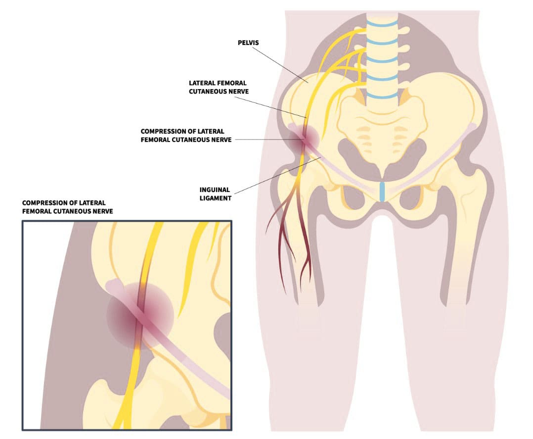
Individuals experiencing pain, numbness, tingling, or a burning sensation in the front and outer thigh could have meralgia paresthetica, a nerve entrapment. Can understanding the condition help healthcare providers develop an effective treatment plan?
Meralgia Paresthetica
Meralgia paresthetica, or MP, is also known as Bernhardt-Roth syndrome, lateral femoral cutaneous nerve syndrome, or lateral femoral cutaneous neuralgia. It occurs when the lateral femoral cutaneous nerve, a sensory nerve that passes over the brim of the pelvis and down the front of the thigh, becomes compressed. The nerve supplies information about sensations over the front and outside of the thigh. This can happen for several reasons, including:
Recent hip injuries, such as from a motor vehicle collision/accident.
Repetitive hip activities, like cycling.
Pregnancy
Weight gain
Wearing tight clothing.
The nerve entrapment condition causes tingling, numbness, and burning pain in the front and/or outer thigh.
Causes
There can be several different causes of this condition, but it is frequently seen in pregnancy, sudden weight gain, wearing tight clothing or belts, and other conditions. (Ivins G. K. 2000) Sometimes, meralgia paresthetica can be caused by medical procedures. For example, the condition can present after an individual has surgery and is in an unusual position for a long period of time, where there is direct external pressure on the nerve. Also, the nerve can become damaged during a surgical procedure. (Cheatham S. W. et al., 2013) This can occur when a bone graft is obtained from the pelvis or anterior hip replacement surgery.
Sensitivity to lightly touching the outside of the thigh.
Worsening of symptoms with certain positions.
Increased symptoms when wearing belts, work belts, or tight-waist clothes.
The symptoms may come and go or be persistent. Some individuals are hardly noticeable and do not impact their lives or activities, while others can be very bothersome and cause significant pain. (Scholz C. et al., 2023)
Treatment
Treatment depends on how long the injury has been present and the frequency and severity of the condition.
Clothing Modifications
If the cause is due to tight clothing, belts, or work belts, then garment modification should alleviate symptoms.
If recent weight gain is thought to contribute to the condition, then a weight loss program may be recommended.
Cortisone Injections
If simple steps do not relieve symptoms, a cortisone injection around the nerve area may be recommended. The goal is to reduce inflammation that contributes to nerve pressure (Houle S. 2012) . Cortisone injections may be a definitive treatment or a temporary treatment.
Chiropractic
Chiropractic care can be an effective, natural, and safe treatment. Adjustments can help relieve pressure on the lateral femoral cutaneous nerve (LFCN) by realigning the spine and restoring nerve function. Chiropractors may also use soft tissue therapies, such as massage, to relieve muscle tension and support the body’s healing process. Other chiropractic techniques that may be used include:
A chiropractic treatment program may include 10–15 treatments over 6–8 weeks, but the number of treatments needed will vary from person to person. If there’s no noticeable progress after 3–4 weeks, it may be time to consult a specialist or surgeon.
Surgery
Surgery is rarely necessary. However, a surgical procedure may be considered when all conservative treatments fail to provide relief. (Schwaiger K. et al., 2018) A surgeon dissects and identifies the nerve, looks for compression locations, and tries to free the nerve from any areas where it may be pinched. Alternatively, some surgeons transect/cut the nerve so it no longer causes problems. If the transection procedure is performed, there will be a permanent area of numbness over the front of the thigh.
Injury Medical Chiropractic and Functional Medicine Clinic works with primary healthcare providers and specialists to develop a customized treatment plan to relieve pain, treat injuries, improve flexibility, mobility, and agility, and help individuals return to optimal function. If other treatments are needed, Dr. Jimenez has teamed up with top surgeons, clinical specialists, medical researchers, and rehabilitation providers to provide the most effective treatments.
Chiropractic Care for Leg Instability
References
Ivins G. K. (2000). Meralgia paresthetica, the elusive diagnosis: clinical experience with 14 adult patients. Annals of surgery, 232(2), 281–286. https://doi.org/10.1097/00000658-200008000-00019
Cheatham, S. W., Kolber, M. J., & Salamh, P. A. (2013). Meralgia paresthetica: a review of the literature. International journal of sports physical therapy, 8(6), 883–893.
Chung, K. H., Lee, J. Y., Ko, T. K., Park, C. H., Chun, D. H., Yang, H. J., Gill, H. J., & Kim, M. K. (2010). Meralgia paresthetica affecting parturient women who underwent cesarean section -A case report-. Korean journal of anesthesiology, 59 Suppl(Suppl), S86–S89. https://doi.org/10.4097/kjae.2010.59.S.S86
Scholz, C., Hohenhaus, M., Pedro, M. T., Uerschels, A. K., & Dengler, N. F. (2023). Meralgia Paresthetica: Relevance, Diagnosis, and Treatment. Deutsches Arzteblatt international, 120(39), 655–661. https://doi.org/10.3238/arztebl.m2023.0170
Hosley, C. M., & McCullough, L. D. (2011). Acute neurological issues in pregnancy and the peripartum. The Neurohospitalist, 1(2), 104–116. https://doi.org/10.1177/1941875211399126
Houle S. (2012). Chiropractic management of chronic idiopathic meralgia paresthetica: a case study. Journal of chiropractic medicine, 11(1), 36–41. https://doi.org/10.1016/j.jcm.2011.06.008
Schwaiger, K., Panzenbeck, P., Purschke, M., Russe, E., Kaplan, R., Heinrich, K., Mandal, P., & Wechselberger, G. (2018). Surgical decompression of the lateral femoral cutaneous nerve (LFCN) for Meralgia paresthetica treatment: Experimental or state of the art? A single-center outcome analysis. Medicine, 97(33), e11914. https://doi.org/10.1097/MD.0000000000011914
Can understanding the location of the funny bone and how pain can be managed after injury help expedite recovery and prevention for individuals who have hit their funny bone?
Elbow Funny Bone Nerve Injury
Behind the elbow is an area known as the “funny bone,” where the ulnar nerve has less tissue and bone protection. This is where part of the ulnar nerve passes around the back of the elbow. Because less tissue and bone protect the nerve in this area, taking a hit like bumping into something can cause an electric shock-like pain and a tingling sensation down the arm and to the outside fingers typical of an irritated nerve. Most injuries to the funny bone resolve quickly, and the pain disappears after a few seconds or minutes, but sometimes, an ulnar nerve injury can lead to more persistent symptoms.
Anatomy
The funny bone is not a bone but the ulnar nerve. The nerve runs down the arm, passing around the back of the elbow. (Dimitrova, A. et al., 2019) Because the ulnar nerve is on top of the elbow and there is very little fatty cushion, lightly bumping this spot can cause pain and tingling sensations down the forearm. Three bones comprise the junction of the elbow that include:
Humerus – arm bone
Ulna and radius – forearm bones
The humerus has a groove that protects and holds the ulnar nerve as it passes behind the joint. This is where the nerve can be injured or irritated when the nerve is hit or pinched against the end of the bone, causing the funny bone pain.
Electrical Pain Sensation
When hitting the ulnar nerve or funny bone where the ulnar nerve provides sensation, pain, and electrical/tingling sensations are experienced from the forearm to the outside fingers. This part of the arm and hand is called the ulnar nerve distribution. (American Academy of Orthopaedic Surgeons. 2024) The ulnar nerve provides sensation into most of the pinky finger and about half of the ring finger. Other nerves, including the median and radial nerve, supply sensation to the rest of the hand.
Treatment
Usually, a sharp jolt to the elbow quickly resolves. Some recommendations to help symptoms improve faster include:
Shaking the forearm and hand out.
Straightening out and bending the elbow to stretch the nerve.
Decreasing mobility of the elbow.
Applying ice to the area.
Taking anti-inflammatory medications.
Treating Long-Lasting Pain
In rare circumstances, injuries to the ulnar nerve can cause more persistent symptoms, a condition known as cubital tunnel syndrome. Cubital tunnel syndrome can happen after an injury or from elbow overuse. Individuals with cubital tunnel syndrome may benefit from wearing a splint at night. Standard-sized splints can be ordered online, but most are fabricated by an occupational or hand therapist. If symptoms become more long-lasting, surgery may be recommended to relieve pressure and tension on the ulnar nerve (American Academy of Orthopaedic Surgeons, 2024). The procedure decompresses the nerve by relieving any tight constrictions around it and releasing them. In severe cases, the nerve is repositioned to an area that doesn’t place as much pressure on the nerve, known as an ulnar nerve transposition.
Injury Medical Chiropractic and Functional Medicine Clinic works with primary healthcare providers and specialists to develop an optimal health and wellness solution that helps individuals return to normal. Our providers create personalized care plans for each patient, including Functional Medicine, Acupuncture, Electro-Acupuncture, and Sports Medicine principles through an integrated approach to treat injuries and chronic pain syndromes to improve ability through flexibility, mobility, and agility programs to relieve pain. If other treatment is needed, Dr. Jimenez has teamed up with top surgeons, clinical specialists, medical researchers, and rehabilitation providers to provide the most effective treatments.
Chiropractic Treatment For Carpal Tunnel Syndrome
References
Dimitrova, A., Murchison, C., & Oken, B. (2019). Local effects of acupuncture on the median and ulnar nerves in patients with carpal tunnel syndrome: a pilot mechanistic study protocol. Trials, 20(1), 8. https://doi.org/10.1186/s13063-018-3094-5
American Academy of Orthopaedic Surgeons. (2024). Ulnar nerve entrapment at the elbow. https://orthoinfo.aaos.org/en/diseases–conditions/ulnar-nerve-entrapment-at-the-elbow-cubital-tunnel-syndrome/
IFM's Find A Practitioner tool is the largest referral network in Functional Medicine, created to help patients locate Functional Medicine practitioners anywhere in the world. IFM Certified Practitioners are listed first in the search results, given their extensive education in Functional Medicine