Can physical therapies help treat a high steppage gait from injury or medical conditions and restore normal gait patterns for individuals who have or are developing one?
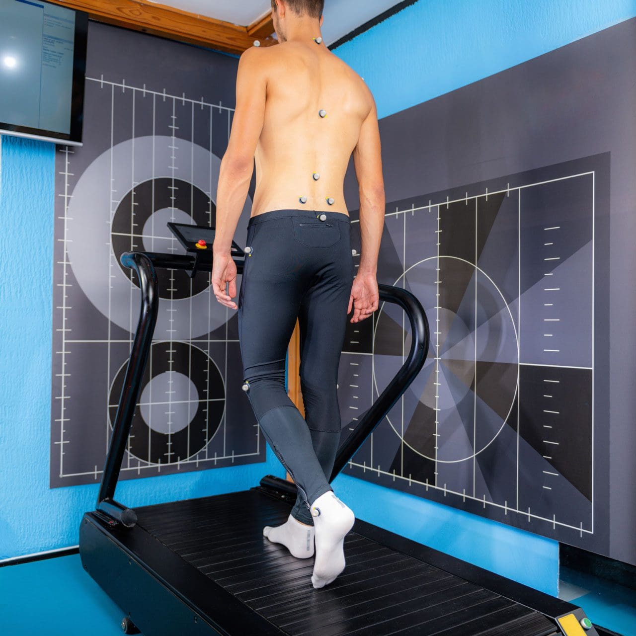
Walking or gait anthropometric analysis on a treadmill
Neuropathic Gait
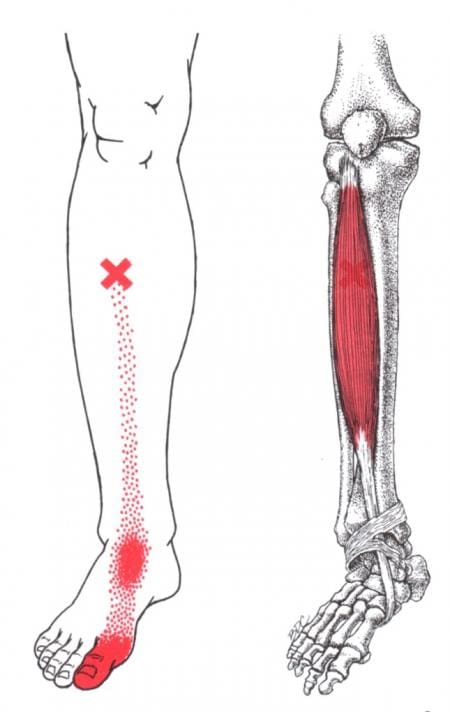
Neuropathic gait, aka equine or high steppage gait, is a type of walking abnormality that causes individuals to raise their hips to lift their legs higher than normal. It occurs when individuals have a foot drop or ankle equinus due to loss of dorsiflexion. As a result, the foot hangs with the toes pointing down, causing the toes to drag while walking. The foot may appear floppy when it drops. Foot drop is caused by weakness or paralysis of the anterior tibialis muscle in front of the shin bone. The anterior tibialis muscle contracts to help flex the foot and ankle while walking, ensuring the foot clears the floor and doesn’t drag. Individuals with anterior tibialis weakness or paralysis may have a neuropathic gait and excessively bend the hip and knee while stepping forward, lifting their leg high off the floor to clear the foot to avoid tripping. A physical therapy team can help with a high steppage gait pattern after illness or injury.
Causes
Conditions that can cause anterior tibialis weakness or paralysis and a high steppage gait pattern include:
Sciatica
Pain caused by compression or irritation of the sciatic nerve starts in the lower back and travels down the back of the leg. (McCabe, F. J., McCabe, J. P. 2016)
Peroneal Nerve Injury
Damage to the peroneal nerve branches from the sciatic nerve that help move the lower leg and foot. (Johns Hopkins Medicine. 2024)
Multiple Sclerosis
An autoimmune disease that damages nerve cells in the brain and spinal cord. (Taylor, P. N. et al., 2016)
Balance exercises will help improve overall proprioception, or the sense of the body’s position and movement.
Neuromuscular electrical stimulation, or NMES, can help improve the function of the muscle. (Hollis, S., McClure, P. 2017)
The electrical stimulation artificially contracts the muscle to restore proper function.
For anterior tibialis weakness caused by sciatica, back decompression exercises may be prescribed to relieve pressure off the sciatic nerve.
The exercises release the nerve to restore normal signal transmission up and down the nerve in the lower back.
Neuromuscular electrical stimulation may also be used to help improve muscle function.
Assistive Walking Devices
A therapist may suggest using an assistive device to help the patient walk properly. This could include a wheeled walker or a quad cane. A temporary solution to anterior tibialis weakness is to elevate the foot while walking with an elastic band. Tie a band around the leg below the knee and secure it around the ball of the foot. When swinging the leg forward, the band pulls the foot up. Using it as a temporary solution may help maintain safe mobility. Sometimes, paralysis of the anterior tibialis muscle can become permanent. In this case, individuals may benefit from a special brace called an ankle-foot orthosis. The brace helps to lift the foot and toes off the ground.
For individuals concerned about losing their balance and falling, there are ways to improve walking patterns to stay safe. A healthcare provider may recommend physical therapy to correct gait, strengthen the anterior tibialis muscle, improve balance, and educate on injury prevention. Individuals should discuss symptoms and conditions with a primary physician, healthcare provider, or specialist to guide them in the right direction and determine the best treatment.
Injury Medical Chiropractic and Functional Medicine Clinic uses an integrated approach personalized to the individual that focuses on what works for them and treats injuries and chronic pain syndromes through personalized care plans that improve ability through flexibility, mobility, and agility programs to relieve pain. If other treatment is needed, Dr. Jimenez has teamed up with top surgeons, clinical specialists, medical researchers, and rehabilitation providers to provide the most effective treatments.
Control Foot Motion and Posture
References
McCabe, F. J., & McCabe, J. P. (2016). An Unusual Presentation of Right-Sided Sciatica with Foot Drop. Case reports in orthopedics, 2016, 9024368. https://doi.org/10.1155/2016/9024368
Kaykisiz, E. K., & Unluer, E. E. (2017). An Unexpected Reason for Isolated Foot Drop: Acute Stroke. Pakistan journal of medical sciences, 33(5), 1288–1290. https://doi.org/10.12669/pjms.335.13593
Taylor, P. N., Wilkinson Hart, I. A., Khan, M. S., & Slade-Sharman, D. E. (2016). Correction of Footdrop Due to Multiple Sclerosis Using the STIMuSTEP Implanted Dropped Foot Stimulator. International journal of MS care, 18(5), 239–247. https://doi.org/10.7224/1537-2073.2015-038
Hollis, S., & McClure, P. (2017). Intramuscular Electrical Stimulation for Muscle Activation of the Tibialis Anterior After Surgical Repair: A Case Report. The Journal of orthopaedic and sports physical therapy, 47(12), 965–969. https://doi.org/10.2519/jospt.2017.7368
For individuals experiencing pelvic pain, it could be a disorder of the pudendal nerve known as pudendal neuropathy or neuralgia that leads to chronic pain. The condition can be caused by pudendal nerve entrapment, where the nerve becomes compressed or damaged. Can knowing the symptoms help healthcare providers correctly diagnose the condition and develop an effective treatment plan?
Pudendal Neuropathy
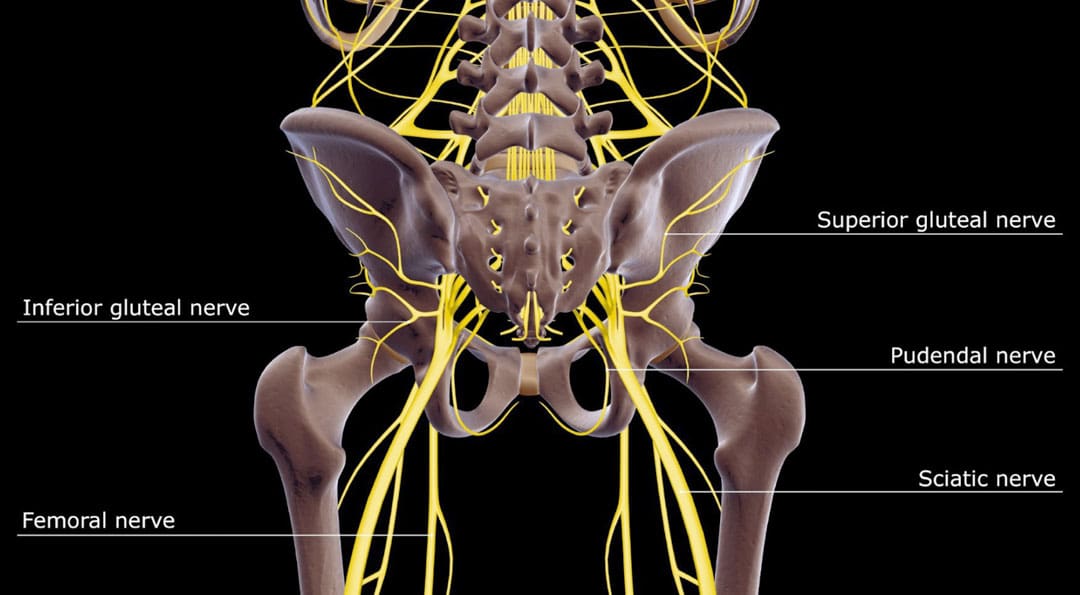
The pudendal nerve is the main nerve that serves the perineum, which is the area between the anus and the genitalia – the scrotum in men and the vulva in women. The pudendal nerve runs through the gluteus muscles/buttocks and into the perineum. It carries sensory information from the external genitalia and the skin around the anus and perineum and transmits motor/movement signals to various pelvic muscles. (Origoni, M. et al., 2014) Pudendal neuralgia, also referred to as pudendal neuropathy, is a disorder of the pudendal nerve that can lead to chronic pelvic pain.
Causes
Chronic pelvic pain from pudendal neuropathy can be caused by any of the following (Kaur J. et al., 2024)
Excessive sitting on hard surfaces, chairs, bicycle seats, etc. Bicyclists tend to develop pudendal nerve entrapment.
Trauma to the buttocks or pelvis.
Childbirth.
Diabetic neuropathy.
Bony formations that push against the pudendal nerve.
Thickening of ligaments around the pudendal nerve.
Symptoms
Pudendal nerve pain can be described as stabbing, cramping, burning, numbness, or pins and needles and can present (Kaur J. et al., 2024)
In the perineum.
In the anal region.
In men, pain in the scrotum or penis.
In women, pain in the labia or vulva.
During intercourse.
When urinating.
During a bowel movement.
When sitting and goes away after standing up.
Because the symptoms are often hard to distinguish, pudendal neuropathy can often be hard to differentiate from other types of chronic pelvic pain.
Cyclist’s Syndrome
Prolonged sitting on a bicycle seat can cause pelvic nerve compression, which can lead to chronic pelvic pain. The frequency of pudendal neuropathy (chronic pelvic pain caused by entrapment or compression of the pudendal nerve) is often referred to as Cyclist’s Syndrome. Sitting on certain bicycle seats for long periods places significant pressure on the pudendal nerve. The pressure can cause swelling around the nerve, which causes pain and, over time, can lead to nerve trauma. Nerve compression and swelling can cause pain described as burning, stinging, or pins and needles. (Durante, J. A., and Macintyre, I. G. 2010) For individuals with pudendal neuropathy caused by bicycling, symptoms can appear after prolonged biking and sometimes months or years later.
Take breaks at least 20–30 seconds after each 20 minutes of riding.
While riding, change positions frequently.
Stand up to pedal periodically.
Take time off between riding sessions and races to rest and relax the pelvic nerves. 3–10 day breaks can help in recovery. (Durante, J. A., and Macintyre, I. G. 2010)
If pelvic pain symptoms are barely starting to develop, rest and see a healthcare provider or specialist for an examination.
Seat
Use a soft, wide seat with a short nose.
Have the seat level or tilted slightly forward.
Seats with cutout holes place more pressure on the perineum.
If numbness or pain is present, try a seat without holes.
Bike Fitting
Adjust the seat height so the knee is slightly bent at the bottom of the pedal stroke.
The body’s weight should rest on the sitting bones/ischial tuberosities.
Keeping the handlebar height below the seat can reduce pressure.
The Triathlon bike’s extreme-forward position should be avoided.
A more upright posture is better.
Mountain bikes have been associated with an increased risk of erectile dysfunction than road bikes.
Shorts
Wear padded bike shorts.
Treatments
A healthcare provider may use a combination of treatments.
The neuropathy can be treated with rest if the cause is excessive sitting or cycling.
Injury Medical Chiropractic and Functional Medicine Clinic care plans and clinical services are specialized and focused on injuries and the complete recovery process. Our areas of practice include Wellness and nutrition, Chronic Pain, Personal Injury, Auto Accident Care, Work Injuries, Back Injury, Low Back Pain, Neck Pain, Migraine Headaches, Sports Injuries, severe sciatica, Scoliosis, Complex Herniated Discs, Fibromyalgia, Chronic Pain, Complex Injuries, Stress Management, and Functional Medicine Treatments. If the individual requires other treatment, they will be referred to a clinic or physician best suited for their condition, as Dr. Jimenez has teamed with the top surgeons, clinical specialists, medical researchers, therapists, trainers, and premiere rehabilitation providers.
Pregnancy and Sciatica
References
Origoni, M., Leone Roberti Maggiore, U., Salvatore, S., & Candiani, M. (2014). Neurobiological mechanisms of pelvic pain. BioMed research international, 2014, 903848. https://doi.org/10.1155/2014/903848
Kaur, J., Leslie, S. W., & Singh, P. (2024). Pudendal Nerve Entrapment Syndrome. In StatPearls. https://www.ncbi.nlm.nih.gov/pubmed/31334992
Durante, J. A., & Macintyre, I. G. (2010). Pudendal nerve entrapment in an Ironman athlete: a case report. The Journal of the Canadian Chiropractic Association, 54(4), 276–281.
Chiaramonte, R., Pavone, P., & Vecchio, M. (2021). Diagnosis, Rehabilitation and Preventive Strategies for Pudendal Neuropathy in Cyclists, A Systematic Review. Journal of functional morphology and kinesiology, 6(2), 42. https://doi.org/10.3390/jfmk6020042
For individuals who have exhausted all other treatment options for low back pain and nerve root compression, can laser spine surgery help alleviate nerve compression and provide long-lasting pain relief?
Laser Spine Surgery
Laser spine surgery is a minimally invasive surgical procedure that uses a laser to cut through and remove spinal structures that are compressing nerves and causing intense pain. The minimally invasive procedure often results in less pain, tissue damage, and faster recovery than more extensive surgeries.
How It Works
Minimally invasive procedures result in less scarring and damage to surrounding structures, often reducing pain symptoms and a shorter recovery time. (Stern, J. 2009) Small incisions are made to access spinal column structures. With open-back surgery, a large incision is made down the back to access the spine. The surgery differs from other surgeries in that a laser beam, rather than other surgical instruments, is used to cut structures in the spine. However, the initial incision through the skin is made with a surgical scalpel. Laser is an acronym for Light Amplification Stimulated by Emission of Radiation. A laser can generate intense heat to cut through soft tissues, especially those with a high water content, like spinal column discs. (Stern, J. 2009) For many spine surgeries, the laser cannot be used to cut through bone as it generates instant sparks that can damage surrounding structures. Rather, laser spine surgery is primarily used to perform a discectomy, which is a surgical technique that removes a portion of a bulging or herniated disc that is pushing against the surrounding nerve roots, causing nerve compression and sciatic pain. (Stern, J. 2009)
Surgical Risks
Laser spine surgery may help resolve the cause of nerve root compression, but there is an increased risk of damage to nearby structures. Associated risks include: (Brouwer, P. A. et al., 2015)
Infection
Bleeding
Blood clots
Remaining symptoms
Returning symptoms
Further nerve damage
Damage to the membrane around the spinal cord.
Need for additional surgery
A laser beam is not precise like other surgical tools and requires practiced mastery and control to avoid damage to the spinal cord and nerve roots. (Stern, J. 2009) Because lasers cannot cut through bone, other surgical instruments are often used around corners and at different angles because they are more efficient and allow greater accuracy. (Atlantic Brain and Spine, 2022)
Purpose
Laser spine surgery is performed to remove structures that are causing nerve root compression. Nerve root compression is associated with the following conditions (Cleveland Clinic. 2018)
Bulging discs
Herniated discs
Sciatica
Spinal stenosis
Spinal cord tumors
Nerve roots that are injured or damaged and constantly send chronic pain signals can be ablated with laser surgery, known as nerve ablation. The laser burns and destroys the nerve fibers. (Stern, J. 2009) Because laser spine surgery is limited in treating certain spinal disorders, most minimally invasive spine procedures do not use a laser. (Atlantic Brain and Spine. 2022)
Preparation
The surgical team will provide more detailed instructions on what to do in the days and hours before surgery. To promote optimal healing and a smooth recovery, it is recommended that the patient stay active, eat a healthy diet, and stop smoking prior to the operation. Individuals may need to stop taking certain medications to prevent excess bleeding or interaction with anesthesia during the operation. Inform the healthcare provider about all prescriptions, over-the-counter drugs, and supplements being taken.
Laser spine surgery is an outpatient procedure at a hospital or outpatient surgical center. The patient will likely go home on the same day of the operation. (Cleveland Clinic. 2018) Patients cannot drive to or from the hospital before or after their surgery, so arrange for family or friends to provide transportation. Minimizing stress and prioritizing healthy mental and emotional well-being is important to lowering inflammation and aiding recovery. The healthier the patient goes into surgery, the easier the recovery and rehabilitation will be.
Expectations
The surgery will be decided by the patient and healthcare provider and scheduled at a hospital or outpatient surgical center. Arrange for a friend or family member to drive to the surgery and home.
Before Surgery
The patient will be taken to a pre-operative room and asked to change into a gown.
The patient will undergo a brief physical examination and answer questions about medical history.
The patient lies on a hospital bed, and a nurse inserts an IV to deliver medication and fluids.
The surgical team will use the hospital bed to transport the patient in and out of the operating room.
The surgical team will assist the patient in getting onto the operating table, and the patient will be administered anesthesia.
The patient may receive general anesthesia, which will cause the patient to sleep for the surgery, or regional anesthesia, injected into the spine to numb the affected area. (Cleveland Clinic. 2018)
The surgical team will sterilize the skin where the incision will be made.
An antiseptic solution will be used to kill bacteria and prevent the risk of infection.
Once sanitized, the body will be covered with sterilized linens to keep the surgical site clean.
During Surgery
For a discectomy, the surgeon will make a small incision less than one inch in length with a scalpel along the spine to access the nerve roots.
A surgical tool called an endoscope is a camera inserted into the incision to view the spine. (Brouwer, P. A. et al., 2015)
Once the problematic disc portion causing the compression is located, the laser is inserted to cut through it.
The cut disc portion is removed, and the incision site is sutured.
After Surgery
After surgery, the patient is brought to a recovery room, where vital signs are monitored as the effects of the anesthesia wear off.
Once stabilized, the patient can usually go home one or two hours after the operation.
The surgeon will determine when the individual is clear to resume driving.
Recovery
Following a discectomy, the individual can return to work within a few days to a few weeks, depending on the severity, but it can take up to three months to return to normal activities. Length of recovery can range from two to four weeks or less to resume a sedentary job or eight to 12 weeks for a more physically demanding job that requires heavy lifting. (University of Wisconsin School of Medicine and Public Health, 2021) During the first two weeks, the patient will be given restrictions to facilitate the spine’s healing until it becomes more stable. Restrictions can include: (University of Wisconsin School of Medicine and Public Health, 2021)
No bending, twisting, or lifting.
No strenuous physical activity, including exercise, housework, yard work, and sex.
No alcohol in the initial stage of recovery or while taking narcotic pain medications.
No driving or operating a motor vehicle until discussed with the surgeon.
The healthcare provider may recommend physical therapy to relax, strengthen, and maintain musculoskeletal health. Physical therapy may be two to three times weekly for four to six weeks.
Process
Optimal recovery recommendations include:
Getting enough sleep, at least seven to eight hours.
Maintaining a positive attitude and learning how to cope and manage stress.
Maintaining body hydration.
Following the exercise program as prescribed by the physical therapist.
Practicing healthy posture with sitting, standing, walking, and sleeping.
Staying active and limiting the amount of time spent sitting. Try to get up and walk every one to two hours during the day to stay active and prevent blood clots. Gradually increase the amount of time or distance as recovery progresses.
Do not push to do too much too soon. Overexertion can increase pain and delay recovery.
Learning correct lifting techniques to utilize the core and leg muscles to prevent increased pressure on the spine.
Discuss treatment options for managing symptoms with a healthcare provider or specialist to determine if laser spine surgery is appropriate. Injury Medical Chiropractic and Functional Medicine Clinic care plans and clinical services are specialized and focused on injuries and the complete recovery process. Dr. Jimenez has teamed with the top surgeons, clinical specialists, medical researchers, therapists, trainers, and premiere rehabilitation providers. We focus on restoring normal body functions after trauma and soft tissue injuries using Specialized Chiropractic Protocols, Wellness Programs, Functional and integrative Nutrition, Agility and mobility Fitness Training, and Rehabilitation Systems for all ages. Our areas of practice include Wellness & Nutrition, Chronic Pain, Personal Injury, Auto Accident Care, Work Injuries, Back Injury, Low Back Pain, Neck Pain, Migraine Headaches, Sports Injuries, Severe Sciatica, Scoliosis, Complex Herniated Discs, Fibromyalgia, Chronic Pain, Complex Injuries, Stress Management, Functional Medicine Treatments, and in-scope care protocols.
The Non-Surgical Approach
References
Stern, J. SpineLine. (2009). Lasers in Spine Surgery: A Review. Current Concepts, 17-23. https://www.spine.org/Portals/0/assets/downloads/KnowYourBack/LaserSurgery.pdf
Brouwer, P. A., Brand, R., van den Akker-van Marle, M. E., Jacobs, W. C., Schenk, B., van den Berg-Huijsmans, A. A., Koes, B. W., van Buchem, M. A., Arts, M. P., & Peul, W. C. (2015). Percutaneous laser disc decompression versus conventional microdiscectomy in sciatica: a randomized controlled trial. The spine journal : official journal of the North American Spine Society, 15(5), 857–865. https://doi.org/10.1016/j.spinee.2015.01.020
Atlantic Brain and Spine. (2022). The Truth About Laser Spine Surgery [2022 Update]. Atlantic Brain and Spine Blog. https://www.brainspinesurgery.com/blog/the-truth-about-laser-spine-surgery-2022-update?rq=Laser%20Spine%20Surgery
Cleveland Clinic. (2018). Can Laser Spine Surgery Fix Your Back Pain? https://health.clevelandclinic.org/can-laser-spine-surgery-fix-your-back-pain/
University of Wisconsin School of Medicine and Public Health. (2021). Home Care Instructions after Lumbar Laminectomy, Decompression or Discectomy Surgery. https://patient.uwhealth.org/healthfacts/4466
Certain neurological disorders can cause acute episodes of peripheral neuropathy, and for individuals diagnosed with chronic peripheral neuropathy, can physical therapy help improve the ability to move around safely along with medications, procedures, and lifestyle adjustments to help control and manage symptoms?
Peripheral Neuropathy Treatments
Peripheral neuropathy treatment includes symptomatic therapies and medical management to help prevent worsening nerve damage.
For acute types of peripheral neuropathy, medical interventions and therapies can treat the underlying process, improving the condition.
For chronic types of peripheral neuropathy, medical interventions and lifestyle factors can help to prevent the condition’s progression.
Chronic peripheral neuropathy treatment focuses on controlling the pain symptoms and protecting areas of diminished sensation from damage or infection.
Self-Care and Lifestyle Adjustments
For individuals who have been diagnosed with peripheral neuropathy or are at risk of developing the condition, lifestyle factors play a significant role in managing symptoms and preventing nerve damage from worsening and can even prevent the condition from developing. (Jonathan Enders et al., 2023)
Pain Management
Individuals can try these self-care therapies and see if and which help reduce their discomfort and then develop a routine that they can work off of. Self-care for pain symptoms include:
Placing a warm heating pad on painful areas.
Placing a cooling pad (not ice) on painful areas.
Covering the area or leaving it uncovered, depending on comfort levels.
Wear loose-fitting clothes, socks, shoes, and/or gloves not made with material that can cause irritation.
Avoid using lotions or soaps that can cause irritation.
Use soothing creams or lotions.
Keeping the painful areas clean.
Injuries Prevention
Diminished sensation is one of the most common effects that can lead to problems like stumbling, difficulty getting around, and injuries. Preventing and regularly checking for injuries can help avoid complications like infected wounds. (Nadja Klafke et al., 2023) Lifestyle adjustments to manage and prevent injuries include:
Wear well-padded shoes and socks.
Inspect feet, toes, fingers, and hands regularly to look for cuts or bruises that may not have been felt.
Clean and cover cuts to avoid infections.
Use extra caution with sharp utensils like cooking and work or gardening tools.
Disease Management
Lifestyle factors can help prevent disease progression and are closely correlated with the risks and underlying causes. To help prevent peripheral neuropathy or its progression can be done by: (Jonathan Enders et al., 2023)
Maintain healthy glucose levels if you have diabetes.
Avoid alcohol for any peripheral neuropathy.
Maintain a well-balanced diet, which can include vitamin supplements, especially for vegetarians or vegans.
Over-the-Counter Therapies
A few over-the-counter therapies can help with painful symptoms and can be taken as needed. Over-the-counter pain therapies include: (Michael Überall et al., 2022)
Topical lidocaine spray, patch, or creams.
Capsaicin creams or patches.
Topical Icy Hot
Non-steroidal anti-inflammatory medications – Advil/ibuprofen or Aleve/naproxen
Tylenol/acetaminophen
These treatments can help relieve painful symptoms of peripheral neuropathy, but they do not help improve diminished sensation, weakness, or coordination problems. (Jonathan Enders et al., 2023)
Prescription Therapies
Prescription therapies for treating peripheral neuropathy include pain medications and anti-inflammatories. Chronic types of peripheral neuropathy include:
Alcoholic neuropathy
Diabetic neuropathy
Chemotherapy-induced neuropathy
The prescription treatments for chronic types differ from the treatments for acute types of peripheral neuropathy.
Pain Management
Prescription treatments can help manage the pain and discomfort. Medications include (Michael Überall et al., 2022)
Sometimes, a prescription strength supplement or vitamin B12 given through injection can help prevent progression when peripheral neuropathy is associated with a severe vitamin deficiency. Prescription treatment can help treat the underlying process in some types of acute peripheral neuropathy. Treatment for acute peripheral neuropathy, such as Miller-Fisher syndrome or Guillain-Barré syndrome, can include:
Corticosteroids
Immunoglobulins – immune system proteins
Plasmapheresis is a procedure that removes the liquid portion of blood, returning the blood cells, which modifies the overactivity of the immune system. (Sanja Horvat et al., 2022)
Researchers believe there is an association between these conditions and inflammatory nerve damage, and modifying the immune system is beneficial for treating symptoms and the underlying disease.
Surgery
In some cases, surgical procedures can benefit individuals who have certain types of peripheral neuropathy. When another condition is exacerbating the symptoms or process of peripheral neuropathy, surgery may help relieve symptoms and prevent disease progression. This has proved effective when nerve entrapment or vascular insufficiency are factors. (Wenqiang Yang et al., 2016)
Complementary and Alternative Medicine
Some complementary and alternative approaches can help individuals cope with the pain and discomfort. These treatments can serve as an ongoing option for those who have chronic peripheral neuropathy. Options can include: (Nadja Klafke et al., 2023)
Acupuncture involves the placement of needles in specific areas of the body to help reduce pain symptoms.
Acupressure involves applying pressure on specific areas of the body to help reduce pain symptoms.
Massage therapy can help relax muscle tension.
Meditation and relaxation therapies can help manage symptoms.
Physical therapy can also serve as an important component of living with chronic peripheral neuropathy and recovering from acute peripheral neuropathy.
Physical therapy can help strengthen weak muscles, improve coordination, and learn how to adapt to sensory and motor changes to get around safely.
Individuals considering complementary or alternative treatment are encouraged to speak with their primary healthcare provider to determine whether it is safe for their condition. Injury Medical Chiropractic and Functional Medicine Clinic will work with the individual’s healthcare provider and/or specialists to develop an optimal health and wellness treatment solution to provide pain relief and improve quality of life.
Peripheral Neuropathy: A Successful Recovery Story
References
Enders, J., Elliott, D., & Wright, D. E. (2023). Emerging Nonpharmacologic Interventions to Treat Diabetic Peripheral Neuropathy. Antioxidants & redox signaling, 38(13-15), 989–1000. https://doi.org/10.1089/ars.2022.0158
Klafke, N., Bossert, J., Kröger, B., Neuberger, P., Heyder, U., Layer, M., Winkler, M., Idler, C., Kaschdailewitsch, E., Heine, R., John, H., Zielke, T., Schmeling, B., Joy, S., Mertens, I., Babadag-Savas, B., Kohler, S., Mahler, C., Witt, C. M., Steinmann, D., … Stolz, R. (2023). Prevention and Treatment of Chemotherapy-Induced Peripheral Neuropathy (CIPN) with Non-Pharmacological Interventions: Clinical Recommendations from a Systematic Scoping Review and an Expert Consensus Process. Medical sciences (Basel, Switzerland), 11(1), 15. https://doi.org/10.3390/medsci11010015
Überall, M., Bösl, I., Hollanders, E., Sabatschus, I., & Eerdekens, M. (2022). Painful diabetic peripheral neuropathy: real-world comparison between topical treatment with lidocaine 700 mg medicated plaster and oral treatments. BMJ open diabetes research & care, 10(6), e003062. https://doi.org/10.1136/bmjdrc-2022-003062
Horvat, S., Staffhorst, B., & Cobben, J. M. G. (2022). Intravenous Lidocaine for Treatment of Chronic Pain: A Retrospective Cohort Study. Journal of pain research, 15, 3459–3467. https://doi.org/10.2147/JPR.S379208
Yang, W., Guo, Z., Yu, Y., Xu, J., & Zhang, L. (2016). Pain Relief and Health-Related Quality-of-Life Improvement After Microsurgical Decompression of Entrapped Peripheral Nerves in Patients With Painful Diabetic Peripheral Neuropathy. The Journal of foot and ankle surgery: official publication of the American College of Foot and Ankle Surgeons, 55(6), 1185–1189. https://doi.org/10.1053/j.jfas.2016.07.004
Can physical therapy treatment protocols aimed at improving range of motion and flexibility around the hip and relieving inflammation around the sciatic nerve help individuals experiencing deep buttock pain or piriformis syndrome?
Deep Buttock Pain
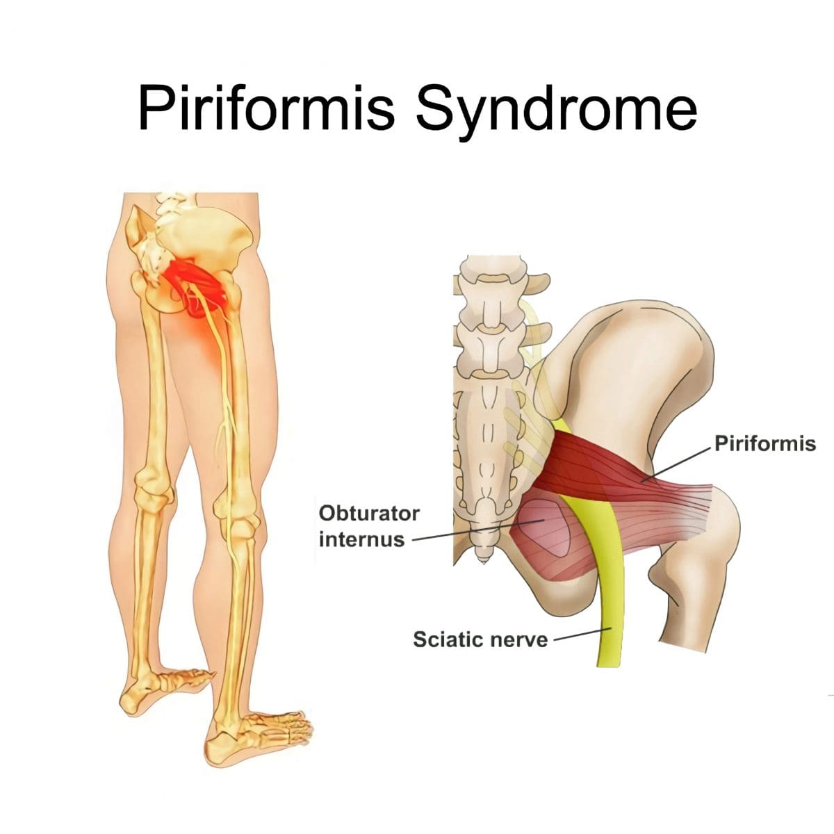
Piriformis syndrome, a.k .a. deep buttock pain, is described as sciatic nerve irritation from the piriformis muscle.
The piriformis is a small muscle behind the hip joint in the buttocks.
It is about one centimeter in diameter and functions in the hip joint’s external rotation or turning outward.
The piriformis muscle and tendon are close to the sciatic nerve, which supplies the lower extremities with motor and sensory functions.
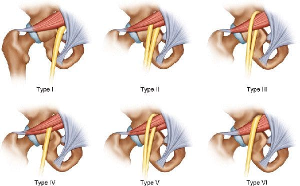
Depending on an individual’s anatomic variation of the muscle and tendon:
The two cross over, under, or through each other behind the hip joint in the deep buttock.
This relationship is thought to irritate the nerve, leading to sciatica symptoms.
Piriformis Syndrome
When diagnosed with piriformis syndrome, it is thought that the muscle and tendon bind to and/or spasm around the nerve, causing irritation and pain symptoms.
The theory supported is that when the piriformis muscle and its tendon tighten, the sciatic nerve becomes compressed or pinched. This decreases blood circulation and irritates the nerve from the pressure. (Shane P. Cass 2015)
Tenderness with pressure on the piriformis muscle.
Discomfort in the back of the thigh.
Deep buttock pain behind the hip.
Electric sensations, shocks, and pains travel down the back of the lower extremity.
Numbness in the lower extremity.
Some individuals develop symptoms abruptly, while others go through a gradual increase.
Diagnosis
Doctors will order X-rays, MRIs, and nerve conduction studies, which is normal.
Because piriformis syndrome can be challenging to diagnose, some individuals with minor hip pain may receive a piriformis syndrome diagnosis even if they don’t have the condition. (Shane P. Cass 2015)
It is sometimes referred to as deep buttock pain. Other causes of this type of pain include back and spinal problems like:
Herniated discs
Spinal stenosis
Radiculopathy – sciatica
Hip bursitis
A piriformis syndrome diagnosis is usually given when these other causes are eliminated.
When the diagnosis is uncertain, an injection is administered in the area of the piriformis muscle. (Danilo Jankovic et al., 2013)
Different medications can be used, but the injection itself is used to help determine the specific location of the discomfort.
When an injection is given into the piriformis muscle or tendon, it is often administered by ultrasound guidance to ensure the needle delivers the medication to the correct location. (Elizabeth A. Bardowski, J. W. Thomas Byrd 2019)
Avoiding activities that cause symptoms for at least a few weeks.
Physical Therapy
Emphasize stretching and strengthening the hip rotator muscles.
Non-Surgical Decompression
Gently pulls the spine to release any compression, allowing optimal rehydration and circulation and taking the pressure off the sciatic nerve.
Therapeutic Massage Techniques
To relax and release muscle tension and increase circulation.
Acupuncture
To help relax the piriformis muscle, sciatic nerve, and surrounding area.
Relieve pain.
Chiropractic Adjustments
Realignment rebalances the spine and musculoskeletal system to alleviate pain.
Anti-Inflammatory Medication
To decrease inflammation around the tendon.
Cortisone Injections
Injections are used to decrease inflammation and swelling.
Botulinum Toxin Injection
Injections of botulinum toxin paralyze the muscle to relieve pain.
Surgery
Surgery can be performed in rare cases to loosen the piriformis tendon, known as a piriformis release. (Shane P. Cass 2015)
Surgery is a last resort when conservative treatments have been tried for at least 6 months with little to no relief.
Recovery can take several months.
Sciatica Causes and Treatment
References
Cass S. P. (2015). Piriformis syndrome: a cause of nondiscogenic sciatica. Current sports medicine reports, 14(1), 41–44. https://doi.org/10.1249/JSR.0000000000000110
Jankovic, D., Peng, P., & van Zundert, A. (2013). Brief review: piriformis syndrome: etiology, diagnosis, and management. Canadian journal of anaesthesia = Journal canadien d’anesthesie, 60(10), 1003–1012. https://doi.org/10.1007/s12630-013-0009-5
Bardowski, E. A., & Byrd, J. W. T. (2019). Piriformis Injection: An Ultrasound-Guided Technique. Arthroscopy techniques, 8(12), e1457–e1461. https://doi.org/10.1016/j.eats.2019.07.033
For individuals looking to improve their spinal health, can understanding the anatomy of the intervertebral foramen help in injury rehabilitation and prevention?
Intervertebral Foramen
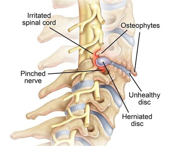
The intervertebral foramen, aka neural foramen, is the opening between the vertebrae through which spinal nerve roots connect and exit to other body areas. If the foramina narrows, it can place added pressure on the nerve roots near and around them, causing pain symptoms and sensations. This is known as neuroforaminal stenosis. (Sumihisa Orita et al., 2016)
Anatomy
The vertebrae comprise the spinal column.
They protect and support the spinal cord and most of the weight placed on the spine.
Foramen is the singular form, and foramina is the plural form.
Structure
The body is the large, round part of the bone that makes up each vertebra.
The body of each vertebra is attached to a bony ring.
Stenosis can occur in the spinal canal, known as central canal stenosis, and the foramina.
Pain brought on by neuroforaminal spinal stenosis and arthritis-related bone growth/bone spurs/osteophytes that are present in one or more foramen rub against the nerve root that passes through the space, causing radicular pain.
Pain accompanied by other sensations, like tingling or numbness, is known as radiculopathy. (Young Kook Choi, 2019)
The main symptom is pain.
Numbness and/or tingling can present depending on the injury.
Neurogenic claudication occurs as a result of ischemia or a lack of blood circulation to the nerves and typically presents with a heaviness in the legs.
It is typically associated with central stenosis rather than foraminal stenosis.
Most individuals with spinal stenosis feel better when flexing or bending forward and worse when arching their backs.
Stenosis treatment aims to relieve pain and prevent nerve symptoms from occurring or worsening. Conservative treatments are recommended and can be highly effective.
These include:
Myelopathy in the neck and/or upper or mid-back (myelopathy symptoms are spinal cord related and occur in central canal stenosis) (Cleveland Clinic. 2021)
Intense incapacitating pain
Different surgical techniques include:
Decompression laminectomy – entails removing the buildup of bone in the spinal canal.
Spinal fusion – when there is instability of the spine or severe foraminal stenosis.
Orita, S., Inage, K., Eguchi, Y., Kubota, G., Aoki, Y., Nakamura, J., Matsuura, Y., Furuya, T., Koda, M., & Ohtori, S. (2016). Lumbar foraminal stenosis, the hidden stenosis including at L5/S1. European journal of orthopaedic surgery & traumatology : orthopedie traumatologie, 26(7), 685–693. https://doi.org/10.1007/s00590-016-1806-7
American Academy of Orthopaedic Surgeons. (2020). Spine Basics (OrthoInfo, Issue. https://orthoinfo.aaos.org/en/diseases–conditions/spine-basics/
American Academy of Orthopaedic Surgeons. (2021). Lumbar spinal stenosis (OrthoInfo, Issue. https://orthoinfo.aaos.org/en/diseases–conditions/lumbar-spinal-stenosis/
Choi Y. K. (2019). Lumbar foraminal neuropathy: an update on non-surgical management. The Korean journal of pain, 32(3), 147–159. https://doi.org/10.3344/kjp.2019.32.3.147
Lee, S. Y., Kim, T. H., Oh, J. K., Lee, S. J., & Park, M. S. (2015). Lumbar Stenosis: A Recent Update by Review of Literature. Asian spine journal, 9(5), 818–828. https://doi.org/10.4184/asj.2015.9.5.818
Lurie, J., & Tomkins-Lane, C. (2016). Management of lumbar spinal stenosis. BMJ (Clinical research ed.), 352, h6234. https://doi.org/10.1136/bmj.h6234
For individuals starting to lift weights, motor units are important for muscle movement. Can building more motor units help build strength and maintain muscle mass?
Motor Units
Motor units control the skeletal muscles and are the force behind every body movement. (C J. Heckman, Roger M. Enoka 2012)
This includes voluntary movements like lifting weights and involuntary movements like breathing. When lifting objects and weights, the body adapts to motor unit needs, meaning that individuals must consistently increase the weight to progress.
Lifting weights regularly trains the body to generate more motor units and force.
General guidelines recommend lifting weights for all muscle groups two to three non-consecutive days a week.
Consistency helps maintain muscle mass.
Regular progression increases the risk of plateauing.
What They Are
Exercise increases the body’s muscle strength, while sedentariness and inactivity weaken them. A motor unit is a single nerve cell/neuron that supplies the nerves to innervate a group of skeletal muscles. The neuron receives signals from the brain that stimulate all the muscle fibers in that particular motor unit to generate movement.
Muscles comprise different fiber types.
They are attached to the bones with connective tissue, which is stronger than the muscle.
Multiple motor units are dispersed throughout the muscle.
The motor units help ensure muscle contraction force is evenly spread throughout the muscle.
Motor units are different sizes and operate differently depending on where and what they do.
Small motor units might only innervate five or ten fibers. For example, to blink or sniff.
Large motor units can comprise hundreds of muscle fibers for swinging or jumping movements.
How They Work
The number of units activated depends on the task. Stronger muscle contractions require more. However, fewer units are needed to accomplish the movement for individuals expending less effort.
Contraction
Once a unit receives a signal from the brain, the muscle fibers contract simultaneously.
The force generated depends on how many units are required to accomplish the task. (Purves D. et al., 2001)
For example, picking up small objects like a pen and paper requires only a few units to generate the force needed.
If picking up a heavy barbell, the body needs more units because more force is needed to lift the heavier load.
The body can generate more force with stronger muscles.
This happens when lifting weights on a regular basis and overloading the muscles with more weight than they can handle.
This process is known as adaptation.
Adaptation
The purpose of lifting weights is to challenge the muscles so they adapt to the new challenge and grow in strength and mass. Motor units are a major part of the adaptation process. (Dr. Erin Nitschke. American Council on Exercise. 2017)
As individuals continue working out, their ability to generate more force increases and units activate more rapidly.
This makes movements more efficient.
Individuals can increase motor unit recruitment by consistently increasing the weight challenge to their muscles.
The development creates movement memory.
A relationship between the brain, muscles, and motor units is established even if the individual stops working out. The pathways are still there no matter how long the individual takes off.
When returning to training, the body will remember how to ride a bike, do a bicep curl, or squat.
However, the muscles will not have the same strength as the strength needs to be built back along with endurance that may have been lost.
It is the movement memory that remains.
Military Training and Chiropractic Care: Maximizing Performance
References
Heckman, C. J., & Enoka, R. M. (2012). Motor unit. Comprehensive Physiology, 2(4), 2629–2682. https://doi.org/10.1002/cphy.c100087
Purves D, Augustine GJ, Fitzpatrick D, et al., editors. (2001). Neuroscience. 2nd edition. Sunderland (MA): Sinauer Associates; 2001. The Motor Unit. Available from: https://www.ncbi.nlm.nih.gov/books/NBK10874/
Dr. Erin Nitschke. American Council on Exercise. (2017). How Muscle Grows (Exercise Science, Issue. https://www.acefitness.org/resources/everyone/blog/6538/how-muscle-grows/
Pete McCall. American Council on Exercise. (2015). 10 Things to Know About Muscle Fibers (Exercise Science, Issue. https://www.acefitness.org/resources/pros/expert-articles/5411/10-things-to-know-about-muscle-fibers/
IFM's Find A Practitioner tool is the largest referral network in Functional Medicine, created to help patients locate Functional Medicine practitioners anywhere in the world. IFM Certified Practitioners are listed first in the search results, given their extensive education in Functional Medicine


 Causes