Broken bones are common injuries. Because children’s bones grow rapidly, they have increased flexibility. When injuries, specifically fractures, occur, they do not always break cleanly across the bone or into pieces. What type of fracture is this, and how are they treated?
Greenstick Fracture
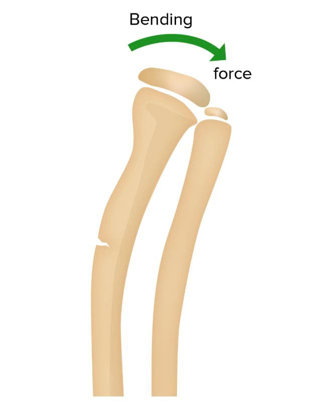
A greenstick fracture is a partial break in a bone that occurs when a bone bends and cracks instead of breaking into separate pieces. (Wolfe J. A. et al., 2019) The term is based on a young green branch that bends and splinters but does not break into pieces when bent. Only one side of the bone is broken, while the other side gets bent. Many children experience at least one fracture during their growing years. This is one of multiple fracture types seen in children. They can happen in adults but are uncommon.
Causes
Greenstick fractures mostly occur in infants or toddlers, sometimes in children during their early adolescent and pre-adolescent years. They are partial-thickness fractures (a break in a bone that doesn’t completely break it) characterized by a break on one side and a bend on the other. Buckle fractures and bow fractures are different types of partial-thickness fractures. Greenstick fractures commonly occur:
In children under 10 years old
Occurs when a child reaches out to break a fall
During motor vehicle collisions
Sports
Direct impacts
Non-accidental trauma
It is more common in long bones, including:
Radius
Ulna
Humerus
Fibula
Tibia
Clavicle
The fracture pattern often indicates a limb’s bending or contortion.
Deformity, such as the affected body part looking crooked or out of alignment.
Treatment
If the bone is not significantly bent out of alignment, a splint or cast may be all that is necessary to treat the break. If the bone is visibly out of alignment, it must be manually straightened before the limb is put into a cast. If the break is severe, surgery may be required. Fortunately, a growing skeleton can remodel bone, so fractured bones can often realign themselves over time with minimal intervention. Healing depends on various factors, including:
Sometimes, the fracture must be bent back and repositioned in a fracture reduction procedure. An anesthetic may be used as the doctor manually realigns the bone into the correct position. After the reduction, a cast or splint will stabilize the bone and maintain proper alignment. Depending on how quickly the bone heals, a cast may be necessary for a few weeks, months, or longer, depending on the patient and/or underlying conditions.
Healing
Healing involves specialized cells that gradually rebuild and fine-tune the new bone.
The average time for a greenstick fracture to heal completely may take four weeks.
Injury Medical Chiropractic & Functional Medicine Clinic
Injury Medical Chiropractic and Functional Medicine Clinic works with primary healthcare providers and specialists to develop an optimal health and wellness solution. We focus on what works for you to relieve pain, restore function, and prevent injury. Regarding musculoskeletal pain, specialists like chiropractors, acupuncturists, and massage therapists can help mitigate the pain through spinal adjustments that help the body realign itself. They can also work with other medical professionals to integrate a treatment plan to resolve musculoskeletal issues.
Building a Stronger Body
References
Wolfe, J. A., Wolfe, H., Banaag, A., Tintle, S., & Perez Koehlmoos, T. (2019). Early Pediatric Fractures in a Universally Insured Population within the United States. BMC pediatrics, 19(1), 343. https://doi.org/10.1186/s12887-019-1725-y
Atanelov, Z., & Bentley, T. P. (2025). Greenstick Fracture. In StatPearls. https://www.ncbi.nlm.nih.gov/pubmed/30020651
Pountos, I., Clegg, J., & Siddiqui, A. (2010). Diagnosis and treatment of greenstick and torus fractures of the distal radius in children: a prospective randomised single-blind study. Journal of children’s orthopaedics, 4(4), 321–326. https://doi.org/10.1007/s11832-010-0269-3
In females, hernia symptoms are often smaller and deeper without a noticeable lump and can mimic gynecological issues, with misdiagnoses being common. Can knowing the risk factors and how female hernias are treated help women get relief?
Female Hernia
A hernia occurs when an internal structure pushes through a weak spot in the abdominal wall, the muscles, and the tissue covering the front of the torso. The more common include:
Groin hernia, known as an inguinal hernia.
Upper thigh or femoral hernia.
However, a hernia can develop anywhere from the ribcage to the upper thigh. Hernias are less common in women, have different symptoms than in men, and are often misdiagnosed. Lower abdominal and pelvic hernias present differently in women than men, who typically have a visible bulge. Instead, female hernias tend to be smaller, deeper, and less noticeable. They can also cause chronic pelvic pressure or pain that can be mistaken for gynecological problems.
Hernia Symptoms For a Woman
Hernias in women tend to be smaller and deeper than male hernias, with no lump showing. Instead, female hernias can cause chronic, deep pelvic pain and occasional sharp, stabbing pain that comes on fast and lingers. (Köckerling F., Koch A., & Lorenz R. 2019) Hernia pain worsens with exercise, laughing, coughing, or straining to evacuate the bowels. The pain is often described as:
Dull
Aching
Pinching
Sharp
Shooting
Burning
Inguinal hernia pain is usually felt at or above the groin and may radiate to the hip, lower back, vulva, or thigh. Many women find the pain increases during their menstrual cycle. The pain can also be exacerbated by any activity that generates extra pressure on the pelvic floor, including:
Prolonged sitting or standing.
Bending
Getting in or out of bed.
Getting in or out of a car.
Sexual intercourse
Emergency
Hernias in the pelvic area are at risk of becoming incarcerated hernias. An incarcerated hernia occurs when a portion of the intestine or other abdominal tissue becomes trapped in the hernial sac, making it impossible to push it back into place. If this gets trapped or strangulated, it can cause tissue death. Strangulated hernias are a medical emergency. Symptoms include:
Deep red or purple tissues.
The hernia bulge does not shrink when you lie down.
Contact a healthcare provider or the emergency room if experiencing any of the above symptoms.
Types
Hernias can occur anywhere on the abdominal wall. They may be caused by:
Internal pressure, such as during pregnancy.
A sports injury
Tissue weakness
Hernias in the lower abdomen or groin are typically indirect inguinal hernias. The inguinal canal comprises multiple layers of muscles and fascia that the thin round ligament threads through. Other groin and pelvic hernias include:
A direct inguinal hernia
A femoral hernia at the top of the inner thigh.
An obturator hernia in the front upper thigh, although this type is rare.
Other common hernias in women are:
Incisional hernia – at the site of a surgical incision
Pregnancy and repeated pregnancies are linked to an increased risk of hernia. Types that are more common in pregnancy include:
Umbilical hernia
Ventral hernia
Inguinal hernia
Umbilical hernias are the most common. However, only a small percentage of pregnant individuals get them. (Kulacoglu H. 2018)
Diagnosis
A hernia diagnosis is made with a physical examination and, if needed, imaging studies. Patients are asked to describe their symptoms precisely, where the pain is located, and any activities that exacerbate it. To check for a hernia, the healthcare provider will palpate for a hernia while the patient sits, stands, or coughs. Imaging tests can include:
Ultrasound
CT scan
Endoscopy – a camera is used to see inside the esophagus and stomach.
Misdiagnoses
Female hernia symptoms can be vague, which often points healthcare providers in the wrong direction. Female hernias are commonly misdiagnosed as: (Köckerling F., Koch A., & Lorenz R. 2019)
Cysts in the reproductive organs
Endometriosis
Fibroid tumors
Treatment
A small hernia that does not cause problems or pain may be treated with a wait-and-evaluate protocol. A hernia often worsens over time and could eventually require surgery. (University of Michigan Health, 2024) Self-care treatments include:
Medical treatments usually start with conservative measures, including physical therapy, stretching, exercise, and rest. Physical therapists often use myofascial release techniques to relieve muscle spasms. Surgery may be needed to repair the weak area of the abdominal wall to relieve symptoms. (University of Michigan Health, 2024) Hernia repair surgery is typically performed as a laparoscopic surgery. (Köckerling F., Koch A., & Lorenz R. 2019) Most patients heal quickly from the surgery and can return to regular activities in a week or two.
Injury Medical Chiropractic and Functional Medicine Clinic
Injury Medical Chiropractic and Functional Medicine Clinic works with primary healthcare providers and specialists to develop an optimal health and wellness solution. We focus on what works for you to relieve pain, restore function, and prevent injury. Regarding musculoskeletal pain, specialists like chiropractors, acupuncturists, and massage therapists can help mitigate the pain through spinal adjustments that help the body realign itself. They can also work with other medical professionals to integrate a treatment plan to resolve musculoskeletal issues.
Lumbar Spine Injuries in Sports: Chiropractic Healing
References
Köckerling, F., Koch, A., & Lorenz, R. (2019). Groin Hernias in Women-A Review of the Literature. Frontiers in surgery, 6, 4. https://doi.org/10.3389/fsurg.2019.00004
Johns Hopkins Medicine. (2025). How to tell if you have a hernia. https://www.hopkinsmedicine.org/health/conditions-and-diseases/how-to-tell-if-you-have-a-hernia
Kulacoglu H. (2018). Umbilical Hernia Repair and Pregnancy: Before, during, after…. Frontiers in surgery, 5, 1. https://doi.org/10.3389/fsurg.2018.00001
University of Michigan Health. (2024). Inguinal hernia: Should I have surgery now, or should I wait? https://www.uofmhealth.org/health-library/za1162
American Academy of Orthopaedic Surgeons. (2022). Sports hernia. https://orthoinfo.aaos.org/en/diseases–conditions/sports-hernia-athletic-pubalgia/
Northeast Georgia Health System. (2022). Living with a hernia. Northeast Georgia Health System Improving the health of our community in all we do. https://www.nghs.com/2022/02/15/living-with-a-hernia
Can the straight leg test help find the cause of back or hamstring pain in individuals experiencing it?
Straight Leg Test
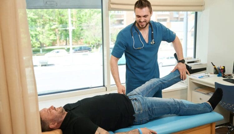
The straight leg raise test is often used to diagnose sciatica/radiculopathy, herniated discs, and other spinal problems. The healthcare provider giving the test performs most of the movement as they assess what’s causing the leg and/or back pain. The patient lies on their back with the legs straight. The provider will have the patient perform specific movements and inform them of how it feels. Then, they’ll raise the leg to see if and at what point symptoms begin to show. Providers often use this test alongside imaging studies.
Some studies suggest the straight leg test helps diagnose sciatica and other causes. (Pesonen J. et al., 2021)
Purpose
The straight leg raise is one of the most common manual tests done during physical exams. The straight leg raise test seeks to reproduce the pain or other symptoms in a controlled fashion to provide clues to what’s happening. It is a manual exam, and the healthcare provider will:
Position the patient
Moves the patient
Create pressure to see how well the patient can resist it
This is often used alongside imaging tests, such as an X-ray or CT scan. (Allegri M. et al., 2016) Its goal is to check for nerve movement and sensitivity of nerve tissue to compression. The straight leg lift test is neurodynamic because it uses movement to diagnose nerve problems. (Baselgia L.T. et al., 2017)
During the Test
Expect to feel some pain during the test, as the whole point is to see what aggravates the symptoms. They may be caused by:
Most of the tests are passive, with the provider doing the lifting. The patient can help achieve the most accurate result by staying as relaxed as possible and being clear about what is felt. (Pande K. 2015) The procedure:
The patient lies on their back with their legs straight.
The provider will ask the patient to turn one of the legs in.
This tells them what hip position affects the lower back symptoms.
They’ll then ask you to bring the leg toward the body’s center.
Then, they’ll lift the straight leg until the patient experiences symptoms.
Pain suggests a herniated disc.
If there is no pain, this also provides valuable information.
The procedure is repeated with the other leg.
Modifications
It’s important to let the examiner know about any limitations. The straight leg raise test has modifications if the patient cannot lift their leg while it’s straight or if they have difficulty lying on their back, which can also help avoid an injury during the test.
Variations
The healthcare provider may repeat the test with the ankle in a dorsiflexed position/raising the foot. Then, they’ll have the patient do it with their chin tucked into their chest. (Young R. et al., 2013) These variations can help check for nerve involvement in specific locations, such as the spinal cord or the dura mater, the membrane covering the brain and spinal cord. (Venne G. et al., 2017) The spinal cord nerves are likely involved and affected if the usual pain is in the back or leg but not the chin, neck, or foot. (Camino Willhuber GO, Piuzzi NS. 2023)
Injury Medical Chiropractic and Functional Medicine Clinic
Injury Medical Chiropractic and Functional Medicine Clinic works with primary healthcare providers and specialists to build optimal health and wellness solutions. We focus on what works for you to relieve pain, restore function, prevent injury, and mitigate issues through adjustments that help the body realign itself. The clinic can also work with other medical professionals to integrate a treatment plan to resolve musculoskeletal problems.
From Injury to Recovery with Chiropractic Care
References
Casiano, V. E., Sarwan, G., Dydyk, A. M., & Varacallo, M. A. (2025). Back Pain. In StatPearls. https://www.ncbi.nlm.nih.gov/pubmed/30844200
Pesonen, J., Shacklock, M., Suomalainen, J. S., Karttunen, L., Mäki, J., Airaksinen, O., & Rade, M. (2021). Extending the straight leg raise test for improved clinical evaluation of sciatica: validity and diagnostic performance with reference to the magnetic resonance imaging. BMC musculoskeletal disorders, 22(1), 808. https://doi.org/10.1186/s12891-021-04649-z
Allegri, M., Montella, S., Salici, F., Valente, A., Marchesini, M., Compagnone, C., Baciarello, M., Manferdini, M. E., & Fanelli, G. (2016). Mechanisms of low back pain: a guide for diagnosis and therapy. F1000Research, 5, F1000 Faculty Rev-1530. https://doi.org/10.12688/f1000research.8105.2
Baselgia, L. T., Bennett, D. L., Silbiger, R. M., & Schmid, A. B. (2017). Negative Neurodynamic Tests Do Not Exclude Neural Dysfunction in Patients With Entrapment Neuropathies. Archives of physical medicine and rehabilitation, 98(3), 480–486. https://doi.org/10.1016/j.apmr.2016.06.019
Pande K. (2015). The Use of Passive Straight Leg Raising Test: A Survey of Clinicians. Malaysian Orthopaedic Journal, 9(3), 44–48. https://doi.org/10.5704/MOJ.1511.012
Young, R., Nix, S., Wholohan, A., Bradhurst, R., & Reed, L. (2013). Interventions for increasing ankle joint dorsiflexion: a systematic review and meta-analysis. Journal of foot and ankle research, 6(1), 46. https://doi.org/10.1186/1757-1146-6-46
Venne, G., Rasquinha, B. J., Kunz, M., & Ellis, R. E. (2017). Rectus Capitis Posterior Minor: Histological and Biomechanical Links to the Spinal Dura Mater. Spine, 42(8), E466–E473. https://doi.org/10.1097/BRS.0000000000001867
Camino Willhuber, G. O., & Piuzzi, N. S. (2025). Straight Leg Raise Test. In StatPearls. https://www.ncbi.nlm.nih.gov/pubmed/30969539
Can physical therapy help individuals with a pinched nerve in the neck?
Pinched Nerve In The Neck
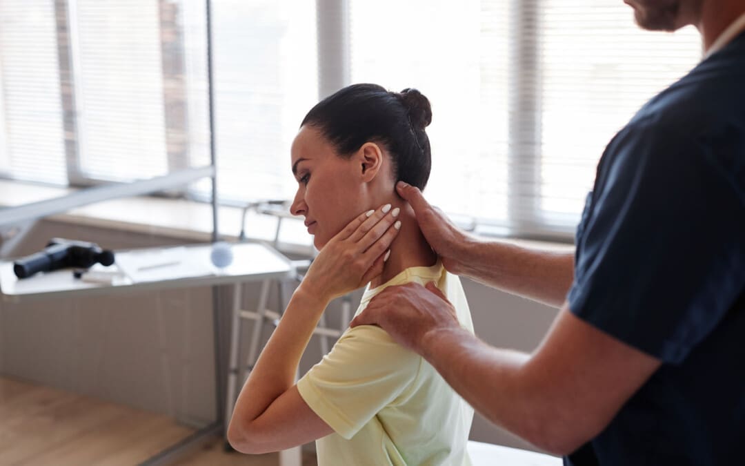
A pinched nerve in the neck can cause pain, numbness, and weakness that extends through the neck, shoulder, and arm. (American Academy of Orthopaedic Surgeons, 2024) Over-the-counter pain medications, resting the muscles, and gentle stretching can help. However, if you’re still in pain after a few days, see a healthcare provider. Common treatments include:
Rest
Over-the-counter (OTC) pain meds
Physical therapy
Steroid injections
Wearing a neck collar
Surgery is rarely needed, but it can provide relief if other treatments don’t help. Most of the time, a pinched nerve resolves within days or weeks.
Sharp pain that extends into the shoulder and arm.
The pain worsens, or there is a shooting sensation when turning the head.
Tingling or feeling of pins-and-needles in the fingers or hand.
Weakness in the arm, shoulder, or hand.
Numbness or loss of feeling.
Often, these symptoms occur only on one side. Some find their pain less when they lift their hand onto their head, which can relieve pressure on the nerve.
Causes
The cervical spine is the spinal cord area around the neck. It’s made up of seven vertebrae. Nerves branch off the spinal cords in the spaces between the vertebrae. Nerve compression occurs when the space between two vertebrae is reduced, putting pressure on the nerve, pinching it, and causing pain. Pinched nerves develop from age because the spinal discs between the vertebrae become compressed over time. Age causes about 70% to 80% of nerve compression. Other factors that cause pinched nerves include: (Harvard Health Publishing, 2021)
Degenerative disc disease
A herniated disc
Injuries like car accidents falls, or other trauma to the spine
Find a comfortable position and try to allow the muscles in your neck to relax and rest.
Heat or Ice
Warmth and coolness can relieve pain and inflammation.
Use a warm or cool compress for 15 minutes at a time.
Over-The-Counter Pain Medications
Pain medications, including nonsteroidal anti-inflammatory drugs (NSAIDs), can help bring relief.
Treatment for Severe Symptoms
If pain doesn’t resolve within a few days, or if it is so bad that you can’t go about daily activities, it’s recommended to see a healthcare provider. They can diagnose a pinched nerve after a physical exam and may also recommend imaging, including an X-ray, CT scan, MRI, or EMG, to reveal what’s causing the symptoms. After diagnosing the condition, the healthcare provider will develop a personalized treatment plan, which may include the following (Harvard Health Publishing, 2021)
Physical Therapy
Physical therapy can help build strength and flexibility in the neck.
This is especially important if there is frequent nerve pain in the same spot.
Cervical Collar
A soft cervical collar is a brace that fits around the neck.
It supports your head so the neck muscles can relax, facilitating healing.
The collar can also keep the head from turning in painful ways.
Oral Corticosteroids
Oral steroids like prednisone can help reduce inflammation.
If inflammation or swelling in the neck puts more pressure on the nerve, they can help.
Steroid Injections
Steroid shots right into the painful tissue reduce inflammation right away.
Muscle Relaxers
These medications keep the muscles in the neck from seizing up.
As the muscles relax, this brings pain relief.
Narcotic Pain Medications
Narcotic pain medications can be used short-term by individuals who have severe pain.
A healthcare provider will inform the patient of the benefits and drawbacks of these medications, which include opiates.
Hold for 20 seconds, then return to a neutral position.
Do this five times.
Eyes to Sky
Lean your head back and look toward the sky.
Hold for 20 seconds, then return to your starting position.
Do this five times.
Side to Side
Turn your head to the right as far as possible, bringing your chin in line with your shoulder.
Hold for 20 seconds, then turn as far as possible toward the left.
Repeat four times.
Ear to Shoulder
Bring your ear down toward your shoulder.
Hold for 20 seconds, then repeat the exercise on the other side.
Alternate between the right and left, stretching each side five times.
While it’s normal for exercises to hurt from stretching the muscles, they should never hurt more than a five on a pain scale of 1 to 10. If they do, stop exercising (National Health Service, 2025)
Healing Time
Healing and recovery depend on the severity of the injury. Some individuals find that the pain from a pinched nerve goes away in days, while for others, it can last for weeks. The pain goes away and then returns. If pain doesn’t go away with the conservative treatments or lasts more than a few days, talk with a healthcare provider or return for a second visit. Rarely do individuals need surgery to bring pain relief. The healthcare provider will discuss whether surgery is the best option and what to expect regarding pain relief. (American Academy of Orthopaedic Surgeons, 2024)
Injury Medical Chiropractic and Functional Medicine Clinic
Injury Medical Chiropractic and Functional Medicine Clinic works with primary healthcare providers and specialists to build optimal health and wellness solutions. We focus on what works for you to relieve pain, restore function, prevent injury, and mitigate issues through adjustments that help the body realign itself. The clinic can also work with other medical professionals to integrate a treatment plan to resolve musculoskeletal problems.
Revitalize and Rebuild with Chiropractic
References
American Academy of Orthopaedic Surgeons. OrthoInfo. (2024). Cervical radiculopathy (pinched nerve). https://orthoinfo.aaos.org/en/diseases–conditions/cervical-radiculopathy-pinched-nerve/
Harvard Health Publishing. Publishing, H. H. (2021). Treating a pinched nerve. https://www.health.harvard.edu/pain/treating-a-pinched-nerve
National Institute of Neurological Disorders and Stroke. (2025). Pinched Nerve Definition. Retrieved from https://www.ninds.nih.gov/health-information/disorders/glossary-neurological-terms#-P-
National Health Service. Service, N. H. (2025). Exercises for neck problems. https://www.nhsinform.scot/illnesses-and-conditions/muscle-bone-and-joints/neck-and-back-problems-and-conditions/exercises-for-neck-problems
While cheddar cheese’s high-calorie count and saturated fat content have nutritional drawbacks, can a moderate amount be an enjoyable part of a healthy diet?
Cheddar Cheese
Cheddar is a hard, cow’s milk cheese known for its dense, layered texture and nutty flavor. It is a favorite cheese served in quesadillas, mac and cheese, or on burgers. However, cheddar cheese nutrition isn’t considered ideal.
Protein performs a variety of functions in the body.
Protein helps build muscle; it’s necessary to produce enzymes, give structure to cells, maintain fluid balance, and more. (Carbone J. W., & Pasiakos S. M. 2019)
Calorie-Dense
Cheddar is calorie-dense, which increases its satisfaction factor.
May Help Weight Loss
There is the belief that cheese causes weight gain; however, the full-fat dairy paradox, which goes against dietary guidelines, is the idea that full-fat dairy products may be healthier than low-fat or fat-free dairy,
Research now suggests removing fat from dairy products may make them more likely to cause weight gain, making full-fat the better choice for weight management. (Soltani S., & Vafa M. 2017)
Compatible With Keto and Low-Carb Diets
Because of cheddar’s high fat percentage, it’s compatible with high-fat nutrition plans.
With zero carbohydrates, cheddar also fits well in low-carb diets.
Low Lactose
Cheddar is one of the harder, aged cheeses that’s quite low in lactose.
This means lactose-intolerant individuals can often eat it without unpleasant symptoms like bloating, stomach upset, and gas.
Allergies
Although it is low in lactose, cheddar still contains casein and whey, two components that can trigger an immune response in individuals with a dairy allergy. (He, M. et al., 2017)
Storage and Safety
Cheddar does not technically require refrigeration. However, storing it in the fridge will help it last longer. According to the USDA, unopened cheddar can last up to six months in the refrigerator, and opened packages can last three to four weeks. Because it’s a hard cheese, cheddar even takes well to freezing, but this will not extend its life more than refrigeration. The USDA estimates cheddar can be frozen for about six months.
Preparation
Cheddar can be added to any number of cheesy dishes or, of course, served alone with crackers. It doesn’t require high heat to become nice and melty. Try adding a whole-grain base and veggies to increase the nutrients in dishes like cheesy casseroles, Mexican dishes, sandwiches, or pasta.
Injury Medical Chiropractic and Functional Medicine Clinic
Injury Medical Chiropractic and Functional Medicine Clinic works with primary healthcare providers and specialists to develop highly effective treatment plans through an integrated approach for each patient and restore health and function to the body through nutrition and wellness, functional medicine, acupuncture, Electroacupuncture, and integrated medicine protocols. We focus on what works for you to relieve pain, restore function, prevent injury, and mitigate issues through adjustments that help the body realign itself. The clinic can also work with other medical professionals to integrate a treatment plan to resolve musculoskeletal problems.
Osteoporosis
References
U.S. Department of Agriculture. FoodData Central. (2019). Cheddar Cheese. Retrieved from https://fdc.nal.usda.gov/food-details/494681/nutrients
Lordan, R., Tsoupras, A., Mitra, B., & Zabetakis, I. (2018). Dairy Fats and Cardiovascular Disease: Do We Really Need to be Concerned?. Foods (Basel, Switzerland), 7(3), 29. https://doi.org/10.3390/foods7030029
Astrup, A., Geiker, N. R. W., & Magkos, F. (2019). Effects of Full-Fat and Fermented Dairy Products on Cardiometabolic Disease: Food Is More Than the Sum of Its Parts. Advances in nutrition (Bethesda, Md.), 10(5), 924S–930S. https://doi.org/10.1093/advances/nmz069
Hirahatake, K. M., Astrup, A., Hill, J. O., Slavin, J. L., Allison, D. B., & Maki, K. C. (2020). Potential Cardiometabolic Health Benefits of Full-Fat Dairy: The Evidence Base. Advances in nutrition (Bethesda, Md.), 11(3), 533–547. https://doi.org/10.1093/advances/nmz132
Malmir, H., Larijani, B., & Esmaillzadeh, A. (2020). Consumption of milk and dairy products and risk of osteoporosis and hip fracture: a systematic review and Meta-analysis. Critical reviews in food science and nutrition, 60(10), 1722–1737. https://doi.org/10.1080/10408398.2019.1590800
Carbone, J. W., & Pasiakos, S. M. (2019). Dietary Protein and Muscle Mass: Translating Science to Application and Health Benefit. Nutrients, 11(5), 1136. https://doi.org/10.3390/nu11051136
Soltani, S., & Vafa, M. (2017). The dairy fat paradox: Whole dairy products may be healthier than we thought. Medical journal of the Islamic Republic of Iran, 31, 110. https://doi.org/10.14196/mjiri.31.110
He, M., Sun, J., Jiang, Z. Q., & Yang, Y. X. (2017). Effects of cow’s milk beta-casein variants on symptoms of milk intolerance in Chinese adults: a multicentre, randomised controlled study. Nutrition journal, 16(1), 72. https://doi.org/10.1186/s12937-017-0275-0
Individuals who have fractured their scaphoid bone may experience pain and swelling in the wrist just below the thumb. Can immobilization with a cast and physical therapy help?
Scaphoid Fracture
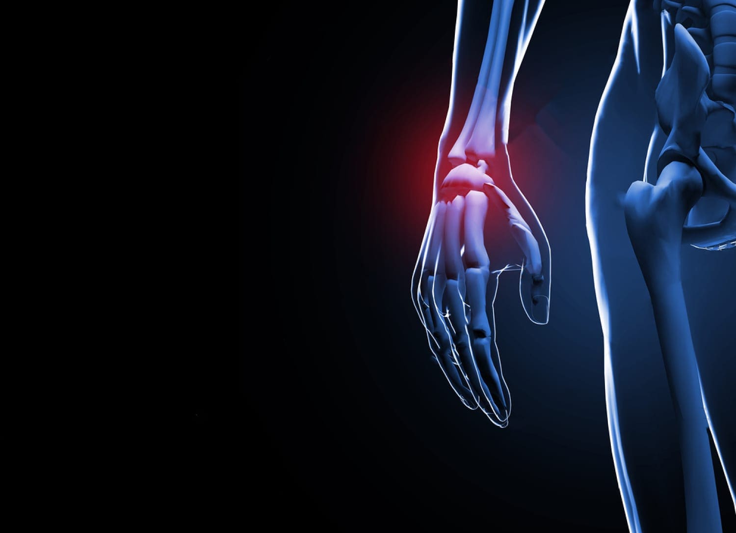
A scaphoid fracture is a break in one of the wrist’s small or carpal bones. This type of fracture occurs most often after a fall onto an outstretched hand. Symptoms typically include swelling and pain in the wrist just below the base of the thumb. These fractures can be difficult to diagnose since they don’t always appear on an X-ray. If the X-ray is negative and the healthcare provider suspects a scaphoid fracture, an MRI may be necessary. Surgery may be required in more severe cases or when the injury is not healing correctly. (American Academy of Orthopaedic Surgeons, 2023)
A Break In The – Navicular Bone
The scaphoid is one of eight carpal bones in the wrist. It is located just below the thumb’s base and is shaped like a kidney bean. This bone can be identified by holding a thumbs-up position and feeling for the hollow between the two tendons below your thumb. The scaphoid is located at the base of the hollow. A break in the scaphoid bone most commonly occurs in the middle of the bone but can also happen at either end. A scaphoid fracture can be displaced or non-displaced (American Academy of Orthopaedic Surgeons, 2023)
Displaced Fracture
It is when the bone fragments have moved out of alignment.
Non-displaced Fracture
It is when the fragments are still in their normal location in the hand.
The scaphoid’s blood supply comes from a small vessel that enters the most distant part of the bone and flows back through the bone. Because of this one small blood supply, a fracture in the center can stop the circulation to the proximal portion of the bone. Because of this, scaphoid fractures need immediate diagnosis and treatment.
Symptoms
Pain or deep aching on the thumb-side of the wrist, typically after a fall on an outstretched arm, could be a scaphoid fracture. Other symptoms experienced include: (American Academy of Orthopaedic Surgeons, 2023)
A healthcare provider will evaluate the hand for tenderness and pain in the hollow and/or the bone. If a break is suspected, they will order an X-ray. (Clementson M., Björkman A., & Thomsen N. O. B. 2020) Many patients are diagnosed with a wrist sprain when they have a fracture. Diagnosis can be difficult because the fracture often doesn’t appear on X-rays until weeks after the healing process starts. Physicians commonly treat a wrist injury as a scaphoid fracture initially and then repeat X-rays within two weeks. (American Academy of Orthopaedic Surgeons, 2023) If the injury doesn’t show on an X-ray, the provider may order an MRI, as these fractures can be easier to see on an MRI. An MRI can help ensure appropriate treatment immediately. (Wong S. B. S., & Peh W. C. G. 2019)
Treatment
If a wrist fracture is diagnosed, the wrist will be immobilized in a cast. However, a healthcare provider may also put the wrist in a cast if the X-ray is negative but they suspect a fracture. This will stabilize the injury until an MRI can be performed. With immobilization and follow-up treatment, scaphoid fractures often heal without surgery. Repeat X-rays are taken over several weeks or months so the provider can make sure the injury is healing correctly. If it is not healing correctly, surgery may be recommended. (Clementson M., Björkman A., & Thomsen N. O. B. 2020) If the fracture is displaced, healing correctly may be a challenge. In this case, a physician may recommend initial surgery to reposition the bones. (Clementson M., Björkman A., & Thomsen N. O. B. 2020) This type of surgery involves pinning the bone in place with screws.
Rehabilitation is an important part of healing because immobilization takes a long time. Wrist range-of-motion exercises can be started, followed by strengthening exercises for the wrist flexors and extensors. Supination, pronation, and grip exercises are also part of physical therapy.
This condition causes degeneration of the cartilage in the joint.
Avascular Necrosis
This is when the blood supply to the bone is reduced or cut off, causing the bone to die.
Injury Medical Chiropractic and Functional Medicine Clinic
Injury Medical Chiropractic and Functional Medicine Clinic works with primary healthcare providers and specialists to build optimal health and wellness solutions. We focus on what works for you to relieve pain, restore function, prevent injury, and mitigate issues through adjustments that help the body realign itself. The clinic can also work with other medical professionals to integrate a treatment plan to resolve musculoskeletal problems.
Skateboarding Injury Treatment
References
American Academy of Orthopaedic Surgeons. (2023). Scaphoid fracture of the wrist. https://orthoinfo.aaos.org/en/diseases–conditions/scaphoid-fracture-of-the-wrist
Clementson, M., Björkman, A., & Thomsen, N. O. B. (2020). Acute scaphoid fractures: guidelines for diagnosis and treatment. EFORT open reviews, 5(2), 96–103. https://doi.org/10.1302/2058-5241.5.190025
Wong, S. B. S., & Peh, W. C. G. (2019). The role of magnetic resonance imaging in the evaluation of scaphoid fractures. Journal of Medical Radiation Sciences, 66(1), 3–4. https://doi.org/10.1002/jmrs.316
Almigdad, A., Al-Zoubi, A., Mustafa, A., Al-Qasaimeh, M., Azzam, E., Mestarihi, S., Khair, Y., & Almanasier, G. (2024). A review of scaphoid fracture, treatment outcomes, and consequences. International orthopaedics, 48(2), 529–536. https://doi.org/10.1007/s00264-023-06014-2
Should individuals experiencing lower left back pain see a healthcare provider if it lasts more than a few weeks?
Left Side Lower Back Pain
Lower left back pain can impact your ability to go about your day. If left-side lower back pain lasts longer than a week, it is considered chronic back pain, which can severely impact one’s quality of life. This type of pain has various causes. Muscle or spine and nerve damage, including sciatica, can cause pain. Organs in the lower back, including the kidneys, can cause pain. Pregnancy-related changes, fibromyalgia, and other conditions can cause lower left-side back pain in females.
Causes
Back pain is common and affects almost everyone. (National Institute of Arthritis and Musculoskeletal and Skin Diseases, 2023) Lower left back pain can have many causes, ranging from muscle and spine issues to organ infections. One way to help tell what’s causing the symptoms is to determine whether there is also sciatica, sharp or burning pain that radiates down one side of the body. It happens when the sciatic nerve gets compressed or irritated. Possible causes include (Penn Medicine, 2020)
Muscle Injury
A muscle injury from an accident or injury can be a cause that can appear with or without sciatica.
If this is the cause, you’ll notice that the pain improves with rest but worsens after you’ve sat for a long time or after getting up from sleep.
There may also be a limited range of motion, tenderness, or swelling.
Arthritis or Bone Conditions
Arthritis and bone issues, like osteoporosis, can also be a cause.
This can happen if the arthritis is in the left hip or the root cause is on the right side, but the body compensates by overusing muscles on the left side of the back.
Unhealthy Posture and Body Positioning
Unhealthy postures and body positioning can contribute to back pain and musculoskeletal problems.
To avoid straining the muscles, try sitting and standing straight and keeping all the joints at a 90-degree angle.
Move around every 20-30 minutes and stretch out.
Kidneys
The kidneys are located in the middle back.
Kidney infections or kidney stones could cause pain on the left side.
Other symptoms include pain when urinating, fever, and nausea.
Ulcerative Colitis
Ulcerative colitis, inflammation of the large intestine, can also cause lower left back pain in some cases.
If this is the cause, there may also be abdominal pain, cramping, nausea, and fatigue.
Uterine-Related Pain
Several conditions related to the uterus can cause back pain symptoms, including PMS, period cramps, endometriosis, and more.
Sometimes, these conditions cause pain on both sides, but some individuals may experience pain just on the left side.
Pregnancy
The weight gain, hormonal changes, and limited movement can also contribute to lower left back pain. (Cedars Sinai, 2024)
Spinal Disease
If one of the discs or vertebrae in the spine slips out of place or becomes damaged, this can cause upper, middle, or lower back pain.
In many cases, over-the-counter anti-inflammatory medications can help with sciatica. If it persists, it is recommended to see your healthcare provider to find the root cause. The causes include: (Aguilar-Shea, A. L. et al., 2022)
Herniated disc
A disc that pops out of place can add pressure to the sciatic nerve.
Spinal Stenosis
Spinal stenosis, or spine narrowing, can also cause sciatic symptoms.
Spondylolisthesis
Occurs when vertebrae are out of alignment, leading to sciatic symptoms.
Pregnancy
Pregnancy-related growth and bodily changes oftentimes lead to sciatic nerve symptoms and sensations.
Muscle Spasms
Spasms like piriformis syndrome, a spasm of the muscle in the buttocks, can cause back pain.
Surgery
It’s normal to have back pain for up to six weeks after a back procedure.
However, if there is new or worsening lower left back pain after surgery, consult the healthcare provider. (Penn Medicine, 2017)
Your healthcare provider may recommend massage, chiropractic care, and acupuncture treatments.
If pain can’t be managed at home, your healthcare provider may suggest prescription medications, including muscle relaxers.
These can allow the tissue to heal and reduce your pain as well.
If you have severe sciatica or vertebrae that have slipped out of place, the healthcare team might recommend a steroid injection into the lower left back to reduce pain by reducing inflammation.
In addition to prescription treatments, your healthcare provider might recommend physical therapy to retrain movements, build strength, and help prevent back pain.
Sleep on a firm mattress that will support your back.
Get a comfortable, ergonomic chair for your job.
If you work on your feet, learn to practice healthy posture and use shoes and insoles to facilitate and maintain correct posture.
Once you’ve healed, building your core strength may help avoid lower back pain in the future.
Injury Medical Chiropractic and Functional Medicine Clinic
Talk with a healthcare provider and request a referral to a specialist who can help with long-term management. Injury Medical Chiropractic and Functional Medicine Clinic works with primary healthcare providers and specialists to build optimal health and wellness solutions. We focus on what works for you to relieve pain, restore function, prevent injury, and mitigate issues through adjustments that help the body realign itself. The clinic can also work with other medical professionals to integrate a treatment plan to resolve musculoskeletal problems.
Lower Back Pain Chiropractor Treatment
References
National Institute of Arthritis and Musculoskeletal and Skin Diseases. (2023). Back pain. Retrieved from https://www.niams.nih.gov/health-topics/back-pain
Penn Medicine, B. T., MD. (2020). 4 reasons you may have back pain on only one side. Penn Medicine. https://www.pennmedicine.org/updates/blogs/musculoskeletal-and-rheumatology/2017/november/back-pain-on-one-side
Cedars Sinai. (2024). Back pain during pregnancy. https://www.cedars-sinai.org/health-library/diseases-and-conditions/b/back-pain-during-pregnancy.html
Aguilar-Shea, A. L., Gallardo-Mayo, C., Sanz-González, R., & Paredes, I. (2022). Sciatica. Management for family physicians. Journal of family medicine and primary care, 11(8), 4174–4179. https://doi.org/10.4103/jfmpc.jfmpc_1061_21
Penn Medicine, V. G., MD. (2017). Back pain that won’t go away—even with surgery. Penn Medicine. https://www.pennmedicine.org/updates/blogs/musculoskeletal-and-rheumatology/2017/november/back-pain-that-wont-go-away
See, Q. Y., Tan, J. B., & Kumar, D. S. (2021). Acute low back pain: diagnosis and management. Singapore Medical Journal, 62(6), 271–275. https://doi.org/10.11622/smedj.2021086
IFM's Find A Practitioner tool is the largest referral network in Functional Medicine, created to help patients locate Functional Medicine practitioners anywhere in the world. IFM Certified Practitioners are listed first in the search results, given their extensive education in Functional Medicine


 Causes