Motivation That Lasts: Fun, Low-Impact Workouts and SMART Goal Strategies
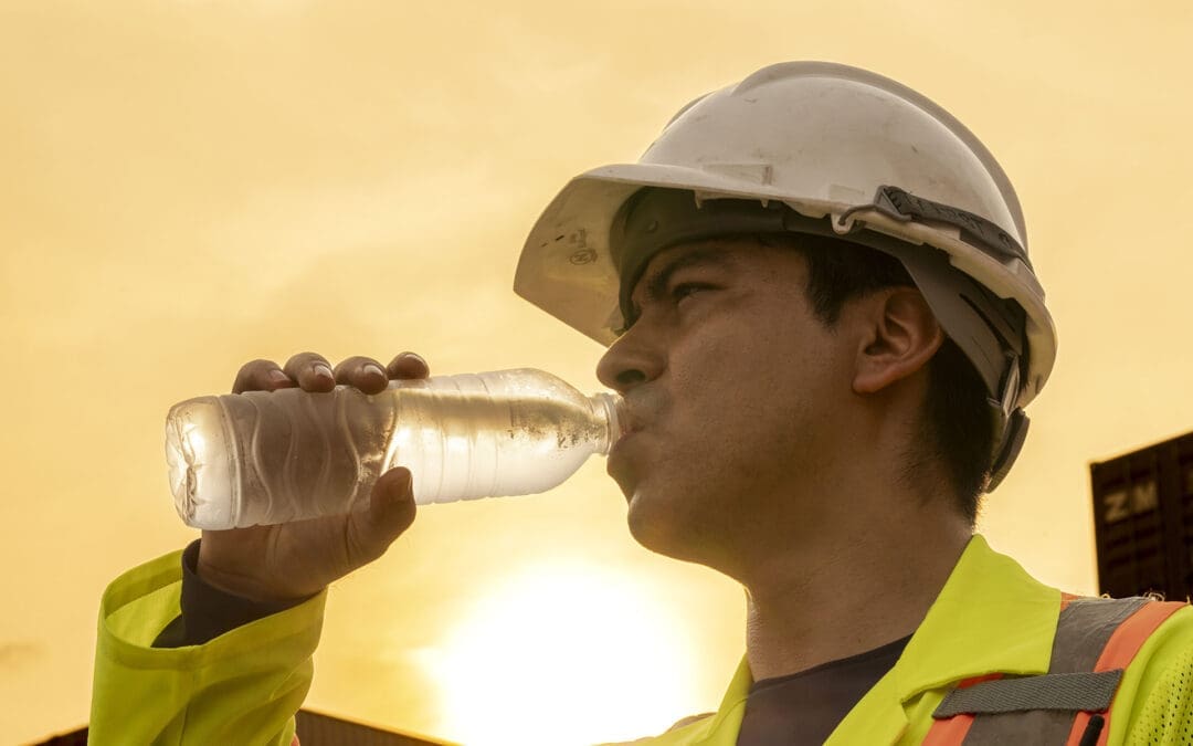
Losing weight does not have to feel impossible, even if back pain, low energy, or busy days get in the way. Many people in El Paso start with easy exercises like short walks or gentle stretches, but staying motivated is what brings real results. The good news is that small, smart steps, plus help from a local expert team, can make all the difference. At El Paso Back Clinic, patients discover how chiropractic care and functional medicine remove roadblocks so basic weight-loss exercises feel safe, doable, and even enjoyable. This guide shares straightforward ways to set goals, track progress, choose fun movement, and get professional support right here in El Paso. You will learn practical tips that fit real life and see how the clinic’s team, led by Dr. Alexander Jimenez, helps turn “I can’t” into steady success.
Basic weight-loss exercises like walking, light yoga, or dancing burn calories without stressing your joints. When your body feels better and pain drops, motivation stays strong. El Paso Back Clinic combines chiropractic adjustments, personalized rehab, and health coaching to make these simple moves part of your everyday routine.
Setting Attainable SMART Objectives for Steady Progress
SMART goals keep your weight-loss journey clear and reachable. SMART means Specific, Measurable, Achievable, Relevant, and Time-bound. Instead of saying “I need to lose weight,” try “I will walk for 15 minutes after dinner, five days this week.” This type of goal is easy to follow and gives quick wins. (Hey Life Training, n.d.; El Paso Back Clinic, n.d.-b)
Here are SMART goal examples perfect for basic weight-loss exercises:
Walk briskly for 15 minutes, five days a week, starting this Monday.
Do gentle yoga stretches for 10 minutes each morning for the next two weeks.
Dance to favorite music for 15 minutes, three evenings a week.
Swim or walk in water for 15 minutes twice a week at a local pool.
Take the stairs instead of the elevator at least five times daily this week.
Start small, so you build confidence fast
At El Paso Back Clinic, health coaches help patients turn these goals into custom plans that match their energy and schedule.
Monitoring progress keeps motivation alive. Use a simple notebook or phone app to log your walks, steps, or how your back feels after movement. Seeing checkmarks add up or a line on a graph climb feels rewarding. Patients at the clinic often say watching their own improvements beats staring at the scale. (Zen Habits, n.d.)
To avoid burnout, pick fun, low-impact activities. Yoga, swimming, and walking ease joints and lift mood through natural feel-good chemicals. These basic exercises become something you look forward to instead of dread. (HelpGuide.org, n.d.)
Find accountability with a workout buddy or the clinic’s support network. Many patients walk with family or join gentle group sessions. Reward small wins with non-food treats like new walking shoes or a relaxing evening. Remember your “why”—more energy for family, better sleep, or less back pain. Read it daily on tough days. (Planet Fitness, n.d.-a)
Easy, Efficient Strategies to Stay Motivated Every Day
Consistency beats intensity when building habits. Here are proven strategies that work well with basic weight-loss exercises:
Start small for lasting consistency: Begin with just 10–15 minutes of movement. This avoids burnout and makes exercise a normal part of your day. (Reddit community insights, 2024)
Track your development: Write down workouts, steps, or how clothes fit. Graphs show real progress and keep you excited. (Zen Habits, n.d.)
Make it fun: Choose dancing, swimming, cycling, or active games. Fun turns movement into “me time.” (HelpGuide.org, n.d.)
Reward yourself: After five good days, celebrate with new socks, a movie, or a quiet bath. (Modern Image Aesthetics, n.d.)
Build accountability: Walk with a friend, pet, or join a beginner class. The clinic’s health coaches provide extra check-ins. (Healthline, n.d.)
Recall your “why”: Focus on deeper reasons like steady energy or pride in your posture. (Planet Fitness, n.d.-b)
Prepare for low-energy days: Have a backup like 10 minutes of gentle stretches at home. (Cleveland Clinic, n.d.)
These steps fit real El Paso life—hot days, long work hours, and family needs. Short walks during lunch or evening strolls add up fast.
Walking Your Way to Better Results: Clinic-Approved Tips
Walking is one of the easiest basic weight-loss exercises, and El Paso Back Clinic shares clear ways to burn more fat while protecting your back. Start with 15 minutes daily, five days a week, then add five minutes each week. Walk at a brisk pace faster than normal, swing your arms, and keep a healthy posture. Add short speed bursts or gentle hills for extra calorie burn without hurting knees. Wear supportive shoes and breathe steadily. (El Paso Back Clinic, n.d.-c)
Benefits include stronger bones, less joint pain, better mood, and reduced belly fat linked to heart health. Even short 15-minute walks several times a day work when time is tight. Patients at the clinic combine walking with chiropractic care for faster mobility gains and steady motivation.
Making Fitness Enjoyable and Part of Your Routine
Pick activities you actually like. If running hurts, try dancing at home, water walking, or bike rides on flat paths. Listen to music or podcasts while moving. Many patients discover they enjoy low-impact options once pain eases. (Medical Beauty and Weight Loss, n.d.)
Social support helps too. Walk with neighbors or join light classes. At El Paso Back Clinic, personalized rehab programs make movement feel safe again, so you stay consistent longer.
How El Paso Back Clinic Boosts Motivation Through Integrative Care
Back pain or low energy often stops people from exercising. El Paso Back Clinic, led by Dr. Alexander Jimenez, DC, APRN, FNP-BC, removes these barriers with chiropractic and functional medicine. Their approach helps thousands of El Paso patients move more freely and lose weight sustainably.
Chiropractic adjustments reduce chronic back, hip, and joint pain, so walking or yoga no longer hurts. Better spinal alignment improves nervous system signals that control metabolism and fat burning. When the body works more smoothly, energy rises, and motivation follows naturally. (El Paso Back Clinic, n.d.-a; Adjusted Life Chiropractic, n.d.)
Dr. Alexander Jimenez has observed over 30 years that fixing spinal misalignments breaks the pain-obesity cycle. Pain leads to less movement and comfort eating; extra weight adds more pain. His team uses gentle adjustments, advanced imaging, and lab tests to address root causes such as inflammation, hormonal imbalances, and gut issues. Patients report less pain, better sleep, steadier moods, and fewer cravings. (Jimenez, n.d.; El Paso Back Clinic, n.d.-a)
Custom low-impact exercise plans are a clinic specialty. Instead of heavy gym work, they recommend practical moves: walking programs, water exercises, light resistance bands, and core stretches that fit daily life. These plans build confidence fast because they feel safe. The clinic’s rehabilitation centers offer guided sessions with trainers who understand back issues. (Robinhood Integrative Health, n.d.; El Paso Back Clinic, n.d.-c)
Functional medicine digs deeper. The team checks for slow metabolism, insulin resistance, or stress hormones that block weight loss. Personalized nutrition advice, supplements, and lifestyle tips clear these hurdles. Health coaches then create step-by-step plans with SMART-style process goals—like “walk three to four times this week”—so patients focus on what they can control. (El Paso Back Clinic, n.d.-b, n.d.-d)
Stress management is built in
High stress raises cortisol and belly fat while lowering motivation. Chiropractic care relaxes tight muscles and calms the nervous system. Many patients report feeling more positive and ready to move on after visits. (Dr. P Chiro, n.d.)
Personalized accountability keeps progress on track. Regular check-ins, body scans, and plan updates show results beyond the scale. Improved posture from adjustments makes patients stand taller and feel stronger—boosting confidence to keep going. (Obesity Action Coalition, n.d.; Westport Chiropractic, n.d.)
Dr. Jimenez often reminds patients that big changes start with small, consistent steps. His team at El Paso Back Clinic offers multiple convenient locations across El Paso, including rehab and fitness centers with 24/7 access. Military discounts, virtual coaching options, and meal-prep support make healthy living easier. Patients with past injuries or long-term back pain often return to activities they once avoided, creating a positive cycle of more movement and faster weight-loss results.
By reducing pain, improving mobility, addressing metabolic issues, and providing expert coaching, El Paso Back Clinic turns basic weight-loss exercises into something patients actually enjoy and stick with long-term.
Putting It All Together for Real, Lasting Success
Begin today with one small change. Choose a SMART goal, schedule a 15-minute walk, and note your “why.” Add music or a friend for fun. If back pain or low energy holds you back, contact El Paso Back Clinic for a personalized evaluation. Dr. Alexander Jimenez and his multidisciplinary team combine chiropractic care, functional medicine, and health coaching to support your goals safely.
Motivation comes and goes—some days feel easier than others, and that is normal. The strategies here—SMART goals, tracking, fun movement, rewards, accountability, and professional help—help you bounce back quickly. Over weeks and months, these habits create real momentum.
Basic weight-loss exercises like daily walking or gentle yoga do more than burn calories. They improve heart health, lift mood, strengthen muscles, ease back pain, and raise self-esteem. With support from El Paso Back Clinic, you gain energy for work, family, and life. Celebrate every step, every stretch, and every healthy choice. You have local experts ready to help—one simple, consistent day at a time.
El Paso Heat Nutrition Guide: Hydrating Foods, Electrolytes, and Light Meals (El Paso Back Clinic)
When El Paso heats up, your body has to work harder to stay cool. You sweat more, lose fluids faster, and burn through key minerals that help your muscles and nerves work right. You might also notice that heavy meals make you feel sluggish, overheated, or even a little nauseated.
At El Paso Back Clinic (https://elpasobackclinic.com/), we see this every year: heat + dehydration + low electrolytes can worsen muscle tightness, trigger cramps, increase headache risk, and add stress to the neck, back, and joints. The goal is not to “eat perfectly.” The goal is to eat and drink in ways that support hydration, steady energy, and recovery during hot weather.
This article explains how to build simple heat-friendly meals using:
High-water foods (fruits and vegetables that add fluid)
Electrolytes (especially sodium, potassium, and magnesium)
Cooling herbs and smart spice use
Light proteins and easy-to-digest meals
Practical El Paso-style food ideas (including lighter taco options)
Throughout, I also include clinical observations from Dr. Alexander Jimenez, DC, APRN, FNP-BC, who often emphasizes hydration, electrolyte support, and recovery habits during intense heat exposure (Jimenez, n.d.).
Why hot weather can worsen fatigue, cramps, and body aches
Heat affects your body in a few big ways:
You lose water through sweat.
You lose electrolytes through sweat.
Your heart and circulation work harder to move blood to the skin so you can cool down.
Digestion can feel heavier, especially after high-fat or fried meals.
If dehydration or electrolyte loss builds up, you may notice:
Headache
Muscle cramps or muscle “pulling”
Dizziness or lightheadedness
Fast heartbeat
Fatigue and brain fog
Dark yellow urine
Severe heat illness is serious and can require urgent medical care (Johns Hopkins Medicine, n.d.). If someone is confused, fainting, has very hot skin, or has symptoms that rapidly worsen, treat it as an emergency (Johns Hopkins Medicine, n.d.).
The El Paso heat strategy: 3 simple goals
When it is hot, your daily plan can be simple:
Hydrate through food and drinks
Replace electrolytes (especially if you sweat a lot)
Choose lighter, easy meals
Community ER guidance often recommends lighter meals and hydration-focused foods during high heat (Community First ER, 2025). Kaiser Permanente also points out that certain foods and spices can help you feel cooler and support hydration habits (Kaiser Permanente, n.d.).
Hydrating and cooling foods that actually help
Water-rich vegetables (easy wins)
Water-rich vegetables add fluid and minerals without making you feel heavy. Many common choices have very high water content.
Great options include:
Cucumbers (very water-rich)
Celery
Zucchini
Tomatoes
Romaine and other lettuces
These types of water-rich foods are commonly recommended in hydration guidance for hot weather (UT Southwestern Medical Center, n.d.; Bass Medical Group, n.d.).
Fast ways to use them:
Cucumber + lime + pinch of salt
Tomato + cucumber + mint salad
Romaine wraps with beans or grilled chicken
Zucchini sliced into a quick “no-cook” salad with lemon
Clinic tip (muscles and cramps): If you are getting cramps, it is not always “just dehydration.” It can be low electrolytes, too. Pair water-rich foods with a little salt and potassium-rich foods (Optum, n.d.).
Melons and berries: hydration + skin support nutrients
In hot weather, fruit is often easier to eat than heavy meals. Watermelon, cantaloupe, strawberries, and citrus are popular for a reason: they hydrate and provide vitamins.
Many medical and wellness sources recommend water-rich fruit during heat stress and after heat exhaustion (UT Southwestern Medical Center, n.d.; Lokmanya Hospitals, n.d.).
Top picks:
Watermelon
Cantaloupe
Strawberries
Grapefruit, oranges, and lemons
Watermelon is also known for plant compounds such as lycopene, which is often discussed for its support of cells and skin (UT Southwestern Medical Center, n.d.).
Easy snack ideas:
Freeze grapes or watermelon cubes
Add citrus slices to cold water
Blend watermelon + mint + ice (no added sugar)
Sunnybrook also suggests simple infused water ideas (like cucumber and citrus) to make hydration easier (Sunnybrook Health Sciences Centre, n.d.).
Light proteins: stay fueled without feeling overheated
Heavy, fried, or very fatty meals can feel worse in the heat, partly because digestion takes work and can increase discomfort (Community First ER, 2025). Instead, use lighter proteins that are easier on the stomach.
Better hot-weather proteins include:
Grilled chicken
Fish
Shrimp
Beans and lentils
Plain, unsweetened yogurt
UT Southwestern highlights that plain yogurt is water-rich and hydrating, and it can work well in smoothies or as a light snack (UT Southwestern Medical Center, n.d.).
Simple meal formula:
Light protein + water-rich produce + salty-acid flavor (lime/lemon)
Example: grilled fish + cucumber/tomato salad + lime + pinch of salt.
Cooling herbs and spices: what helps and why
Mint: “cooling” sensation that can make hydration easier
Mint can trigger cold receptors in the mouth, creating a cooling feeling and making water and light meals more enjoyable (Kaiser Permanente, n.d.).
Try:
Mint + cucumber + lemon water
Mint stirred into yogurt
Mint on tacos with fresh salsa
Spicy foods: yes, they can help you cool down
This surprises many people: spicy foods can increase sweating, and when sweat evaporates, it cools the skin. Kaiser Permanente explains this effect with foods such as ginger and chile (Kaiser Permanente, n.d.).
Use spicy foods smartly:
Start small if you are not used to spicy heat.
Do not push spicy foods if you already feel sick or dehydrated
Pair spice with hydrating foods (cucumber, fruit, salsa)
Electrolytes: the missing piece for many people
Electrolytes are minerals that help control fluid balance and support muscle and nerve function. When you sweat a lot, you can lose electrolytes along with water (Optum, n.d.; Ally Medical, n.d.).
The big ones are:
Sodium
Potassium
Magnesium
Signs you may need electrolyte support
Not everyone needs electrolyte powders every day, but you might benefit if you have:
Heavy sweating (workouts, outdoor work, long time in the sun)
Muscle cramps or twitching
Frequent headaches with heat exposure
Low energy that improves after salty fluids
Heat exhaustion recovery guidance often includes electrolyte replacement and easy-to-digest foods (Lokmanya Hospitals, n.d.).
Food-first electrolyte support
Before supplements, start with food and simple options:
Water-rich produce (helps hydration)
Beans, leafy greens, fruits (potassium support)
Light soups or broths (fluid + sodium)
Coconut water (check sugar levels)
El Paso Wellness Associates also discusses “electrolytes without the junk” approaches for hydration routines (El Paso Wellness Associates, n.d.).
Supplements for hot weather: what may help (and how to be safe)
Supplements are not required for everyone. But for some people, especially those who sweat a lot, certain supplements may help with comfort and recovery. Several wellness and health sources discuss summer supplementation, including electrolytes, omega-3s, and antioxidants (Physical Dimensions IHG, 2024; Optum Perks, n.d.; Life Extension, n.d.).
Magnesium (often discussed for cramps and muscle function)
Many summer supplement guides mention magnesium for electrolyte support and muscle comfort (Physical Dimensions IHG, 2024; Optum Perks, n.d.).
Common forms people tolerate include magnesium glycinate, but needs vary.
Potassium
Potassium supports fluid balance and muscle function. Food sources are often the safest starting point unless your clinician recommends otherwise (Optum, n.d.).
Vitamin C
Vitamin C supports antioxidant defenses and is often recommended in summer wellness guides (Physical Dimensions IHG, 2024). Food sources include citrus, strawberries, tomatoes, and peppers.
Omega-3 fatty acids
Omega-3s are often discussed for their role in inflammation balance, which may help overall recovery and comfort during stressors like heat (Optum Perks, n.d.; Physical Dimensions IHG, 2024).
Vitamin B12
Some guides discuss B12 and fatigue, including summer fatigue support (NDL Pro-Health, n.d.; Physical Dimensions IHG, 2024). If fatigue is persistent, testing is often smarter than guessing.
Liquid chlorophyll
Some local wellness resources promote chlorophyll drops in water as a refreshing habit that helps people drink more (El Paso Wellness Associates, n.d.). Think of this as a hydration helper, not a cure.
Important safety note: If you have kidney disease, heart rhythm issues, uncontrolled blood pressure, or you take medications that affect electrolytes (diuretics, ACE inhibitors, ARBs), talk to your clinician before using electrolyte supplements or high-dose minerals.
El Paso-friendly tips you can follow today
Eat smaller, more frequent meals
Large meals can make you feel hotter and heavier. Smaller meals are often better during high heat (Community First ER, 2025).
Try a pattern like:
Morning: yogurt + berries
Midday: lettuce wraps + beans
Afternoon: frozen fruit + electrolyte water if needed
Evening: grilled protein + salad + citrus
Drink smart, not just “more”
Helpful habits include:
Sip water consistently, not only when thirsty (Ally Medical, n.d.)
Limit heavy alcohol use in extreme heat (Ally Medical, n.d.)
Use electrolytes during heavy sweating or long periods of outdoor activity (Optum, n.d.).
Freeze fruit for quick cooling hydration
Frozen grapes
Frozen watermelon chunks
Frozen orange slices for flavored water
Use urine color as a simple hydration check
A common, practical sign:
Clear to light yellow urine often suggests good hydration
Dark yellow can mean you need more fluids (Ally Medical, n.d.)
Local flavors that fit the heat: light El Paso-style taco ideas
You do not need to give up flavor to eat heat-smart. Lighter taco builds can be a great fit.
PushASRx highlights nutritious Mexican-style options like soft tortillas, grilled proteins, avocado, onions, fresh salsa, and lighter toppings (PushASRx, n.d.).
Heat-friendly taco build:
Soft tortilla
Grilled chicken, fish, or shrimp (or beans)
Lettuce/cabbage + salsa + avocado
Lime + pinch of salt
Optional: mint or cilantro
Try to limit during extreme heat:
Fried shells
Heavy creamy sauces
Very greasy meats at midday
Clinical observations from Dr. Alexander Jimenez (DC, APRN, FNP-BC)
Dr. Alexander Jimenez’s educational posts often reinforce a practical heat-season message: hydration and mineral balance matter, especially when people are active or spending time outdoors in the El Paso heat (Jimenez, n.d.). He often stresses:
Hydration is foundational for energy and recovery during high temperatures (Jimenez, n.d.).
Electrolytes can be lost through sweat, and low electrolyte levels can contribute to cramps and fatigue (Jimenez, n.d.).
Heat symptoms should be taken seriously, especially when dizziness, weakness, or confusion appear (Jimenez, n.d.; Johns Hopkins Medicine, n.d.).
This aligns with broader medical guidance on dehydration and heat illness risk (Johns Hopkins Medicine, n.d.).
How El Paso Back Clinic fits into summer health
At El Paso Back Clinic (https://elpasobackclinic.com/), we think about summer heat as part of the full picture of pain and function. Hydration and electrolytes can influence:
Muscle tone and cramping risk
Headache patterns
Energy and sleep quality
Recovery from workouts or physical work
How stiff or sore you feel after heat exposure
If you notice that your neck, back, or muscle tightness gets worse in the heat, it is worth adjusting your hydration strategy and meal choices. Small changes can make a big difference.
Quick grocery list for hot El Paso days
Hydrating produce
Cucumbers, lettuce, tomatoes, zucchini (UT Southwestern Medical Center, n.d.; Bass Medical Group, n.d.)
Watermelon, strawberries, grapefruit, oranges (UT Southwestern Medical Center, n.d.)
Light proteins
Chicken, fish, shrimp, beans (Community First ER, 2025; PushASRx, n.d.)
Plain yogurt (UT Southwestern Medical Center, n.d.)
Hydration flavor
Mint, lemons/limes, salsa, ginger/chile (Kaiser Permanente, n.d.; Sunnybrook Health Sciences Centre, n.d.)
Chiropractic Care and Gut Health Support at El Paso Back Clinic®
Digestive symptoms can be frustrating because they often feel unpredictable. You may eat “right,” take probiotics, and still deal with reflux, bloating, constipation, or IBS-like flare-ups. One reason is that digestion is not just about food—it is also about how well your nervous system regulates the gut, how your body handles stress, and how your posture and spinal mechanics affect breathing and pressure patterns through the abdomen. This is where an integrative chiropractic approach can be a helpful part of a broader plan.
At El Paso Back Clinic®, the care model described in their wellness content blends chiropractic, functional medicine, and nutrition-based strategies to support whole-body recovery—not just symptoms. The goal is practical: help the body move better, regulate stress more effectively, and create conditions that support improved gut function.
This article explains the key ways chiropractic care may support gut health—especially when digestive symptoms overlap with posture strain, chronic pain, and stress physiology—and how an integrative clinic may pair adjustments with nutrition and lifestyle guidance.
Important: Chiropractic care can be supportive, but it does not replace medical evaluation. If you have severe or persistent symptoms, unexplained weight loss, blood in stool, fever, vomiting, or trouble swallowing, seek medical care promptly.
The Gut–Brain–Spine Connection (Why Digestion Is Not “Just the Stomach”)
Your digestive system is closely linked with your nervous system. The “gut–brain axis” is the two-way communication between your brain and your GI tract through nerves, hormones, immune signals, and the gut microbiome. When your nervous system is stressed, digestion can shift too—motility changes, sensitivity increases, and symptoms can feel worse.
Many people notice patterns like these:
Stressful week → more reflux or belly tightness
Poor sleep → constipation or loose stools
Long hours sitting → bloating or slower digestion
Neck/back pain flare → gut flare
Integrative chiropractic sources often describe that spinal tension and restricted movement can add “noise” to the nervous system. They propose that improving spinal mechanics may help the body shift into a better-regulated state that supports digestion.
Key Way #1: Reducing Physical Stress Load That Can Keep the Body in “Alarm Mode”
A stressed body does not digest as smoothly. Physical stress includes more than emotions—it also includes:
Chronic neck and back pain
Poor posture and muscle guarding
Shallow breathing patterns
Limited daily movement
Long sitting or repetitive work strain
Many chiropractic gut-health articles describe adjustments as a way to reduce musculoskeletal tension and improve joint motion, which may help calm the body’s overall stress response.
At El Paso Back Clinic®, the broader philosophy discussed in their blog is holistic and recovery-focused—helping patients restore function after injury and addressing lifestyle factors that affect healing.
What this can mean in real life:
Less back tightness → easier walking after meals
Less ribcage stiffness → deeper breathing (better “rest-and-digest” support)
Less pain → better sleep (which supports digestion and appetite regulation)
Key Way #2: Supporting Nervous System Regulation (Including the Gut–Brain Axis)
Many clinics explain the digestive benefits of chiropractic care by noting that the spine influences nervous system signaling to the body, including the digestive tract.
Even if you describe it in simple terms, the concept is straightforward:
The brain and gut constantly communicate.
When the nervous system is overloaded, digestion can become less predictable.
If care reduces pain and tension and improves movement patterns, the nervous system may become less reactive.
Several chiropractic resources you provided describe chiropractic adjustments as supporting the nervous system’s “control” of digestion and helping to normalize digestive movement.
At El Paso Back Clinic®, gut-focused posts use similar language—describing the nervous system as a key driver of gut function and positioning chiropractic care as part of a “reset” strategy paired with nutrition and detox-style lifestyle support.
Key Way #3: Thoracic (Mid-Back) Function, Rib Motion, and Reflux-Like Symptoms
Reflux and heartburn are not only about stomach acid. They can also worsen when:
Posture is collapsed (rounded shoulders, forward head)
The rib cage doesn’t expand well
Breathing becomes shallow and upper-chest dominant
Abdominal pressure patterns increase (especially after meals)
Some chiropractic sources discuss thoracic spine and upper abdominal mechanics in relation to digestion and reflux. They suggest that improving spinal mobility and reducing tension patterns may help some individuals experience smoother digestion.
Supportive strategies often paired with care include:
Posture coaching for desk work and driving
Gentle thoracic mobility work
Meal timing (avoiding late heavy meals when reflux is an issue)
Breathing drills that encourage diaphragmatic expansion
El Paso Back Clinic® also emphasizes combining chiropractic with nutrition and wellness planning, which fits well with reflux management strategies (food triggers, timing, and stress load).
Key Way #4: Lumbar (Low Back) and Pelvic Mechanics That Can Affect “Sluggish” Motility
Constipation and slow motility usually involve several factors at once:
Hydration and fiber intake
Daily movement and walking
Stress and nervous system tone
Pelvic floor coordination
Medication side effects
Pain and guarding patterns
Some chiropractic resources propose that addressing lower back and pelvic mechanics supports more normal digestive movement by reducing tension and supporting nervous system regulation.
There is also published clinical literature on chiropractic care and gastrointestinal symptoms, including reports and studies in which some patients reported improvement. The evidence varies in quality, and results are not guaranteed, but it supports why this topic continues to be explored.
If constipation is persistent, do not guess—get evaluated. Chronic constipation can sometimes point to thyroid issues, medication effects, pelvic floor dysfunction, or other medical problems that need specific care.
Key Way #5: Breathing Mechanics, the Diaphragm, and Abdominal Pressure
Breathing is not just for oxygen—it also affects the “pressure system” of the trunk, including the abdomen and pelvic floor.
When someone is stuck in shallow breathing, they may experience:
Higher neck and chest tension
Reduced diaphragm motion
More bracing through the belly
Less core stability during movement
A stress pattern that can aggravate gut symptoms
Integrative chiropractic articles often connect spinal tension, stress regulation, and digestion—suggesting that improving mobility and reducing pain may help people return to healthier breathing patterns that support “rest-and-digest” physiology.
At El Paso Back Clinic®, the integrative style described in gut-focused and nutrition-focused posts supports this whole-body logic: address mechanics, address stress, and support healing habits.
Key Way #6: Integrative Chiropractic + Nutrition Support (Where Results Often Improve)
One of the strongest points across your resources is that chiropractic care is often most effective for gut goals when paired with nutrition guidance and daily habits.
El Paso Back Clinic® specifically highlights nutrition and functional medicine-style planning as part of their wellness approach, including digestive health support through diet, stress management, and personalized routines.
Examples of gut-supportive nutrition habits that many clinics focus on:
More whole, fiber-rich foods (vegetables, beans, berries, oats—if tolerated)
Adequate protein for tissue repair and stable energy
Hydration consistency (not just “some water”—daily enough to support motility)
Fermented foods or probiotics when appropriate (and tolerated)
This is also consistent with the “nutrition + digestion + whole-body wellness” emphasis described in El Paso Back Clinic® content.
Key Way #7: The Gut–Liver Connection (Detox Is a Process, Not a Trend)
El Paso Back Clinic® also publishes content on the gut–liver connection, emphasizing that digestion and detoxification are linked through bile flow, gut barrier function, and metabolic processing.
A grounded way to think about it:
Your liver processes and packages substances for elimination.
Your gut helps move waste out of the body.
If motility is slow or the gut barrier is irritated, you may feel worse.
Their clinic content frames chiropractic and integrative care as supportive tools within a broader plan that includes nutrition and lifestyle strategies.
What Chiropractic Can (and Can’t) Claim for Gut Issues
To keep this honest and helpful:
Chiropractic care may help support
Stress-related digestive flare-ups
Tension patterns that affect breathing and abdominal pressure
Motility support for some people when paired with movement and nutrition
Overall regulation by improving pain, posture, and mobility
Chiropractic care does not replace
Workups for GERD, ulcers, gallbladder disease, IBD, celiac disease, infections, or anemia
Imaging/labs when symptoms are severe or persistent
Medication decisions (always coordinate with a prescribing clinician)
Some clinic resources discuss improvements in reflux, constipation, and IBS symptoms, but responses vary by person and by the underlying cause of the symptoms.
A Practical “El Paso Back Clinic® Style” Support Plan (Simple and Actionable)
If you want the best chance of success, use a layered plan instead of a single tactic.
Step 1: Track your patterns for 14 days
Write down:
What you eat and when
Stress level (1–10)
Sleep (hours + quality)
Symptoms (reflux, bloating, constipation, pain)
Movement (walked after meals or not)
Step 2: Address mechanics + regulation
Supportive options commonly used in integrative chiropractic settings include:
Spinal adjustments (as appropriate)
Mobility work (thoracic spine, hips)
Soft tissue work for tension patterns
Breathing drills to downshift stress response
Step 3: Make digestion easier with “boring basics”
Hydration daily
Protein + fiber consistency
Walk 10 minutes after meals (if tolerated)
Reduce late-night heavy meals if reflux is present
Step 4: Reassess honestly
Better? Keep what works and build gradually.
Not better? Escalate evaluation and get medical guidance. Don’t keep guessing.
Incorporating Dr. Alexander Jimenez’s Clinical Observations (Integrative Lens)
El Paso Back Clinic® content describes Dr. Alex Jimenez as providing integrative, whole-body wellness insights—often linking musculoskeletal function, gut health, nutrition, and recovery planning.
His dual-scope background (DC + APRN/FNP) is presented across related clinic and professional profiles as supporting a broader clinical perspective—especially when symptoms involve multiple systems at once.
In the gut-health articles on El Paso Back Clinic®, the clinical message is consistent:
Digestion is connected to nervous system regulation,
Chiropractic care can reduce stress load and support function,
Nutrition and lifestyle strategies help make the improvements “stick.”
Conclusion
Gut health is not only a food issue—it is also a regulation issue. When your body is tense, inflamed, sleep-deprived, or stuck in poor movement patterns, digestion often becomes more reactive. Chiropractic care may support gut health by improving spinal mechanics, reducing physical stress load, and helping the nervous system shift toward a calmer “rest-and-digest” state—especially when paired with nutrition and lifestyle strategies.
At El Paso Back Clinic®, the care approach described in their wellness content emphasizes integrative recovery: chiropractic support, nutrition planning, and whole-body habits aimed at restoring function and resilience.
How Detoxing Can Boost Your Energy Levels: A Simple Guide
Many people feel tired all the time. They drag through the day, relying on coffee or snacks to keep going. But what if there was a way to feel more awake and alert without those quick fixes? Detoxing might be the answer. Detoxing means helping your body get rid of harmful stuff that builds up over time. This can come from the air we breathe, the food we eat, or even stress. When you detox, you lighten your body’s load. This can lead to more energy and better thinking. Let’s explore how this works.
Detoxing can boost your energy levels! Absolutely! By lightening the load on your liver and cutting back on foods that can make you feel tired, you might find yourself with more energy and clearer thinking. Your liver is like a filter for your body. It cleans out bad things. When it’s overloaded, you feel sluggish. Detoxing helps by giving it a break. You do this by eating cleaner foods and drinking more water.
Think about the toxins around us. They come from pollution, processed foods, and chemicals. These can pile up and make you tired. Detoxing clears them out. This lets your liver and kidneys work better. When they do, your body absorbs nutrients more easily. Stable blood sugar means no big crashes after meals. Better oxygen flow helps, too. All this adds up to more energy.
But detoxing isn’t just about feeling less tired. It fights inflammation, the body’s swelling that drains your energy. Toxins cause this swelling, leading to fatigue. Getting rid of them makes your body run more smoothly. It improves how you take in food’s good stuff and boosts energy at the cellular level, in the form of ATP. ATP is like fuel for your cells. More of it means you feel stronger.
Experts like those in chiropractic care, functional medicine, and nursing help with this. They have titles like DC, MSACP, APRN, and IFMCP. They create plans based on science to detox safely. These plans restore balance in your body, reduce swelling, and boost energy. They look for why you’re low on energy, like hidden health issues. Instead of quick fixes, they offer custom solutions backed by research.
Dr. Alexander Jimenez, DC, APRN, FNP-BC, is one such expert. At his clinic in El Paso, Texas, he uses functional medicine to help people detoxify and boost energy. He checks for root causes, such as gut problems or stress. His patients report better sleep, less pain, and more daily energy after following his plans. On LinkedIn, he shares how detox helps with energy production and fights oxidative stress. His approach combines chiropractic adjustments with nutrition to make detoxification more effective.
What Are Toxins and How Do They Affect Energy?
Toxins are harmful things that enter your body. They can be from outside, like car fumes or pesticides on food. Alternatively, toxins can enter your body through internal sources such as stress or unhealthy eating habits. Over time, they build up. This makes your body work harder to stay healthy. The result? You feel worn out.
Environmental toxins, such as heavy metals and pollution, can slow down your cells.
Processed food waste: Sugary treats and junk food create waste that clogs your system.
Daily stress: It adds to the load, making detox harder.
When toxins stay, they cause inflammation. This is your body’s way of fighting back, but it uses up energy. You end up with fatigue, brain fog, and low mood. Detoxing removes these, so your energy comes back.
How Detoxing Works to Boost Energy
Your body has natural ways to detox. The liver, kidneys, skin, and gut all help. But sometimes they need support. Detoxing helps through diet, exercise, and habits.
Detoxing boosts energy by clearing built-up toxins and waste. This eases chronic inflammation and improves nutrient absorption. Stable blood sugar stops energy dips. Better oxygen flow means cells work well.
Here are key ways detox helps:
Clears the liver: Less work for it means more energy for you.
Improves digestion: A better gut means more nutrients for energy.
Functional medicine experts like Dr. Jimenez focus on this. They test for toxins and make plans. This includes foods like garlic and greens to support detox.
Benefits of Detoxing for Energy
People who detox often say they feel renewed. Energy is a big win. But it’s not magic. It’s about better body function.
More daily stamina: No afternoon slump.
Clearer mind: Less fog, better focus.
Better sleep: Detox fixes rhythms for restful nights.
Less fatigue: Your body is efficient, and you feel vital.
One study-like view from experts shows detox can balance hormones, too. This affects energy. But remember, not all detoxes are safe. Some extreme ones tire you more.
Myths and Facts About Detoxing
Not everyone agrees on detox. Some say your body does it on its own. That’s true, but lifestyle helps. Myths say detox diets clean you fast. Facts: They can help when done right, but there are dangers.
Myth: Detox removes all toxins forever. Fact: It’s ongoing.
Myth: You need fancy juices. Fact: Whole foods work best.
Fact: Cutting junk boosts energy from better habits.
Groups like the British Dietetic Association warn against strict detoxes. They can cause low energy due to a lack of food. MD Anderson says switch to healthy eating for real gains, not myths.
Functional Medicine and Personalized Detox
Functional medicine looks at the whole you. Experts find out why energy is low. They use tests for toxins or imbalances. Plans are tailored to each individual’s needs, rather than being universal.
Dr. Jimenez uses this. He combines chiropractic with detox. Patients get more energy from addressing gut or hormone issues. His background in nursing and functional medicine backs this.
Tips from experts:
Eat greens and fiber to help your liver.
Drink water to flush toxins.
Exercise to sweat it out.
Use supplements like milk thistle safely.
Safe Ways to Start Detoxing for Energy
Start slow. Talk to a doctor first. This is especially important if you are dealing with health issues.
Cut back on sugar, alcohol, and processed foods.
Add veggies, fruits, and nuts.
Stay hydrated.
Try sauna or baths for sweat detox.
Get good sleep.
Detox days can reset you. Focus on clean eating one day a week. This builds energy over time.
Potential Side Effects and How to Handle Them
Detox can make you feel worse first. This is due to toxins leaving. Symptoms: Headache, tiredness.
Drink more water.
Eat small meals.
Rest.
If serious, stop and see a pro like Dr. Jimenez.
Long-Term Energy from Detoxing
Detox isn’t a one-time thing. Make it a habit. Eat well, move, manage stress. This keeps energy high.
Patients of functional pros report lasting vitality. It’s about balance, not extremes.
In summary, detoxing boosts energy by clearing toxins, reducing inflammation, and improving body functions. With expert help, it’s safe and effective. Try it for more pep in your step.
Understanding Chiropractic Wedges: Their Role in Pain Relief and Spinal Health
Chiropractic care helps people feel better by fixing problems in the spine and body without surgery or strong medicines. One tool that chiropractors often use is called a wedge. These are simple, triangle-shaped blocks made from foam or other firm materials. They are placed on parts of the body, such as the neck, hips, or feet. The idea is to use gravity—the Earth’s natural pull—to gently stretch and align the body. This can help correct spinal curves, ease pain, and improve overall body function (Diamond State Chiropractic, n.d.).
Wedges are not like hard adjustments where the chiropractor pushes on the spine. Instead, they let the body relax and correct itself slowly. Patients lie on them for a few minutes, and gravity does the work. This makes them good for people who want gentle care, such as older adults or pregnant individuals. They can help with back pain, neck strain, and even headaches by improving the body’s alignment (Tiger Lily Chiropractic, n.d.).
In this article, we’ll look at how these wedges work, the different types, and why they fit into a bigger picture of health care. We’ll also discuss how clinics that combine different treatments can improve patient outcomes.
What Are Chiropractic Wedges, and How Do They Work?
Chiropractic wedges are basic tools that look like small ramps. They come in different sizes and shapes, but most are firm enough to support the body’s weight. When a person lies on one side, the wedge lifts a specific area, such as the neck or pelvis. This creates a gentle pull that stretches tight muscles and helps bones return to their proper positions.
The main goal is to restore the spine’s natural curves. The spine isn’t straight; it has gentle bends that help us stand tall and move easily. If these curves become flat or twisted due to poor posture, injuries, or daily stress, it can lead to pain. Wedges use the body’s own weight to fix this over time (Core Chiropractic, n.d.).
Here’s how they typically work:
Placement: The chiropractor places the wedge at the right spot based on the body’s needs.
Time: Patients relax on it for 5 to 10 minutes, sometimes longer, as they get used to it.
Gravity’s Role: No pushing or twisting—just letting gravity pull things into alignment.
Safety: Always start slow to avoid strain, and stop if it hurts (Pure Health, n.d.).
This passive method means no sudden moves, making it comfortable for most people. It’s often part of a plan that includes other care, such as exercises or advice on sitting better.
Types of Chiropractic Wedges
There are a few main kinds of wedges, each for a different part of the body. They target specific issues but can help the whole body feel better.
Neck Wedges (Cervical Wedges)
These are for the upper spine, which includes the neck. Many people lose the natural curve in their neck from looking down at phones or computers all day. This is called forward head posture, and it puts extra pressure on the neck and shoulders.
To use a neck wedge:
Lie on your back on a flat surface.
Place the wedge so the flat side is against your shoulders, and your head rests on the sloped part.
Relax for 5-10 minutes, letting gravity stretch the neck.
Start with short times and build up (YouTube – Cordova & Siegmund, n.d.).
Benefits include less neck pain, fewer headaches, and better posture. It can even help with things like dizziness or tingling in the arms by taking stress off nerves (Pure Health, n.d.). One clinic notes that consistent use, along with adjustments, helps the curve come back and makes changes last longer (Chiropractic First, n.d.).
Pelvic Wedges or SOT Blocks
These are used in the Sacro Occipital Technique (SOT). They go under the hips or pelvis while the person lies face down. The wedges act like a see-saw, using gravity to balance the lower spine and hips.
How they’re placed:
Two wedges under the hips, angled to fix tilts or twists.
The patient lies still, and gravity corrects imbalances.
They are beneficial for conditions such as low back pain, sciatica, or uneven hips (Tiger Lily Chiropractic, n.d.).
They help with conditions like scoliosis or coccydynia (tailbone pain) by aligning the pelvis without hard thrusts. This is ideal for people who can’t tolerate stronger adjustments, such as those with acute pain or older individuals (Walkley Chiropractic Group, n.d.). Dr. Alexander Jimenez, a chiropractor with over 30 years of experience, notes that misaligned hips can cause pain that spreads to the back, legs, and even the knees. He uses non-invasive methods, such as decompression, to fix this, which pairs well with wedge techniques (Jimenez, n.d.a; Jimenez, n.d.b).
Foot Wedges
These smaller wedges go under the feet or in shoes. They fix problems with how the feet roll in or out, called pronation or supination. Bad foot mechanics can affect the knees, hips, and spine.
Uses include:
Placing them to encourage better foot movement.
Helping with pain in the feet, ankles, or higher up the body.
Unlike stiff inserts, they promote natural motion (PhysioFlexx Ayrshire, n.d.).
They can ease nagging aches or prevent injuries by improving the body’s overall movement. For example, if one foot turns in too much, it might tilt the pelvis and cause back issues (Boroondara Osteopathy, n.d.).
Benefits of Using Wedges in Chiropractic Care
Wedges offer many advantages because they’re simple and effective. They don’t require fancy equipment, and patients can often use them at home after learning how to use them.
Key benefits:
Pain Relief: They reduce pressure on nerves and joints, helping with back, neck, and hip pain (Diamond State Chiropractic, n.d.).
Better Alignment: Restore natural spine curves to improve posture and reduce strain (Core Chiropractic, n.d.).
Gentle for Everyone: Safe for pregnant people, older individuals, or those recovering from injuries (Walkley Chiropractic Group, n.d.).
No Side Effects: Unlike pills, they work naturally without risks (National Center for Complementary and Integrative Health [NCCIH], n.d.).
Long-Term Help: When used regularly, they help adjustments last and prevent problems from recurring (Pure Health, n.d.).
Studies show that about 11% of U.S. adults used chiropractic care in 2022, often for pain, and tools like wedges play a big role (NCCIH, n.d.).
Conditions Treated with Wedges
Wedges aren’t a cure-all, but they help with many common issues. Chiropractors check the body first to see if they’re right for you.
Common conditions:
Neck and Shoulder Pain: From poor posture or stress (YouTube – Cordova & Siegmund, n.d.).
Low Back Pain and Sciatica: By balancing the pelvis (Tiger Lily Chiropractic, n.d.).
Scoliosis: Gentle corrections to ease curves (Diamond State Chiropractic, n.d.).
Coccydynia (Tailbone Pain): Using cushions or wedges to reduce pressure while sitting or lying (El Paso Chiropractor Blog, 2019).
Headaches: Less tension in the neck means fewer migraines (Integrated Chiropractic of Boca, n.d.).
Hip Misalignment: Fixes uneven hips that cause limping or leg pain (Jimenez, n.d.a).
Dr. Jimenez notes that hip issues often stem from daily habits, such as carrying heavy bags on one side. He combines alignments with lifestyle changes for better results (Jimenez, n.d.b).
Integrative Clinics and Holistic Approaches
Many chiropractic clinics now take a holistic view, meaning they look at the whole person—not just the spine. This includes mixing wedges with other treatments for better healing.
In an integrative clinic, highly trained experts work together. They might use:
Manual adjustments to move bones.
Physical therapy for strength and flexibility.
Acupuncture to ease pain and inflammation.
Nutritional advice to support the body’s repair (Involve Health, n.d.).
This team approach helps mobility, reduces pain, and boosts quality of life. It’s like what the NCCIH describes: care that combines different methods for overall wellness (NCCIH, n.d.; All Cure Spine and Sports, n.d.).
For example, a patient with back pain might get wedge sessions, then exercises, and tips on eating anti-inflammatory foods. Clinics like Nexus Chiropractic even offer seat wedges for better sitting posture, helping people who work at desks (Nexus Chiropractic, n.d.).
Dr. Jimenez’s practice in El Paso, Texas, shows this well. As a DC, APRN, and FNP-BC, he blends chiropractic with functional medicine. He looks at factors such as diet, stress, and genes to address root causes. For sciatica, he uses adjustments and self-massage tools, including wedge-like supports. His patients report less pain and better movement after integrative plans (Jimenez, n.d.a; Jimenez, n.d.b).
Other benefits of multidisciplinary care:
Faster Healing: Combining therapies speeds up recovery (Dallas Accident and Injury Rehab, n.d.).
Less Medication: Natural methods cut down on pills, including opioids (All Cure Spine and Sports, n.d.).
Personalized Plans: Care fits your life, like adding positive psychology for stress (Involve Health, n.d.).
Prevention: Learn habits to stay healthy in the long term (Poets Corner Medical Centre, n.d.).
Medical doctors often see chiropractors as helpful partners. They value how chiropractic restores movement without surgery (AICA, n.d.).
How to Use Wedges Safely at Home
Some chiropractors teach patients to use wedges at home. Videos show simple steps, like for lumbar or neck stretches (Facebook – West Chiropractic, n.d.; YouTube – Pelvic Wedges, n.d.).
Tips:
Always get checked by a pro first.
Start with 1-2 minutes and add time slowly.
Use on a firm surface, not a soft bed.
Relax fully—don’t tense up.
Stop if you feel pain and talk to your doctor (Pure Health, n.d.).
Consistency matters. Using them daily, along with healthy habits, leads to big changes.
Clinical Observations from Dr. Alexander Jimenez
Dr. Alexander Jimenez has seen thousands of patients over 30 years. He notes that many pains start with small imbalances, such as in the hips or spine. In his clinic, he uses digital X-rays to spot issues, then non-invasive fixes like decompression. While he doesn’t always mention wedges, his focus on gentle alignment aligns with their use. For example, in treating sciatica, he combines adjustments with home tools like foam rollers, which are similar to wedges for pressure relief (Jimenez, n.d.b).
He stresses integrative care: “Addressing the whole person—body, nutrition, and mind—leads to lasting health.” His work with veterans and athletes shows how these methods improve life without drugs (Jimenez, n.d.a).
Conclusion
Chiropractic wedges are a smart, gentle way to support the body’s healing. They fix alignments, ease pain, and fit into bigger health plans. Whether for neck curves, pelvic balance, or foot mechanics, they offer real benefits. In integrative clinics, like Dr. Jimenez’s, they team up with other therapies for the best results. If you’re dealing with pain, talk to a chiropractor—they can show if wedges are right for you.
Healthy Valentine’s Day Snacks & Meals: Heart-Healthy Ideas Backed by El Paso Back Clinic
A delighted couple sits on the couch at night after winning at video games on television.
Valentine’s Day is the perfect excuse to spoil the person you love—and yourself—with food that actually feels good. Skip the heavy candies and sugary desserts that leave you sluggish. Instead, fill the day with bright red fruits, dark chocolate, lean proteins, and fresh veggies that support your heart, reduce inflammation, and keep energy steady.
At El Paso Back Clinic, Dr. Alex Jimenez and his team help patients build simple, realistic habits that improve how they feel every day. Their integrated chiropractic health coaches create personalized nutrition plans, teach anti-inflammatory eating, and suggest fun, real-life movement ideas. Whether you want a romantic dinner or healthier daily choices, the clinic’s functional medicine approach makes it easy and enjoyable.
Here are practical, delicious ideas you can make at home. Everything uses nutrient-dense ingredients that love your heart and pair beautifully with a cozy celebration.
Why These Foods Are Heart-Healthy
Dark chocolate (80% cacao or higher) contains flavonoids that help blood vessels relax and improve circulation. Red berries deliver antioxidants and vitamin C to fight inflammation. Salmon and other fatty fish supply omega-3s that keep arteries clear. Avocados, nuts, beets, asparagus, and leafy greens add healthy fats, fiber, and natural nitrates that support blood flow.
Top Heart-Smart Foods to Use This Valentine’s Day
Dark chocolate (80%+ cacao)
Strawberries, raspberries, cherries
Salmon or other fatty fish
Avocados
Beets and asparagus
Almonds, walnuts, seeds
Spinach, kale, and other greens
These ingredients are easy to find and quick to prepare, and they make everything look festive with red and pink hues.
Healthy Valentine’s Day Breakfast Ideas
Start the morning with something sweet yet nourishing. These options provide steady energy rather than a sugar crash.
Easy Breakfasts You’ll Both Love
Chocolate-Covered Strawberry Smoothie: Blend frozen strawberries, half an avocado, almond milk, and a tablespoon of dark cocoa. Creamy, chocolatey, and full of good fats.
Strawberry-Banana Baked Oatmeal: Mix oats, mashed banana, fresh strawberries, and cinnamon; bake until warm.
Red-Velvet Beet Pancakes: Grate beets into the almond-flour batter for a natural pink hue and added blood-flow benefits.
Strawberry-Vanilla Chia Pudding: Soak chia seeds in almond milk with vanilla and chopped berries overnight.
Serve with coffee or fresh juice and enjoy a slow morning together.
Festive & Shareable Snacks
Snacks should be colorful, fun to eat, and light enough to leave room for dinner.
Simple Snack Ideas
Red Fruit Kabobs: Skewers of strawberries, raspberries, cherries, and melon; drizzle with melted dark chocolate.
Beet Hummus with Veggie Sticks: Bright pink dip made from beets, chickpeas, garlic, and tahini; serve with carrots, cucumbers, and red peppers.
Red Pepper Hummus: Roasted red peppers blended to a smooth consistency; pair with whole-grain crackers.
Heart-Healthy Trail Mix: Dried cherries, raw almonds, walnuts, and dark chocolate chips—portion into small bowls.
These are perfect for couch cuddling or a quick picnic-style date.
Romantic Heart-Healthy Dinners
Keep dinner light, flavorful, and easy to cook together.
Cozy Dinner Options
Baked Salmon with Asparagus: Lemon-garlic salmon roasted with asparagus spears—omega-3s plus circulation-boosting asparagus.
Garlic Shrimp Zucchini Noodles: Sauté shrimp with garlic and olive oil; toss with spiralized zucchini and cherry tomatoes.
Butternut Squash Vegan Lasagna: Layers of roasted squash, spinach, and cashew “ricotta.”
Shrimp-Stuffed Pasta Shells: Whole-grain shells filled with shrimp, spinach, and herbs.
Cooking side by side turns dinner into quality time.
Decadent Yet Healthy Desserts
End the night sweetly without feeling heavy.
Guilt-Free Treats
Dark Chocolate Avocado Mousse: Blend avocados, cocoa powder, a touch of maple syrup, and vanilla extract.
5-Ingredient Chocolate-Strawberry Truffles: Melted dark chocolate mixed with strawberry puree and coconut oil; roll and chill.
Flourless Honey-Almond Cake: Almond flour, eggs, and honey; top with fresh berries.
Classic Chocolate-Covered Strawberries: Large berries dipped in 80% dark chocolate.
These desserts satisfy cravings while delivering antioxidants and healthy fats.
How El Paso Back Clinic’s Integrated Health Coaches Can Help
Dr. Alex Jimenez, DC, APRN, FNP-BC, leads El Paso Back Clinic’s multidisciplinary team. With dual licensure in chiropractic medicine and family practice nursing, plus certifications in functional medicine and clinical nutrition, he and his coaches consider the whole picture—nutrition, movement, stress, and spinal health.
What a Health Coach at the Clinic Can Do for You
Create a custom Valentine’s menu that fits your needs (heart-healthy, gluten-free, vegetarian, etc.).
Teach anti-inflammatory food choices that reduce swelling and support better blood flow.
Suggest active date ideas like partner yoga, dancing, or a romantic walk to keep your body moving.
Connect nutrition to spinal alignment and stress management so you finish the day energized instead of drained.
Patients at the clinic receive in-person or virtual coaching, personalized meal plans, and practical tools to turn a single romantic day into lasting, healthy habits.
This Valentine’s Day, celebrate love and wellness together. Simple, colorful, nutrient-rich foods plus guidance from El Paso Back Clinic make it easy to feel your best—together.
Fitness Optimization in El Paso, TX: How to Organize a Weekly Workout Plan With Warm-Ups, Cool-Downs, and Integrative Chiropractic Support
A woman doing her weekly workout
A weekly workout plan should do two things at the same time:
Help you get stronger, fitter, and more mobile
Help you stay consistent without getting hurt or burned out
That balance matters even more in El Paso, Texas, where heat, dry air, and busy schedules can make training feel harder than expected. A smart plan incorporates strength training, cardio, mobility, and recovery—and includes warm-ups and cool-downs in every session.
This guide is written for real life. It is geared to the El Paso Back Clinic approach: improving movement quality, addressing posture and joint mechanics, and supporting safer training through an integrative model that blends chiropractic and clinical assessment. ()
Why most people struggle with weekly workout planning
Many people start with motivation, then hit one of these problems:
They do too much too fast (and flare up pain)
They skip warm-ups and feel stiff or strained
They train hard but don’t recover well
They repeat the same muscle groups without enough rest
They don’t have a simple weekly structure that they can repeat
A better plan is not “perfect.” It is repeatable.
A common starting target for beginners and intermediate exercisers is 3–5 workout days per week, depending on schedule, recovery, and current fitness level. (Mayo Clinic, 2023; EōS Fitness, 2024) ()
What a balanced weekly workout plan includes
A strong weekly plan usually includes these building blocks:
Strength training (2–3 days/week)
Cardio (2–3 days/week)
Mobility (most days, even 5–10 minutes helps)
Recovery (at least 1 full rest day, plus lighter days)
Many gyms and fitness instructors recommend alternating training styles throughout the week—such as upper body, lower body, and cardio—to give muscles time to recover while you stay active. (Grinder Gym, 2025; ISSA, 2022)
El Paso-specific training: heat, hydration, and timing
El Paso’s climate can change how workouts feel, especially if you train outdoors. Dry air can increase fluid loss, and heat can accelerate fatigue.
Simple El Paso-friendly adjustments:
Train early morning or later evening outdoors when possible
Build hydration into your plan, not as an afterthought
Hydration tip: If you sweat heavily or train longer, you may need electrolytes—especially during hot weather—based on your personal needs and health status. (American College of Sports Medicine, 2007)
Warm-ups and cool-downs: the 5–10 minute habit that protects progress
If you only change one thing in your training week, make it this:
Warm up for 5–10 minutes (dynamic movement)
Cool down for 5–10 minutes (gradual slowdown + stretching/breathing)
Why warm-ups matter
Warm-ups help your body transition from rest to work. Mayo Clinic explains that warm-ups prepare the cardiovascular system, raise temperature, increase blood flow to muscles, and may lower injury risk. (Mayo Clinic, 2023) ()
Why cool-downs matter
Cooling down helps your body transition back toward rest. Mayo Clinic Press emphasizes that cooldown supports recovery and helps the body transition out of high-intensity exercise more smoothly. (Mayo Clinic Press, 2025) ()
A simple warm-up you can reuse for almost any workout (5–10 minutes)
Keep it easy. The goal is to feel warmer, looser, and more “ready,” not exhausted.
Warm-up (choose this as your default):
2 minutes of easy movement
brisk walk, light bike, easy row
2 minutes dynamic mobility (pick 3–4)
arm circles
hip circles
ankle rocks
thoracic (upper back) rotations
2–4 minutes workout-specific prep
strength day: 1–2 lighter sets of your first lift
cardio day: start slower and gradually build pace
Mayo Clinic Press notes that warm-up duration depends on intensity, but 5–10 minutes is a solid baseline for many people, with longer warm-ups for higher-intensity work. (Mayo Clinic Press, 2025) ()
A simple cool-down you can reuse (5–10 minutes)
Cool-downs work best when they are consistent.
Cool-down template:
3–5 minutes gradual slowdown
walk slowly, easy cycling, gentle movement
2–5 minutes stretching + breathing
hamstrings
hip flexors
calves
chest/shoulders
gentle low back rotation (if comfortable)
Mayo Clinic explains that warm-ups and cool-downs are often the same activity, performed at a lower intensity before and after the workout. (Mayo Clinic, 2023) ()
The best weekly workout schedules for beginners and intermediates (3–5 days/week)
Below are three schedules you can choose from. Pick the one you can follow most weeks.
Option A: 3-day plan (simple and sustainable)
This is perfect if you are starting again, staying consistent, or managing pain flare-ups.
Day 1 (Mon): Full-body strength + short walk
Day 2 (Wed): Cardio + mobility
Day 3 (Fri): Full-body strength + core
Weekend: 1 light activity day + 1 full rest day
Many weekly workout guides recommend 2–3 strength sessions and at least one rest day for recovery. (Health, n.d.) ()
Option B: 4-day plan (upper/lower split + cardio)
This is a popular plan for steady progress.
Mon: Lower body strength
Tue: Upper body strength
Thu: Lower body strength + core
Sat: Cardio + mobility (or a class)
Splitting upper/lower body helps prevent repeating the same muscle groups on back-to-back days and makes recovery easier to manage. (ISSA, 2022; Grinder Gym, 2025) ()
Option C: 5-day plan (shorter sessions, more frequency)
This works well if you like shorter workouts and a daily structure.
Mon: Strength (full body)
Tue: Cardio
Wed: Strength (upper)
Thu: Mobility + easy cardio
Fri: Strength (lower)
Sat: Optional class or easy walk
Sun: Rest
EōS Fitness emphasizes building a weekly plan based on your goals and starting level, often incorporating strength, cardio, and recovery. (EōS Fitness, 2024) ()
What to do inside each strength workout (so it’s organized)
A clean structure keeps you from wandering around the gym and doing random exercises.
Strength session structure (45–60 minutes):
Warm-up: 5–10 minutes
Main lift: 10–15 minutes
Assistance work: 15–25 minutes
Core: 5–10 minutes
Cool-down: 5–10 minutes
Main lift examples:
squat pattern (leg press or squat)
hinge pattern (deadlift variation or hip hinge)
press (dumbbell press)
pull (row or pulldown)
Assistance work examples:
glute bridges or hip thrusts
split squats or step-ups
face pulls or band work for shoulders
hamstring curls
carries (farmer carry)
This aligns with structuring training days around major patterns (push/pull/lower) to build balanced strength and avoid overuse. (Grinder Gym, 2025; ISSA, 2022) ()
Cardio planning: simple is better than perfect
Cardio should support your life, not crush you.
Great El Paso-friendly cardio options:
incline treadmill walking (easy on joints)
stationary bike
rowing machine
brisk outdoor walking (timing matters in heat)
Easy weekly cardio goals:
2 days of steady cardio (20–40 minutes)
1 optional interval day (shorter, only if you tolerate it)
Health.com outlines weekly schedules that combine strength and cardio while protecting recovery. (Health, n.d.) ()
Mobility and recovery: the glue that holds the week together
Recovery is not “doing nothing.” It is training your body to stay ready for the next workout.
Recovery habits that work:
sleep consistency
hydration plan
protein and balanced meals
walking on rest days
mobility work for hips, ankles, upper back, and shoulders
Simple mobility “micro-dose” (5 minutes):
1 minute hip flexor stretch (each side)
1 minute calf stretch (each side)
1 minute thoracic rotations
1 minute shoulder mobility
This kind of daily movement keeps joints from stiffening, especially if you sit a lot.
How integrative chiropractic supports routine optimization
Many people don’t need more willpower. They need:
better joint motion
better movement patterns
better recovery
fewer flare-ups
The El Paso Back Clinic approach: integrative care and movement-focused support
The El Paso Back Clinic describes an integrated model led by Dr. Alex Jimenez, DC, APRN, FNP-BC, combining chiropractic care and clinical assessment within a multidisciplinary setting. (El Paso Back Clinic, n.d.)
From a routine-optimization standpoint, that integrative approach can help people who struggle with:
recurring neck or low back tightness during training
posture-related strain (desk work, long driving, “tech neck”)
limited hip or shoulder mobility
compensation patterns (one side always “takes over”)
The clinic also discusses advanced collaboration and diagnostics, including imaging relationships when needed for complex cases—especially when symptoms do not match what someone expects from “normal soreness.” (El Paso Back Clinic, n.d.) ()
Clinical observations from Dr. Jimenez (fitness-focused takeaways)
Across the clinic’s educational content, Dr. Jimenez emphasizes:
improving posture and movement quality to reduce repeated strain patterns (El Paso Back Clinic, n.d.) ()
using mobility and functional training to build resilience and prevent re-injury (El Paso Back Clinic, n.d.) ()
integrating training structure with recovery so people can stay consistent long-term (El Paso Back Clinic, n.d.) ()
In simple terms: train with a plan, move better, recover better.
A weekly “checklist” you can follow
Use this to keep your week on track:
✅ 3–5 workouts completed (based on your plan)
✅ Warm-up done every workout (5–10 minutes) (Mayo Clinic, 2023)
pain that worsens with training, even after deloading
trouble figuring out what movements are safe for your body
If you want clinic support, El Paso Back Clinic provides contact and appointment options, including online scheduling information listed on their site. (El Paso Back Clinic, n.d.) ()
IFM's Find A Practitioner tool is the largest referral network in Functional Medicine, created to help patients locate Functional Medicine practitioners anywhere in the world. IFM Certified Practitioners are listed first in the search results, given their extensive education in Functional Medicine