Personal Injury
Back Clinic Personal Injury Chiropractic Team. Injuries from an accident can not only cause physical harm to you or a loved one, being involved in a personal injury case can often be a complicated and stressful situation to handle. These types of circumstances are unfortunately fairly common and when the individual is faced with pain and discomfort as a result of trauma from an accident or an underlying condition that has been aggravated by the injury, finding the right treatment for their specific issue can be another challenge on its own.
Dr. Alex Jimenez’s compilation of personal injury articles highlights a variety of personal injury cases, including automobile accidents resulting in whiplash, while also summarizing various effective treatments, such as chiropractic care. For more information, please feel free to contact us at (915) 850-0900 or text to call Dr. Jimenez personally at (915) 540-8444.
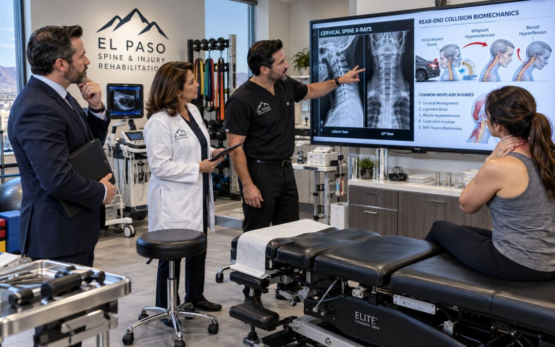
by Dr Alex Jimenez DC, APRN, FNP-BC, CFMP, IFMCP | Auto Accident Injuries, Herniated Disc, Med-Legal Corner, Personal Injury, Spinal Decompression Treatments
Why Mechanism-to-MRI Linkage of Disc Injury Is Required to Justify Lumbar Causation at Trial
Abstract
Defense counsel routinely labels annular tears and modest disc-height loss as “pre-existing degeneration.” A precise mechanism-to-MRI linkage—correlating specific highway force vectors (axial compression, lateral shear, rotation) to the level, side, and morphology of the imaging findings—is required to establish clinical and proximate causation under Texas law and to support special damages for future non-surgical care. This article shows attorneys how structural MRI, timed to the collision and interpreted against the Texas Eggshell Plaintiff framework, neutralizes those challenges while future-care projections for extracorporeal shockwave therapy (ESWT) and spinal decompression document ongoing structural needs.
Imagine the defense expert in an El Paso courtroom pointing at a lumbar MRI and stating, “The annular fissure and 2 mm height loss are age-related.” Without a clear biomechanical bridge from the crash forces to those exact findings, the jury is left with competing medical opinions, and the claim is vulnerable. Mechanism-to-MRI linkage supplies that bridge. It converts a radiologic observation into courtroom-ready evidence of traumatic aggravation.
The Trial Reality of Annular Tears and Disc-Height Challenges
Annular tears (more precisely termed annular fissures in NASS nomenclature) and measured disc-height loss are the two findings most often attacked as pre-existing. Defense radiologists emphasize that high-intensity zones on T2-weighted images can appear in asymptomatic individuals and that modest height loss is common after age 40 (Fardon et al., 2014). Attorneys therefore need more than the finding’s mere presence; they must show that the morphology, laterality, and timing match the collision’s known force vectors and differ from multilevel, symmetric baseline degeneration.
Texas law does not require a perfectly healthy spine. Under the Eggshell Plaintiff doctrine (PJC 28.8 series), the defendant takes the plaintiff as found. If the collision created a new fissure, extended a pre-existing one, or accelerated previously asymptomatic height loss, the resulting functional impairment and future care are compensable (Texas Pattern Jury Charges). The clinical task is to document that acceleration with objective imaging and a reasoned causation narrative.
Correlating Highway Force Vectors to Structural MRI
A high-speed impact on an El Paso corridor transmits forces in milliseconds. Pure axial compression from a rear-end or frontal collision loads the disc vertically, increasing intradiscal pressure and stressing the annulus. Lateral or oblique impacts (common on Loop 375 and similar highways) add shear and rotational components that preferentially load the posterolateral fibers—the anatomically weakest region of the annulus (NCBI StatPearls, 2023). When those forces exceed tissue tolerance, a radial or transverse fissure forms, nuclear material migrates or dehydrates, and measurable height loss appears at the loaded level.
Key imaging correlates that support causation include:
- Single- or dominant-level abnormality matching the predicted segment (commonly L4–L5 or L5–S1 after low-lumbar loading).
- Asymmetric high-intensity zone or height reduction greater on the side of the force vector.
- Temporal relationship: symptoms beginning immediately or within days, with MRI performed after the collision and compared, when available, to any pre-accident imaging.
- Distinction from baseline: the traumatic pattern is focal and asymmetric, while degenerative change is typically multilevel and symmetric.
Clinic notes at Injury Medical Clinic PA capture this linkage in plain language: “The left posterolateral L4–L5 annular high-intensity zone and 2 mm asymmetric disc-height reduction are morphologically and temporally consistent with the lateral shear and rotational forces of the collision. These findings differ from the mild, symmetric multilevel desiccation representing age-related baseline change.” Such statements survive Colossus review and deposition because they rest on biomechanics, not advocacy.
For deeper discussion of how chiropractic and medical records establish causation elements (mechanism, temporal association, biological plausibility, and exclusion of alternatives), see Causation: Understanding Its Role With Chiropractic Care. Attorneys preparing expert reports will also find practical guidance in Medical Legal Causation Opinions for Attorneys Explained.
Future-Care Projections That Support Special Damages
Once the mechanism-to-MRI link is established, the same objective findings justify future non-surgical interventions that restore disc hydration and height. Spinal decompression reduces intradiscal pressure, promotes nutrient inflow, and has been shown to improve height and pain in selected lumbar disc cases (Choi et al., 2017). Extracorporeal shockwave therapy (ESWT) stimulates neovascularization and extracellular-matrix remodeling; recent laboratory and clinical observations indicate it can attenuate endplate degeneration under compressive stress and facilitate resorption of herniated material (Frontiers in Bioengineering and Biotechnology, 2026; Orthopedic Reviews case report, 2026).
These modalities are not speculative. When serial MRI or functional testing demonstrates persistent height loss or annular signal that correlates with ongoing symptoms, a future-care spreadsheet listing a defined course of decompression and ESWT is supported by the same causation evidence that proves the injury. The resulting impairment evidence is packaged inside medical-legal causation documentation standards at https://dralexjimenez.com.
Bioethical Integration of Care
Beneficence is realized when chiropractic spinal adjustments, decompression, and ESWT are combined with MD/NP-directed laboratory and imaging oversight to restore structural integrity at the disc level rather than merely mask symptoms.
Non-maleficence is achieved by prioritizing these non-invasive, drug-free options, thereby delaying or avoiding the risks of surgery or prolonged opioid exposure.
Autonomy places the patient in control of the imaging timeline and treatment decisions. The clinic orders serial MRI scans when clinically indicated, explains results in plain language, and lets the patient coordinate care with any existing medical team.
What Makes the Clinic Unique
- Dr. Jimenez’s Injury Medical Chiropractic and Functional Medicine Clinic is the only practice in El Paso, Texas, that integrates a board-certified internal medicine medical director with a dual-licensed chiropractor/nurse practitioner specializing in soft-tissue and disc injury.
- Dr. Maria Guadalupe Cardenas, MD (Texas License #J2933, NPI 1164426748), provides medical direction, advanced laboratory panels, and risk stratification for complex metabolic comorbidities that influence disc healing.
- Dr. Alex Jimenez, DC, APRN, FNP-BC, CFMP, IFMCP (Texas License #1191402, Prescriptive Authority #59628, NPI 1205907805), delivers structural and regenerative interventions—chiropractic adjustments, spinal decompression, ESWT, and electroacupuncture—while maintaining complete collaborative notes that meet clinical and legal standards. This dual-credentialed, single-clinic model eliminates fragmented records and supplies the continuous, objective documentation attorneys require for Letters of Protection and trial.
From Injury to Recovery (with Chiropractic Care) | El Paso, TX
Practical Documentation Checklist for Counsel
- Accident reconstruction or black-box data establishing direction and approximate magnitude of force.
- Immediate or near-immediate symptom onset recorded in emergency or early clinical notes.
- MRI protocol that includes T2 sagittal and axial images with measurements of height loss and description of annular signal laterality.
- Comparison to any pre-accident imaging or, in its absence, clear language distinguishing focal traumatic morphology from multilevel degenerative baseline.
- Functional testing (range of motion, neurologic exam, patient-reported outcome scores) timed to imaging.
- Future-care cost projections tied to the same structural findings that support causation.
When these elements are present, annular tears and disc-height loss stop being defense talking points and become the foundation of special damages. Patients regain mobility and sleep quality through non-invasive care, while attorneys get courtroom-ready proof that the collision—not ordinary aging—caused measurable structural change.
Schedule a comprehensive evaluation at Injury Medical Clinic PA. The team will map the collision’s force vectors to current MRI findings, design a plan to restore disc hydration and height, and prepare the objective documentation needed for trial or settlement.
References
Choi, J., Lee, S., & Hwangbo, G. (2017). Influences of spinal decompression therapy and general traction therapy on the pain, disability, and straight leg raising of patients with intervertebral disc herniation. Journal of Physical Therapy Science, 29(4), 697–699. https://doi.org/10.1589/jpts.29.697
Fardon, D. F., Williams, A. L., Dohring, E. J., Murtagh, F. R., Rothman, S. L. G., & Sze, G. K. (2014). Lumbar disc nomenclature: Version 2.0: Recommendations of the combined task forces of the North American Spine Society, the American Society of Spine Radiology, and the American Society of Neuroradiology. The Spine Journal, 14(11), 2525–2545. https://doi.org/10.1016/j.spinee.2014.04.022
Frontiers in Bioengineering and Biotechnology. (2026). ESWT attenuates compressive stress-induced cartilaginous endplate degeneration via modulation of the SOST/Wnt/β-catenin signaling pathway. Frontiers in Bioengineering and Biotechnology, 14, Article 1769920. https://doi.org/10.3389/fbioe.2026.1769920
StatPearls. (2023). Annular disc tear. StatPearls Publishing. https://www.ncbi.nlm.nih.gov/books/NBK459235/
Texas Pattern Jury Charges—General Negligence, Intentional Personal Torts & Workers’ Compensation. (2024). Committee on Pattern Jury Charges, State Bar of Texas.
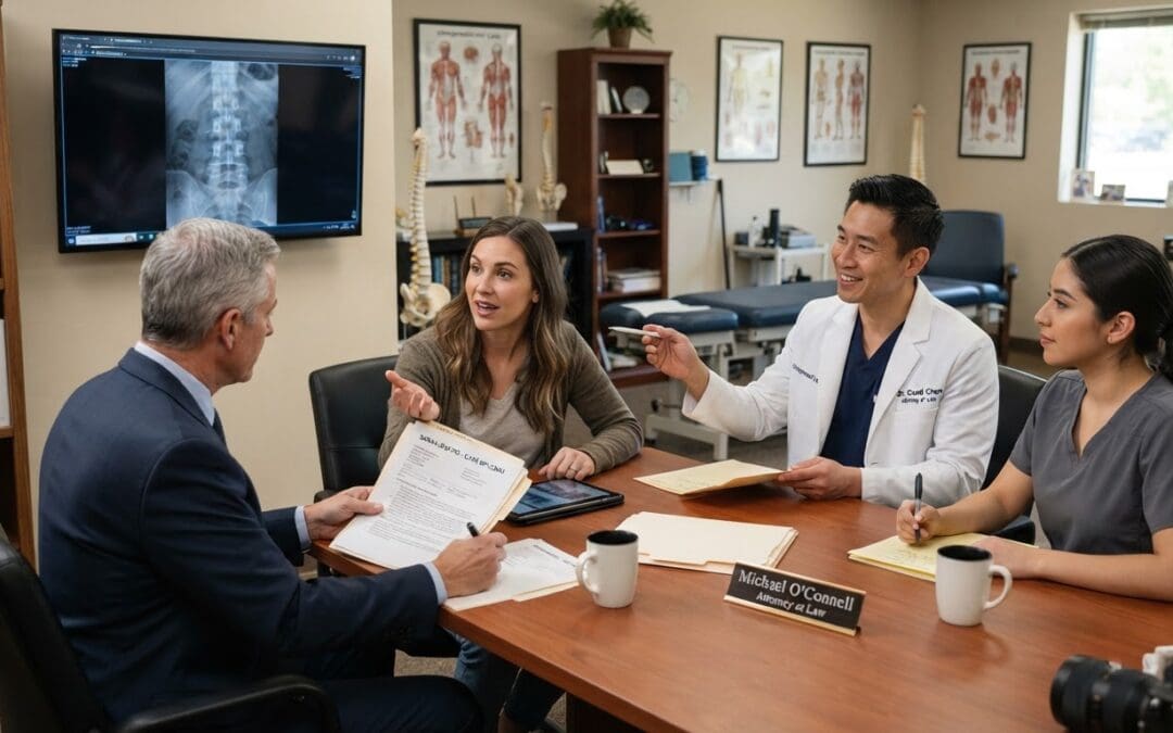
by Dr Alex Jimenez DC, APRN, FNP-BC, CFMP, IFMCP | Chiropractic, Chronic Pain, Health, Injury Care, Personal Injury, Treatments, Viscerosomatic Reflex
Explore the role of causation in personal injury combined with chiropractic care and how it can affect your recovery journey.
Abstract
Causation forms the indispensable legal and clinical bridge that connects a specific traumatic event—such as a motor vehicle collision, workplace incident, or slip-and-fall—to the musculoskeletal injuries that entitle an individual to fair recovery of damages in personal injury claims. Without rigorous demonstration that the incident caused or materially contributed to the diagnosed harm, even substantial pain, functional loss, and documented disability may fail to support full compensation. This educational post, written for attorneys, clinicians, and patients, explains why causation matters in court, how evidence-based standards, including the Bradford Hill criteria and reasonable medical probability, support it, and how modern chiropractic care provides critical contemporaneous clinical insight that strengthens both causation opinions and patient recovery. Chiropractic care is defined as a licensed, evidence-based profession focused on the neuromusculoskeletal system that restores joint motion, modulates pain signaling, reduces local inflammation, and supports natural healing without defaulting to surgery or prolonged pharmaceuticals. Drawing on recent research from leading investigators, the post details the physiological underpinnings of spinal and extremity adjustments—including afferent bombardment of mechanoreceptors, segmental and descending pain modulation, improved local circulation, and effects on inflammatory mediators such as TNF-α, IL-6, and BDNF. It further explains how chiropractic interventions reduce musculoskeletal injuries and their common comorbidities, including central sensitization, compensatory strain, sleep disruption, reduced activity, and opioid reliance. The narrative integrates the multidisciplinary model at Injury Medical Clinic PA (also known as Mission Plaza Injury Medical Clinic) in El Paso, Texas, where Dr. Maria Guadalupe Cardenas, MD, Board Certified in Internal Medicine (NPI #1164426749, Texas MD License #J2933), with more than 40 years of experience as an internist, serves as Medical Director and Collaborative Physician. This setup combines Dr. Jimenez’s chiropractic and advanced-practice nursing expertise with internal medicine oversight, functional medicine, rehabilitation, and non-surgical regenerative options. Clinical observations from decades of practice, along with resources at elpasobackclinic.com and related professional profiles, show how the team delivers durable functional recovery and the objective documentation attorneys need to establish causation to a reasonable degree of medical probability. The journey from injury mechanism through evidence-based conservative care to courtroom-ready insight is presented in clear, sequential sections so readers can follow the logic from biomechanics and physiology to practical legal and clinical application.
Why Causation Is Essential and Required in the Courtroom for Injury Claims
In every personal injury action grounded in negligence, the plaintiff must prove by a preponderance of the evidence—more likely than not—that the defendant owed a duty, breached that duty, and that the breach was both the actual and proximate cause of the claimed injuries and damages. Actual cause, often tested by the “but-for” inquiry, asks whether the injury would have occurred in the absence of the incident. Proximate or legal cause asks whether the harm was a reasonably foreseeable result of the conduct and whether the causal chain is close enough to justify imposing liability. Medical causation sits at the center of this proof: expert testimony is typically required to establish that the trauma produced the specific diagnoses, that the mechanism is biologically plausible, and that alternative explanations are less likely.
Courts and insurance evaluators demand this linkage because pain and functional limitation alone do not establish liability. Pre-existing degenerative changes, prior injuries, or unrelated conditions can produce similar symptoms. Without contemporaneous records that capture the mechanism of injury, the precise timing of symptom onset or aggravation, objective findings such as restricted range of motion, positive orthopedic tests, and correlated imaging, and a treatment response consistent with expected tissue-healing phases, claims are vulnerable to minimization or dismissal. Delayed symptom onset does not defeat causation in soft-tissue injuries. Strains, sprains, facet capsule injuries, and annular disc disruptions often generate progressive inflammation and protective muscle guarding that peak hours to days later as chemical mediators accumulate. Thorough documentation of impact direction, occupant position, restraint use, and progressive clinical findings distinguishes trauma-related injury from the natural history of pre-existing conditions.
The Bradford Hill criteria, originally articulated by epidemiologist Sir Austin Bradford Hill in 1965, supply a structured framework for evaluating whether an observed association is likely causal. These include strength of association, consistency, specificity, temporality (exposure must precede outcome), biological gradient, plausibility, coherence with existing knowledge, experimental evidence, and analogy. In musculoskeletal personal injury, temporality and biological plausibility carry particular weight. Symptoms that begin or clearly intensify after a documented collision whose forces are consistent with known tissue tolerances support the causal inference. When the same treating provider examines the patient early, records baseline objective measures, follows the response to care through the inflammatory, proliferative, and remodeling phases of healing, and rules out more probable alternative causes, the resulting opinion rests on direct clinical observation rather than retrospective speculation.
Recent appellate decisions in multiple jurisdictions confirm that properly qualified chiropractors may offer causation testimony within the scope of their practice. In Virginia, courts have recognized that authorizing chiropractors to testify about etiology and diagnosis within the scope of chiropractic expressly permits opinions on the causation of human physical injury. Michigan appellate rulings have held that licensed chiropractors may diagnose musculoskeletal and nervous system conditions related to subluxations, misalignments, and joint dysfunctions and may therefore develop and testify about the underlying cause of such conditions when a proper foundation is laid. These rulings align with longstanding principles that expert opinions are admissible when they fall within the witness’s specialized knowledge and rest on reliable methodology. Clinical causation (the injury is consistent with the mechanism) must be distinguished from legal causation (more likely than not that the incident caused or substantially contributed to the injury). Treating providers who document both the acute presentation and the recovery trajectory supply the continuity that strengthens the latter.
Legal Justification of Causation Through Evidence-Based Clinical Documentation
The justification for requiring and accepting causation evidence rests on both fairness and scientific reliability. Defendants should not be held liable for conditions that pre-existed or arose independently; plaintiffs should not be denied recovery for genuine trauma-related harm simply because soft-tissue injuries lack dramatic radiographic findings. Expert testimony must meet reliability standards such as those articulated in Daubert v. Merrell Dow Pharmaceuticals, which evaluate whether the theory can be tested, has been subjected to peer review and publication, has a known or potential error rate, is subject to controlling standards, and is generally accepted in the relevant community. Biomechanical analysis that correlates impact severity, occupant kinematics, and tissue-specific tolerances with clinical diagnoses supplies one such reliable methodology.
In the personal injury setting, serial clinical records create a temporal map. An initial examination that records the patient’s description of the mechanism, immediate or progressive symptoms, and objective findings such as limited cervical rotation, positive Spurling’s or straight-leg-raise tests, and postural deviations establishes the baseline. Subsequent visits documenting decreasing pain scores, increasing goniometric ranges of motion, improved strength, and reduced functional limitations show a recovery trajectory consistent with the expected phases of soft-tissue healing. When these improvements occur under conservative care and alternative explanations have been considered and found less likely, the treating provider’s opinion that the incident caused or materially contributed to the injury carries substantial weight. Courts and juries give particular credence to contemporaneous observations by providers who examined the patient, rather than opinions formed solely from later chart review.
Pre-existing conditions do not automatically defeat causation. Aggravation of a previously asymptomatic or minimally symptomatic degenerative spine is compensable when the incident produces new or substantially increased symptoms and functional loss. Documenting the pre-incident baseline—through history, prior records when available, and a clear temporal correlation with worsening—supports the aggravation theory. Gaps in treatment, non-compliance, or inconsistent histories undermine credibility; conversely, consistent, objective, and progressive documentation strengthens it.
What Chiropractic Care Is and How It Supplies Causation Insight in Personal Injury
Chiropractic care is a licensed healthcare profession focused on diagnosing, treating, and preventing disorders of the neuromusculoskeletal system, with particular attention to the spine and extremities. Doctors of Chiropractic complete rigorous graduate education emphasizing anatomy, biomechanics, neurology, and manual therapy. In contemporary, evidence-based practice, the emphasis is on restoring normal joint motion, reducing abnormal mechanical stress and afferent input, modulating pain signaling, and facilitating the body’s capacity for recovery. High-velocity, low-amplitude adjustments and graded mobilizations are core techniques, often combined with soft-tissue methods, rehabilitative exercise, and patient education.
Modern chiropractic practice is grounded in biomechanics and neurophysiology rather than outdated systemic theories. Clinical practice guidelines from multiple organizations support spinal manipulative therapy as part of multimodal care for acute, subacute, and chronic low back pain, neck pain, and certain headache presentations. Systematic reviews and randomized trials demonstrate reductions in pain intensity and disability when chiropractic care is integrated with exercise and education. In the personal injury context, chiropractic care is frequently the patient’s earliest and most continuous contact after trauma. Early evaluation identifies red flags requiring imaging or medical referral, establishes objective baselines, and initiates conservative management while the inflammatory phase remains active.
Chiropractic care and causation proof reinforce each other. The initial examination captures the mechanism in the patient’s words and correlates it with clinical findings—for example, restricted cervical rotation and positive orthopedic tests after a rear-end impact. Serial measurements of range of motion, muscle strength, and functional capacity create a clear timeline. Objective data improve in parallel with expected tissue-healing phases, supporting the conclusion that the trauma produced a treatable injury. Documenting the absence of prior similar symptoms, or the clear aggravation of pre-existing conditions, is essential when the defense argues natural progression of degenerative change. The treating chiropractor’s ongoing relationship provides continuity that a single independent examination cannot. When the same provider observes both the acute presentation and the response to care, the causation opinion rests on direct observation. Consistency across chiropractic and medical notes within an integrated clinic further strengthens the record.
Physiological Underpinnings of Chiropractic Care for Musculoskeletal Injuries
Trauma disrupts joint mechanics and generates continuous abnormal input from mechanoreceptors and nociceptors. This input facilitates segmental spinal cord neurons, expands pain receptive fields, and maintains protective muscle co-contraction that further restricts motion. High-velocity, low-amplitude manipulation produces a brief, high-frequency burst of discharge from muscle spindle primary afferents and other dynamically sensitive receptors. Animal and human studies show that this afferent volley can reduce the excitability of dorsal horn neurons involved in pain transmission and modulate descending inhibitory pathways originating in the brainstem and higher centers. Segmental effects include attenuation of dorsal-horn neuron hyperexcitability; heterosegmental and supraspinal effects involve the periaqueductal gray and cortical networks that process pain and motor control.
Beyond neural effects, restoring joint motion improves local circulation and reduces concentrations of inflammatory mediators in peri-articular tissues. When synovial fluid redistributes and capsular tension normalizes, the chemical environment that sustains peripheral sensitization improves. Soft-tissue techniques address myofascial trigger points and adhesions that develop from muscle guarding. Together, these changes reduce the mechanical and chemical drivers of pain, allowing patients to resume more normal movement patterns that further promote healing.
Recent pragmatic randomized controlled trials have examined biomarker changes after chiropractic spinal adjustments. One 2025 study reported that 12 weeks of care was associated with increased blood brain-derived neurotrophic factor and interleukin-6 levels and decreased tumor necrosis factor-alpha, supporting modulation of neuroplasticity and inflammation. Other work has shown reductions in pro-inflammatory cytokines and increases in anti-inflammatory interleukin-10 following spinal manipulation in models of neuropathic and postoperative pain. These findings align with clinical observations that patients experience not only local pain reduction but also improvements in sleep, mood, and overall function—outcomes consistent with central nervous system modulation.
For disc-related injuries, controlled distraction or flexion-distraction techniques can reduce intradiscal pressure and may improve nutrient diffusion into the avascular disc. Combined with core stabilization exercise, this strategy restores load-sharing among the spine’s passive (ligaments and discs), active (muscles), and neural control subsystems. Patients who regain segmental motion early and then progress through graded loading recover function more reliably than those who remain immobilized or rely solely on passive modalities.
How Chiropractic Care Reduces Musculoskeletal Injuries and Their Comorbidities
Musculoskeletal injuries from trauma commonly initiate a cascade of secondary problems. Restricted motion leads to muscle atrophy and altered recruitment patterns. Persistent nociceptive input can contribute to central sensitization, sleep fragmentation, and reduced physical activity. Reduced activity then worsens cardiovascular fitness, glycemic control, and mood. Chiropractic care interrupts this cascade at several points by restoring motion, decreasing abnormal afferent drive, and enabling earlier graded activity.
Randomized trials show that adding chiropractic care to usual medical management improves low-back-pain intensity and disability, increases patient satisfaction, and reduces self-reported pain-medication use. Observational studies of older adults and Medicare beneficiaries with chronic low back pain and multiple comorbidities have found lower overall costs and lower rates of subsequent surgery among those who received chiropractic manipulative treatment. Systematic reviews support spinal manipulative therapy as part of multimodal care for neck pain and cervicogenic headache. Meta-analyses indicate that receipt of chiropractic care is associated with substantially lower odds of receiving prescription opioids, with greater reductions when care begins early.
In personal injury populations, frequently observed comorbidities include compensatory shoulder or hip strain from altered gait or posture, tension-type or cervicogenic headache secondary to upper cervical dysfunction, and sleep disruption that prolongs recovery. By restoring cervical and lumbar motion, reducing muscle hypertonicity, and enabling earlier return to graded activity, chiropractic care decreases the likelihood that these secondary problems become entrenched. Patients who recover segmental mobility and core endurance report better tolerance for work and daily activities, which supports mental health and reduces the risk of prolonged opioid exposure. Recent cohort data further associate initial spinal manipulative therapy for low back pain with markedly lower rates of subsequent opioid use disorder diagnosis compared with initial ibuprofen.
Integrative Chiropractic Care Within Non-Surgical Treatment Models
Non-surgical management of traumatic musculoskeletal injury is most effective when mechanical, neurological, and systemic factors are addressed together. Integrative chiropractic care combines joint mobilization or manipulation with rehabilitative exercise, soft-tissue therapy, nutritional support for tissue repair, and, when indicated, regenerative options such as platelet-rich plasma under appropriate medical direction. The reasoning is straightforward: restoring motion reduces abnormal loading and afferent drive, while progressive loading stimulates collagen remodeling and motor-pattern retraining. Functional medicine evaluation can identify co-factors—such as vitamin D insufficiency, sleep disruption, or inflammatory dietary patterns—that slow healing even when local mechanics are improved.
At Injury Medical Clinic PA in El Paso, Texas, Dr. Alexander Jimenez, DC, APRN, FNP-BC, CFMP, IFMCP, ATN, CCST, and Dr. Maria Guadalupe Cardenas, MD, bring this model to life through collaboration. Dr. Cardenas, board-certified in Internal Medicine with more than 40 years of experience as an internist (NPI #1164426749, Texas MD License #J2933), serves as Medical Director and Collaborative Physician. This multidisciplinary arrangement is common in integrative injury-care clinics: the physician provides medical direction, orders or interprets advanced diagnostics when needed, manages systemic conditions or medication requirements, and ensures that care remains within appropriate medical standards, while the chiropractor focuses on biomechanical restoration, soft-tissue recovery, and functional rehabilitation. The team integrates chiropractic adjustments and mobilizations, nurse-practitioner-level evaluation, functional medicine assessment, targeted rehabilitation protocols, nutritional optimization, soft-tissue techniques, spinal decompression, and, when appropriate, consultations for regenerative therapies. Documentation remains coordinated so that clinical findings, treatment responses, and medical oversight appear in a consistent record that supports both recovery and causation analysis.
Clinical Observations from Dr. Alexander Jimenez and the Injury Medical Clinic Team
Over more than three decades of practice in El Paso, clinical observations at Injury Medical Clinic PA have repeatedly shown that early restoration of segmental motion, combined with progressive loading and attention to systemic factors, produces more reliable functional recovery than prolonged passive care or immediate reliance on pharmaceuticals alone. Patients presenting after motor vehicle collisions frequently exhibit restricted cervical or lumbar ranges of motion, positive orthopedic tests consistent with soft-tissue strain or facet irritation, and compensatory postural shifts. Serial goniometric measurements and functional outcome tools document progressive improvement that aligns with the phases of tissue healing. When Dr. Cardenas’s medical oversight addresses concurrent internal-medicine issues—such as hypertension, diabetes, or inflammatory markers that may delay recovery—the overall trajectory improves.
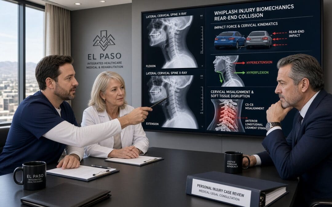
These observations align with published evidence on biomechanical mechanisms of whiplash and soft-tissue injury. Rear-impact collisions produce characteristic S-shaped cervical kinematics with lower-segment extension and upper-segment flexion, loading facet capsules, annular fibers, and supporting ligaments beyond normal physiologic strains in a substantial fraction of cases. Early chiropractic intervention that restores motion and reduces protective muscle guarding interrupts the cycle of ongoing abnormal loading and afferent input. Patients who progress from passive modalities to active stabilization and graded activity report better return-to-work outcomes and fewer secondary complaints. Documenting the mechanism, baseline findings, and serial improvement provides the temporal and clinical consistency attorneys require to establish causation to a reasonable degree of medical probability.
How the Integrative Team Supports Personal Injury Insight and Durable Recovery
The combination of chiropractic expertise and internal-medicine oversight produces records that address both clinical recovery and legal requirements. Objective measurements, treatment responses, and correlated medical findings create a coherent narrative. When alternative causes are considered and found less probable, and when improvement follows expected healing timelines under coordinated care, the resulting expert opinions carry weight. Attorneys benefit from providers who understand the distinction between clinical consistency and the legal standard of reasonable medical probability, document thoroughly from the first visit, and coordinate across disciplines to keep the record internally consistent.
Non-surgical integrative care further reduces the likelihood of escalation to invasive procedures when conservative measures succeed. By addressing biomechanics, inflammation, motor control, and systemic co-factors simultaneously, the approach supports durable function while generating the contemporaneous evidence that justifies causation in the courtroom. Patients recover mobility and reduce pain; the documentation simultaneously strengthens the causal link between the incident and the injuries.
Conclusion
Causation remains the essential requirement that links traumatic events to compensable musculoskeletal injuries in the courtroom. Its justification rests on fairness, scientific reliability, and the need for opinions grounded in reliable methodology and contemporaneous clinical observation. Modern, evidence-based chiropractic care provides this insight by documenting mechanism, objective findings, and recovery trajectories while reducing pain, restoring motion, modulating inflammation and neural signaling, and interrupting the cascade toward comorbidities such as central sensitization, compensatory strain, and opioid reliance. When paired with medical oversight from a board-certified internist such as Dr. Maria Guadalupe Cardenas at Injury Medical Clinic PA, the model delivers durable non-surgical recovery and the coordinated documentation attorneys require. Drawing on the latest research in biomechanics, neurophysiology, and clinical outcomes, this approach offers a clear, evidence-informed pathway from injury event through functional restoration to courtroom-ready causation support.
References
American Medical Association. (n.d.). Maria Guadalupe Cardenas, MD. AMA Find a Doctor. https://find-doctor.ama-assn.org/doctor/maria-guadalupe-cardenas-md/13591899
Basques, B. A., et al. (2025). Chiropractic care and spinal manipulation: Evidence, risks, and referral considerations for spine surgeons. JBJS Reviews, 13(12), e25.00196. https://doi.org/10.2106/JBJS.RVW.25.00196
Bronfort, G., Meier, E. N., Leininger, B., et al. (2025). Spinal manipulation and clinician-supported biopsychosocial self-management for acute back pain: The PACBACK randomized clinical trial. JAMA. Advance online publication. https://doi.org/10.1001/jama.2025.21990
Gevers-Montoro, C., Provencher, B., Descarreaux, M., Ortega de Mues, A., & Piché, M. (2021). Neurophysiological mechanisms of chiropractic spinal manipulation for spine pain. European Journal of Pain, 25(7), 1429–1448. https://doi.org/10.1002/ejp.1773
Jimenez, A. D. (2026). Causation and chiropractic care: Addressing pain and mobility. El Paso Back Clinic. https://elpasobackclinic.com/causation-and-chiropractic-care-addressing-pain-and-mobility/
Lisi, A. J., et al. (2025). The impact of chiropractic care on opioid prescriptions in Veterans Health Administration patients receiving low back pain care. Journal of General Internal Medicine. Advance online publication.
Song, X. J., et al. (2016). Attenuation effect of spinal manipulation on neuropathic and postoperative pain through activating endogenous anti-inflammatory cytokine interleukin 10 in rat spinal cord. Journal of Manipulative and Physiological Therapeutics, 39(1), 42–53. https://doi.org/10.1016/j.jmpt.2015.12.004
Trager, R. J., et al. (2025). Association between spinal manipulative therapy for low back pain with or without sciatica and opioid use disorder: A retrospective cohort study. Health Science Reports, 8, e71267.
Virginia Lawyers Weekly. (2025, November 4). Chiropractor can testify on causation of man’s injuries. https://valawyersweekly.com/2025/11/04/virginia-chiropractor-expert-testimony-new-trial/
SEO Tags
causation in personal injury, chiropractic care for musculoskeletal injuries, evidence-based chiropractic personal injury, integrative non-surgical treatment El Paso, Dr Alexander Jimenez DC APRN, Dr Maria Guadalupe Cardenas MD, Injury Medical Clinic PA, Bradford Hill criteria musculoskeletal, spinal manipulation inflammation biomarkers, opioid reduction chiropractic, whiplash biomechanics, expert testimony chiropractor causation, functional medicine injury recovery, non-surgical rehabilitation trauma, El Paso chiropractic, personal injury documentation
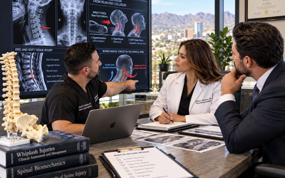
by Dr Alex Jimenez DC, APRN, FNP-BC, CFMP, IFMCP | Auto Accident Injuries, Collision & Injury Dynamics, Med-Legal Corner, Personal Injury, Spinal Decompression Treatments
Annular Tear and Disc Height Loss After Loop 375 Lateral Impact: Mechanism-to-MRI Linkage
A high-speed side impact on El Paso’s Loop 375 can transmit focused lateral shear, axial compression, and rotational forces through the lumbar or cervical spine in a fraction of a second. When those force vectors exceed the tensile strength of the annulus fibrosus—particularly its relatively weaker posterolateral fibers—an acute annular tear can form, allowing nuclear material to migrate and producing measurable disc height loss visible on MRI. At Injury Medical Clinic PA in El Paso, the multidisciplinary team of Dr. Alex Jimenez, DC, APRN, FNP-BC, CFMP, IFMCP (Texas License #1191402, Prescriptive Authority #59628, NPI 1205907805) and Medical Director Dr. Maria Guadalupe Cardenas, MD (Board Certified Internal Medicine, Texas License #J2933, NPI 1164426748), systematically links these biomechanical mechanisms to objective imaging so that clinical and proximate causation are clear for patients and attorneys.
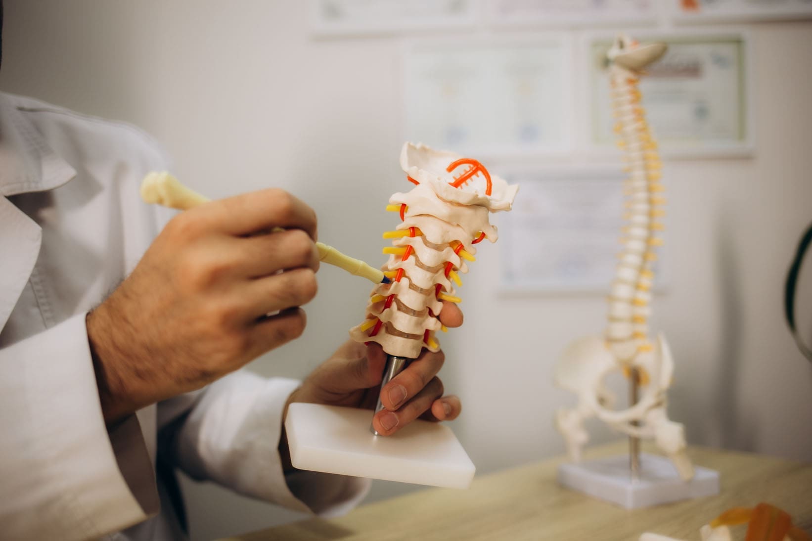
How a Loop 375 Lateral Impact Generates Disc Injury
Loop 375 (Joe Battle Boulevard and related segments) is a high-volume corridor where lane changes, side-swipes, and T-bone or angle collisions are common. In a pure lateral impact, the striking vehicle imparts a force vector roughly perpendicular to the struck vehicle’s longitudinal axis. Occupants experience rapid lateral torso acceleration while the pelvis and lower extremities lag or rotate, creating shear across the intervertebral discs and facet joints. At the same time, the sudden change in velocity produces axial compression and, often, a twisting moment as the body rebounds against the seatbelt or door.
The annulus fibrosus consists of concentric lamellae whose fiber orientation alternates for strength; however, the posterolateral region contains a higher proportion of vertically oriented fibers and is a known site of relative weakness (StatPearls, 2023). When lateral shear and rotation are superimposed on compression, these fibers can separate, producing a radial or transverse annular tear. Nuclear material may then extrude or lose hydration and volume, resulting in a measurable reduction in disc height at the affected level. Because the forces are asymmetric, the tear and height loss are often unilateral or more pronounced on the side of impact—an important morphologic clue when compared with the more uniform, multilevel pattern of ordinary age-related degeneration.
MRI Linkage: Reading the Images Through the Force Vectors
On T2-weighted MRI, an acute annular tear typically appears as a high-intensity zone (HIZ)—a focal bright signal within the normally dark annulus caused by increased water content and inflammatory fluid (StatPearls, 2023; Teraguchi et al., 2022). Supporting signs of acute traumatic change, as opposed to a chronic degenerative baseline, include:
- A single or dominant level of abnormality that matches the predicted biomechanics of the impact (for example, L4-L5 or L5-S1 after a low lumbar lateral load, or mid-cervical levels after a higher side-impact).
- Asymmetric height loss or annular signal that is greater on the side of the force vector.
- Associated soft-tissue findings such as ligamentous edema, facet fluid, or paraspinal muscle signal change at the same or adjacent levels.
- Higher T2 hyperintensity and involvement of the outer third of the annulus are more consistent with traumatic disruption than the lower-intensity, inner-two-thirds fissures typical of slow degenerative breakdown (Khurana et al., 2025).
Degenerative disc disease is extremely common and increases with age; imaging studies of asymptomatic volunteers show annular fissures in roughly 19–29% of adults depending on the decade (Brinjikji et al., as cited in clinical radiology reviews). The clinic therefore never equates the mere presence of an HIZ or reduced disc height with trauma. Instead, the team correlates the precise morphology, laterality, and associated acute soft-tissue changes with the documented mechanism, the patient’s immediate post-collision symptoms, and serial clinical examinations. When pre-existing degeneration is present, the records explicitly separate the baseline findings from the new traumatic aggravation—language that prevents defense arguments that “the MRI only shows wear and tear.”
Distinguishing Acute Traumatic Change from Degenerative Baseline
Clear, plain-language documentation is essential. An example of phrasing used in clinic notes and narrative reports reads: “The left posterolateral L4-L5 annular high-intensity zone and 2 mm asymmetric disc-height reduction are morphologically and temporally consistent with the lateral shear and rotational forces of the 14 July Loop 375 side-impact collision. These findings differ from the mild, symmetric multilevel disc desiccation and small inner annular fissures that represent age-related baseline change.” Such statements rest on the mechanism-to-MRI linkage rather than on the radiologist’s impression alone. Digital imaging, functional testing (range-of-motion, neurologic, and strength measures), and Colossus-compliant electronic records further corroborate that the structural changes produce the patient’s objective functional deficits.
Future-Care Modeling with ESWT and Non-Surgical Decompression
Once causation is established, the clinic models evidence-based future care to address both the annular tear and height loss. Non-surgical spinal decompression creates controlled negative intradiscal pressure that can increase disc height and reduce pain; one retrospective cohort demonstrated a statistically significant correlation between restored disc height and decreased low-back pain scores (Apfel et al., 2010). Additional studies confirm improvements in radicular pain and disc morphology after motorized decompression protocols (Choi et al., 2022; Egyptian Rheumatology and Rehabilitation, 2019).
Extracorporeal shock-wave therapy (ESWT) supplies a complementary biologic stimulus. In laboratory models, low-energy ESWT combined with low-tension traction has been shown to restore disc height, improve rehydration, and reduce matrix-degrading enzymes (MMP-3, MMP-13, ADAMTS-4), thereby reshaping the microenvironment for repair (Spine Journal, 2021). Clinical series report pain reduction and facilitation of herniation resorption when ESWT is integrated into staged conservative care (Orthopedic Reviews, 2026; Rehabilitation Medicine, 2024). Because both modalities are non-invasive and drug-free, they align with non-maleficence: they lower the risk of progression to surgery or long-term opioid dependence while still delivering measurable structural and symptomatic benefit.
Unlocking Pain Relief: How We Assess Motion to Alleviate Pain | El Paso, TX
In the future-care cost spreadsheet these interventions appear as a defined course of decompression sessions (typically 20–30 over 6–8 weeks) plus a series of ESWT treatments, accompanied by supportive chiropractic adjustments, neurofunctional acupuncture or electroacupuncture, and functional-medicine laboratory monitoring directed by Dr. Cardenas. The combination targets root-cause healing—restoring disc height and annular integrity where possible, reducing inflammatory cytokines, and improving segmental stability—rather than merely masking pain (beneficence). Patients receive clear education about expected timelines, self-care strategies, and how the plan coordinates with any outside specialists they already see, preserving autonomy and informed decision-making.
Documentation That Withstands Scrutiny
Every element—mechanism narrative, MRI description distinguishing acute from baseline change, functional deficits, and itemized future-care costs—is recorded in a manner suitable for letters of protection and deposition. The dual-licensed perspective of Dr. Jimenez (structural alignment plus advanced practice medical diagnostics), together with Dr. Cardenas’s internal-medicine oversight, supplies both the biomechanical and medical foundation attorneys require. The result is a transparent, mechanism-linked record that supports the patient’s recovery while meeting El Paso personal-injury litigation evidentiary standards.
Patients and counsel seeking this level of integrated causation analysis and future-care planning may contact Injury Medical Clinic PA for a coordinated evaluation.
References
Apfel, C. C., Cakmakkaya, O. S., Martin, W., Richmond, C., Macario, A., George, E., Schaefer, M., & Pergolizzi, J. V. (2010). Restoration of disk height through non-surgical spinal decompression is associated with decreased discogenic low back pain: A retrospective cohort study. BMC Musculoskeletal Disorders, 11, Article 155. https://doi.org/10.1186/1471-2474-11-155
Choi, E., Gil, H. Y., Ju, J., Han, W. K., Nahm, F. S., & Lee, P. B. (2022). Effect of nonsurgical spinal decompression on intensity of pain and herniated disc volume in subacute lumbar herniated disc. International Journal of Clinical Practice, 2022, Article 6343837. https://doi.org/10.1155/2022/6343837
Khurana, B., Khorasani, M., & et al. (2025). Lumbar disc annular tear versus annular fissure: Distinguishing traumatic from degenerative findings on MRI. [PDF]. https://img1.wsimg.com/blobby/go/b1a39d28-bc5a-416f-9f65-e6d871130b13/downloads/f8cc02aa-ed0e-4623-a8fc-400307687027/Khurana%20et%20al%2C%2C%202025%20-%20Lumbar%20disc%20annular%20tea.pdf
National Center for Biotechnology Information. (2023). Annular disc tear. In StatPearls. StatPearls Publishing. https://www.ncbi.nlm.nih.gov/books/NBK459235/
Orthopedic Reviews. (2026). Spontaneous resorption of prolapsed lumbar disc herniation facilitated by extracorporeal shock wave therapy: A case report. Orthopedic Reviews, 17(1). https://orthopedicreviews.openmedicalpublishing.org/article/172374-spontaneous-resorption-of-prolapsed-lumbar-disc-herniation-facilitated-by-extracorporeal-shock-wave-therapy-a-case-report
Rehabilitation Medicine. (2024). Effect of extracorporeal shock wave therapy on different types of lumbar disc herniation. Rehabilitation Medicine, 34(1), 84–89.
Spine Journal. (2021). Low energy extracorporeal shock wave therapy combined with low tension traction can better reshape the microenvironment in degenerated intervertebral disc regeneration and repair. The Spine Journal, 21(1), 160–177. https://doi.org/10.1016/j.spinee.2020.08.012
Teraguchi, M., Cheung, J. P. Y., & Samartzis, D. (2022). High-intensity zones and annular tears. In Spine phenotypes (pp. 187–201). Academic Press. https://doi.org/10.1016/B978-0-12-822778-7.00016-X
El-Sayed, A. M., et al. (2019). Spinal decompression therapy as an alternative modality for management of low back pain and radicular pain caused by lumbar disc herniation or protrusion. Egyptian Rheumatology and Rehabilitation, 46, 161–168. https://doi.org/10.4103/err.err_34_18
Brinjikji, W., Luetmer, P. H., Comstock, B., Bresnahan, B. W., Chen, L. E., Deyo, R. A., Halabi, S., Turner, J. A., Avins, A. L., James, K., Wald, J. T., Kallmes, D. F., & Jarvik, J. G. (2015). Systematic literature review of imaging features of spinal degeneration in asymptomatic populations. American Journal of Neuroradiology, 36(4), 811–816. https://doi.org/10.3174/ajnr.A4173
by Dr Alex Jimenez DC, APRN, FNP-BC, CFMP, IFMCP | Auto Accident Injuries, Back Pain, Herniated Disc, Imaging & Diagnostics, Lawyers, Med-Legal Corner, Spinal Decompression Treatments
Segmental Disc Compression Patterns Generated by Loop 375 Lateral Impacts
Abstract
Loop 375 lateral impacts in El Paso generate rapid, side-directed force vectors that compress lumbar segments, tear the annulus, and accelerate disc height loss visible on structural MRI. These patterns create clear mechanism-to-injury linkages that support clinical and proximate causation for lumbar claims. At Injury Medical Clinic PA, the dual-licensed team of Dr. Alex Jimenez, DC, APRN, FNP-BC, and collaborating medical director Dr. Maria Guadalupe Cardenas, MD, documents these objective findings with Colossus-compliant notes, functional testing, and imaging timelines. Non-invasive care—chiropractic decompression and ESWT—restores disc hydration and height, reduces surgical risk, and keeps patients in control of their recovery decisions. The resulting impairment ratings are packaged inside the medical-legal causation documentation framework at https://dralexjimenez.com.

Highway-side impacts on El Paso’s Loop 375 do more than crumple doors. They deliver concentrated lateral force vectors that simultaneously load lumbar discs in compression, shear, and side-bending. The result is a recognizable pattern of segmental disc compression: annular fissures, progressive height loss, and altered load-sharing that shows up clearly on structural MRI. For personal-injury attorneys and patients, this pattern clearly links crash mechanics to documented objective injury.
How Lateral Force Vectors Create Segmental Compression
In a typical Loop 375 side-impact or side-swipe, the struck vehicle’s occupant experiences a rapid lateral acceleration. The lumbar spine, already bearing axial load, is forced into lateral bending while the upper body lags. Finite-element and cadaver studies show that axial compression plus lateral bending produces peak stresses along the posterolateral annulus—the same region where clinical annular tears most often appear.
The outer annulus, whose fibers run nearly vertical in the posterolateral quadrant, is mechanically weakest in this direction. Once a tear begins, nuclear material can migrate, and the disc loses height. Sequential MRI demonstrates the progression: an initially bright T2 fissure evolves into a broader annular disruption accompanied by measurable reduction in disc height at the involved segment (commonly L4–L5 or L5–S1).
MRI Correlation That Withstands Scrutiny
Structural MRI is the cornerstone of objective documentation. High-resolution T2 sequences reveal hyperintense annular fissures; sagittal and axial measurements quantify disc-height loss relative to adjacent normal levels. When these imaging findings are correlated with the known crash kinematics—ΔV, principal direction of force, and seat-belt usage—the clinical causation statement becomes precise: the lateral impact produced the observed segmental compression pattern and annular disruption.
Digital diagnostic imaging, functional range-of-motion and strength testing, and detailed electronic notes are packaged so they align with Colossus valuation factors. These records also support Letters of Protection and provide a clear timeline that patients and counsel can share.
Beneficence: Restoring Disc Hydration and Height
The clinical goal is root-cause recovery, not symptom suppression. Non-surgical spinal decompression creates negative intradiscal pressure that draws nuclear material centrally and allows water and nutrients to re-enter the disc. When low-energy extracorporeal shockwave therapy (ESWT) is combined with controlled traction, animal and clinical data show improved disc height, rehydration, and favorable remodeling of the annular matrix.
At the clinic, Dr. Jimenez (DC, APRN, FNP-BC, CFMP, IFMCP; Texas License #1191402, Prescriptive Authority #59628, NPI 1205907805) integrates these mechanical therapies with targeted regenerative options under the medical direction of Dr. Cardenas, MD (Board Certified Internal Medicine, Texas License #J2933, NPI 1164426748). The combination addresses both the structural compression and any concurrent metabolic or inflammatory factors that slow healing.
Non-Maleficence: Delaying or Avoiding Surgery
Invasive procedures carry risks of infection, hardware, and adjacent-segment disease. By restoring disc height and reducing annular stress through decompression and ESWT, many patients experience durable pain relief and improved mobility without immediate surgical intervention. This approach minimizes iatrogenic harm while still producing the objective measurements insurers and defense counsel expect.
Autonomy: Patient-Owned Imaging and Decision Making
Patients retain full access to their imaging timeline and functional data. They review the mechanism-to-injury linkage with both the clinical team and their attorney, then choose the care pathway that fits their goals. Coordination with existing medical providers remains seamless; the clinic’s dual-licensure model keeps chiropractic, acupuncture/electroacupuncture, and advanced medical diagnostics within one coordinated plan.
Future-Care Projections That Drive Case Value
Documented residual disc-height loss and annular disruption support projections for ongoing decompression sessions, periodic ESWT, and monitoring MRI. These future-care items are stated in plain language and tied directly to the original lateral-impact mechanism, so they withstand Colossus review and support appropriate impairment ratings.
The resulting impairment ratings are packaged inside the medical-legal causation documentation framework at https://dralexjimenez.com.
Detailed Biomechanics of Loop 375 Lateral Impacts
Loop 375’s high-speed corridors and frequent lane-change geometry produce a distinctive set of force vectors. When a vehicle is struck broadside or sideswiped, the occupant’s torso is accelerated laterally while the pelvis remains partially restrained by the seat. The resulting moment arm loads the lumbar spine in combined compression, lateral bending, and shear.
Peak annular stresses concentrate at the posterolateral annulus of L4–L5 and L5–S1. In vitro studies using complex loading protocols that include lateral bending demonstrate endplate junction failures and isolated outer-annulus disruptions—exactly the lesions seen clinically after side impacts.
These mechanics explain why patients present with segmental rather than diffuse disc changes. The MRI shows focal height loss and annular high-intensity zones at the levels that experienced the greatest lateral moment. Correlating the police report’s principal direction of force with these segmental findings supplies the proximate-causation statement required for both medical and legal purposes.
Structural MRI Protocols and Quantification
Standard lumbar MRI protocols are augmented with thin-slice T2 sequences and precise mid-sagittal height measurements. Disc height is compared with the adjacent normal disc and with published normative data. We grade annular tears by radial extent and T2 signal intensity. When serial imaging is available, the timeline shows progressive height loss beginning after the documented impact rather than from age-related change alone.
These measurements are entered into the electronic record in a Colossus-ready format: diagnosis codes, objective findings, and functional limitations appear in structured fields. The same images are released to counsel so the patient retains ownership of the visual evidence.
Clinical Examination and Functional Correlates
The physical examination maps MRI findings to clinical signs: restricted lateral flexion, positive straight-leg or femoral-nerve tension, and segmental motion restriction. Instrumented strength and range-of-motion testing quantifies the deficit. These functional data corroborate imaging and show that segmental compression produces real activity limitations—information that directly influences impairment ratings and future care needs.
Multidisciplinary Treatment: Decompression and ESWT
Chiropractic decompression tables apply computer-controlled distraction forces that cycle between hold and rest phases, generating negative intradiscal pressures sufficient to retract nuclear material and promote rehydration. ESWT delivers acoustic energy that, when combined with traction, down-regulates matrix-degrading enzymes and stimulates annular repair.
Sessions are scheduled according to the severity of height loss and annular disruption. We track progress with clinical scores and, when indicated, follow-up MRI. The dual-licensed model allows Dr. Jimenez to integrate these mechanical therapies with medical oversight from Dr. Cardenas, ensuring concurrent management of any metabolic factors.
Spinal Decompression in Depth | El Paso, TX
Bioethical Framework in Practice
Beneficence is realized when disc height and hydration improve and patients regain mobility and sleep. Non-maleficence is served by prioritizing non-invasive options that delay or eliminate the need for fusion or discectomy. Autonomy is preserved because every patient receives a clear explanation of the mechanism, imaging findings, and expected outcomes of each treatment choice; the patient retains possession of the imaging timeline and shares it with counsel at the patient’s direction.
Documentation That Survives Defense Scrutiny
Every encounter generates Colossus-compliant electronic notes that link the original crash kinematics to current objective findings. Digital imaging is archived with measurement annotations. Functional testing results are dated and signed. The complete package supports both the Letter of Protection and the impairment rating that is later incorporated into the medical-legal causation report hosted at https://dralexjimenez.com.
Impairment Ratings and Future-Care Projections
Residual disc-height loss and annular disruption are translated into AMA Guides impairment percentages when appropriate. Future-care projections specify the number and frequency of decompression and ESWT sessions needed to maintain restored height and prevent further annular progression. These projections are written in plain language and tied explicitly to the Loop 375 lateral-impact mechanism.
Coordination with Legal Counsel and Existing Providers
The clinic supplies counsel with a concise mechanism-to-injury summary, annotated imaging, and the functional data set. When a patient already has a primary-care or specialty physician, the dual-licensure team coordinates with that relationship rather than replacing it, preserving continuity while adding the structural and regenerative expertise needed for the injury claim.
Multidisciplinary Call to Action and Reference List
Patients injured in Loop 375 lateral impacts and their attorneys can obtain a coordinated evaluation that links crash mechanics to MRI-documented segmental disc compression, restores disc height non-surgically through decompression and ESWT, and packages the entire record for legal use.
Call to Action
Schedule a comprehensive evaluation at Injury Medical Clinic PA in El Paso. Dr. Alex Jimenez, DC, APRN, FNP-BC, and Dr. Maria Guadalupe Cardenas, MD, will map the force vectors of your collision to your current MRI findings, design a drug-free plan to restore disc hydration and height, and prepare the objective documentation your counsel needs. Contact the clinic or visit https://dralexjimenez.com to begin.
References
- Che, Y.-J., Guo, J.-B., Wang, Y.-F., Zhu, L.-M., Zhou, C.-X., & Liu, Y.-L. (2021). Low energy extracorporeal shock wave therapy combined with low tension traction can better reshape the microenvironment in degenerated intervertebral disc regeneration and repair. The Spine Journal, 21(1), 91–102. https://doi.org/10.1016/j.spinee.2020.08.004
- Ghavam, K., & Standring, S. (2023). Annular disc tear. In StatPearls. StatPearls Publishing. https://www.ncbi.nlm.nih.gov/books/NBK459235/
- Disc injury and spine loads in low-to-moderate-severity frontal impacts. (2025). PMC. https://pmc.ncbi.nlm.nih.gov/articles/PMC12457475/
- Stress evaluation along the posterior annular circumferential tears on the L5-S1 spinal unit as an index of tear progression. (2025). ScienceDirect. https://www.sciencedirect.com/science/article/pii/S0972978X25000261
- Influence of complex loading conditions on intervertebral disc failure. (2016). PubMed. https://pubmed.ncbi.nlm.nih.gov/27187053/
- Posttraumatic changes of the intervertebral disc. (2017). In Wheeless’ Textbook of Orthopaedics. https://www.wheelessonline.com/ISSLS/section-17-chapter-7-posttraumatic-changes-of-the-intervertebral-disc/
- Regional variations of mechanical responses of IVD to 7 different motions: An in vivo study combined with FEA and DFIS. (2024). ScienceDirect. https://www.sciencedirect.com/science/article/abs/pii/S175161612400417X
- Effect of extracorporeal shock wave therapy on different types of lumbar disc herniation. (2024). ResearchGate. https://www.researchgate.net/publication/385851112

by Dr Alex Jimenez DC, APRN, FNP-BC, CFMP, IFMCP | Lower Back Pain, Posture, Spine Care, Work Related Injuries
Why Your Low Back Burns After Standing at a Counter or Workbench
Abstract: Standing still at a counter or workbench loads the low back differently from walking. This post explains static lumbar loading, trunk endurance, hip position, and forward reach, then outlines chiropractic assessment and ways to vary position and build standing tolerance for data-center technicians, Amazon workers, lab and network technicians, and hybrid employees.
The burn starts quietly. You finish a server-rack check, a packing-station count, or a lab-bench setup, and your low back feels tight and hot. Walking the aisle felt fine. Standing still at the counter, however, did not feel fine. Static standing holds the lumbar spine under continuous load without the regular movement that walking provides.
Data-center technicians, Amazon fulfillment associates, lab personnel, network technicians, and hybrid employees often move between desk time and standing tasks. The low back may tolerate walking across the floor, then complain once the body locks in place at the workbench. Building better standing tolerance starts with understanding that difference, not simply being told to “stand straight.”
How Static Standing Loads the Lumbar Spine
When you walk, the pelvis tilts slightly with each step, and the back extensors alternate between brief work and brief rest. Standing still removes that rhythm. The lumbar extensors must hold an isometric contraction to keep the trunk upright. That continuous demand is called static lumbar loading.
Research shows that people who develop low back pain during prolonged standing often start with lower trunk endurance and different muscle activation patterns. One study found that participants who reported pain after two hours of standing had shorter side-bridge endurance times and higher co-activation of the gluteus medius muscles (Marshall et al., 2011). Another analysis linked higher estimated intervertebral disc compressive force during quiet standing with low back pain (Hasegawa et al., 2018). The load isn’t huge compared with heavy lifting, yet it stays on the tissues without the brief unloading movement provides.
Reaching forward at a counter or workbench increases the moment arm, so the back extensors work harder to keep the trunk from falling forward. Leaning onto the counter can feel like a rest, yet it often shifts the pelvis and may increase uneven loading if the hips are already tight from earlier desk time. Hybrid workers who sit for long blocks and then stand at a bench often carry that sitting posture into standing.
Why Standing Still Feels Different from Walking
Walking activates the calf muscle pump and repeatedly changes spinal posture. Standing still reduces that activity and keeps joints in the same positions for minutes at a time. A laboratory comparison found that standing produced less lower-leg swelling and muscle fatigue than walking (Wall et al., 2020). The same principle applies higher up: the low back receives less recovery when the body remains fixed.
People who develop standing-related low back pain also tend to move less early in a standing period and show higher gluteus medius co-activation (Khoshroo et al., 2023). Less movement means the tissues stay under the same stress longer. For an Amazon associate at a packing station or data center technician at a raised bench, those minutes add up across a shift.
Trunk Endurance and Hip Position Matter More Than “Stand Straight”
Telling someone to stand straight often increases effort if trunk endurance is low or if the hips cannot easily reach a neutral position. After hours at a desk, many hybrid employees and network technicians have shortened hip flexors and reduced glute endurance. When they stand, the pelvis may tip forward, increasing lumbar lordosis and the demand on the back extensors. Side-bridge endurance is a practical marker of the capacity that helps people tolerate standing without early pain (Marshall et al., 2011).
Isometric trunk training that builds endurance while keeping the spine relatively neutral has been shown to reduce pain and disability in chronic low back pain. The goal is the ability to hold useful positions for the minutes a work task requires, not maximum strength in one lift.
Chiropractic Assessment for Standing Tolerance
A focused chiropractic evaluation looks at how you stand, how your hips move, how long you can hold a side-bridge or similar endurance position, and whether forward reach or counter-lean changes your symptoms. We check lumbar joint motion, pelvic alignment, and your ability to shift weight because limited joint motion can force muscles to work harder.
Dr. Alex Jimenez, DC, APRN, FNP-BC, CFMP, IFMCP, brings dual licensure as a chiropractor and board-certified family nurse practitioner. At Injury Medical Clinic PA in El Paso, he works with Dr. Maria Guadalupe Cardenas, MD, the medical director with more than forty years of internal medicine experience. Together, they match structural findings with any medical factors that could affect recovery. Beneficence means the plan exists to improve function and reduce unnecessary risk. Non-maleficence is served by starting with non-invasive options—joint motion restoration, endurance work, and position changes. Autonomy is protected when you understand the findings and help decide which habits fit your shifts.
Building Standing Tolerance with Position Variation and Conditioning
Position variation supports the research on movement and recovery. Useful options include:
- Shifting weight from one foot to the other every minute or two.
- Placing one foot on a low step or the bottom shelf of a workbench for short periods.
- Taking two or three short steps in place or away from the counter before returning.
- Alternating a slight lean with an upright hold rather than locking into one angle.
- Avoiding locked knees and continuous forward reach when the task allows a closer stance.
These are not substitutes for conditioning. Progressive hip and trunk work builds the endurance linked to better standing tolerance. Side bridges, bird-dog holds, glute bridges, and controlled hip-hinge patterns can be practiced in short sessions. The target is the ability to stand for a typical task block with less burn, not a perfect posture held for an entire shift.
For data-center technicians moving between racks and benches, Amazon workers at packing stations, lab personnel at fixed counters, and hybrid employees alternating desk blocks with standing work, the same principles apply. The surface and reach distance change; the need for endurance and variation does not.
When to Seek Coordinated Care
Most standing-related low back pain improves with better load management and capacity. Red flags such as progressive leg weakness, bowel or bladder changes, or pain that wakes you from sleep need prompt medical review. Dr. Cardenas provides medical oversight for complex cases, while Dr. Jimenez coordinates the mechanical plan. Many group insurance plans cover the initial evaluation; the clinic can help clarify benefits.
The aim is to raise the threshold at which the low back begins to burn so you can finish the shift with less residual tightness. That outcome comes from matching assessment to your work demands, then practicing the variation and conditioning that support them.
Call 915-850-0900 or visit the clinic scheduler to arrange an evaluation focused on your standing tolerance and work tasks. You remain the decision-maker; the team supplies the information you need to choose the next step.
Spinal Decompression in Depth | El Paso, TX
References
Hasegawa, T., Katsuhira, J., Oka, H., Fujii, T., & Matsudaira, K. (2018). Association of low back load with low back pain during static standing. PLOS ONE, 13(12), e0208877. https://doi.org/10.1371/journal.pone.0208877
Khoshroo, F., Seidi, F., Bayattork, M., & Minoonejad, H. (2023). Distinctive characteristics of prolonged standing low back pain developers’ and the associated risk factors: Systematic review and meta-analysis. Scientific Reports, 13, 6392. https://doi.org/10.1038/s41598-023-33590-5
Marshall, P. W. M., Patel, H., & Callaghan, J. P. (2011). Gluteus medius strength, endurance, and co-activation in the development of low back pain during prolonged standing. Human Movement Science, 30(1), 63–73. https://doi.org/10.1016/j.humov.2010.08.017
Wall, R., Garcia, G., Läubli, T., Seibt, R., Rieger, M. A., Martin, B., & Steinhilber, B. (2020). Physiological changes during prolonged standing and walking considering age, gender and standing work experience. Ergonomics, 63(5), 579–592. https://doi.org/10.1080/00140139.2020.1725145

by Dr Alex Jimenez DC, APRN, FNP-BC, CFMP, IFMCP | Lower Back Pain, Power & Strength, Spine Care, Work Related Injuries
Why Repeated Small Bends Can Bother Your Back More Than One Heavy Lift
Abstract
Repeated small bends for packages, cables, laundry, or drawers can irritate the lower back more than one controlled heavy lift. This article explains cumulative spinal loading, how fatigue and reach distance change forces on the discs and ligaments, and why limited hip mobility and trunk endurance shift more work onto the lumbar spine. It outlines a chiropractic assessment process, progressive hip and trunk conditioning, practical task changes, and a plan to build bending tolerance safely rather than avoid spinal flexion. The goal is better function for Amazon associates, data center technicians, parents, warehouse workers, and desk professionals.
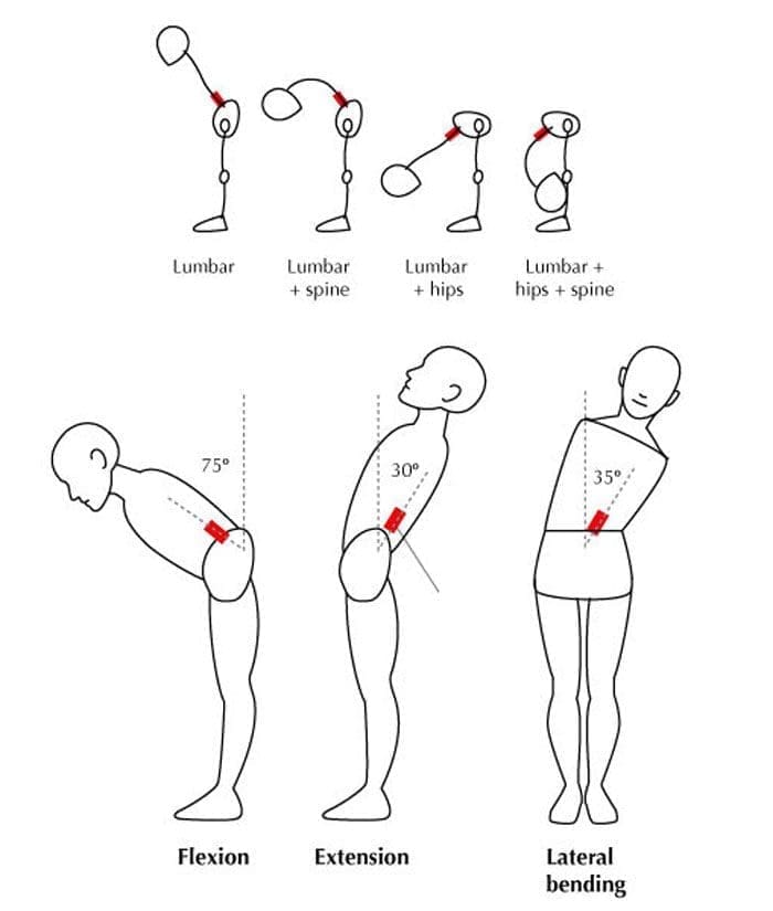
Maria finishes her Amazon shift without a single heavy box. She reaches into totes and bends for packages dozens of times an hour. By the time she gets home, her low back feels tight. At home, she loads the washer and picks up toys. One controlled lift earlier never bothered her as much. The pattern shows up in data center technicians, parents, warehouse workers, and desk professionals who repeatedly bend for cables, laundry, tools, or drawers.
The idea that only heavy objects matter misses how the spine handles repeated work. A single well-executed lift is one event. One hundred lighter bends spread the work across tired muscles, changing angles, and longer reaches. The tissues never fully recover between cycles, so the result can feel different—and sometimes worse.
The Myth of the Heavy Object
Advice to “lift with your legs” and “never bend your back” protects against sudden overload. It does not fully describe repeated bending for hours. Research shows both the size of each force and the number of cycles matter. High-frequency moderate loads still contribute substantially to total spinal stress (Coenen et al., 2012).
A controlled heavy lift usually uses a planned technique and finishes before fatigue sets in. Repeated light bends are hurried, done at the end of a reach, and performed after the trunk muscles have already worked. The spine sees a different pattern of compression, shear, and bending moment.
What Cumulative Loading Means for Your Spine
Each bend compresses and bends the lumbar discs and ligaments. When erector spinae muscles fatigue, they allow more lumbar flexion. Volunteers who lifted a 10 kg weight 100 times showed increased peak lumbar flexion and a 36 percent rise in estimated bending moment by the end of the task (Dolan & Adams, 1998).
Fatigue failure studies reinforce the point. Motion segments cyclically loaded to simulate lifting a 9 kg weight failed after far fewer cycles as torso flexion increased. Neutral posture allowed thousands of cycles; 45 degrees of flexion reduced survival to a few hundred cycles on average (Gallagher et al., 2005). Spinal posture during bending changes tissue tolerance.
Fatigue and Creep
As back muscles tire, the spine relies more on passive tissues. Repeated flexion produces creep—a slow increase in flexibility. After 20 minutes of sustained flexion, peak flexion rose by about 5.5 degrees, and full stiffness recovery took longer than the loading period (McGill & Brown, 1992). Short rests do not always fully reset the tissues. Residual creep can leave the spine more flexible for the next cycle.
Reach Distance and the Moment Arm
How far the hands are from the body matters as much as the weight. Holding a package at arm’s length multiplies the lumbar bending moment even if the package is light. Reaching into a rack, tote, or across a bed lengthens the lever arm. Bringing the object closer or raising the work surface shortens that distance and reduces demand.
How Hips and Trunk Endurance Share the Work
The lumbar spine favors stability; the hips handle large ranges of flexion. Limited hip mobility forces earlier and deeper lumbar flexion. Over dozens of bends, this shifts cumulative load onto the discs and ligaments.
Trunk endurance matters as much as peak strength. Someone who can deadlift once may still fatigue during 40 minutes of light bending if deep stabilizers and hip extensors lack endurance. Building that capacity protects the spine later in the shift.
Chiropractic Assessment: Finding the Real Pattern
A thorough evaluation at El Paso Back Clinic looks beyond the single heavy lift. Dr. Alex Jimenez, DC, APRN, FNP-BC, CFMP, IFMCP, and the team examine how the patient moves during the tasks that bother them. Assessment covers lumbar and hip range of motion, hip-hinge ability with a relatively neutral lumbar spine, trunk and hip endurance, control during repeated bending, and postural habits. The evaluation promptly identifies red-flag signs that require imaging or referral.
The evaluation identifies limited hip mobility, poor trunk endurance, excessive reach distance, residual stiffness, or a combination. Medical Director Dr. Maria Guadalupe Cardenas, MD, provides collaborative oversight, interpreting structural findings alongside medical factors when needed. Accurate diagnosis lets treatment address the real pattern. Beneficence guides the process: coordinated care improves safety, function, and recovery.
Low Back Pain Chiropractic Care | El Paso, TX
Building Bending Tolerance Instead of Avoiding Flexion
Fear of spinal flexion can create its own problems. Avoiding all bending leads to deconditioning. The tissues that are never loaded gradually lose capacity. A better approach is to restore control and then gradually reintroduce bending under conditions the spine can tolerate.
Chiropractic care restores restricted joint motion and reduces protective guarding. Adjustments and soft-tissue work free the hips and thoracic spine so the lumbar spine compensates less. Once motion improves, focus shifts to endurance work that matches real tasks:
- Hip-hinge practice with light loads held close to the body
- Controlled step-ups or partial squats that train the hips to share work
- Timed trunk endurance holds in positions that match the job
- Progressive increases in the number of bends per set, starting well below the threshold that produces next-day stiffness
The plan matches the job. An Amazon associate who bends 200 times per shift needs a different progression from a parent who bends mainly after a desk day. Progress is measured by how the back feels during and after the real task. Non-invasive options reduce risks of surgical complications or heavy medication dependence.
Practical Task Modifications for Daily Work and Home
While capacity builds, small changes reduce the total load the spine sees each day:
- Bring packages, cables, or laundry closer before lifting or lowering them.
- Use a low stool or kneel for tasks that would otherwise require deep lumbar flexion.
- Alternate sides and change stance so one pattern does not dominate.
- Take brief standing or walking breaks after longer periods of repeated bending.
- Raise frequently used items so the average bend is shallower.
- For parents, place a sturdy surface at hip height for folding or packing.
These changes reduce peak moments and high-flexion cycles while improving capacity. Patients remain the informed decision-makers, choosing modifications that fit their shift and family schedule.
The Multidisciplinary Path Forward
Integrative care at Injury Medical Clinic PA combines chiropractic structural work with medical oversight from Dr. Cardenas. Dr. Jimenez’s dual licensure supports coordinated attention to mechanical alignment and contributing factors. The emphasis remains on non-invasive strategies that restore function and reduce the need for surgery. Many patients with excellent group insurance coverage find coordinated care covered when it addresses documented functional limits.
If repeated bending has become the part of your day you dread, a focused assessment can clarify the drivers and how to rebuild tolerance safely. Call 915-850-0900 or visit the clinic in El Paso. The aim is to keep the spine working for the tasks you actually do.
References
Coenen, P., Kingma, I., Boot, C. R. L., Bongers, P. M., & van Dieën, J. H. (2012). The contribution of load magnitude and number of load cycles to cumulative low-back load estimations: A study based on in-vitro compression data. Clinical Biomechanics, 27(9), 871–877.
Dolan, P., & Adams, M. A. (1998). Repetitive lifting tasks fatigue the back muscles and increase the bending moment acting on the lumbar spine. Journal of Biomechanics, 31(8), 713–721.
Gallagher, S., Marras, W. S., Litsky, A. S., & Burr, D. (2005). Torso flexion loads and the fatigue failure of human lumbosacral motion segments. Spine, 30(20), 2265–2273.
McGill, S. M., & Brown, S. (1992). Creep response of the lumbar spine to prolonged full flexion. Clinical Biomechanics, 7(1), 43–46.

by Dr Alex Jimenez DC, APRN, FNP-BC, CFMP, IFMCP | Lower Back Pain, Power & Strength, Spine Care, Work Related Injuries
Why the First Heavy Lift of the Shift Can Feel Worse Than the Twentieth
Abstract
The first demanding lift after sitting or standing still can feel stiff or surprisingly heavy. That does not automatically mean the back is damaged. It often reflects a rapid transition from inactivity to loaded movement. This article explains how temperature, coordination, readiness, fatigue, and conditioning shape that transition—and how workers can prepare without fearing lifting.

You badge in, reach your station, and the first real task is a heavy tote, server component, tool case, cable spool, or awkward package. The weight is familiar, yet your hips feel slow, and your back feels tight.
Twenty lifts later, the job may feel smoother. That difference can be unsettling, especially if you’ve had lower back pain before. But the body does not move identically from the first repetition to the twentieth. The goal is not to fear the early lift. It is to prepare for the demand.
The Body Has to Change Gears
A commute or computer session requires less force than lifting, carrying equipment, or pulling inventory. When demand rises quickly, muscles and joints must coordinate faster.
Warm-up research helps explain part of this effect. Higher muscle temperature is associated with improved rate-dependent muscle function, including faster force production and power, although not necessarily greater maximum strength (Wilson et al., 2025). Appropriate warm-ups can also improve physical performance (Fradkin et al., 2010).
A “cold” muscle is not automatically unsafe. Progressive movement can help the body reach the speed and coordination the job requires.
Why Sitting Before Work Can Matter
Many workers begin physical work after a commute, meeting, or computer task.
After inactivity, the first deep hip bend or loaded reach may simply feel unfamiliar. The hips flex, the trunk controls the load, the feet create a stable base, and the upper body manages the object. Problems are more likely when demand suddenly exceeds what the worker can comfortably coordinate or tolerate that day.
Readiness also changes with sleep, soreness, injury, stress, and object shape. Good preparation, therefore, needs more than one lifting slogan.
There Is No Single Perfect Lift
Workers are often told to “lift with the legs” and never bend the back. That sounds simple, but research shows that technique training alone does not reliably prevent lower back pain in material-handling workers (Verbeek et al., 2012).
Technique still matters—in context. Useful principles can include:
- bringing the load closer when practical;
- creating a stable stance before pulling or lifting;
- using the hips and knees when the task allows;
- avoiding sudden twisting with a demanding load;
- using team lifting or mechanical help when needed;
- changing strategy when space, fatigue, or object shape changes.
Lifting is a skill, not one perfect pose. The best strategy is controlled, repeatable, and adaptable.
Warm-Up Effects and Fatigue Are Different
Feeling better by the twentieth lift does not mean endless repetitions are protective. Early in a shift, increasing temperature and repeated practice may improve movement efficiency. Later, fatigue can reduce control, slow reactions, and make familiar loads feel harder.
Think of the shift as a curve: preparation early, efficiency in the middle, and smarter pacing or assistance later.
“I loosen up after a few lifts” should not become “I should ignore pain.” Progressive readiness is useful; sharp pain, increasing numbness, weakness, or loss of coordination deserves clinical attention.
A Five-Minute Work-Readiness Sequence
A practical warm-up should resemble the work ahead.
1. Walk and breathe
Walk briskly for one or two minutes to transition out of inactivity.
2. Move the hips and trunk
Perform several comfortable hip hinges, shallow squats, or controlled reaches without forcing range.
3. Add light load
Practice a few lifts with a light object and focus on smooth control.
4. Rehearse the task
If work involves carrying, pulling, stepping, or lifting from knee height, rehearse that pattern at lower effort.
5. Build toward harder work
When workflow allows, use a few graded repetitions before the heaviest or most awkward task.
When the First Lift Repeatedly Hurts
If the first heavy lift repeatedly causes back pain, evaluation should go beyond “stretch more.” Assessment can examine spinal and hip motion, neurological findings, strength, tolerance, previous injury, and triggering work demands.
For persistent lower back pain, exercise is supported as part of conservative care and can improve pain and function for many patients (Hayden et al., 2021). The World Health Organization also includes structured exercise and spinal manipulative therapy as options within person-centered care for chronic primary lower back pain (World Health Organization, 2023).
At El Paso Back Clinic, chiropractic care should not teach patients that their spine is fragile. The aim is to improve comfortable movement and pair hands-on care with strength, mobility, and work-specific rehabilitation.
Building Capacity Beats Avoiding Work
Workers who lift, carry, kneel, reach, or handle equipment need the capacity to do those tasks. Long-term progress may include hip strength, trunk endurance, leg strength, grip, loaded carries, squats, hinges, step-ups, and graded exposure to realistic work demands.
The practical target is simple: enough mobility to reach the task, enough strength to control it, and enough endurance to repeat it without unnecessary guarding or fear.
Progress the load rather than fear it. Exercise therapy has evidence for chronic lower back pain, but programs should match the individual rather than treating every painful back the same way (Hayden et al., 2021).
This supports beneficence by restoring function, non-maleficence by emphasizing non-invasive care before unnecessary procedures when appropriate, and autonomy by helping patients understand options and coordinate decisions with their medical team.
Integrated Care When Stiffness Is Not the Whole Story
Radiating pain, numbness, weakness, recurrent injury, or stalled recovery may require broader evaluation.
Dr. Alex Jimenez, DC, APRN, FNP-BC, CFMP, IFMCP, combines chiropractic and advanced practice nursing perspectives. Dr. Maria Guadalupe Cardenas, MD, provides internal medicine oversight for complex medical factors and coordinated care. Depending on findings, care may include chiropractic treatment, rehabilitation, diagnostic testing, acupuncture, electroacupuncture, or other appropriate options. Advanced therapies should follow a clear diagnosis and should not replace strength and movement capacity.
Start the Shift Ready, Not Afraid
The first heavy lift may feel worse because your body has not transitioned from stillness to demanding movement. That doesn’t mean lifting is dangerous.
Give your body time to adjust. Build strength for your actual work. Adjust strategy as load or fatigue changes. If pain keeps coming back, get assessed instead of guessing.
The goal is not to avoid lifting. The goal is to be better prepared to lift, carry, bend, and work with confidence.
Multidisciplinary Call to Action
If lower back pain is limiting your lifts or full shift, schedule an evaluation with the El Paso Back Clinic team. A chiropractic assessment, rehabilitation, and medical oversight can build a plan focused on function, capacity, and independence.
Can Core Exercises Help with Back Pain? | El Paso, TX
References
Fradkin, A. J., Zazryn, T. R., & Smoliga, J. M. (2010). Effects of warming-up on physical performance: A systematic review with meta-analysis. Journal of Strength and Conditioning Research, 24(1), 140–148. https://doi.org/10.1519/JSC.0b013e3181c643a0
Hayden, J. A., Ellis, J., Ogilvie, R., Malmivaara, A., & van Tulder, M. W. (2021). Exercise therapy for chronic low back pain. Cochrane Database of Systematic Reviews, 2021(9), CD009790. https://doi.org/10.1002/14651858.CD009790.pub2
Verbeek, J. H., Martimo, K. P., Kuijer, P. P. F. M., Karppinen, J., Viikari-Juntura, E., & Takala, E. P. (2012). Proper manual handling techniques to prevent low back pain, a Cochrane systematic review. Work, 41(Suppl 1), 2299–2301. https://doi.org/10.3233/WOR-2012-0455-2299
Wilson, C. J., Nunes, J. P., & Blazevich, A. J. (2025). The effect of muscle warm-up on voluntary and evoked force-time parameters: A systematic review and meta-analysis with meta-regression. Journal of Sport and Health Science, 14, 101024. https://doi.org/10.1016/j.jshs.2025.101024
World Health Organization. (2023). WHO guideline for non-surgical management of chronic primary low back pain in adults in primary and community care settings. World Health Organization.