Can individuals with osteoarthritis incorporate spinal decompression therapy to restore spinal mobility and quality of life?
Introduction
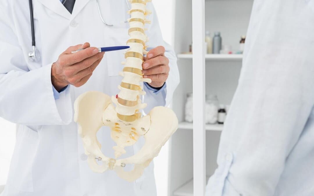
As the body ages, so does the spine, as the spinal disc between the joints and the bones starts dehydrating from constant compression through repetitive motions. The many environmental factors contributing to this degenerative disorder can vary within the person and lead to arthritic conditions within the upper and lower extremities. One of the most common types of arthritis is osteoarthritis, and it can affect many people worldwide. Dealing with osteoarthritis in their joints can cause numerous pain-like symptoms that correlate with other body conditions, causing referred pain. However, many treatments can help slow the process of osteoarthritis and relieve the body from the pain-like symptoms of the joints. Today’s article looks at how osteoarthritis affects spinal mobility and how treatments can restore spinal mobility from the effects of osteoarthritis. We talk with certified medical providers who utilize our patients’ information to provide various treatments to reduce the impact of osteoarthritis on the joints. We also inform patients how multiple treatments can help slow down the degenerative process of osteoarthritis. We encourage our patients to ask their associated medical providers intricated and important questions about the pain-like symptoms they are experiencing from osteoarthritis. Dr. Jimenez, D.C., incorporates this information as an academic service. Disclaimer.
How Does Osteoarthritis Affect Spinal Mobility?
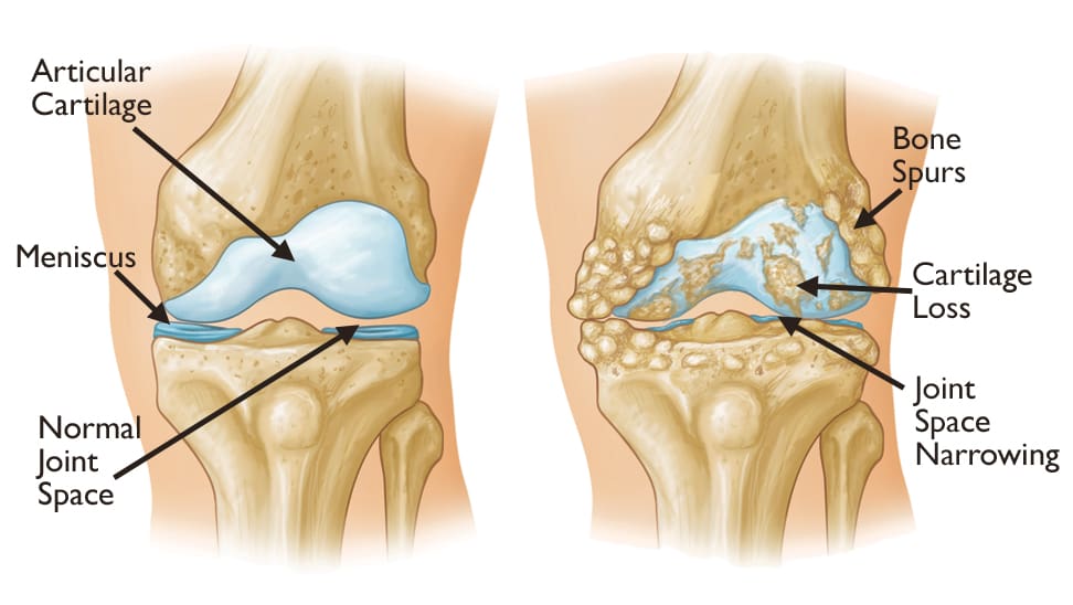
Have you noticed morning stiffness after a good night’s rest? Do you feel tenderness in your joints after some light pressure? Or do you feel limited mobility in your joints, causing a restricted range of motion? Many of these pain-like scenarios are correlated with osteoarthritis, a degenerative joint disorder that has affected many individuals, including older adults. As stated earlier, when the body ages, so do the joints, bones, and spine. Regarding osteoarthritis, the joints will degenerate through natural wear and tear around the cartilage. Osteoarthritis affects multiple joints like the hips and knees, which are the most common, and the spine, and causes numerous sensory-motor dysfunctions. (Yao et al., 2023) When the cartilage around the affected joints starts to deteriorate, the pathogenesis of osteoarthritis causes a disturbed cytokine balance of the proinflammatory cytokines to initiate a vicious cycle that causes cartilage and other intra-articular structure damage around the joint. (Molnar et al., 2021) What this does is that when osteoarthritis starts to affect the joints, it can lead to numerous referred pain-like symptoms.
However, although osteoarthritis can affect the joints, naturally, numerous environmental factors do play a part in the development of osteoarthritis. Physical inactivity, obesity, bone deformities, and joint injuries are some of the causes that can progress the degenerative process. The symptoms that are associated with these environmental factors include:
Pain
Joint stiffness
Tenderness
Inflammation
Swelling
Grating sensation
Bone spurs
Many individuals dealing with pain-like symptoms caused by osteoarthritis will explain to their primary doctors that the pain varies in duration, depth, type of occurrence, impact, and rhythm. This is because the pain from osteoarthritis is complex and multifactorial. (Wood et al., 2022) However, many individuals can look for the help they need to reduce the pain-like issues caused by osteoarthritis through treatments that can slow down the degenerative progress.
An In-depth Look At Spinal Decompression-Video
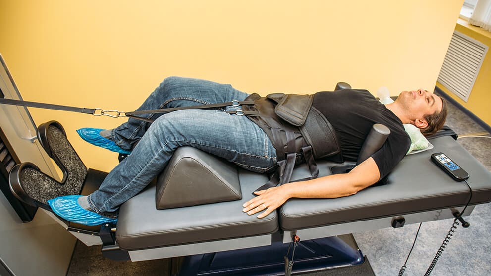
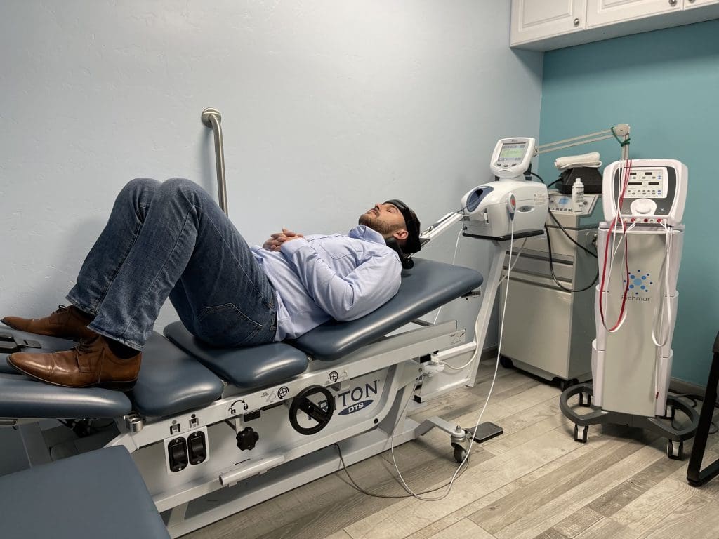
When it comes to seeking treatment to reduce the effects of osteoarthritis, many individuals seek out treatments that are cost-effective and safe for older individuals. Non-surgical treatments could be the solution many individuals seek to reduce the progress of osteoarthritis. When people experiencing osteoarthritis go to non-surgical treatments, they find out that the pain is decreased, their range of motion is increased, and their physical function has improved. (Alkhawajah & Alshami, 2019) At the same time, non-surgical treatments can be combined with other therapies to the individual’s personalized treatment plan. No-surgical treatments can range from chiropractic care to spinal decompression as they work on gently realigning the spine through traction and help reduce joint and muscle pain. The video above gives an in-depth look at spinal decompression and how it can benefit individuals who are in pain.
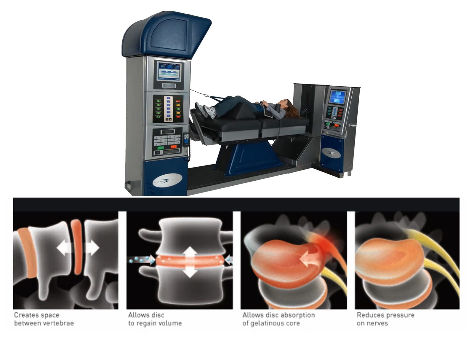
Spinal Decompression Restoring Spinal Mobility From Osteoarthritis
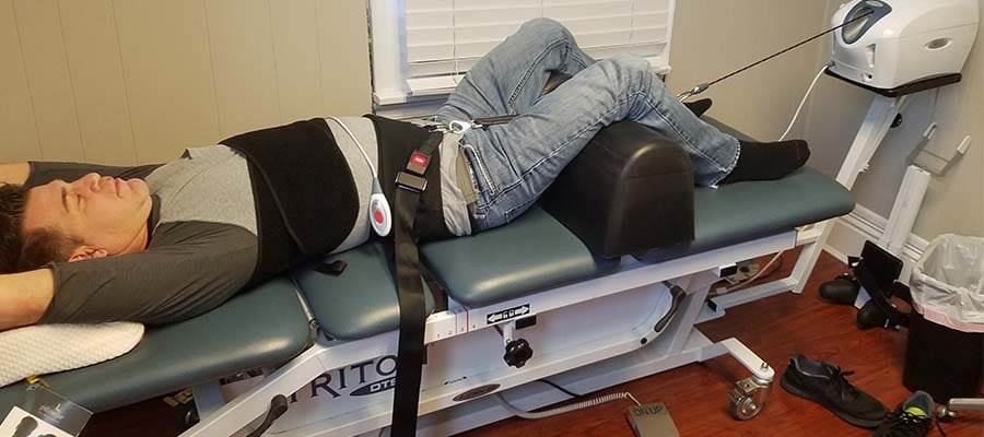
Since spinal decompression is a form of non-surgical treatment, it can help slow down the process of osteoarthritis. Spinal decompression incorporates traction to gently pull on the spine, allowing the discs and joints to be lubricated and permitting the natural healing process to occur. This is because the surrounding muscles that protect the joints are being stretched gently and the vertebral disc space is being increased to allow the disc to be rehydrated and the protrusion to recede back to its original position. (Cyriax, 1950) Spinal decompression can help slow down the degenerative process of osteoarthritis, and when combined with physical therapy, the surrounding muscles, tissues, and ligaments are strengthened.
In contrast, joint and spinal mobility and flexibility are increased. Spinal decompression can also help many individuals reduce their chances of surgery, as consecutive sessions can help provide pain relief and functional improvement to the spine. (Choi et al., 2022) When people regain their spinal mobility back to their bodies from spinal decompression, they can make small changes in their daily routine to slow down the degenerative process of osteoarthritis.
References
Alkhawajah, H. A., & Alshami, A. M. (2019). The effect of mobilization with movement on pain and function in patients with knee osteoarthritis: a randomized double-blind controlled trial. BMC Musculoskelet Disord, 20(1), 452. https://doi.org/10.1186/s12891-019-2841-4
Choi, E., Gil, H. Y., Ju, J., Han, W. K., Nahm, F. S., & Lee, P. B. (2022). Effect of Nonsurgical Spinal Decompression on Intensity of Pain and Herniated Disc Volume in Subacute Lumbar Herniated Disc. International Journal of Clinical Practice, 2022, 6343837. https://doi.org/10.1155/2022/6343837
Molnar, V., Matisic, V., Kodvanj, I., Bjelica, R., Jelec, Z., Hudetz, D., Rod, E., Cukelj, F., Vrdoljak, T., Vidovic, D., Staresinic, M., Sabalic, S., Dobricic, B., Petrovic, T., Anticevic, D., Boric, I., Kosir, R., Zmrzljak, U. P., & Primorac, D. (2021). Cytokines and Chemokines Involved in Osteoarthritis Pathogenesis. Int J Mol Sci, 22(17). https://doi.org/10.3390/ijms22179208
Wood, M. J., Miller, R. E., & Malfait, A. M. (2022). The Genesis of Pain in Osteoarthritis: Inflammation as a Mediator of Osteoarthritis Pain. Clin Geriatr Med, 38(2), 221-238. https://doi.org/10.1016/j.cger.2021.11.013
Yao, Q., Wu, X., Tao, C., Gong, W., Chen, M., Qu, M., Zhong, Y., He, T., Chen, S., & Xiao, G. (2023). Osteoarthritis: pathogenic signaling pathways and therapeutic targets. Signal Transduct Target Ther, 8(1), 56. https://doi.org/10.1038/s41392-023-01330-w
Can individuals dealing with myofascial pain syndrome in their bodies find the relief they are looking for through acupuncture?
Introduction
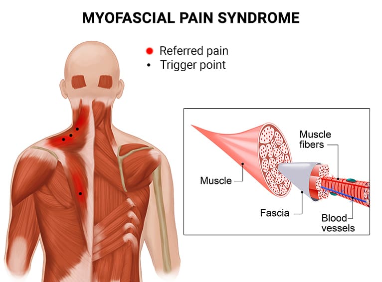
The musculoskeletal system has numerous ligaments, joints, soft tissues, and muscles that allow the body to be in motion without feeling pain or discomfort. The musculoskeletal system has upper and lower body portions, and each quadrant has a specific job it needs to do. The head works with the neck for the upper body portions to allow it to turn and be mobile. The shoulders work with the arms and hands to allow flexibility while stabilizing the neck. For the lower body portions, the hips and legs stabilize the upper body’s weight and help the different quadrants flex, extend, and rotate without pain. However, when traumatic or normal forces start to affect the body, it can lead to pain and discomfort, depending on the severity. When this happens, it can cause the muscle fibers from the upper and lower body portions to become tight and form tiny nodules known as trigger points to cause myofascial pain. This causes many individuals to be in constant discomfort and feel pain in different body locations. However, there are numerous treatments that can reduce the pain from the trigger points and restore muscle function to the body. Today’s article examines how myofascial pain syndrome affects the body, how non-surgical treatments like acupuncture can reduce trigger point pain, and how acupuncture can restore body function. We speak with certified medical providers who incorporate our patients’ information to provide various treatments to reduce the effects of myofascial pain syndrome on the body. We also inform patients how non-surgical therapies like acupuncture can help restore body function that is caused by myofascial pain. We encourage our patients to ask intricated questions to our associated medical providers about the pain-like symptoms they are experiencing from myofascial pain syndrome that is affecting their bodies. Dr. Alex Jimenez, D.C., utilizes this information as an academic service. Disclaimer.
Myofascial Pain Syndrome Affecting The Body
Do you feel pain radiating in certain locations in your body, affecting your daily routine? Are you feeling any complaints in your back, knees, elbows, or shoulders? Or do you have mobility issues that are affecting your gait and making you feel unstable? Many of these issues that people are experiencing are known as myofascial pain syndrome, and it can cause overlapping risk profiles in the musculoskeletal system. Myofascial pain syndrome is a problematic musculoskeletal pain originating from the muscles and surrounding fascia. (Tantanatip & Chang, 2023) This common musculoskeletal condition causes localized pain in certain body areas or referred pain to various muscle locations. When a person is dealing with myofascial pain syndrome, their muscles in the upper or lower body quadrants will become overstretched and tight through repetitive motions that can cause tiny nodules known as trigger points that can be a source of where the pain might originate from. When people are dealing with myofascial pain syndrome in their bodies, they will inform their primary doctors that they are experiencing pain in different locations in their bodies that are causing them pain. The doctor then will ask the individual numerous questions and examine where the pain is occurring. The doctor will also take note of the person’s daily routine, allowing the doctor to diagnose that myofascial pain syndrome is at play.
When myofascial pain syndrome affects body function, it can come in the forms of nociceptive pain and neuropathic pain. When the muscle fibers in the upper and lower extremities are dealing with trigger points, the surrounding nerve roots that provide the sensory-motor function to the arms and legs can become irritated, causing spot tenderness, referred pain, and nerve root compression that can cause the muscles to succumb to muscle trauma and muscle overload. (Fernandez-de-Las-Penas et al., 2023) To that point, myofascial pain syndrome can affect a person’s quality of life as the mechanisms of the comorbidities combined with the psychological stressors that are impacting the muscles might sensitize the trigger points. (Sabeh et al., 2020) However, when the pain becomes excruciating in the muscles that are caused by myofascial pain syndrome, many individuals will start to seek treatment to not only reduce the pain but also restore their body functionality.
The Non-Surgical Approach To Wellness- Video
Have you been dealing with radiating or localized pain in different body locations? How about experiencing mobility issues when moving your upper or lower extremities? Or do you experience stability issues from walking from one location to another? Many of these pain-like scenarios are associated with myofascial pain syndrome that is affecting the musculoskeletal system. When the human body is dealing with myofascial pain syndrome, it can be difficult to diagnose due to the affected muscles causing referred pain. At the same time, when a person is dealing with myofascial pain syndrome is not finding treatment to reduce the pain, it can lead to major health problems like:
Mobility impairment
Muscle pain and hypersensitivity
Nerve issues
Neurological issues
When people with myofascial pain syndrome are looking for treatment, they are looking for cost-effective treatments that can be combined with other therapies that can help reduce the pain and restore body function to the extremities. Non-surgical treatments can be the solution for many individuals because they are cost-effective and can be personalized. The video above shows how non-surgical treatments like chiropractic care can help restore the body through manual and mechanical manipulation that can stretch and locate the trigger points while restoring body function to the extremities.
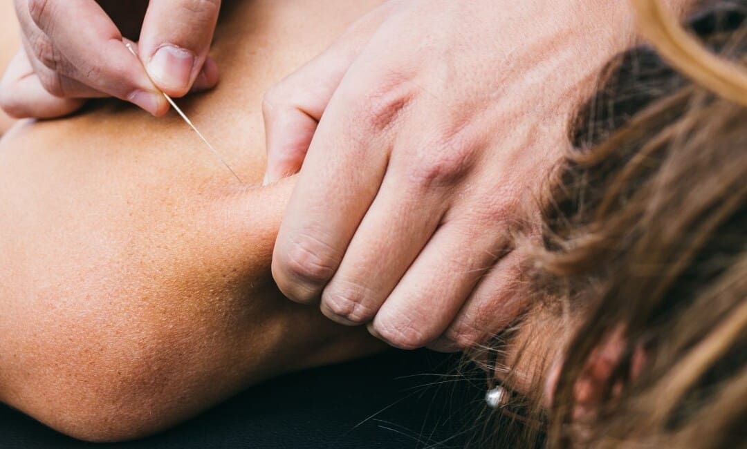
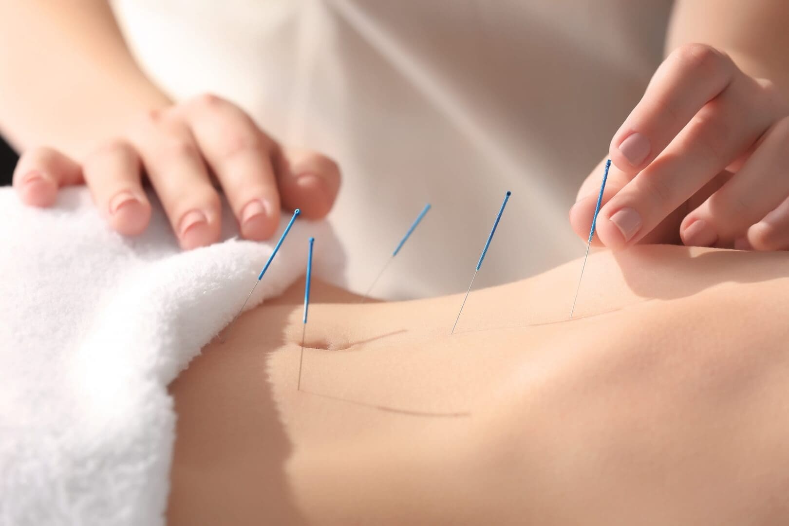
Acupuncture Reducing Trigger Point Pain
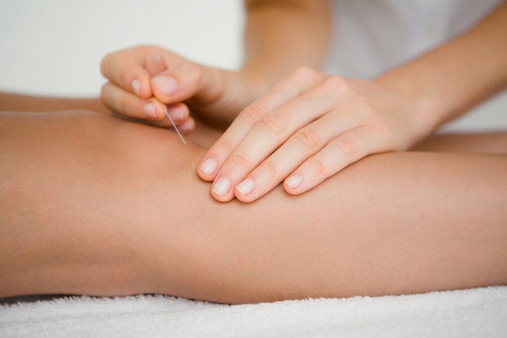
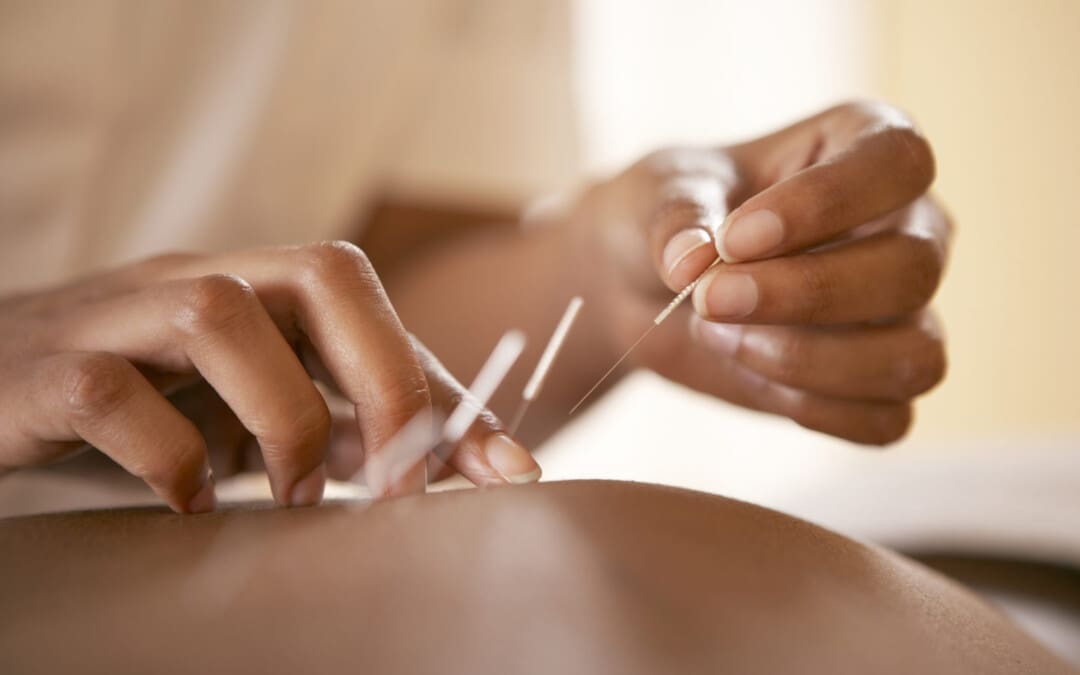
When a person goes in for non-surgical treatments for their myofascial pain syndrome, acupuncture could be the answer. Acupuncture is an Eastern medical practice from China performed by highly trained licensed professionals. So, how can acupuncture help alleviate trigger point pain from myofascial pain syndrome? Acupuncturists use solid, super thin needles to place them in specific points in the body to reduce the referred pain-like symptoms causing issues. This causes the myofascial trigger points to become inactive and, reduces intramuscular hypertension, relieves uneven musculoskeletal pull, and improves mechanical balance in the upper and lower extremities. (Lin et al., 2022)
Acupuncture Restoring Body Function
Acupuncture is traditionally used to restore the normal flow of energy to the body. Still, in the modern era, it has been shown to have multiple positive effects on the central and peripheral nervous systems. So, since myofascial pain syndrome is both nociceptive and neuropathic, the effects of acupuncture can change the pain’s perspective while causing the body’s natural healing process to be restored. (Kelly & Willis, 2019) At the same time, acupuncturists can gently prick and stimulate the affected tendon with the myofascial to induce a muscle twitch to relax the tight muscle. (Qiu et al., 2023) This allows the body to relax and helps reduce the chances of trigger points returning to the muscle facia. For individuals who are looking for treatment for their myofascial pain syndrome, incorporating acupuncture could be the solution to restore body function to the musculoskeletal system.
References
Fernandez-de-Las-Penas, C., Nijs, J., Cagnie, B., Gerwin, R. D., Plaza-Manzano, G., Valera-Calero, J. A., & Arendt-Nielsen, L. (2023). Myofascial Pain Syndrome: A Nociceptive Condition Comorbid with Neuropathic or Nociplastic Pain. Life (Basel), 13(3). https://doi.org/10.3390/life13030694
Kelly, R. B., & Willis, J. (2019). Acupuncture for Pain. American Family Physician, 100(2), 89-96. https://www.ncbi.nlm.nih.gov/pubmed/31305037
Lin, X., Li, F., Lu, H., Zhu, M., & Peng, T. Z. (2022). Acupuncturing of myofascial pain trigger points for the treatment of knee osteoarthritis: A systematic review and meta-analysis. Medicine (Baltimore), 101(8), e28838. https://doi.org/10.1097/MD.0000000000028838
Qiu, X. H., Yang, X. Y., Wang, Y. Y., Tian, S. L., Yan, Y. B., Xu, A. P., Fu, F., Wen, F. Y., Yang, Y., Zhang, Y., Zhang, Y. Q., Yang, Z. W., Xu, C., Sun, Q. H., Wu, X. L., Dai, X. Y., Li, N., & Cheng, K. (2023). Myofascial acupuncture versus routine acupuncture for mechanical neck pain: a protocol for a multicentre randomised controlled trial. BMJ Open, 13(8), e068129. https://doi.org/10.1136/bmjopen-2022-068129
Sabeh, A. M., Bedaiwi, S. A., Felemban, O. M., & Mawardi, H. H. (2020). Myofascial Pain Syndrome and Its Relation to Trigger Points, Facial Form, Muscular Hypertrophy, Deflection, Joint Loading, Body Mass Index, Age and Educational Status. J Int Soc Prev Community Dent, 10(6), 786-793. https://doi.org/10.4103/jispcd.JISPCD_328_20
For individuals experiencing pelvic pain, can incorporating acupuncture help alleviate and reduce low back pain?
Introduction
In the musculoskeletal system, the upper and lower body portions have jobs to allow the host to be in motion. The lower body portions provide stability and maintain proper posture, which can help the surrounding muscles be strong and protect the vital organs. The skeletal joints in the body help ensure that the person’s body weight is evenly distributed. For the musculoskeletal system, the pelvic region in the lower body portion helps with stabilization and provides normal urinary function to the body. However, when normal and traumatic factors begin to affect the lower portions of the body, it can lead to pain-like issues that can cause some visceral referred pain to the lower back, and it can make many individuals think they are experiencing lower back pain, which is one of the symptoms associated with pelvic pain. When many individuals are experiencing pelvic pain associated with lower back pain, many will opt to seek treatment to reduce the pain-like symptoms and restore their body function. Today’s article looks at how pelvic pain is associated with low back pain and how treatments like acupuncture can help reduce pelvic pain associated with low back pain and provide relief. We speak with certified medical providers who incorporate our patients’ information to provide various treatments to ease low back pain correlated with pelvic pain. We also inform patients how non-surgical therapies like acupuncture can help reduce the effects of pelvic pain. We encourage our patients to ask intricated questions to our associated medical providers about the pain-like symptoms they are experiencing correlating with pelvic pain that is also causing issues in their lower backs. Dr. Alex Jimenez, D.C., utilizes this information as an academic service. Disclaimer.
How Pelvic Pain Is Associated With Low Back Pain?
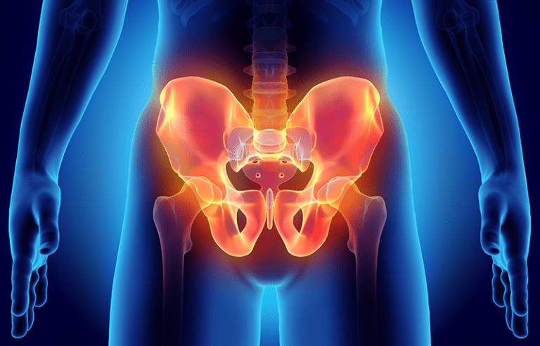
Have you experienced excruciating pain from excessive sitting that is causing pain in your lower back or pelvic region? Do you feel stiffness in your lower back and pelvic region due to poor posture? Or are you experiencing intense cramping around your pelvic area? When many individuals are dealing with these pain-like issues, it is correlated with pelvic pain. Now, pelvic pain is a common, disabling, persistent pain that is associated with comorbidities that are multifactorial and are often centralized pain. (Dydyk & Gupta, 2023) At the same time, pelvic pain is a challenge to diagnose due to being multifactorial and sharing the numerous nerve roots that are spread out and intertwined with the lumbar region. To this point, this causes referred pain to the lower back and causes many individuals to think they are experiencing lower back pain when, in actuality, they are dealing with pelvic pain. This is due to the pelvic floor muscles becoming weak, which can cause many individuals to develop poor posture, leading to low back pain over time.
Additionally, when the pelvic region is misaligned due to repetitive motions that cause lower back pain, it can cause the surrounding muscles to be overstretched and loose around the sacroiliac joints. (Mutaguchi et al., 2022) When this happens, the surrounding muscles surrounding the hips and lower back may weaken, leading to anterior pelvic tilt and causing alterations to the lumbopelvic area.
Since the lumbopelvic area is in the lower body portions, it can cause alterations to the body’s skeletal structure, leading to lower back pain. When an increasing number of individuals deal with spinal deformity, they will maintain a standing position while preventing their central gravity from moving forward by using their pelvic muscles to compensate for their weight. (Murata et al., 2023) When this happens, it causes the surrounding core muscles and back muscles to overstretch, which then causes the accessory muscles to produce more energy and do the primary muscles’ jobs. This causes urinary and muscle issues that cause tomato-visceral referred pain in the musculoskeletal system. However, there are numerous ways to reduce pelvic pain associated with low back pain while restoring pelvic function and restoring muscle strength to the surrounding core muscles in the pelvic region.
Is Motion Key To Healing- Video
Have you been experiencing any muscle stiffness around your hips, lower back, or pelvic region? Do you feel you have a limited range of motion in the morning, only for it to feel better throughout the day? Or are you experiencing bladder issues that are correlated with low back pain? Many of these pain-like scenarios are associated with pelvic pain and can cause common back pain issues that cause many individuals to be hunched over and be in constant pain. Since pelvic pain is a multifactorial musculoskeletal disorder, it can be associated with comorbidities that can cause issues to the lumbar region of the spine and affect the body’s mobility. However, numerous treatments can reduce the effects of pelvic pain and restore low back mobility to the body. When it comes to looking for treatments, many individuals will look for therapies that are cost-effective and can help reduce the referred pain that is associated with low back and pelvic pain. The video above shows how non-surgical treatments can help restore mobility to the lower extremities.
Acupuncture For Pelvic & Low Back Pain
When it comes to non-surgical treatments, many individuals will seek cost-effective treatments. Treatments like chiropractic care, spinal decompression, and massage therapy can help reduce low back pain, but for pelvic pain, many individuals will seek out acupuncture. Acupuncture is a medical practice performed by a highly trained professional that uses solid but thin needles in specific body areas. So, for individuals dealing with pelvic pain, acupuncture can help restore the balance of energy that is associated with the internal organs that are causing the pain. (Yang et al., 2022) Acupuncture can help restore power to the pelvic region by redirecting the energy to the body and helping reduce impairment and functional disorders. (Pan et al., 2023) Acupuncture can minimize low back pain by selecting certain trigger points that can influence the areas between the hips and back to unblock circulation back to the muscle. (Sudhakaran, 2021) When many people start incorporating acupuncture as part of their personalized treatment plan, they can utilize it with other therapies to feel better and improve their health.
Murata, S., Hashizume, H., Tsutsui, S., Oka, H., Teraguchi, M., Ishomoto, Y., Nagata, K., Takami, M., Iwasaki, H., Minamide, A., Nakagawa, Y., Tanaka, S., Yoshimura, N., Yoshida, M., & Yamada, H. (2023). Pelvic compensation accompanying spinal malalignment and back pain-related factors in a general population: the Wakayama spine study. Sci Rep, 13(1), 11862. https://doi.org/10.1038/s41598-023-39044-2
Mutaguchi, M., Murayama, R., Takeishi, Y., Kawajiri, M., Yoshida, A., Nakamura, Y., Yoshizawa, T., & Yoshida, M. (2022). Relationship between low back pain and stress urinary incontinence at 3 months postpartum. Drug Discov Ther, 16(1), 23-29. https://doi.org/10.5582/ddt.2022.01015
Pan, J., Jin, S., Xie, Q., Wang, Y., Wu, Z., Sun, J., Guo, T. P., & Zhang, D. (2023). Acupuncture for Chronic Prostatitis or Chronic Pelvic Pain Syndrome: An Updated Systematic Review and Meta-Analysis. Pain Res Manag, 2023, 7754876. https://doi.org/10.1155/2023/7754876
Can working individuals with low back pain incorporate nonsurgical treatments to reduce limited mobility and provide relief?
Introduction
Many working individuals will slowly develop low back pain due to excessive standing or sitting, physical demands that cause them to lift heavy objects, or improper footwear that causes them to be imbalanced. Since the spine is part of the musculoskeletal system, the spinal discs in the lumbar region are the most susceptible to being compressed. They can be one of the issues why many individuals tend to develop lower back pain. Low back pain is common for working individuals and is a multifactorial musculoskeletal disorder that causes many working people to miss out on work. However, many people with low back pain often seek treatment to reduce the pain and help them get back to work. Today’s article looks at the causes of low back pain and how nonsurgical treatments can help reduce low back pain and restore mobility to the body. We speak with certified medical providers who incorporate our patients’ information to provide various treatments to ease low back pain. We also inform patients how nonsurgical treatments can help restore mobility to the body while giving numerous techniques to reduce the chances of low back pain returning. We encourage our patients to ask intricated questions to our associated medical providers about the pain-like symptoms they are experiencing correlating with their backs. Dr. Alex Jimenez, D.C., utilizes this information as an academic service. Disclaimer.
The Causes For Low Back Pain
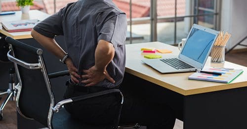
Do you feel stiffness in your lower back after a hard workday? Do you experience muscle aches or pain in your lower back after picking up a heavy object? Or do you experience limited mobility and stiffness over time after excessive standing or sitting at your job? Many individuals in these pain-like scenarios have experienced low back pain at some point in their lives, and it has impacted them to miss out on work. Since many people worldwide have experienced low back pain at some point in their lives, it has become a common problem that has become the leading cause of disability and is often associated with high cost. (Chou, 2021) Low back pain is a multifactorial condition that is specific or non-specific depending on the severity of the person’s experience. Non-specific low back pain often refers to when there isn’t a particular disease or structural reason for the pain to occur. This causes many people to go into early retirement due to losing their ability to work and become a socio-economic burden when seeking treatment. (Chenot et al., 2017) Specific low back pain is due to repetitive trauma and overusing the surrounding muscles that can cause the spine and spinal disc to be constantly compressed. This causes musculoskeletal pain symptoms and affects the rest of the lower extremities. (Will et al., 2018)
Some of the causes that low back pain is associated with can range from normal environmental factors to traumatic injuries that many working individuals have endured. Since low back pain is one of the leading causes of lost workdays around the world, some of the common causes that are contributed to low back pain include:
Mechanical strain
Obesity
Poor body mechanics
Trauma
Repetitive motions (twisting, bending, or lifting)
Herniated disc
Spinal stenosis
These pain-like causes can affect the upper and lower extremities and, when not being treated, lead to pain-like symptoms from radiating pain to limited mobility. However, when many people decide that enough is enough and want to get the treatment they need, they will seek out something that is not only affordable but can reduce the pain while restoring mobility.
The Power Of Chiropractic Care-Video
Nonsurgical Treatments For Low Back Pain
When it comes to seeking treatment for low back pain, many individuals are looking for something that is not only cost-effective but can help reduce the pain-like symptoms associated with the lower back. Nonsurgical treatments can help reduce low back pain and are cost-effective for many individuals including working individuals. Treatments like acupuncture, chiropractic care, and spinal decompression have various techniques and methods to provide pain relief to many individuals dealing with low back pain. Knowing the prevalence of the multiple pathologies of low back pain, a detailed history, and physical examination maneuvers allow doctors to accurately and quickly classify the most common causes of low back pain. (Kinkade, 2007) This will give them a better understanding of what kind of low back pain treatment they need to restore mobility to their bodies.
Chiropractic Care
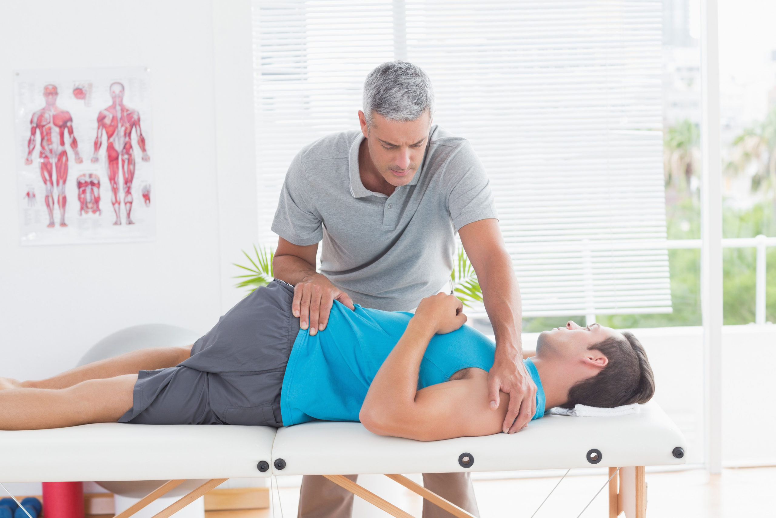
Chiropractic care is a nonsurgical treatment that incorporates manual and mechanical manipulation to realign the body out of subluxation from low back pain. Chiropractic care can be incorporated into a person’s health and wellness treatment plan as it can help improve pain and disability associated with low back pain. (Bussieres et al., 2018) Chiropractors combine various techniques to stretch and strengthen weak muscles around the lower back and reduce low back pain intensity and disability. (Vining et al., 2020) Chiropractic care can also work with other forms of therapies to reduce the chances of low back pain from returning.
Spinal Decompression
Spinal decompression is another form of nonsurgical treatment that can help the lumbar spine through gentle traction and help decompress affected spinal discs from causing mechanical back pain. Spinal decompression can also alleviate the referred pain-like symptoms from the nerve roots involved in the lumbar region while rehydrating herniated discs. Spinal decompression can also help many individuals have their lumbar range of motion back and improve their pain and endurance while restoring their quality of life. (Amjad et al., 2022) Just like chiropractic care, spinal decompression can be combined with other therapies to strengthen the surrounding muscles and ligaments.
Acupuncture
With low back pain being a common problem for many individuals, sometimes it could be due to aggravated nerve roots along the surrounding muscles that are causing referred trigger pain correlating with low back pain. When that happens, many individuals will seek out acupuncture to reduce the pain and improve their quality of life. (Baroncini et al., 2022) Acupuncture can reduce the inflammatory effects caused by inflammation associated with low back pain and can increase mobility in the sacroiliac joint to improve mobility. (Sudhakaran, 2021) Depending on the source of pain in the back, acupuncture can help reduce the pain and provide relief. Many individuals seeking treatment for their lower back can incorporate these treatments to improve their health and restore their quality of life.
References
Amjad, F., Mohseni-Bandpei, M. A., Gilani, S. A., Ahmad, A., & Hanif, A. (2022). Effects of non-surgical decompression therapy in addition to routine physical therapy on pain, range of motion, endurance, functional disability and quality of life versus routine physical therapy alone in patients with lumbar radiculopathy; a randomized controlled trial. BMC Musculoskelet Disord, 23(1), 255. https://doi.org/10.1186/s12891-022-05196-x
Baroncini, A., Maffulli, N., Eschweiler, J., Molsberger, F., Klimuch, A., & Migliorini, F. (2022). Acupuncture in chronic aspecific low back pain: a Bayesian network meta-analysis. J Orthop Surg Res, 17(1), 319. https://doi.org/10.1186/s13018-022-03212-3
Bussieres, A. E., Stewart, G., Al-Zoubi, F., Decina, P., Descarreaux, M., Haskett, D., Hincapie, C., Page, I., Passmore, S., Srbely, J., Stupar, M., Weisberg, J., & Ornelas, J. (2018). Spinal Manipulative Therapy and Other Conservative Treatments for Low Back Pain: A Guideline From the Canadian Chiropractic Guideline Initiative. J Manipulative Physiol Ther, 41(4), 265-293. https://doi.org/10.1016/j.jmpt.2017.12.004
Chenot, J. F., Greitemann, B., Kladny, B., Petzke, F., Pfingsten, M., & Schorr, S. G. (2017). Non-Specific Low Back Pain. Dtsch Arztebl Int, 114(51-52), 883-890. https://doi.org/10.3238/arztebl.2017.0883
Vining, R., Long, C. R., Minkalis, A., Gudavalli, M. R., Xia, T., Walter, J., Coulter, I., & Goertz, C. M. (2020). Effects of Chiropractic Care on Strength, Balance, and Endurance in Active-Duty U.S. Military Personnel with Low Back Pain: A Randomized Controlled Trial. J Altern Complement Med, 26(7), 592-601. https://doi.org/10.1089/acm.2020.0107
Can individuals dealing with neck and back pain find the relief they need from the effects of spinal decompression therapy?
Introduction
Across the world, many individuals deal with neck or back pain from excessive sitting or standing, poor posture, or lifting heavy objects that cause their spine and muscles to ache constantly. Since the body is in constant movement, the spine is being compressed through repetitive movement that can cause the spinal discs to pop out of their original position and aggravate the surrounding nerves to cause pain-like symptoms in the neck and back regions. Many people start to complain about their necks and backs hurting and feeling referred pain in different locations in the upper and lower body portions. This can range from acute to chronic, depending on the severity of the pain. When people are experiencing these musculoskeletal pain disorders in their bodies, many will seek treatment to alleviate the pain in their necks and backs to return to their daily routines. Hence why, treatments like spinal decompression can have a positive effect on providing the relief that many individuals deserve. Today’s article looks at why the neck and back in the human body are the most common pain areas many people endure and how spinal decompression can reduce neck and back pain. We speak with certified medical providers who incorporate our patients’ information to provide various techniques to relieve neck and back pain from the body. We also inform patients how treatments like decompression can reduce musculoskeletal pain disorders from the neck and back. We encourage our patients to ask intricated questions to our associated medical providers about the pain-like symptoms they are experiencing correlating with their neck and back. Dr. Alex Jimenez, D.C., utilizes this information as an academic service. Disclaimer.
Why Are The Neck & Back Common Pain Areas?
Do you feel muscle tension in your neck after being hunched on the computer or your phone for a long time? Do you feel aches and pains in your back after carrying or lifting a heavy object? Or do you feel tingling or numbness in your arms or legs? Many of these pain-like symptoms are often correlated with neck and back pain that can be a nuisance to many individuals. So why is it that the neck and back of the human body are the most common pain areas that many people worldwide endure? Many people with highly demanding jobs often perform normal movements repetitively, which causes stress on the surrounding muscles, ligaments, and joints, and the accessory muscles will begin to be overworked and tight. Neck and back pain are amongst the most common symptom-related complaints that contribute to high levels of lost workdays, disability, and health care use. (Corwell & Davis, 2020) This causes many individuals to have unwanted socio-economic stress when they visit their primary care doctors. Additionally, neck and back pain are non-neurologic causes in the musculoskeletal system; these can generate pain in the muscles, tendons, ligaments, spinal discs, articular cartilage, and bone. (Meleger & Krivickas, 2007) To that point, when neck and back pain are not treated right away, it can lead to correlating pain symptoms that can lead to a life of disability. Since the spine has multiple structures, from the neck to the lower back, when a person is in pain, it can lead to various pain generators that can cause some visceral pain. (Patel et al., 2015) Hence why, neck and back pain are multi-factorial and lead to numerous disorders.
When it comes to reducing neck and back pain from the body, many individuals will seek medical treatment to relieve themselves from the pain. However, many primary care doctors will assess their patients to determine what the root cause of their pain by taking notes of their daily routine. Many normal causes of neck and back pain can be due to:
Poor Posture
Stress
Physical Inactivity
Trauma/Injuries
Excessive sitting/standing
Lifting/carrying heavy objects
These causes can lead to a life of disability and affect a person’s quality of life; however, luckily, many individuals have researched and looked for treatment that is cost-effective and can help reduce the pain they are experiencing.
Understanding Academic Low Back Pain- Video
Do you feel aches and pains in your neck and back? Do you feel stress in your muscles that cause you to feel miserable? Or do you feel pain in your upper or lower body portions affecting your daily routine? Many of these scenarios correlate with neck and back pain, a common issue many individuals experience. If not treated right away, it can lead to a life of disability and, for working individuals, lose a day of work. However, many individuals seek cost-effective treatments that can help reduce the pain affecting their necks and back. Treatments like chiropractic care, traction therapy, massage therapy, and spinal decompression are all non-surgical, affordable, and can help reduce pain-like symptoms associated with neck and back pain. The video above explains the causes of academic low back pain and how non-surgical treatments like chiropractic care can work with additional therapies to prevent back and neck pain from returning. At the same time, when individuals begin to reduce their workload and educate themselves on what to do to avoid neck and back pain from returning, they can start feeling better. (Tyrdal et al., 2022)
The Effects Of Decompression On Neck & Back Pain
As part of the non-surgical treatments, spinal decompression can help many individuals dealing with neck and back pain. What spinal decompression does is incorporate gentle traction on the spine to decompress the affected spinal disc that can be associated with neck and back pain. When the spine is being treated with spinal decompression, the gravitational traction pull helps produce a greater disc space on the spine to decrease intradiscal pressure and pain. (Vanti et al., 2021) This allows all the nutrients and fluids to return to the spine and spinal discs while promoting the body’s natural healing process.
Additionally, many individuals with neck and back pain will begin to notice a huge reduction in their pain and disability through consecutive treatment. (Vanti et al., 2023) By incorporating healthy habits to reduce the chances of neck and back pain from returning, many individuals can make small changes to their daily routine. This allows them to have a positive outlook and continue their health and wellness journey.
References
Corwell, B. N., & Davis, N. L. (2020). The Emergent Evaluation and Treatment of Neck and Back Pain. Emerg Med Clin North Am, 38(1), 167-191. https://doi.org/10.1016/j.emc.2019.09.007
Patel, V. B., Wasserman, R., & Imani, F. (2015). Interventional Therapies for Chronic Low Back Pain: A Focused Review (Efficacy and Outcomes). Anesth Pain Med, 5(4), e29716. https://doi.org/10.5812/aapm.29716
Tyrdal, M. K., Veierod, M. B., Roe, C., Natvig, B., Wahl, A. K., & Stendal Robinson, H. (2022). Neck and back pain: Differences between patients treated in primary and specialist health care. J Rehabil Med, 54, jrm00300. https://doi.org/10.2340/jrm.v54.363
Vanti, C., Saccardo, K., Panizzolo, A., Turone, L., Guccione, A. A., & Pillastrini, P. (2023). The effects of the addition of mechanical traction to physical therapy on low back pain? A systematic review with meta-analysis. Acta Orthop Traumatol Turc, 57(1), 3-16. https://doi.org/10.5152/j.aott.2023.21323
Vanti, C., Turone, L., Panizzolo, A., Guccione, A. A., Bertozzi, L., & Pillastrini, P. (2021). Vertical traction for lumbar radiculopathy: a systematic review. Arch Physiother, 11(1), 7. https://doi.org/10.1186/s40945-021-00102-5
Can individuals incorporate decompression to reduce spinal disc pressure on their lower backs to restore their quality of life?
Introduction
The spine has a wonderful relationship with the human body as it is part of the musculoskeletal system. The spine has many components allow the body to be mobile and help stabilize the different muscle groups around the upper and lower portions. When the body is in motion, the spine starts to compress the spinal discs between the spinal column, which helps reduce the vertical axial load. Many people with highly demanding jobs will often use repetitive motions that cause the spinal disc to be constantly compressed. When the spinal disc starts to be continuously compressed, it can eventually crack over time from the immense pressure. It can aggravate the surrounding nerves that can cause referred pain-like symptoms in the upper and lower extremities. To that point, it can lead to a life of disability if it is not treated right away. Luckily, numerous treatments can help reduce the immense pressure from the spinal discs and reduce the pain-like symptoms from the upper and lower extremities. Today’s article looks at how spinal pressure affects the lower back and how decompression can help reduce spinal pressure on the lower back. We speak with certified medical providers who incorporate our patients’ information to provide various solutions to relieve spinal pressure on the spine. We also inform patients how treatments like decompression can reduce vertical axial pressure on the lower back. We encourage our patients to ask intricated and educational questions to our associated medical providers about the pain-like symptoms they are experiencing correlating with spinal pressure affecting their lower back. Dr. Alex Jimenez, D.C., utilizes this information as an academic service. Disclaimer.
How Does Spinal Pressure Affect The Lower Back?
Have you felt any muscle aches or stiffness in your lower back after bending down to pick up an object? What about feeling excruciating pain in your lower back that is radiating to your neck or your legs? Or do you feel pain in one location of your back that is not going away after rest? When many individuals are in pain, and home remedies are not providing the relief they deserve, they could be dealing with spinal pressure that is affecting their back. When people start to do repetitive motions to their bodies, the spinal disc will begin to crack and shrink depending on the environmental factor the pain is associated with.
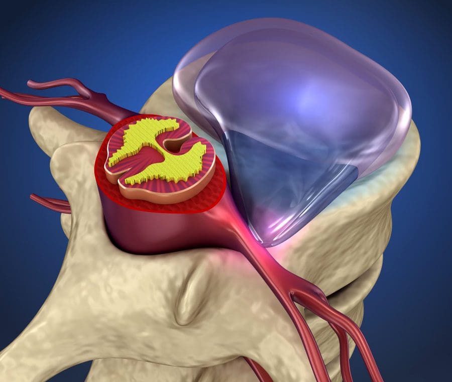
Regarding spinal pressure in the lower back, the disc is thicker and the most susceptible to injury. When it comes to spinal pressure related to disc herniation, it can lead to many individuals dealing with lower back pain and can affect their quality of life. One of the symptoms of disc herniation that are correlated with spinal pressure is that the displacement of the spinal disc can cause pain and disability in the spine as a result of a traumatic injury or degenerative changes due to the natural aging process. (Chu et al., 2023) When working, individuals put constant pressure on their spines, which can speed up the development of lower back pain.
Additionally, when there is immense spinal pressure on the spine, many pain-like issues that individuals don’t normally have will begin to pop up. This is due to a focal displacement of the intervertebral disc material that is beyond the normal limit of the spine and compresses one or more nerve roots, which can cause musculoskeletal issues to arise. (Trager et al., 2022) This, in turn, causes radiating extremity pain on the upper and lower body portions, sensory disturbances, muscle weakness, and even diminished muscle stretch reflexes as pain-like symptoms in the lower back. At the same time, when individuals are experiencing low back pain associated with spinal pressure, their truck muscles have an abnormal tilt when sitting, standing, and walking. (Wang et al., 2022) When this happens, it can cause them to develop poor posture, and when they are in an upright position, they will feel pain in their lower backs due to weak truck muscles. However, there are ways to relieve spinal pressure from aggravating the nerve roots affecting the lower back.
The Non-Surgical Approach To Wellness-Video
When looking for the right treatment, many individuals want to look for something that is cost-effective and relieves their pain. Non-surgical treatments are cost-effective and utilize various techniques to help reduce musculoskeletal pain through mechanical and manual motions to strengthen weakened muscles, relieve spinal pressure off the disc, and help realign the body to promote healing properties. The video above shows how non-surgical treatments like chiropractic care can help many individuals get their foot on the right on their health and wellness journey. At the same time, spinal decompression is another form of non-surgical treatment as it incorporates gentle traction on the spine to reduce intervertebral pressure during active and passive traction. (Andersson et al., 1983) When the spine is gently pulled, the herniated disc starts to return to its original position back to the spine, which then allows the fluids and nutrients to return to the disc and rehydrate them.
Decompression Reducing Spinal Pressure On Lower Back
So, how does spinal decompression help reduce disc pressure off the spine when dealing with low back pain? As stated earlier, spinal decompression incorporates gentle traction on the spine to be gently pulled to stretch weak surrounding muscles in the lower back. This causes an inverse relationship as the pressure within the nucleus pulposus of the herniated disc can help improve posture for many individuals with low back pain. (Ramos & Martin, 1994) Similarly, when many people incorporate decompression and chiropractic, the pain intensity is significantly reduced in all body parts, and many individuals will begin to feel the relief they deserve. (Ljunggren et al., 1984) When many individuals listen to their bodies and get the treatment they deserve, they will start to notice how decompression can help restore their bodies and positively improve their health.
References
Andersson, G. B., Schultz, A. B., & Nachemson, A. L. (1983). Intervertebral disc pressures during traction. Scand J Rehabil Med Suppl, 9, 88-91. https://www.ncbi.nlm.nih.gov/pubmed/6585945
Chu, E. C., Lin, A., Huang, K. H. K., Cheung, G., & Lee, W. T. (2023). A Severe Disc Herniation Mimics Spinal Tumor. Cureus, 15(3), e36545. https://doi.org/10.7759/cureus.36545
Ljunggren, A. E., Weber, H., & Larsen, S. (1984). Autotraction versus manual traction in patients with prolapsed lumbar intervertebral discs. Scand J Rehabil Med, 16(3), 117-124. https://www.ncbi.nlm.nih.gov/pubmed/6494835
Ramos, G., & Martin, W. (1994). Effects of vertebral axial decompression on intradiscal pressure. J Neurosurg, 81(3), 350-353. https://doi.org/10.3171/jns.1994.81.3.0350
Trager, R. J., Daniels, C. J., Perez, J. A., Casselberry, R. M., & Dusek, J. A. (2022). Association between chiropractic spinal manipulation and lumbar discectomy in adults with lumbar disc herniation and radiculopathy: retrospective cohort study using United States’ data. BMJ Open, 12(12), e068262. https://doi.org/10.1136/bmjopen-2022-068262
Can individuals with herniated pain associated with low back pain find relief through spinal decompression to restore mobility?
Introduction
Many people worldwide have experienced pain in the back region and often complain that it affects their mobility when doing their normal routine. The musculoskeletal system has various muscles, soft tissues, joints, ligaments, and bones that help surround the spine and protect the vital organs. The spine consists of bones, joints, and nerve roots that have an outstanding relationship with the central nervous system and musculoskeletal system as the spinal cord is protected by the spinal joints and discs that have the nerve roots spread out and help provide the sensory-motor function to the upper and lower extremities. When various pathogens or environmental factors start to cause the spine to compress the spinal discs constantly, it can lead to herniation and affect the body’s mobility over time. Individuals, both young and old, will notice that the pain is not going away from home remedies and may have to seek out treatment if the pain is too much. However, it can lead to dealing with unnecessary stress when looking for affordable treatment. Today’s article looks at how herniation can affect low back mobility and how treatments like decompression can help restore the spine. We speak with certified medical providers who incorporate our patients’ information to provide various solutions to restore low back mobility to the spine. We also inform patients how treatments like decompression can restore the spine’s mobility to the body. We encourage our patients to ask intricated and educational questions to our associated medical providers about the pain-like symptoms they are experiencing correlating with disc herniation affecting the spine. Dr. Alex Jimenez, D.C., utilizes this information as an academic service. Disclaimer.
Disc Herniation Affecting Low Back Mobility
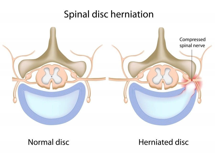
Do you often experience stiffness or limited mobility in your lower back that causes you to walk a little slower than usual? Do you feel pain in your lower back muscles from stretching or bending down to pick up an object? Or do you feel numbness or tingling sensations down your legs that feel uncomfortable? When many individuals start to do repetitive motions, that can cause their spinal discs to compress over time and eventually become herniated. When many individuals overwork their bodies, their spinal discs can eventually crack, causing the inner portion to protrude and press on the surrounding nerve root. This causes the disc tissue to have a central ballon-type cyst that causes degenerative changes, leading to low back pain and herniation. (Ge et al., 2019)
At the same time, when many individuals start to deal with lower back pain from herniated discs, they will begin to lose mobility in their lower backs. This could be due to weak abdominal muscles combined with limited mobility. When many individuals do not have strong core muscles to provide support and mobility to their lower backs, it can start with simple muscle aches, leading to constant lower back pain without treatment and negatively impacting their quality of life. (Chu, 2022) However, dealing with low back pain does not have to be tedious as numerous therapies can reduce the effects of low back pain correlated with disc herniation while restoring low back spinal mobility.
The Science Of Motion-Video
Have you ever experienced unquestionable muscle aches that radiate from your lower back and travel down your legs? Do you feel stiffness when bending down to pick up an object that causes muscle strain on your lower back? Or do you feel pain in your lower back from excessive sitting or standing? When many people are dealing with these pain-like issues in their lower backs, it can lead to a life of disability while affecting their quality of life. This is due to a disc herniation that affects a person’s lower back mobility and, when not treated right away, can lead to chronic issues. However, many individuals will seek treatment for their lower back pain and find the relief they need. Many therapeutic exercises combined with non-surgical treatments can help retrain the weakened trunk muscles to stabilize the lower back better and help reduce lower back pain. (Hlaing et al., 2021) When individuals start to think about their health and wellness, especially when they are dealing with low back pain affecting their mobility, they will find that most of the pain is from normal, repetitive factors that cause their spinal disc to be compressed and herniated. Hence, applying traction to the lumbar spine can help reduce lumbar disc protrusion that causes low back pain. (Mathews, 1968) Treatments like chiropractic care, traction therapy, and spinal decompression are all non-surgical treatments that are cost-effective and gentle on the spine. They help realign the body and help kick start the body’s natural healing factor to rehydrate the spinal discs. When many individuals start to do continuous treatment to reduce their lower back pain associated with herniated discs, they will begin to see improvements in their spinal mobility and their pain diminished. Check out the video above to look at how non-surgical treatments can help restore mobility to the body and reduce pain-like symptoms.
Decompression Restoring The Spine
When it comes to reducing pain-like symptoms caused by disc herniation that is causing limited mobility and low back pain, spinal decompression could be the answer that many individuals are looking for to incorporate into their health and wellness routine. Since lumbar herniated spinal discs are a common cause of low back pain and radiculopathy, spinal decompression can help gently pull the herniated disc back to its original position to promote healing. Since spinal decompression and lumbar traction are part of the physiotherapy treatment, they can help decrease the pain intensity from the spine and reduce the size of the herniated disc. (Choi et al., 2022) When many individuals feel relief from the gentle pull from spinal decompression, they will notice that their mobility is back. After consecutive treatment, their pain will be diminished as their spinal disc is completely healed. (Cyriax, 1950) With many individuals who are looking for numerous treatments to reduce their lower back pain and regain their sense of life, incorporating these treatments can provide beneficial results to their musculoskeletal system.
References
Choi, E., Gil, H. Y., Ju, J., Han, W. K., Nahm, F. S., & Lee, P. B. (2022). Effect of Nonsurgical Spinal Decompression on Intensity of Pain and Herniated Disc Volume in Subacute Lumbar Herniated Disc. International Journal of Clinical Practice, 2022, 6343837. https://doi.org/10.1155/2022/6343837
Chu, E. C. (2022). Large abdominal aortic aneurysm presented with concomitant acute lumbar disc herniation – a case report. J Med Life, 15(6), 871-875. https://doi.org/10.25122/jml-2021-0419
Ge, C. Y., Hao, D. J., Yan, L., Shan, L. Q., Zhao, Q. P., He, B. R., & Hui, H. (2019). Intradural Lumbar Disc Herniation: A Case Report and Literature Review. Clin Interv Aging, 14, 2295-2299. https://doi.org/10.2147/CIA.S228717
Hlaing, S. S., Puntumetakul, R., Khine, E. E., & Boucaut, R. (2021). Effects of core stabilization exercise and strengthening exercise on proprioception, balance, muscle thickness and pain related outcomes in patients with subacute nonspecific low back pain: a randomized controlled trial. BMC Musculoskelet Disord, 22(1), 998. https://doi.org/10.1186/s12891-021-04858-6
IFM's Find A Practitioner tool is the largest referral network in Functional Medicine, created to help patients locate Functional Medicine practitioners anywhere in the world. IFM Certified Practitioners are listed first in the search results, given their extensive education in Functional Medicine