Discover the connection between chiropractic care and bone health. Enhance your musculoskeletal system with expert insights.
Chiropractic Care: A Comprehensive Guide to Musculoskeletal Pain Relief and Bone Health
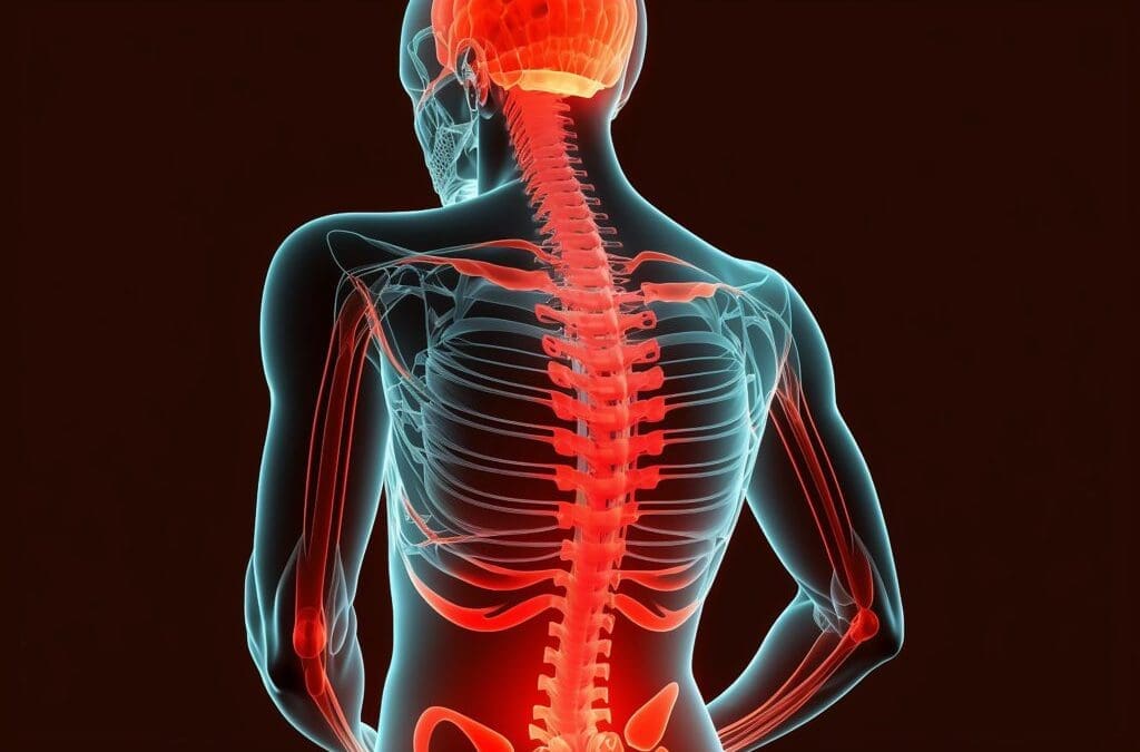
When you think about your body, it’s easy to take for granted the complex machinery that keeps you moving—your muscles, bones, and joints working together like a well-oiled (or sometimes creaky) machine. The musculoskeletal system is the unsung hero of daily life, allowing you to run, lift, dance, or even just scroll through your phone without toppling over. But when pain strikes—whether from a fender-bender, a clumsy tumble, or just the wear and tear of life—it can feel like your body’s betraying you. Enter chiropractic care, the hands-on hero that helps get you back on track. In this comprehensive guide, we’ll dive into how chiropractic care, led by experts like Dr. Alexander Jimenez, DC, APRN, FNP-BC, at El Paso Back Clinic, can reduce musculoskeletal pain, boost bone health, and help you reclaim your quality of life. Plus, we’ll sprinkle in a bit of humor to keep things light—because who said healing can’t come with a chuckle?
The Musculoskeletal System: Your Body’s Framework
The musculoskeletal system is like the scaffolding of a building—it holds everything together, provides structure, and lets you move without collapsing into a heap. It’s made up of bones, muscles, tendons, ligaments, and other connective tissues that work in harmony. Bones provide the framework, muscles give you the power to move, and joints act like hinges, allowing flexibility. When everything’s working smoothly, you barely notice it. But when something goes wrong—like a strained muscle, a misaligned spine, or a bone that’s not as strong as it used to be—life can get uncomfortable fast.
Why Bone Health Matters
Bones aren’t just static structures; they’re living tissues that constantly remodel themselves. Strong bones are crucial for mobility, protecting vital organs, and even producing blood cells. Poor bone health, like osteoporosis, can lead to fractures, chronic pain, and reduced quality of life. According to research, maintaining bone health involves a balance of proper nutrition, exercise, and avoiding environmental factors that weaken bones (Baim, 2014).
Environmental factors—like poor posture, sedentary lifestyles, or repetitive stress—can wreak havoc on your musculoskeletal system. For instance, sitting at a desk all day can strain your spine, while heavy lifting without proper form can lead to joint injuries. Add in accidents like car crashes or slips, and you’ve got a recipe for musculoskeletal mayhem. Chiropractic care steps in to address these issues, not just by treating pain but by restoring balance and supporting bone health.
Reference:
Baim, S. (2014). Osteoporosis prevention, screening, and treatment: A review. Journal of Clinical Densitometry, 17(3), 371-378. https://pubmed.ncbi.nlm.nih.gov/24709112/
Chiropractic Care: The Science Behind the Snap, Crackle, Pop
Chiropractic care is like giving your body a tune-up, but instead of a wrench, chiropractors use their hands (and sometimes a bit of tech) to adjust misaligned joints and relieve pressure on nerves. The goal? Restore proper alignment, reduce pain, and improve function. It’s not just about cracking your back—though, let’s be honest, that satisfying pop can feel like a mini-victory. The real magic happens when the musculoskeletal system is realigned, allowing your body to heal naturally.
Clinical Rationale for Pain Relief
Chiropractic care works by addressing misalignments (subluxations) in the spine and joints that can cause pain, inflammation, and reduced mobility. For example, spinal manipulative therapy (SMT) has been shown to reduce low back pain by improving spinal mobility and reducing muscle tension (Blanchette et al., 2016). A systematic review found that SMT is effective for acute low back pain, providing relief comparable to other treatments like medication but with fewer side effects (Paige et al., 2017).
Dr. Alexander Jimenez, a chiropractor at El Paso Back Clinic, emphasizes a holistic approach. His methods focus on restoring joint and biomechanical function, which can alleviate pain from conditions like sciatica, whiplash, or degenerative arthritis. By using advanced imaging and diagnostic evaluations, he identifies the root cause of pain, ensuring targeted treatment. For instance, a misaligned spine from a car accident can pinch nerves, causing radiating pain. Chiropractic adjustments realign the spine, relieving pressure and promoting healing.
Reference:
Blanchette, M. A., Stochkendahl, M. J., Borges Da Silva, R., Boruff, J., Harrison, P., & Bussières, A. (2016). Effectiveness and economic evaluation of chiropractic care for the treatment of low back pain: A systematic review of pragmatic studies. PLoS One, 11(8), e0160037. https://pubmed.ncbi.nlm.nih.gov/27487116/
Paige, N. M., Miake-Lye, I. M., Booth, M. S., Beroes, J. M., Mardian, A. S., Dougherty, P., … & Shekelle, P. G. (2017). Association of spinal manipulative therapy with clinical benefit and harm for acute low back pain: Systematic review and meta-analysis. JAMA, 317(14), 1451-1460. https://pubmed.ncbi.nlm.nih.gov/28399251/
Bone Health Benefits
Chiropractic care doesn’t just help with pain—it can also support bone health. By improving spinal alignment, chiropractic adjustments reduce stress on bones and joints, which can prevent wear and tear that contributes to conditions like osteoarthritis. Additionally, techniques like electrical stimulation, often used in chiropractic settings, can promote bone healing by stimulating osteogenesis (bone formation) (Ciombor & Aaron, 1996). This is particularly beneficial for individuals recovering from fractures or dealing with osteoporosis.
Dr. Jimenez incorporates therapies like massage and durable medical equipment (e.g., braces or supports) to enhance recovery. These tools stabilize joints, reduce inflammation, and support bone health, especially after injuries like those from motor vehicle accidents (MVAs).
Reference:
Ciombor, D. M., & Aaron, R. K. (1996). Stimulators of bone healing: Biologic and biomechanical. Clinical Orthopaedics and Related Research, 327, 12-20. https://pubmed.ncbi.nlm.nih.gov/8653943/
Five Common Musculoskeletal Issues and Chiropractic Solutions
Let’s break down five common musculoskeletal problems and how chiropractic care can help. Think of these as the “usual suspects” when it comes to body aches and pains.
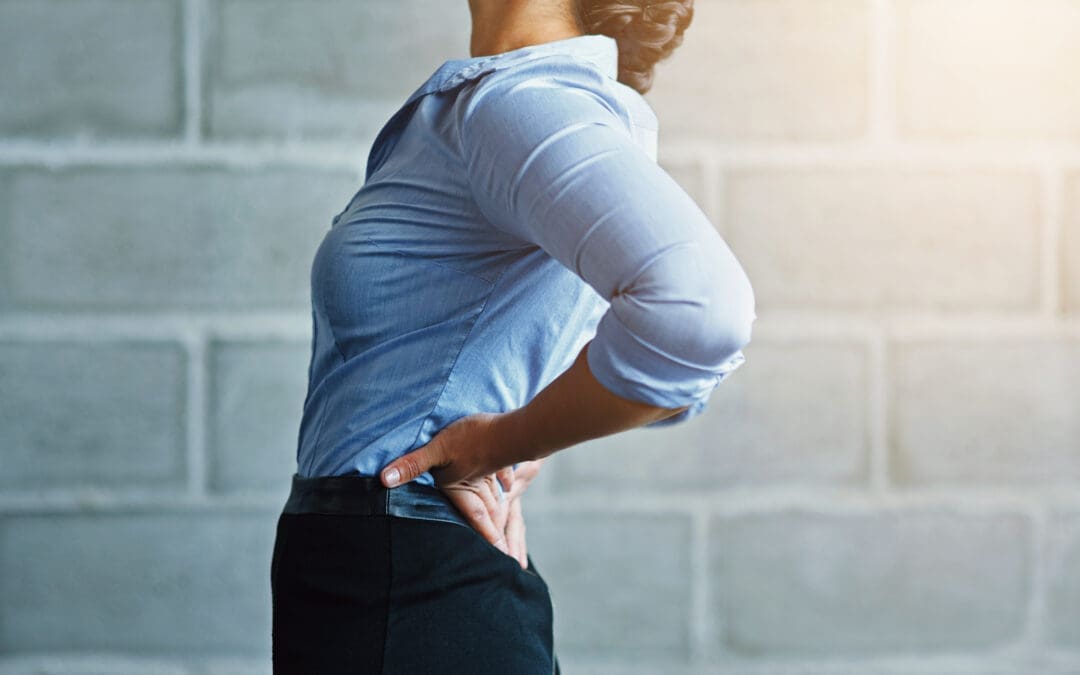
1. Low Back Pain
Low back pain is the bane of many people’s existence—whether from sitting too long, lifting something heavy, or just sleeping in a weird position. It’s like your spine’s way of saying, “Hey, pay attention to me!” Chiropractic care, particularly spinal manipulative therapy, has been shown to reduce pain and improve function in active-duty military personnel with low back pain (Goertz et al., 2018).
Dr. Jimenez uses advanced imaging to pinpoint misalignments or disc issues, then applies targeted adjustments to restore mobility. He might also recommend exercises to strengthen core muscles, which act like a natural corset for your spine.
Reference:
Goertz, C. M., Long, C. R., Vining, R. D., Pohlman, K. A., Walter, J., & Coulter, I. (2018). Effects of chiropractic care on strength, balance, and endurance in active-duty U.S. military personnel with low back pain: A randomized controlled trial. Journal of Alternative and Complementary Medicine, 24(7), 669-676. https://pubmed.ncbi.nlm.nih.gov/29470104/
2. Neck Pain and Whiplash
Ever get a stiff neck from staring at your phone too long? Now imagine that pain amplified by a car accident. Whiplash-associated disorders (WAD) are common after MVAs, causing neck pain, headaches, and restricted movement. Chiropractic care can help by realigning the cervical spine and reducing muscle tension. Dr. Jimenez’s dual-scope approach—combining chiropractic adjustments with massage therapy—can speed up recovery by improving blood flow and reducing inflammation.
3. Sciatica
Sciatica is like an unwelcome guest that shoots pain down your leg, often caused by a pinched nerve in the lower back. Chiropractic adjustments can relieve pressure on the sciatic nerve, while exercises prescribed by Dr. Jimenez strengthen supporting muscles to prevent recurrence.
4. Joint Injuries
Whether from sports or a slip on a rainy day, joint injuries (like sprained ankles or shoulders) can limit mobility. Chiropractic care restores joint alignment and reduces inflammation, while integrative therapies like acupuncture (available at El Paso Back Clinic) enhance healing.
5. Degenerative Arthritis
Arthritis is like rust on your joints—it builds up over time, making movement painful. Chiropractic care can’t cure arthritis, but it can improve joint function and reduce pain through gentle adjustments and therapies like electrical stimulation, which supports bone health (Ciombor & Aaron, 1996).
Reference:
Ciombor, D. M., & Aaron, R. K. (1996). Stimulators of bone healing: Biologic and biomechanical. Clinical Orthopaedics and Related Research, 327, 12-20. https://pubmed.ncbi.nlm.nih.gov/8653943/
Understanding Ligamentous Injuries- Video
Environmental Factors and Their Impact
Your daily routine can be a minefield for your musculoskeletal system. Here are some common culprits:
Poor Posture: Slouching at your desk or hunching over your phone can misalign your spine, leading to pain. Chiropractic adjustments correct these misalignments, while Dr. Jimenez’s team offers ergonomic advice to prevent future issues.
Sedentary Lifestyle: Lack of movement weakens muscles and bones, increasing the risk of osteoporosis (Baim, 2014). Regular chiropractic care, combined with exercise plans, keeps your musculoskeletal system strong.
Accidents: MVAs, bicycle crashes, or 18-wheeler collisions can cause severe injuries. Dr. Jimenez’s expertise in personal injury cases ensures comprehensive care, from diagnostics to rehabilitation.
Repetitive Stress: Jobs involving repetitive motions (like typing or lifting) can strain joints and muscles. Chiropractic care reduces inflammation and restores function.
Reference:
Baim, S. (2014). Osteoporosis prevention, screening, and treatment: A review. Journal of Clinical Densitometry, 17(3), 371-378. https://pubmed.ncbi.nlm.nih.gov/24709112/
Dr. Alexander Jimenez: El Paso’s Personal Injury Expert
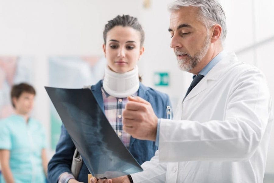
In El Paso, personal injury cases—like those from car accidents or workplace injuries—are a significant concern. Dr. Alexander Jimenez stands out as a distinguished practitioner, blending chiropractic expertise with advanced diagnostic skills. At El Paso Back Clinic, he uses tools like X-rays, MRIs, and dual-scope procedures to assess injuries accurately. His ability to connect medical findings with legal documentation makes him a vital liaison for patients navigating personal injury claims.
For example, after an 18-wheeler accident, Dr. Jimenez might use imaging to identify spinal misalignments or soft tissue damage, then create a tailored treatment plan combining adjustments, massage, and nutritional guidance. His holistic approach ensures patients recover physically while supporting their legal cases with detailed medical reports.
Chiropractic Care in Action: Real-World Applications
Let’s paint a picture: You’re driving home, and bam—a fender-bender leaves you with a sore neck and a grumpy spine. Or maybe you’re a cyclist who took a tumble, and now your shoulder’s acting like it’s auditioning for a horror movie. These scenarios are where chiropractic care shines.
Motor Vehicle Accidents (MVAs)
MVAs can cause everything from whiplash to gastrointestinal injuries. Dr. Jimenez’s team uses a combination of chiropractic adjustments, massage therapy, and durable medical equipment to address these issues. For example, trigger point therapy can release muscle knots caused by MVAs, while nutritional plans support internal healing.
Bicycle Accidents
Bicyclists face unique risks, like collisions with cars or falls. Chiropractic care restores joint function, while integrative medicine (like acupuncture) reduces inflammation. Dr. Jimenez’s comprehensive approach ensures cyclists get back on the road safely.
18-Wheeler Crashes
These accidents are no joke—think of an 18-wheeler as a giant rolling pin flattening your musculoskeletal system. Chiropractic care, combined with advanced diagnostics, helps address spinal and joint injuries, while therapies like electrical stimulation promote bone healing.
Nutrition and Lifestyle: The Unsung Heroes
You can’t talk about musculoskeletal health without mentioning nutrition. A diet rich in calcium, vitamin D, and anti-inflammatory foods (like leafy greens and fatty fish) supports bone health and reduces inflammation (Health Coach Clinic, n.d.). Dr. Jimenez’s team at El Paso Back Clinic creates personalized nutrition plans to aid recovery, especially after MVAs.
Lifestyle changes, like regular exercise and proper ergonomics, also play a role. Think of it like giving your body a daily pep talk—it needs movement and care to stay strong. Chiropractic care complements these efforts by ensuring your spine and joints are aligned, making exercise more effective.
The Chiropractic Identity: A Framework for Healing
Chiropractic care isn’t just a treatment—it’s a philosophy of health that emphasizes the body’s ability to heal itself when properly aligned. Research highlights spinal care as a cornerstone of chiropractic identity, focusing on restoring function and preventing chronic issues (Gliedt et al., 2015). Dr. Jimenez embodies this philosophy, combining evidence-based techniques with personalized care to address each patient’s unique needs.
Reference:
Gliedt, J. A., Hawk, C., Anderson, M., Ahmad, K., Bunn, D., Cambron, J., … & Schneider, M. J. (2015). Spine care as a framework for the chiropractic identity. Journal of Chiropractic Humanities, 22(1), 14-21. https://pubmed.ncbi.nlm.nih.gov/26770177/
A Dash of Humor: Keeping It Light
Let’s face it—talking about back pain and bone health can feel like a lecture from a grumpy science teacher. So, imagine your spine as a cranky old neighbor who just needs a little TLC to stop complaining. Chiropractic care is like inviting that neighbor over for a cup of tea and a good stretch—suddenly, they’re a lot less cranky! Whether it’s a quick adjustment or a full-on rehab plan, Dr. Jimenez’s team at El Paso Back Clinic knows how to make your musculoskeletal system smile again.
Conclusion
Chiropractic care is a powerful tool for managing musculoskeletal pain and supporting bone health. From low back pain to whiplash, Dr. Alexander Jimenez and his team at El Paso Back Clinic offer evidence-based solutions that address the root cause of discomfort. By combining spinal adjustments, advanced diagnostics, and integrative therapies like massage and nutrition, they help patients recover from injuries and improve their quality of life. For personal injury victims in El Paso, Dr. Jimenez’s expertise ensures comprehensive care and legal support, making him a trusted ally in recovery.
Disclaimer: This blog post is intended for informational purposes only and should not be taken as medical advice. Always consult a qualified healthcare professional, such as a chiropractor or physician, before starting any treatment. The information provided is based on current research and clinical insights, but does not replace professional medical evaluation.
Understand the role of chiropractic care for herniated discs in relieving pain and restoring function for a healthier spine.
Chiropractic Care for Low Back Pain: A Deep Dive into Herniated Discs, Spinal Decompression, and Recovery with Dr. Alex Jimenez
Mon cher, picture this: your spine, that elegant column of bones, is like a grand chandelier in the Addams Family mansion—beautiful, complex, but oh so prone to a flicker or two when things go awry! When a herniated disc sneaks into the lumbar spine, it’s like Gomez Addams tripping over a loose floorboard, sending chaos through the household. But fear not, for chiropractic care, led by the masterful Dr. Alexander Jimenez in El Paso, Texas, is here to restore harmony with a twirl and a flourish!
Low back pain is a common complaint, affecting millions worldwide, with herniated discs often playing the villain in this spine-tingling drama. This blog post explores the clinical rationale behind chiropractic care and spinal decompression as effective treatments for low back pain caused by herniated discs. We’ll dive into the anatomy of the lumbar spine, how herniated discs disrupt daily life, and why Dr. Alex Jimenez, DC, APRN, FNP-BC, stands out as a beacon of hope for personal injury victims in El Paso. With advanced imaging, diagnostic evaluations, and his unique dual-scope approach, Dr. Jimenez bridges the gap between medical care and legal documentation, ensuring patients recover while navigating the complexities of personal injury cases. So, grab a seat—preferably not on a wobbly one—and let’s unravel this tale of spinal recovery with a dash of Gomez Addams’ charm!
The Lumbar Spine: The Backbone of Your Daily Grind
The lumbar spine, or lower back, is the unsung hero of your body, supporting the weight of your upper torso while allowing you to bend, twist, and tango like Gomez with Morticia. It consists of five vertebrae (L1-L5), sturdy bones stacked like a tower of Gothic bricks, connected by intervertebral discs that act as shock-absorbing cushions. These discs, with their tough outer layer (annulus fibrosus) and jelly-like center (nucleus pulposus), are designed to handle pressure, much like a well-crafted torture device from the Addams Family—resilient but not invincible.
When a disc herniates, the nucleus pulposus bulges or ruptures through the annulus fibrosus, often pressing on nearby spinal nerves. This can happen due to aging, wear and tear, or sudden trauma, like lifting a heavy coffin or surviving a fender-bender in El Paso’s bustling streets. The result? Pain, numbness, or weakness that can radiate from the lower back into the buttocks, thighs, or calves, often mimicking the electric jolt Gomez feels when Morticia speaks French.
How Herniated Discs Affect Daily Life
A herniated disc in the lumbar spine, particularly at the L4-L5 or L5-S1 levels, can turn everyday activities into a comedy of errors—minus the laughs. Imagine trying to tie your shoes but feeling like Lurch is sitting on your back. Common symptoms include:
Low Back Pain: A dull ache or sharp, stabbing pain that worsens with movement, making bending or lifting as daunting as facing Uncle Fester’s experiments.
Sciatica: Pain radiating down the leg, caused by nerve root compression, often described as a burning or electric sensation. It’s like Gomez’s fencing foil zapping you unexpectedly.
Numbness or Tingling: A pins-and-needles feeling in the legs or feet, disrupting your ability to walk or stand without feeling like you’re on a bed of nails.
Weakness: Muscles served by affected nerves may weaken, causing stumbling or difficulty lifting objects, as if Pugsley swapped your weights for marshmallows.
These symptoms can severely limit daily routines. Sitting at a desk, driving to work, or even sleeping can become painful, leading to missed workdays, reduced productivity, and a dampened zest for life. For El Paso residents, who often lead active lifestyles and demanding jobs, a herniated disc can feel like a betrayal by their spine.
Chiropractic care, much like Gomez’s passionate dance moves, is all about restoring balance and flow. It focuses on the musculoskeletal system, particularly the spine, to correct misalignments (subluxations) that disrupt nerve function and cause discomfort. For herniated discs, chiropractic care offers a non-surgical, evidence-based approach to relieve pain, reduce nerve compression, and restore mobility. Here’s why it works:
Spinal Manipulation: The Chiropractic Tango
Spinal manipulation, also known as adjustments, involves the precise and controlled application of force to the spine to correct misalignments. Think of it as Gomez gently nudging Morticia back into step during a waltz. By realigning the vertebrae, chiropractors reduce pressure on the herniated disc and compressed nerves, alleviating pain and improving function. A 2020 study in the Spine Journal found that spinal manipulative therapy significantly reduces pain and disability in patients with chronic low back pain (Rubinstein et al., 2020, as cited in).
For patients with MRI-confirmed lumbar disc herniation and sacroiliac joint hypomobility, spinal manipulation has shown promising results. A quasi-experimental study in Chiropractic & Manual Therapies demonstrated that patients receiving spinal manipulation experienced significant pain reduction and improved mobility compared to the control group (Shokri et al., 2018). This is because adjustments restore joint function, reduce inflammation, and enhance blood flow, helping the body heal naturally.
Spinal Decompression: Stretching the Spine with Flair
Non-surgical spinal decompression is like stretching out a tightly wound Addams Family tapestry. This therapy uses a motorized table to gently elongate the spine, creating negative pressure within the disc. This negative pressure can help retract the herniated nucleus pulposus, reducing nerve compression and promoting disc healing. A 2017 study in the Journal of Physical Therapy Science found that spinal decompression significantly reduced pain and disability in patients with lumbar disc herniation (Choi et al., 2017, as cited in).
Dr. Alex Jimenez, a leading chiropractor in El Paso, emphasizes that spinal decompression not only alleviates pain but also rehydrates the disc by improving nutrient delivery. “It’s like giving your spine a refreshing sip of water after a long, dry day,” he notes on his website (El Paso Back Clinic, n.d.). By increasing disc height and reducing herniation volume, decompression therapy restores spinal flexibility, allowing patients to move without wincing.
Functional Medicine: A Holistic Twist
Dr. Jimenez’s practice extends beyond adjustments, incorporating functional medicine to address underlying issues such as inflammation and nutritional deficiencies. For instance, dietary changes can reduce systemic inflammation, accelerating recovery from disc injuries. A 2019 meta-analysis in Pain Physician confirmed that regenerative therapies, like platelet-rich plasma (PRP), can complement chiropractic care by reducing lumbar pain (Sanapati et al., 2019, as cited in). This holistic approach ensures that the body heals from the inside out, much like Gomez nurturing his beloved carnivorous plants.
Shokri, M., et al. (2018). Spinal manipulation in the treatment of patients with MRI-confirmed lumbar disc herniation and sacroiliac joint hypomobility: A quasi-experimental study. Chiropractic & Manual Therapies, 26, 16. https://chiromt.biomedcentral.com/articles/10.1186/s12998-018-0185-z
Rubinstein, S. M., et al. (2020). Spinal manipulative therapy for chronic low-back pain. Spine Journal, 20(4), 489–502.
Dr. Alex Jimenez: El Paso’s Chiropractic Maestro
In El Paso, Texas, Dr. Alexander Jimenez is the Gomez Addams of chiropractic care—passionate, skilled, and dedicated to his craft. With over 25 years of experience as a Doctor of Chiropractic (DC) and a board-certified Family Nurse Practitioner (APRN, FNP-BC), Dr. Jimenez brings a dual-scope approach to treating herniated discs and personal injury cases. His practice at El Paso Back Clinic (https://elpasobackclinic.com/) is a haven for those seeking relief from low back pain, sciatica, and other musculoskeletal woes.
Advanced Imaging and Diagnostics
Dr. Jimenez utilizes state-of-the-art imaging techniques, including MRI and CT scans, to precisely identify the location and severity of a herniated disc. These tools provide a clear picture of soft tissues, revealing disc bulges or nerve compression that X-rays might miss (Personal Injury Doctor Group, 2017). By combining imaging with physical exams, such as the straight leg raise test, he confirms diagnoses with precision, ensuring treatments are tailored to each patient’s individual needs.
Dual-Scope Procedures
What sets Dr. Jimenez apart is his ability to blend chiropractic and medical expertise. His dual-scope approach involves:
Chiropractic Assessments: Identifying spinal misalignments and nerve compression through hands-on evaluations.
Medical Evaluations: Assessing systemic factors, like inflammation or hormonal imbalances, that may hinder healing (Jimenez, 2023, as cited in).
This comprehensive method enables him to create personalized treatment plans that address both the biomechanical and physiological aspects of a herniated disc. For example, he might use spinal adjustments to relieve nerve pressure while recommending nutritional changes to reduce inflammation, ensuring a holistic recovery.
Bridging Medical and Legal Needs
In personal injury cases, such as those from auto accidents, Dr. Jimenez shines as a liaison between medical care and legal documentation. His detailed reports, backed by advanced diagnostics, provide critical evidence for insurance claims or court cases, ensuring patients receive fair compensation. “My goal is to help patients heal while protecting their rights,” Dr. Jimenez shares on his LinkedIn profile (Jimenez, n.d., https://www.linkedin.com/in/dralexjimenez/). His expertise in documenting injuries, from whiplash to complex herniated discs, makes him a trusted practitioner for El Paso’s personal injury victims.
Personal Injury in El Paso: Why Chiropractic Care Matters
El Paso, a vibrant city with a bustling economy, sees its fair share of personal injuries, particularly from motor vehicle accidents (MVAs). These incidents often result in herniated discs, whiplash, or nerve compression, leaving victims in pain and struggling to navigate insurance claims or legal battles. Chiropractic care, especially under Dr. Jimenez’s guidance, is a cornerstone of recovery for these individuals.
The Impact of MVAs
MVAs can cause sudden trauma to the lumbar spine, leading to disc herniation or nerve injuries. For instance, a rear-end collision might whip the spine, causing the nucleus pulposus to bulge and compress the sciatic nerve, resulting in debilitating pain. Dr. Jimenez’s clinic specializes in these cases, using non-invasive techniques like spinal decompression and adjustments to restore function without surgery.
Legal Documentation and Medical Care
Personal injury cases require meticulous documentation to prove the extent of injuries. Dr. Jimenez’s dual licensure as a chiropractor and nurse practitioner enables him to provide comprehensive medical reports that meet legal standards. His use of advanced imaging ensures that injuries are documented, strengthening patients’ cases while guiding their recovery. This dual role is particularly valuable in El Paso, where personal injury claims are common due to the high volume of traffic and industrial activity.
References
El Paso Back Clinic. (2016, September 29). El Paso, TX: Wellness Chiropractic Care Clinic. https://elpasobackclinic.com/
Spinal decompression is a star player in the chiropractic playbook, especially for herniated discs. By gently stretching the spine, this therapy creates a vacuum effect that pulls the herniated disc material back into its proper position, thereby reducing pressure on the nerves. It’s like coaxing a wayward bat back into the Addams Family attic—gentle but effective.
How It Works
During a decompression session, patients lie on a specialized table that alternates between traction and relaxation. This process:
Reduces Disc Pressure: Negative pressure within the disc helps retract the herniated material, relieving nerve compression.
Promotes Healing: Increased blood flow delivers oxygen and nutrients to the disc, aiding rehydration and repair.
Restores Mobility: By alleviating pain and stiffness, decompression allows patients to move freely again.
A 2022 study on PubMed found that non-surgical spinal decompression reduced pain and herniated disc volume in patients with subacute lumbar disc herniation, supporting its efficacy (Choi et al., 2022, https://www.ncbi.nlm.nih.gov/pmc/articles/PMC9473337/). Dr. Jimenez’s clinic leverages this therapy to help patients avoid surgery, with many reporting significant relief after a six-week course (El Paso Back Clinic, 2022).
Rehydration: The Disc’s Fountain of Youth
As we age, spinal discs lose water content, becoming less flexible and more prone to herniation. Spinal decompression counteracts this by improving nutrient exchange, effectively “rehydrating” the disc. Dr. Jimenez likens it to “watering a parched plant, bringing it back to life” (El Paso Back Clinic, n.d.). This process not only reduces pain but also enhances disc resilience, preventing future injuries.
References
Choi, J., et al. (2022). Effect of nonsurgical spinal decompression on intensity of pain and herniated disc volume in subacute lumbar herniated disc. International Journal of Clinical and Experimental Medicine, 15(4), 159–167. https://www.ncbi.nlm.nih.gov/pmc/articles/PMC9473337/
The effectiveness of chiropractic care for herniated discs is grounded in science, not just Gomez’s theatrical flair. Here’s a closer look at the mechanisms:
Nerve Root Compression Relief
Herniated discs often compress nerve roots, causing radiculopathy—pain, numbness, or weakness radiating along the nerve’s path. Chiropractic adjustments and decompression reduce this compression by realigning the spine and retracting disc material. A French study highlighted that nerve root compression due to lumbar disc herniation is a significant cause of sciatica, and non-surgical interventions, such as chiropractic care, can effectively address it (Valat et al., 2010, https://www.ncbi.nlm.nih.gov/pmc/articles/PMC2912793/).
Reducing Inflammation
Inflammation exacerbates disc-related pain. Chiropractic care, when combined with functional medicine, helps reduce inflammation through adjustments, targeted nutrition, and lifestyle modifications. Dr. Jimenez’s approach includes dietary plans to reduce systemic inflammation, which supports disc healing (Jimenez, 2023).
Enhancing Biomechanics
Misaligned vertebrae or sacroiliac joint hypomobility can worsen disc issues. Spinal manipulation corrects these misalignments, improving biomechanics and reducing stress on the disc. This is particularly effective for patients with both disc herniation and joint dysfunction (Shokri et al., 2018).
Shokri, M., et al. (2018). Spinal manipulation in the treatment of patients with MRI-confirmed lumbar disc herniation and sacroiliac joint hypomobility: A quasi-experimental study. Chiropractic & Manual Therapies, 26, 16. https://chiromt.biomedcentral.com/articles/10.1186/s12998-018-0185-z
Practical Tips for Managing Herniated Disc Pain
While chiropractic care is a powerful tool, patients can support their recovery with these practical tips, sprinkled with a touch of Addams Family mischief:
Stay Active (Carefully): Gentle movements, such as walking or stretching, keep the spine limber. Avoid heavy lifting—leave that to Lurch!
Mind Your Posture: Sit and stand like Gomez, proud and upright, to reduce spinal stress.
Apply Heat or Ice: Ice reduces inflammation, while heat soothes muscle spasms. Alternate them like Morticia’s mood swings.
Follow Dr. Jimenez’s Nutrition Advice: Anti-inflammatory foods, like berries or fatty fish, support healing. Avoid processed foods—they’re as harmful as Pugsley’s pranks.
Dr. Jimenez’s practice is a beacon for El Paso’s injury victims, offering a blend of compassion and expertise. His clinic, El Paso Back Clinic, provides:
Personalized Care: Tailored treatment plans based on advanced diagnostics.
Holistic Approach: Combining chiropractic, functional medicine, and rehabilitation.
Legal Support: Detailed documentation for personal injury claims, ensuring fair compensation.
Community Trust: Patient testimonials highlight his transformative impact (Jimenez, 2023).
His dual licensure and certifications (IFMCP, CFMP) make him uniquely qualified to address complex cases, from sciatica to chronic pain, with a focus on restoring function and quality of life.
My dear reader, we’ve danced through the shadowy halls of herniated discs and chiropractic care with the grace of Gomez Addams, but now it’s time to dim the candelabra and speak plainly. Low back pain from herniated discs is a serious condition that can disrupt daily life, but chiropractic care, including spinal manipulation and decompression, offers a proven, non-surgical solution. Dr. Alexander Jimenez, with his dual expertise and advanced diagnostic tools, stands out as a trusted practitioner in El Paso, particularly for personal injury cases. His ability to bridge medical care and legal documentation ensures patients recover physically and financially.
Disclaimer: This blog post is for informational purposes only and is not a substitute for professional medical advice. Always consult a qualified healthcare provider, such as Dr. Alex Jimenez, DC, APRN, FNP-BC, before starting any treatment. Individual results may vary, and chiropractic care may not be suitable for all conditions. For personalized guidance, contact El Paso Back Clinic at 915-850-0900 or visit https://elpasobackclinic.com/.
Choi, J., et al. (2022). Effect of nonsurgical spinal decompression on intensity of pain and herniated disc volume in subacute lumbar herniated disc. International Journal of Clinical and Experimental Medicine, 15(4), 159–167. https://www.ncbi.nlm.nih.gov/pmc/articles/PMC9473337/
Shokri, M., et al. (2018). Spinal manipulation in the treatment of patients with MRI-confirmed lumbar disc herniation and sacroiliac joint hypomobility: A quasi-experimental study. Chiropractic & Manual Therapies, 26, 16. https://chiromt.biomedcentral.com/articles/10.1186/s12998-018-0185-z
Understand the benefits of chiropractic care for those suffering from five musculoskeletal issues and regain your quality of life.
Chiropractic Care: A Comprehensive Guide to Managing Common Musculoskeletal Issues
Musculoskeletal issues can turn everyday activities into a real pain in the neck—literally! Just like Herman Munster, who often lumbered around with a stiff back and a creaky neck, many of us deal with aches and pains that make us feel like we’ve got a bolt loose. Fortunately, modern chiropractic care provides a non-invasive, drug-free approach to regaining our optimal health. In this blog post, we’ll explore five common musculoskeletal conditions—subluxation, scoliosis, disk degeneration, carpal tunnel syndrome, and Achilles tendonitis—and explain how chiropractic care, particularly from experts like Dr. Alexander Jimenez in El Paso, Texas, can help. We’ll also delve into the critical role chiropractic plays in personal injury cases, with a touch of humor to keep things light, but rest assured, we’ll conclude with a serious note.
Subluxation—“When Your Spine’s Out of Line”
What is Subluxation?
Imagine your spine as a stack of building blocks. If one block slips out of place, the whole tower wobbles, causing all sorts of trouble. In chiropractic terms杀了subluxation refers to a misalignment or dysfunction of a vertebra in the spine, which can put pressure on nerves, leading to pain, stiffness, or even headaches. Common causes include trauma (like a car accident), poor posture, arthritis, or muscle spasms. Neck subluxations are especially common, affecting many people due to prolonged sitting or stress.
Subluxations can make daily tasks, such as turning your head or bending over, feel like a scene from a horror movie. You might feel sharp pain, stiffness, or even tingling in your arms or legs if nerves are compressed. Left untreated, these misalignments can lead to chronic issues, much like ignoring a creaky door until it falls off its hinges.
How Chiropractic Care Helps
Chiropractors, like skilled architects, use spinal adjustments to nudge those wayward vertebrae back into place. These gentle, precise movements aim to restore proper alignment, reduce nerve interference, and promote the body’s natural healing. Think of it as straightening out a kink in a garden hose to let the water flow freely again. By improving spinal function, chiropractic care can alleviate pain, enhance mobility, and even boost overall wellness.
Clinical Rationale
The spine houses the spinal cord, a key part of the nervous system that controls bodily functions. A misaligned vertebra can irritate nerves, disrupting communication between the brain and body. Chiropractic adjustments aim to correct these misalignments, potentially reducing pain and improving function. While the term “subluxation” is debated in mainstream medicine due to limited empirical evidence, patient-reported outcomes often highlight significant relief from chiropractic care (Terlep Chiropractic, 2023).
Dr. Jimenez’s Approach
Dr. Alexander Jimenez, a leading chiropractor in El Paso, utilizes advanced diagnostic tools, including X-rays, to accurately identify subluxations. His evidence-based approach involves tailored adjustments and functional medicine to address underlying causes, such as poor posture or muscle imbalances, thereby ensuring long-term relief.
Scoliosis is like your spine deciding to take a scenic route, curving sideways in an “S” or “C” shape. This abnormal curvature often develops in childhood due to unknown causes (idiopathic scoliosis), trauma, or abnormal bone growth. It can lead to back pain, uneven shoulders or hips, and difficulty moving, making everyday activities like carrying a backpack or sitting for long periods uncomfortable.
How Chiropractic Care Helps
While chiropractic care may not fully correct a scoliotic spine, it can significantly reduce pain and improve function. Chiropractors use posture evaluations and manual manipulations to enhance spinal alignment and mobility. Specific exercises, such as those in the ScoliSMART method, can complement adjustments to strengthen supporting muscles and improve posture, making daily life more manageable.
Clinical Rationale
Research suggests that chiropractic care can provide short-term pain relief and improve mobility in patients with scoliosis, although it doesn’t correct the curvature itself (Healthline, 2024). Adjustments can help reduce muscle tension and improve joint function, while rehabilitation programs may slow the progression of a curve in mild cases.
Dr. Jimenez’s Expertise
At El Paso’s Chiropractic Rehabilitation Clinic, Dr. Jimenez employs a multidisciplinary approach, combining chiropractic adjustments with physical therapy and nutritional counseling. His personalized care plans, informed by detailed imaging, help patients with scoliosis manage their symptoms and maintain an active lifestyle.
Disk Degeneration—“When Your Shock Absorbers Wear Out”
What is Disk Degeneration?
As we age, the spinal disks—those cushy pads between vertebrae—lose fluid and elasticity, much like the shock absorbers in an old car. This degenerative disc disease (DDD) is common in people over 55 and can cause neck or back pain, stiffness, and reduced flexibility. Everyday tasks, such as sitting, lifting, or twisting, can become painful, making simple movements a challenge.
How Chiropractic Care Helps
Chiropractic care for DDD involves spinal adjustments to improve joint mechanics and reduce nerve compression, thereby enhancing overall spinal health. Techniques like flexion-distraction gently stretch the spine, relieving pressure on affected disks. This can make getting out of bed or sitting at a desk feel less like a wrestling match with your spine.
Clinical Rationale
By improving spinal alignment and reducing inflammation, chiropractic adjustments can alleviate pain and enhance mobility in early-stage DDD (Advantage Spinal Dynamics, 2024). Studies, such as one involving a geriatric patient with DDD, showed significant pain reduction and improved ambulation after chiropractic treatment (Daniels et al., 2012).
Dr. Jimenez’s Approach
Dr. Jimenez uses advanced imaging to assess disk degeneration and tailors treatments to each patient’s needs. His clinic offers spinal decompression and manual therapies to reduce stress on disks, helping patients regain comfort and function.
Carpal Tunnel Syndrome—“A Traffic Jam in Your Wrist”
What is Carpal Tunnel Syndrome?
Carpal tunnel syndrome (CTS) is akin to a traffic jam in your wrist, where the median nerve becomes compressed, resulting in numbness, tingling, and weakness in the hand. Often triggered by repetitive motions like typing or assembly line work, CTS can make gripping a coffee mug or buttoning a shirt feel like a Herculean task.
How Chiropractic Care Helps
Chiropractors use wrist, elbow, and cervical spine adjustments to relieve pressure on the median nerve. Soft tissue therapies and therapeutic exercises can further reduce inflammation and restore hand function. It’s like clearing the traffic to let nerve signals flow smoothly again.
Clinical Rationale
A case study demonstrated that chiropractic manipulations resulted in increased grip strength and normalized nerve function in a patient with CTS (Davis et al., 1994). By addressing misalignments in the spine and wrist, chiropractic care can offer lasting relief without the need for surgery.
Dr. Jimenez’s Expertise
Dr. Jimenez’s sports medicine background enhances his ability to treat CTS, especially in athletes or workers with repetitive strain injuries. His clinic offers targeted therapies, including stretches and adjustments, to improve wrist mobility and reduce symptoms.
Achilles Tendonitis—“When Your Heel’s Got a Grudge”
What is Achilles Tendonitis?
Achilles tendonitis is an overuse injury of the tendon connecting your calf muscles to your heel bone, common among runners and weekend warriors. It causes pain and stiffness in the heel, making walking, running, or even standing a real challenge. If left untreated, it can lead to serious complications, such as tendon rupture.
How Chiropractic Care Helps
Chiropractic care for Achilles tendonitis involves adjustments to the foot, ankle, and spine to correct misalignments that contribute to stress on the tendon. Techniques like Active Release Technique (ART) and Graston reduce scar tissue and inflammation, promoting faster healing. It’s like giving your tendon a much-needed vacation.
Clinical Rationale
Chiropractic adjustments and soft tissue therapies can enhance blood flow and alleviate tension in the Achilles tendon, thereby aiding in recovery (Stamford Spine, 2024). These treatments address biomechanical issues, such as misaligned joints, that exacerbate tendonitis.
Dr. Jimenez’s Approach
Dr. Jimenez’s holistic approach includes functional movement analysis to identify and correct biomechanical errors. His clinic offers manual therapies and rehabilitation exercises to restore function and prevent recurrence, helping patients regain their mobility—literally.
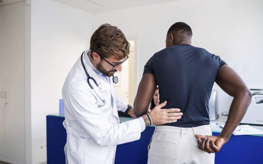
Personal Injury Cases—“Getting Back on Track After an Accident”
The Role of Chiropractic Care
Personal injuries from auto accidents, workplace incidents, or sports can result in musculoskeletal issues like whiplash, back pain, or joint injuries. Chiropractic care is essential for recovery, providing non-invasive treatments that alleviate pain, restore mobility, and prevent chronic issues. In El Paso, where car accidents are a common cause of injury, chiropractors play a vital role in helping victims regain their quality of life.
Dr. Jimenez’s Expertise
Dr. Alexander Jimenez stands out as a leading practitioner for personal injury cases in El Paso. With qualifications as a Doctor of Chiropractic, Advanced Practice Registered Nurse, and Family Nurse Practitioner, he brings a unique blend of expertise to his practice. His clinic, El Paso’s Chiropractic Rehabilitation Clinic & Integrated Medicine Center, utilizes advanced imaging techniques (e.g., X-rays, MRIs) and comprehensive diagnostic evaluations to assess injuries accurately. Dr. Jimenez employs a multidisciplinary approach, combining chiropractic adjustments, spinal decompression, acupuncture, and functional medicine to create personalized treatment plans.
Clinical and Legal Liaison
Dr. Jimenez’s ability to associate patient injuries with detailed medical documentation makes him an invaluable asset in personal injury cases. His comprehensive reports, supported by advanced diagnostics, provide clear evidence for insurance claims and legal proceedings. By bridging medical care and legal documentation, he ensures patients receive both effective treatment and the support needed for fair compensation.
Clinical Rationale
Chiropractic care can accelerate recovery from personal injuries by addressing soft tissue damage, spinal misalignments, and nerve irritation, thereby promoting overall well-being. For example, a study on whiplash injuries showed that chiropractic treatment significantly reduced pain and improved range of motion (Woodward et al., 1996). Dr. Jimenez’s use of dual-scope procedures, which combine chiropractic and functional medicine, enhances outcomes by addressing both structural and systemic issues.
Gluteal Muscle Activation and Low Back Pain: Research indicates that activating the gluteal muscles during walking can reduce low back pain, which may be related to disk degeneration and subluxation treatments (Bullock-Saxton et al., 1993). Chiropractic care often includes exercises to strengthen these muscles, enhancing spinal stability.
Mitochondria and Osteoarthritis: Mitochondrial dysfunction can contribute to osteoarthritis, which may overlap with disk degeneration in the spine (Blanco et al., 2011). Chiropractic care’s focus on reducing inflammation may support joint health.
Brain Plasticity and Spinal Injuries: Studies on brain plasticity suggest that spinal adjustments may influence neurological function, potentially aiding recovery from severe injuries (Haavik & Murphy, 2012).
References
Bullock-Saxton, J. E., Janda, V., & Bullock, M. I. (1993). Reflex activation of gluteal muscles in walking: An approach to restoration of muscle function for patients with low-back pain. Spine, 18(6), 704–708. https://pubmed.ncbi.nlm.nih.gov/8516697/
Blanco, F. J., Rego, I., & Ruiz-Romero, C. (2011). The role of mitochondria in osteoarthritis. Nature Reviews Rheumatology, 7(3), 161–169. https://pubmed.ncbi.nlm.nih.gov/21200395/
Haavik, H., & Murphy, B. (2012). Brain plasticity in patients with spinal cord injuries: A systematic review. Journal of Neurotrauma, 29(10), 1681–1695. https://pubmed.ncbi.nlm.nih.gov/22471998/
Conclusion
Chiropractic care offers a promising, non-invasive approach to managing subluxation, scoliosis, disk degeneration, carpal tunnel syndrome, and Achilles tendonitis. By addressing spinal and joint misalignments, reducing nerve compression, and promoting natural healing, chiropractors like Dr. Alexander Jimenez help patients regain comfort and function. In personal injury cases, Dr. Jimenez’s expertise in diagnostics and documentation ensures comprehensive care and legal support, making him a trusted practitioner in El Paso.
Disclaimer: This blog post is for informational purposes only and is not a substitute for professional medical advice, diagnosis, or treatment. Always consult a qualified healthcare provider for personalized guidance on musculoskeletal conditions or injuries.
Rainy Weather and Motor Vehicle Accidents: Understanding the Risks and Recovery
Rainy weather can make driving more hazardous, increasing the likelihood of minor car accidents that may result in injuries such as spinal misalignment or soft tissue damage. These accidents, while often not severe, can still cause significant health issues if not treated properly. This article examines how rain creates hazardous driving conditions, how these conditions contribute to minor accidents, and how such accidents can lead to injuries that impact spinal health. It also highlights the expertise of Dr. Alexander Jimenez, a chiropractor and nurse practitioner in El Paso, Texas, who specializes in treating auto accident injuries using a unique combination of chiropractic care, medical diagnostics, and integrative medicine to promote recovery and overall health.
How Rainy Weather Increases Accident Risks
Rainy weather creates a cascade of challenges for drivers, making roads more dangerous and increasing the likelihood of minor accidents. Wet roads reduce tire traction, making it harder for vehicles to stop or steer effectively. According to the Texas Department of Insurance, rain is a leading cause of weather-related accidents due to slippery surfaces and reduced visibility (Texas Department of Insurance, n.d.). When tires lose grip on wet roads, vehicles may slide or hydroplane, especially during sudden stops or turns, which can lead to low-speed collisions, such as fender-benders or rear-end accidents (Chicago Lawyer, 2023).
Reduced visibility is another major issue. Heavy rain can obscure a driver’s view, even with windshield wipers on high, making it difficult to see other vehicles, pedestrians, or road signs (Lawyer Schwartz, n.d.). This limited visibility often forces drivers to react more slowly, increasing the risk of minor accidents, such as bumping into another car while braking or changing lanes (Knowles Law Firm, n.d.). Additionally, rain can make roads uneven by creating puddles or washing out gravel, which affects vehicle control and alignment, further contributing to accidents (Springs Auto, n.d.).
These conditions create a chain reaction: wet roads and poor visibility lead to compromised driving conditions, which in turn increase the likelihood of minor accidents. Even low-speed collisions can cause jolts to the body, resulting in injuries that affect spinal alignment and soft tissues, such as muscles and ligaments (Joyce & Macdonald, n.d.). For example, a sudden stop on a slippery road can cause a rear-end collision, leading to whiplash, a common injury where the neck jerks forward and backward, straining muscles and misaligning the spine (Salinas Trial Law, n.d.).
The Link Between Minor Accidents and Spinal Injuries
Minor car accidents, such as those caused by rainy weather, may seem harmless, but they can still cause significant injuries. The sudden forces from even a low-speed collision can strain the body, particularly the spine and surrounding tissues. Whiplash is one of the most common injuries, occurring when the head and neck are jolted rapidly, stretching muscles, ligaments, and tendons beyond their normal range (South Sound Law Group, n.d.). This can lead to spinal misalignment, where the vertebrae shift out of their proper position, potentially pressing on nerves and causing pain or discomfort (Jimenez, 2025).
Soft tissue injuries, such as sprains or strains in the neck, back, or shoulders, are also common. These injuries occur when the muscles or ligaments are stretched or torn due to the impact (Because You Want to Win, n.d.). Over time, untreated soft tissue damage can contribute to spinal misalignment by creating uneven tension around the spine, which can lead to chronic pain or reduced mobility (HSI, n.d.). For instance, a minor rear-end collision on a wet road might cause a driver to tense up, resulting in muscle strain that pulls the spine out of alignment.
Spinal misalignments, or subluxations, can disrupt the nervous system, leading to symptoms like headaches, numbness, or tingling in the arms or legs (Jimenez, 2025). If left untreated, these injuries can worsen, causing long-term issues like chronic back pain or reduced range of motion. Early intervention is crucial in preventing minor injuries from developing into chronic conditions, and professionals like Dr. Alexander Jimenez specialize in identifying and treating these issues before they escalate.
Dr. Alexander Jimenez: A Leader in Auto Accident Recovery
Dr. Alexander Jimenez, a chiropractor and board-certified family nurse practitioner in El Paso, Texas, has over 25 years of experience treating injuries from motor vehicle accidents (MVAs). His unique dual licensure allows him to combine chiropractic expertise with medical diagnostics, offering a comprehensive approach to recovery (Jimenez, 2025). At his Injury Medical & Chiropractic Clinic, Dr. Jimenez uses advanced tools and integrative medicine to address both the symptoms and root causes of injuries, helping patients regain mobility and prevent long-term complications.
Clinical Correlation and Dual Diagnosis
Dr. Jimenez’s approach involves a dual diagnosis process, combining chiropractic assessments with medical evaluations. He utilizes advanced imaging techniques, including X-rays, MRIs, and CT scans, to detect spinal misalignments, disc herniations, and soft tissue damage (Jimenez, 2025). These tools help him correlate a patient’s symptoms, like neck pain or numbness, with objective findings, ensuring accurate treatment plans. For example, a patient with whiplash might show cervical misalignment on an MRI, which Dr. Jimenez can address with targeted spinal adjustments.
His nurse practitioner training enables him to assess systemic issues, such as inflammation or hormonal imbalances, that may impede recovery. By combining chiropractic and medical perspectives, Dr. Jimenez creates personalized treatment plans that address both biomechanical (spine and muscle) and physiological (body-wide) factors (Jimenez, 2025). This dual approach ensures that injuries are treated holistically, thereby reducing the risk of chronic pain.
Advanced Diagnostics and Imaging
Dr. Jimenez relies on advanced diagnostics to guide his treatments. X-rays and MRIs reveal structural issues, such as misaligned vertebrae or herniated discs, while electromyography (EMG) and functional movement screens assess nerve damage or muscle dysfunction (Jimenez, 2025). These tools are especially important for minor accident injuries, which may not be apparent in standard exams but can cause significant problems if left untreated. For instance, a low-speed collision might cause a subtle ligament tear that can only be detected by an MRI, allowing Dr. Jimenez to tailor his treatment to the specific injury.
Chiropractic and Integrative Medicine
Chiropractic care is central to Dr. Jimenez’s approach, using spinal adjustments to correct misalignments and relieve nerve pressure. These adjustments restore joint mobility and reduce pain, enabling patients to recover without the need for surgery or long-term medication (Jimenez, 2025). He also incorporates soft tissue therapies, such as massage and myofascial release, to reduce muscle tension and improve circulation, which accelerates the healing process.
Integrative medicine enhances his treatments by addressing overall health and well-being. Dr. Jimenez provides nutritional guidance to reduce inflammation, offers acupuncture to alleviate pain, and recommends rehabilitation exercises to strengthen muscles and improve flexibility (Jimenez, 2025). For example, a patient with a soft tissue injury might receive spinal adjustments to realign the spine, nutritional advice to reduce inflammation, and exercises to restore mobility, ensuring a comprehensive recovery.
Legal Paperwork and Personal Injury Cases
Dr. Jimenez’s dual licensure makes him uniquely qualified to handle the legal aspects of personal injury cases. He provides detailed medical reports that document injuries, treatments, and progress, which are essential for insurance claims or legal proceedings (Jimenez, 2025). His reports include objective evidence from imaging and diagnostic tests, giving attorneys and insurance companies clear proof of injury. This thorough documentation helps patients secure fair compensation while focusing on their recovery and rehabilitation.
Recovering from Minor Accidents with Chiropractic Care
Chiropractic care is highly effective for recovering from minor accident injuries, especially those caused by rainy weather. Spinal adjustments correct misalignments, reducing pressure on nerves and alleviating pain (Jimenez, 2025). Soft tissue therapies, like massage, help heal strained muscles and ligaments, while rehabilitation exercises strengthen the body to prevent future injuries. Dr. Jimenez’s integrative approach also includes acupuncture and nutritional counseling to support overall health, addressing inflammation and stress that can worsen injuries.
Early intervention is crucial. Seeking care within 72 hours of an accident can prevent minor injuries from becoming chronic (Jimenez, 2025). For instance, a patient with whiplash resulting from a rainy-day collision may receive adjustments to correct cervical misalignment, soft tissue therapy to reduce muscle tension, and nutritional guidance to combat inflammation, ultimately leading to a faster recovery and improved long-term health.
References
Jimenez, A. (2025). Spinal alignment pain relief for motor vehicle injuries. Retrieved from https://dralexjimenez.com/
Conclusion
Rainy weather increases the risk of minor car accidents by creating slippery roads and reducing visibility, which can lead to collisions that may cause spinal misalignments and soft tissue injuries. These injuries, if untreated, can lead to chronic pain or reduced mobility. Dr. Alexander Jimenez’s expertise as a chiropractor and nurse practitioner enables him to provide comprehensive care, utilizing advanced diagnostics, chiropractic adjustments, and integrative medicine to address both the symptoms and underlying causes of injuries. His ability to handle medical and legal aspects ensures patients recover fully while navigating personal injury cases. By seeking early treatment, individuals can restore their health and prevent long-term complications.
El Paso, TX’s Leading Spine and Back Injury Specialist: Dr. Alex Jimenez, DC, APRN, FNP-C, IFMCP
Welcome to El Paso’s premier destination for advanced spine and back injury care, led by Dr. Alex Jimenez, a board-certified Family Practice Nurse Practitioner (FNP-C) and Chiropractor (DC). At www.dralexjimenez.com and www.chiromed.com, Dr. Jimenez and his team deliver cutting-edge, evidence-based treatments that integrate chiropractic care, functional medicine, and advanced rehabilitation protocols to address complex neuromusculoskeletal conditions. Our mission is to restore mobility, alleviate pain, and empower patients to live vibrant, pain-free lives through personalized, holistic care.
Specialized Spine and Back Injury Care
Dr. Jimenez’s practice is renowned for its expertise in treating intricate spine and back injuries, including herniated discs, severe sciatica, scoliosis, spinal stenosis, and chronic low back pain. By combining his chiropractic expertise with his advanced training as a Family Practice Nurse Practitioner and Institute for Functional Medicine Certified Practitioner (IFMCP), Dr. Jimenez offers a unique, integrative approach to spine care that addresses both symptoms and underlying causes.
Complex Spine Care Treatments
Dr. Jimenez utilizes cutting-edge techniques to address complex spine conditions in his chiropractic and rehabilitation clinic.
Chiropractic Spinal Adjustments use precise manual and instrument-assisted adjustments to restore spinal alignment, reduce nerve compression, and improve mobility. A 2020 study in Spine Journal found that spinal manipulative therapy significantly reduces pain and disability in patients with chronic low back pain (Rubinstein et al., 2020).
Decompression Therapy: Non-surgical spinal decompression to relieve pressure on herniated discs and pinched nerves, promoting disc healing and pain relief. Research in Journal of Physical Therapy Science (2017) supports its efficacy for lumbar disc herniation (Choi et al., 2017).
Regenerative Therapies: Platelet-rich plasma (PRP) and stem cell support therapies to enhance tissue repair and reduce inflammation in degenerative spine conditions. A 2019 meta-analysis in Pain Physician confirmed PRP’s effectiveness in managing lumbar pain (Sanapati et al., 2019).
Electro-Acupuncture: Targeted electrical stimulation of acupuncture points to reduce pain and promote healing in sciatica and chronic back pain. A 2018 study in Evidence-Based Complementary and Alternative Medicine demonstrated its benefits for neuropathic pain (Li et al., 2018).
Customized Rehabilitation Programs: Tailored exercises focusing on flexibility, core strength, and spinal stability to prevent re-injury and enhance long-term recovery.
As a Family Practice Nurse Practitioner, Dr. Jimenez offers comprehensive medical evaluations and functional medicine protocols to complement spine care:
Functional Medicine Assessments: In-depth evaluations using the Living Matrix Functional Medicine Assessment to identify root causes of chronic spine pain, including inflammation, nutritional deficiencies, and hormonal imbalances. A 2021 study in Frontiers in Medicine highlighted functional medicine’s role in improving outcomes for chronic pain patients (Beidelschies et al., 2021).
Nutritional Therapy: Personalized nutrition plans to reduce inflammation and support spinal health, incorporating anti-inflammatory diets rich in omega-3s and antioxidants. Research in Nutrients (2020) supports dietary interventions for reducing chronic pain (Kaushik et al., 2020).
Hormone Optimization: Addressing hormonal imbalances that exacerbate pain and delay healing, such as cortisol dysregulation in chronic stress. A 2019 study in Journal of Clinical Endocrinology & Metabolism linked cortisol imbalances to chronic pain syndromes (Hannibal et al., 2019).
Lifestyle Medicine: Guidance on stress management, sleep optimization, and ergonomic adjustments to support spine health and prevent injury recurrence.
Why Choose Dr. Jimenez for Spine and Back Injury Care?
Dr. Jimenez’s dual expertise as a chiropractor and nurse practitioner, combined with his IFMCP certification, sets him apart as El Paso’s top spine care specialist. Key differentiators include:
Holistic, Evidence-Based Approach: Integrating chiropractic care, functional medicine, and advanced diagnostics to address the whole person, not just symptoms.
Non-Invasive Protocols: Natural, non-surgical solutions should be prioritized to avoid the risks and recovery time associated with invasive procedures. A 2022 study in The Lancet emphasized the efficacy of non-invasive treatments for chronic low back pain (Foster et al., 2022).
Collaborative Care Network: Partnerships with leading orthopedic surgeons, neurologists, and rehabilitation specialists ensure seamless referrals when advanced interventions are needed.
Patient-Centered Plans: Using advanced diagnostics like MRI analysis, electromyography (EMG), and functional health assessments to create targeted treatment plans tailored to each patient’s unique needs.
Conditions Treated
Our clinic specializes in a wide range of spine- and back-related conditions, including:
Herniated Discs: Comprehensive care to reduce disc protrusion, alleviate nerve compression, and restore spinal function.
Severe Sciatica: Targeted therapies to relieve radiating leg pain and improve mobility.
Scoliosis: Customized bracing and exercise programs to manage spinal curvature and prevent progression.
Spinal Stenosis: Decompression and strengthening protocols to alleviate pain and improve quality of life.
Chronic Low Back Pain: Multifaceted treatment plans addressing biomechanical, inflammatory, and lifestyle factors.
Transform Your Spine Health Today
Whether you’re recovering from a traumatic spine injury, managing chronic back pain, or seeking preventive care, Dr. Alex Jimenez and his team are here to guide you. Contact us today at www.dralexjimenez.com or www.chiromed.com to schedule a consultation and discover how our integrative approach can transform your health. With evidence-based protocols and a compassionate, patient-centered philosophy, we help you live pain-free and thrive in El Paso’s vibrant community.
What are the advantages of having a team of nurse practitioners and chiropractors help maintain the health of your spine after a car accident?
Benefits of Chiropractic and Nurse Practitioners for Motor Vehicle Collisions
One of the main causes of spinal injuries, such as whiplash, herniated discs, and soft tissue injury, which can cause severe pain and impair movement, is motor vehicle collisions (MVCs). For both short-term symptoms and long-term rehabilitation, these injuries frequently necessitate a multimodal therapy strategy. While nurse practitioners, as advanced practice registered nurses, conduct medical evaluations, write prescriptions, and oversee overall health management, chiropractors focus on musculoskeletal care, including spine adjustments and manual therapies. These professionals’ collaboration aims to provide a comprehensive, patient-centered strategy for spine health following MVC. (Kent, R., et al., 2023)
For those recuperating from auto accident injuries, a chiropractic and nurse practitioner team can offer thorough spinal health care with an emphasis on pain management and increased mobility.
A chiropractic and nurse practitioner team can offer a comprehensive approach to spinal health after a car accident by addressing pain, improving mobility, and facilitating faster recovery.
Chiropractors focus on spinal alignment and joint mobility.
Nurse practitioners provide broader medical oversight and patient education.
The team approach can lead to more effective and personalized care for individuals recovering from car accident injuries. (Riva, J. J., et al., 2010)
Key advantages of this collaborative approach
A chiropractor and nurse practitioner (NP) therapy team can combine their skills to provide comprehensive care for spine health following a motor vehicle collision (MVC) and address acute and long-term requirements.
Care that is multidisciplinary and holistic
Collaboration between chiropractors and NPs to address structural and systemic issues enhances treatment outcomes, particularly for spine injuries related to motor vehicle collisions (MVC), as well as for chronic headaches and neck discomfort. (Riva, J. J., et al., 2010)
Plans for Treatment That Are Unique to You
Chiropractors and NPs create personalized patient treatment plans, focusing on their specific injuries and overall health, including pre-existing conditions and medication needs. This approach enhances outcomes by tailoring care to the patient’s unique circumstances.
Managing Pain Without Relying Too Much on Drugs
By using non-invasive methods to alleviate pain, chiropractic therapy may help reduce the use of opioids. NPs can prescribe short-term pain relief and monitor side effects, ensuring safe use and reducing dependency risks. Natural pain management combined with medical supervision lessens dependence and side effects. (Prater, C., Tepe, M., & Battaglia, P. 2020)
Quicker Recuperation and Rehabilitation
As demonstrated in the treatment of auto accidents, chiropractic adjustments can lessen muscle spasms and restore joint function. By referring patients to physical therapy and tracking their progress, NPs can hasten recovery and reduce the likelihood of developing persistent back pain. This integrated therapy not only reduces chronic back pain and other long-term problems, but it also accelerates healing.
Help with Insurance and the Law
Chiropractic and medical providers must carefully record injuries and treatments for insurance claims or legal cases after an MVC to ensure just reimbursement and coverage for care.
Why It Works After MVC
Following a motor vehicle collision (MVC), a chiropractor and nurse practitioner team offers a patient-centered approach to spine health. This team enhances recovery, lowers chronic risks, and improves patient outcomes by fusing NP’s medical management with chiropractic knowledge. This method ensures rapid alleviation and long-term health, especially helpful for complex spine injuries due to MVC.
Injury, Chiropractic, and Functional Medicine Clinic
Dr. Jimenez, a nurse practitioner, uses medical knowledge and chiropractic care to treat various conditions. The clinic provides tailored care programs incorporating functional medicine, acupuncture, electroacupuncture, and sports medicine. The clinic focuses on strength, agility, and flexibility for treating chronic pain syndromes and injuries. Patients of all ages and abilities benefit from comprehensive care plans and in-person and virtual health coaching, ensuring tailored treatment and wellness outcomes.
Personal Injury Rehabilitation
References
Kent, R., Cormier, J., McMurry, T. L., Johan Ivarsson, B., Funk, J., Hartka, T., & Sochor, M. (2023). Spinal injury rates and specific causation in motor vehicle collisions. Accident; analysis and prevention, 186, 107047. https://doi.org/10.1016/j.aap.2023.107047
Riva, J. J., Muller, G. D., Hornich, A. A., Mior, S. A., Gupta, A., & Burnie, S. J. (2010). Chiropractors and collaborative care: An overview illustrated with a case report. The Journal of the Canadian Chiropractic Association, 54(3), 147–154.
Prater, C., Tepe, M., & Battaglia, P. (2020). Integrating a Multidisciplinary Pain Team and Chiropractic Care in a Community Health Center: An Observational Study of Managing Chronic Spinal Pain. Journal of primary care & community health, 11, 2150132720953680. https://doi.org/10.1177/2150132720953680
What is pseudoarthrosis of the cervical and lumbar spine?
Pseudoarthrosis of the cervical and lumbar spine
Individuals may need a spinal fusion to treat a fractured vertebra, scoliosis, or conditions like spinal stenosis, degenerative disc disease, and spondylolisthesis/slipped vertebrae. A spinal fusion reduces pain and stabilizes the spine by limiting movement between vertebrae. Pseudoarthrosis happens when the bones don’t heal after a fracture or bone surgery. When pseudoarthrosis affects the cervical or lumbar spine, it means that two vertebrae did not heal and grow together after spinal surgery to fuse them (spinal fusion). Reasons for a failed spinal fusion include:
Issues with the instruments used to stabilize the bone
Lack of bone growth
The number of vertebrae being fused.
The patient’s health and lifestyle play a role in failed fusions, which can include
Diabetes
Inflammatory health conditions increase the risk
Smoking
Long-term steroid use
In many cases, revision surgery is needed.
Surgery-Related
During a spinal fusion, surgeons insert a bone graft between two vertebrae and then apply spinal fixation hardware (instrumented spinal fusion) that includes:
Plates
Rods
Screws
The bone graft promotes growth between the two bones.
The hardware stabilizes the vertebrae and prevents movement while they fuse and grow together.
The hardware goes inside, or internal fixation.
Although rare, a severe spinal fracture or deformity may need external fixation.
A rigid frame secured outside the body helps to stabilize the bones.
If the fusion fails, it could be caused by one or more of the following surgical issues:
The surgeon must carefully plan and use the right hardware.
The type of hardware used during a spinal fusion may influence bone healing.
The instruments can come loose or break, interfering with the fusion process.
Spinal osteoporosis, having thin, weak bones, can affect fixation.
Even with the optimal surgical preparedness, weak bones significantly increase the chance of the instruments loosening and pseudoarthrosis developing.
Bone Graft
The type of bone graft used may affect the fusion.
For example, in cervical/neck spinal fusions, an autograft, which uses a small piece of bone from the patient’s body, has a higher success rate. (Verla T. et al., 2021)
Other graft options include specialized steel cages that fit between vertebrae and contain bone growth factors.
The surgeon recommends the optimal bone graft for the type of surgery, the number of vertebrae involved, and risk factors.
Risk Factors
The patient’s overall health and lifestyle impact the results of spinal fusion. Smoking increases the risk. (Berman D. et al., 2017)
Nicotine restricts blood circulation, decreases bone density, reduces new bone formation, and delays bone healing. (Hernigou J., & Schuind F., 2019)
The primary sign of pseudoarthrosis is pain in the same area as before the fusion surgery.
If the bones pinch a spinal nerve, one arm may experience pain, tingling, burning, or numbness.
Rarely does a pinched nerve affect both arms.
The pain may return shortly after the procedure.
The pain may develop gradually or not appear for many months.
However, it’s more likely to appear after several months when the individual returns to their usual activities.
Diagnosis
The healthcare provider will learn about symptoms and perform a physical exam to evaluate the back.
They’ll assess mobility and the type of movement that causes pain.
Then, they order diagnostic imaging to see the spine and identify the cause of pain.
Individuals may need a CT scan, MRI, and/or X-rays to evaluate the spinal structures and instrumentation fully.
Treatment
Treatment for pseudoarthrosis will likely start with:
Physical therapy
Pain management – especially in cases where it is important to rule out other sources of back or neck pain.
Medication
Injections
If symptoms don’t improve with conservative care or if there is severe pain, the healthcare provider may recommend revision surgery.
Revision surgery is another procedure to treat complications or correct issues that arise after the initial pseudoarthrosis surgery.
Injury Medical Chiropractic and Functional Medicine Clinic
As a Family Practice Nurse Practitioner, Dr. Jimenez combines advanced medical expertise with chiropractic care to address various conditions. Our clinic integrates Functional Medicine, Acupuncture, Electro-Acupuncture, and Sports Medicine to create customized care plans that promote natural healing, mobility, and long-term wellness. By focusing on flexibility, agility, and strength, we empower patients to thrive, regardless of age or health challenges. At El Paso’s Chiropractic Rehabilitation Clinic & Integrated Medicine Center, we passionately focus on treating patients after injuries and chronic pain syndromes. We focus on improving your ability through flexibility, mobility, and agility programs tailored for all age groups and disabilities. We use in-person and virtual health coaching and comprehensive care plans to ensure every patient’s personalized care and wellness outcomes.
Enhancing Health Together
References
Boonsirikamchai, W., Wilartratsami, S., Ruangchainikom, M., Korwutthikulrangsri, E., Tongsai, S., & Luksanapruksa, P. (2024). Pseudarthrosis risk factors in lumbar fusion: a systematic review and meta-analysis. BMC musculoskeletal disorders, 25(1), 433. https://doi.org/10.1186/s12891-024-07531-w
Verla, T., Xu, D. S., Davis, M. J., Reece, E. M., Kelly, M., Nunez, M., Winocour, S. J., & Ropper, A. E. (2021). Failure in Cervical Spinal Fusion and Current Management Modalities. Seminars in plastic surgery, 35(1), 10–13. https://doi.org/10.1055/s-0041-1722853
Berman, D., Oren, J. H., Bendo, J., & Spivak, J. (2017). The Effect of Smoking on Spinal Fusion. International journal of spine surgery, 11(4), 29. https://doi.org/10.14444/4029
Hernigou, J., & Schuind, F. (2019). Tobacco and bone fractures: A review of the facts and issues that every orthopaedic surgeon should know. Bone & joint research, 8(6), 255–265. https://doi.org/10.1302/2046-3758.86.BJR-2018-0344.R1
Scoliosis Research Society. (2023). Pseudoarthrosis. https://www.srs.org/Patients/Conditions/Pseudoarthrosis
Torres, H. M., Arnold, K. M., Oviedo, M., Westendorf, J. J., & Weaver, S. R. (2023). Inflammatory Processes Affecting Bone Health and Repair. Current osteoporosis reports, 21(6), 842–853. https://doi.org/10.1007/s11914-023-00824-4
Jiao, H., Xiao, E., & Graves, D. T. (2015). Diabetes and Its Effect on Bone and Fracture Healing. Current osteoporosis reports, 13(5), 327–335. https://doi.org/10.1007/s11914-015-0286-8
IFM's Find A Practitioner tool is the largest referral network in Functional Medicine, created to help patients locate Functional Medicine practitioners anywhere in the world. IFM Certified Practitioners are listed first in the search results, given their extensive education in Functional Medicine