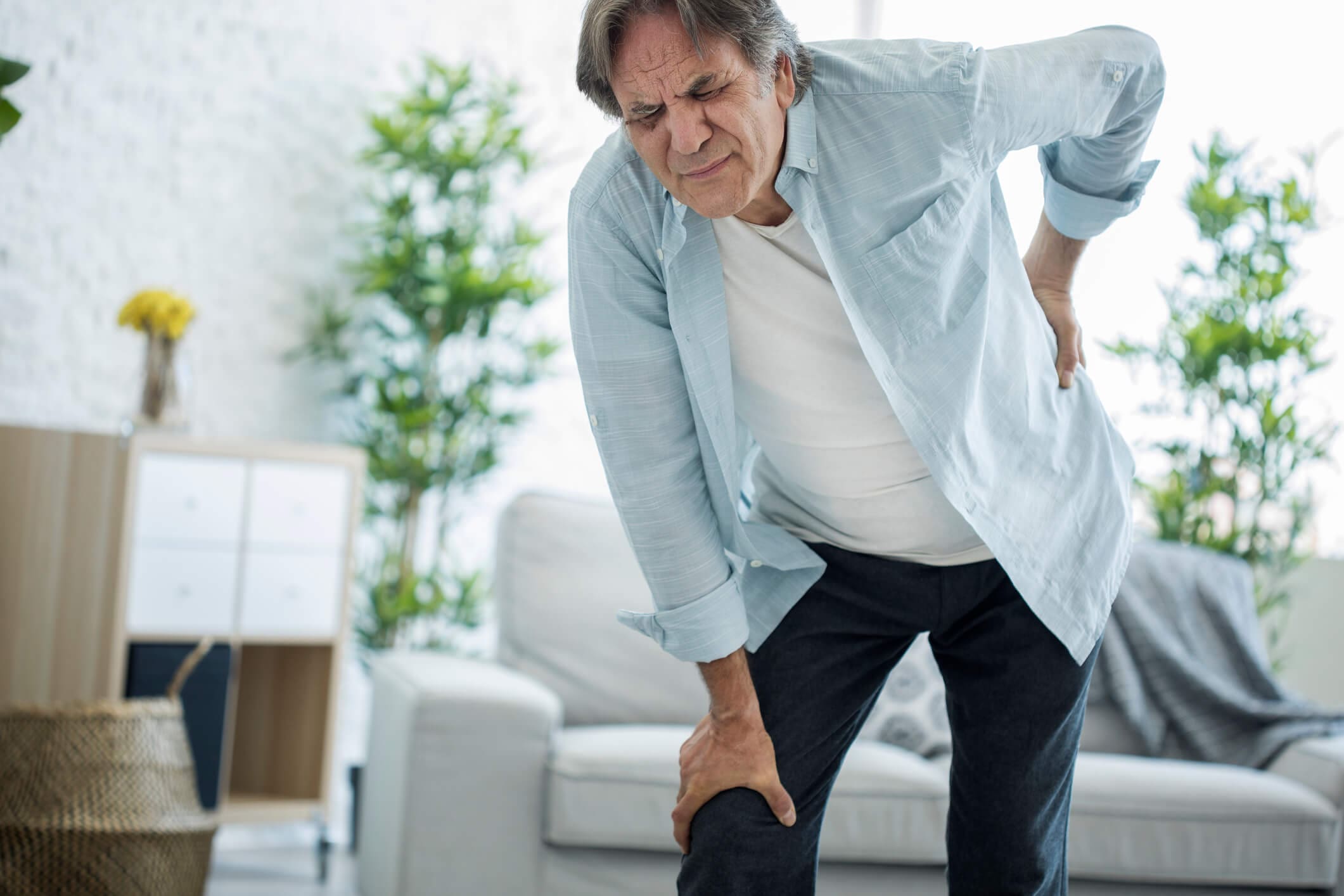
For individuals suffering from back pain, can knowing basic chiropractic terminology help in understanding diagnosis and treatment plan development?
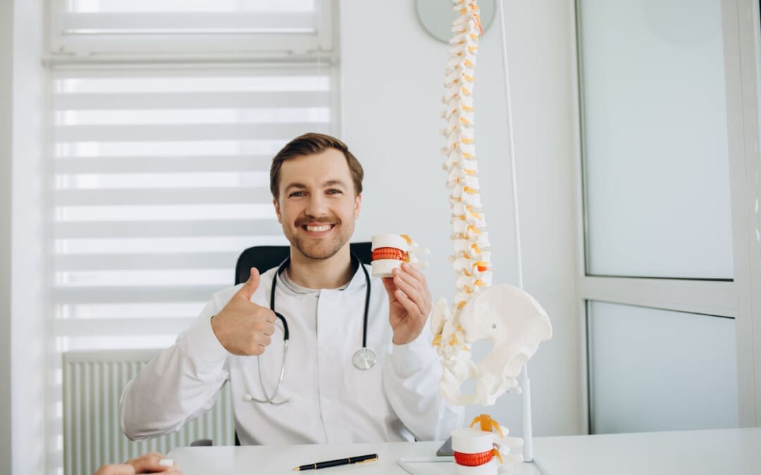
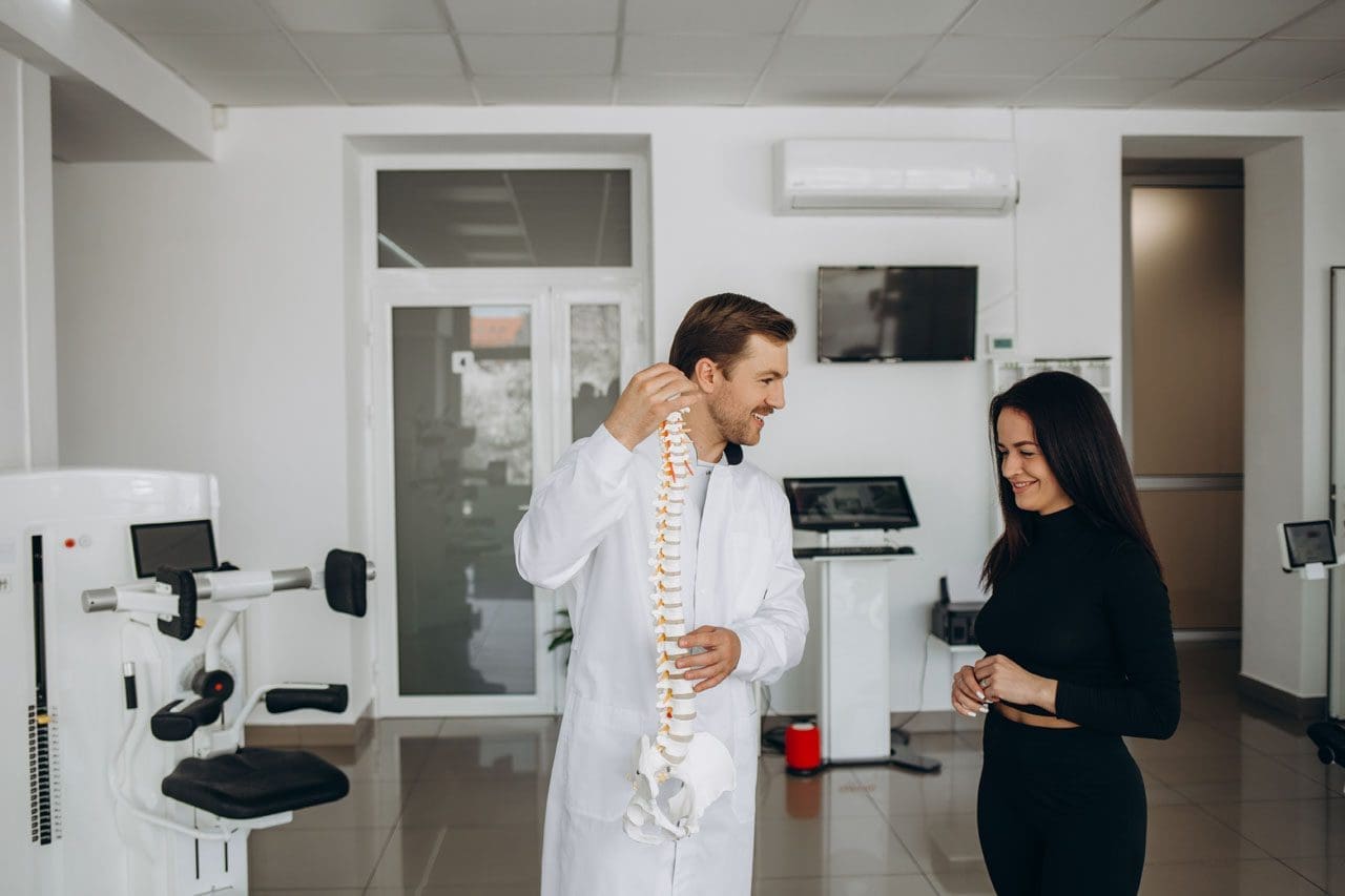
Chiropractic Terminology
The chiropractic principle is that a properly aligned spine positively affects an individual’s overall health. One of the main aspects of chiropractic care is applying calculated force to the spinal joints to restore correct spinal alignment. Chiropractic terminology describes specific types of techniques and care.
General Subluxation
A subluxation can mean different things for various doctors. In general, a subluxation is a significant structural displacement or an incomplete or partial dislocation of a joint or organ.
To medical doctors, a subluxation refers to a partial dislocation of a vertebrae.
This is a serious condition, usually brought on by trauma, that can result in a spinal cord injury, paralysis, and/or death.
X-rays show a conventional subluxation as an obvious disconnect between the vertebrae.
Chiropractic Subluxation
The chiropractic interpretation is more subtle and refers to the misalignment of adjacent spinal vertebrae.
Subluxation in this context refers to position changes in the joints and soft tissues of the spine.
Vertebral misalignment is believed to lead to pain and abnormal intervertebral joint motion.
This difference between the serious subluxation medical condition and the chiropractic version may cause individuals to dismiss seeking back pain treatments.
Motion Segment
Chiropractors and surgeons use it as a technical term.
Motion segment refers to two adjacent vertebrae and the intervertebral disc between them.
This is the area chiropractors assess and adjust.
Adjustment
The chiropractor performs a spinal manual adjustment to realign joint subluxations.
Adjustments involve applying force to motion segments to bring them back into a centered alignment.
The goal for adjustments and realigning the vertebrae includes:
Spinal manipulation is a technique used by chiropractors to provide relief for musculoskeletal pain related to the back and neck. Manipulation provides mild to moderate relief and works as well as some conventional treatments like pain-relieving medications. (Sidney M. Rubinstein et al., 2012)
Spinal manipulation is divided into grades of mobilization.
Depending on their training, practitioners of various medical disciplines may be licensed to perform grade 1 to grade 4 mobilizations.
Only physical therapists, osteopathic physicians, and chiropractors are licensed to perform grade 5 mobilizations, which are high-velocity thrust techniques.
Most massage therapists, athletic trainers, and personal trainers are not licensed to perform spinal manipulations.
Based on a systematic review, the effectiveness of these treatments found that there is quality evidence that manipulation and mobilization can help reduce pain and improve function for individuals with chronic low back pain, with manipulation appearing to produce a more profound effect than mobilization. Both therapies are safe, with multimodal treatments potentially being an effective option. (Ian D. Coulter et al., 2018)
As with any treatment, results vary from person to person and with different chiropractors. There are also potential risks with spinal manipulation. Though rare, cervical, carotid, and vertebral artery dissections have occurred with cervical/neck manipulation. (Kelly A. Kennell et al., 2017) Individuals with osteoporosis may be advised to avoid chiropractic adjustments or manipulation because of the increased risk of injury. (James M. Whedon et al., 2015)
Many individuals choose chiropractic treatment for a variety of conditions. Understanding chiropractic terminology and reasoning allows individuals to ask questions as they discuss their symptoms to develop a personalized treatment plan and restore function and wellness.
What Causes Disc Herniation?
References
Henderson C. N. (2012). The basis for spinal manipulation: chiropractic perspective of indications and theory. Journal of electromyography and kinesiology : official journal of the International Society of Electrophysiological Kinesiology, 22(5), 632–642. https://doi.org/10.1016/j.jelekin.2012.03.008
Blanchette, M. A., Stochkendahl, M. J., Borges Da Silva, R., Boruff, J., Harrison, P., & Bussières, A. (2016). Effectiveness and Economic Evaluation of Chiropractic Care for the Treatment of Low Back Pain: A Systematic Review of Pragmatic Studies. PloS one, 11(8), e0160037. https://doi.org/10.1371/journal.pone.0160037
Rubinstein, S. M., Terwee, C. B., Assendelft, W. J., de Boer, M. R., & van Tulder, M. W. (2012). Spinal manipulative therapy for acute low-back pain. The Cochrane database of systematic reviews, 2012(9), CD008880. https://doi.org/10.1002/14651858.CD008880.pub2
Coulter, I. D., Crawford, C., Hurwitz, E. L., Vernon, H., Khorsan, R., Suttorp Booth, M., & Herman, P. M. (2018). Manipulation and mobilization for treating chronic low back pain: a systematic review and meta-analysis. The spine journal : official journal of the North American Spine Society, 18(5), 866–879. https://doi.org/10.1016/j.spinee.2018.01.013
Kennell, K. A., Daghfal, M. M., Patel, S. G., DeSanto, J. R., Waterman, G. S., & Bertino, R. E. (2017). Cervical artery dissection related to chiropractic manipulation: One institution’s experience. The Journal of family practice, 66(9), 556–562.
Whedon, J. M., Mackenzie, T. A., Phillips, R. B., & Lurie, J. D. (2015). Risk of traumatic injury associated with chiropractic spinal manipulation in Medicare Part B beneficiaries aged 66 to 99 years. Spine, 40(4), 264–270. https://doi.org/10.1097/BRS.0000000000000725
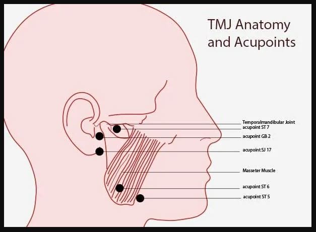
Can individuals with jaw pain find relief in acupuncture therapy to reduce pain and improve jaw mobility in the upper body portions?
Introduction
The head is part of the upper musculoskeletal body quadrant supported by the neck area, which consists of the skull, various muscles, and vital organs that provide stability, mobility, and functionality. Around the head, the different facial features include the mouth, nose, eyes, and jaw to allow the host to eat, speak, smell, and see. While the head provides sensory and motor function, the neck includes motor stability to ensure no injuries or trauma affect the head. Located below the eyes is the jaw, which allows motor function with various muscles and joints to hyperextend without pain or discomfort. However, multiple factors can affect the jaw muscles and joints to invoke pain and discomfort, which can cause radiating referred pain down to the neck muscles. Today’s article looks at how jaw pain can affect the upper body, how non-surgical treatments can help with jaw pain, and how treatments like acupuncture can help restore jaw mobility. We talk with certified medical providers who consolidate our patients’ information to provide treatments to reduce jaw pain affecting their jaw and neck area. We also inform and guide patients on how acupuncture and non-surgical treatments can benefit many individuals with pain correlating with the jaw. We encourage our patients to ask their associated medical providers intricate and important questions about how their pain affects their quality of life and reduces jaw pain. Dr. Jimenez, D.C., includes this information as an academic service. Disclaimer.
Jaw Pain Affecting The Upper Body
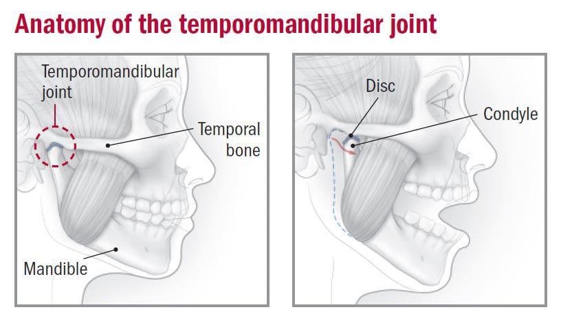
Do you feel muscle soreness in your jaw and neck muscles throughout the day? Have you constantly rubbed or massaged your jaw muscles to reduce tension? Or have you been dealing with headaches or neck pain continually that affects your daily routine? Many individuals experiencing these pain-like symptoms are dealing with jaw pain or TMJ (temporomandibular joint syndrome). The jaw consists of mastication muscles on each side that help provide various functions like chewing, swallowing, or talking. When multiple traumatic or ordinary factors start to affect the jaw, it can disrupt the sensory-motor function of the upper body. For individuals, jaw pain is common worldwide, and with TMJ, it can become an issue as the pain seems to affect the jaw’s motor control while being accompanied by restricted mouth opening and impaired max bite force. (Al Sayegh et al., 2019) Additionally, TMJ affects not only the mastication muscles but also the temporomandibular joint, the joint that connects the jaw to the skull, which becomes inflamed and causes more issues.
So, how would TMJ affect the upper body? Well, when TMJ affects the mastication muscles and the temporomandibular joint, many individuals will experience various symptoms like:
Difficulty moving mouth when chewing
Popping/cracking sensation when opening or closing the jaw
Headaches/Migraines
Ear pain
Tooth pain
Neck and shoulder pain
This causes myofascial and intraarticular disorders that affect the muscles and joints of the jaw, which are linked to the skull. (Maini & Dua, 2024) To that point, many individuals will be experiencing referred pain, thinking they are dealing with a toothache when it is due to trigger points in the mastication muscles. This is when TMJ is accompanied by muscle-joint pain in the neck or upper back or if teeth issues accompany TMJ, but it depends on the individual and situation they are under. However, numerous treatments can reduce jaw pain and its associated symptoms that affect the jaw and the neck.
The Non-Surgical Approach To Wellness- Video
Non-Surgical Treatments For Jaw Pain
When reducing jaw pain, many individuals seek treatment to minimize the pain-like effects and regain mobility back to their jaws. It can be challenging and complex when people are dealing with jaw pain. It is a multifactorial issue that can affect the neck and back areas. So, when people speak with their primary doctors about their jaw pain, they will get an evaluation of where their pain is located and if they have any complaints correlating with the jaw pain. Afterward, many doctors will refer to musculoskeletal specialists to relieve the jaws’ pain. Treatments and techniques used by chiropractors, massage therapists, and physiotherapists can help ease the inflamed and tense mastication muscles. Techniques like soft tissue mobilization can help relax the masticatory muscles by lengthening them to the extent of releasing the trigger points in the muscles. (Kuc et al., 2020) At the same time, physiotherapy can help the jaw muscle through various relaxing techniques to increase the range of motion while strengthening the jaw to reduce pain and stress. (Byra et al., 2020) Many of these treatments are non-surgical, which means they are non-invasive and effective for the person’s pain while affordable.
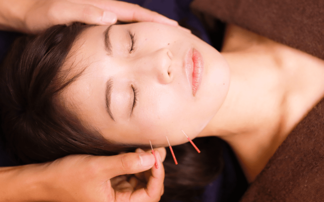
Acupuncture To Restore Jaw Mobility
When it comes to non-surgical treatments, one of the oldest forms is acupuncture, which can help reduce the pain-like effects of jaw pain and restore mobility. Acupuncture originates from China, and highly trained medical professionals use thin, solid needles to be placed in acupoints on the body to disrupt the pain signal and provide relief. For jaw pain, acupuncturists will put needles on the acupoints of the jaw or the surrounding muscles to reduce mechanical hypersensitivity of the nerve cells that are causing pain while improving the sensory-motor function with a positive response. (Teja & Nareswari, 2021) Additionally, when dealing with ear pain associated with TMJ affecting the neck muscles, acupuncture can help enhance the neck’s range of motion by placing the needles on the trigger points of the cervical muscles. (Sajadi et al., 2019) When acupuncture treatment helps many individuals with jaw pain affecting their necks and heads, they can provide beneficial, positive results through consecutive treatment and improve jaw mobility function.
References
Al Sayegh, S., Borgwardt, A., Svensson, K. G., Kumar, A., Grigoriadis, A., & Christidis, N. (2019). Effects of Chronic and Experimental Acute Masseter Pain on Precision Biting Behavior in Humans. Front Physiol, 10, 1369. https://doi.org/10.3389/fphys.2019.01369
Byra, J., Kulesa-Mrowiecka, M., & Pihut, M. (2020). Physiotherapy in hypomobility of temporomandibular joints. Folia Med Cracov, 60(2), 123-134. https://www.ncbi.nlm.nih.gov/pubmed/33252600
Kuc, J., Szarejko, K. D., & Golebiewska, M. (2020). Evaluation of Soft Tissue Mobilization in Patients with Temporomandibular Disorder-Myofascial Pain with Referral. Int J Environ Res Public Health, 17(24). https://doi.org/10.3390/ijerph17249576
Sajadi, S., Forogh, B., & ZoghAli, M. (2019). Cervical Trigger Point Acupuncture for Treatment of Somatic Tinnitus. J Acupunct Meridian Stud, 12(6), 197-200. https://doi.org/10.1016/j.jams.2019.07.004
Teja, Y., & Nareswari, I. (2021). Acupuncture Therapies for Addressing Post Odontectomy Neuropathy. Med Acupunct, 33(5), 358-363. https://doi.org/10.1089/acu.2020.1472
For individuals dealing with lower back pain, it could be quadricep muscle tightness causing the symptoms and posture problems. Can knowing the signs of quadricep tightness help prevent pain and avoid injury?
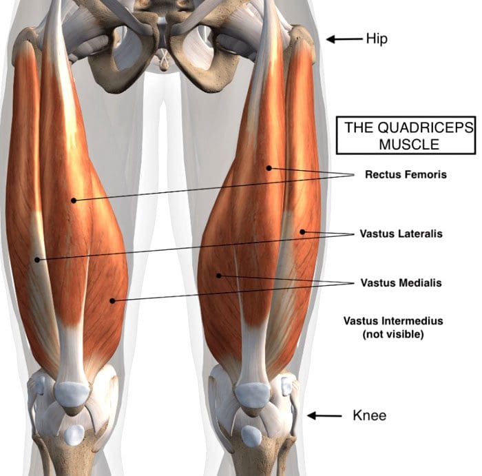
Quadriceps Tightness
Quadriceps muscles are in the front of the thigh. Forces that could be creating chronic pain and posture problems could be happening at the same time are:
Quadricep tightness causes lower back pain as the pelvis gets pulled down.
Tight quadriceps lead to weakened hamstring muscles.
These are the opposing muscles behind the thigh.
Stress and pressure on the hamstrings can cause back pain and problems.
The rectus femoris attaches to the pelvis at the anterior superior iliac spine, which is the front part of the hip bone.
The rectus femoris is the only muscle in the group that crosses over the hip joint, which also affects movement.
When the quadriceps, especially the rectus femoris, become tight, they pull down on the hips.
The pelvis tilts downward or forward, technically referred to as the anterior tilt of the pelvis. (Anita Król et al., 2017)
The spine is between the pelvis, and if the pelvis tilts forward, the lumbar spine compensates by arching.
A larger arch in the lower back is referred to as excessive lordosis and often causes tightness and pain in the back muscles. (Sean G. Sadler et al., 2017)
Hamstring Compensation
When the quadriceps tighten and the pelvis gets pulled down, the back has an abnormal lift. This puts the hamstring on a consistent stretch that can cause pain symptoms.
Healthy posture and hamstring muscle tone help maintain correct pelvic positioning in the back.
This is correct because it helps maintain a comfortable position.
Quadricep tightness can set off a reaction as the pelvis tilts down in front and up in the back while overly stretching the hamstrings.
Pain and soreness are the usual result
Lack of hamstring strength and quadriceps stretching can cause the hamstrings to lose their ability to support correct pelvic and spinal positions. (American Council on Exercise. 2015)
Knowing When Quads Are Tightening
Individuals often don’t realize their quadriceps are tight, especially those who spend most of the day sitting.
The more time spent in a chair can cause the quadriceps and lower back muscles to tighten steadily.
Individuals can try a few tests at home:
Standing Up
Push the hips forward.
Push from the sitting bones so you’re at the correct level.
How far forward do the hips go?
What is felt?
Pain could indicate tight quadriceps.
In A Lunge Position
With one leg forward and bent in front of the other.
The back leg is straight.
How far forward does the leg go?
What is felt?
How does the front of the hip on the back leg feel?
Standing Bent Leg
Stand with the front leg bent and the back leg straight.
Discomfort in the back leg could mean tight quadriceps.
In A Kneeling Position
Arch the back
Grab the ankles
Modify the position to adjust for any pain or joint issues.
If you have to prop yourself up or modify the pose to reduce pain, it could be tight quadriceps.
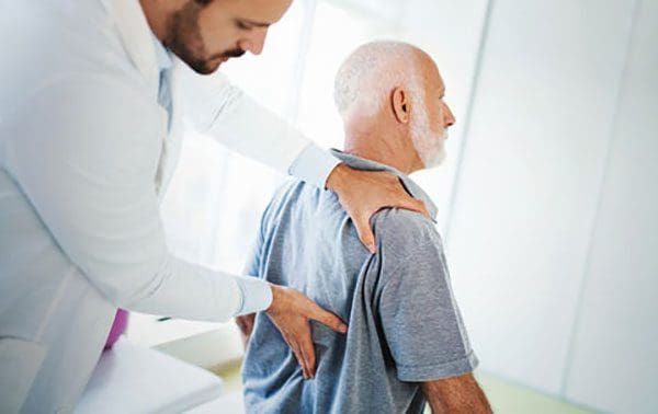
Helping to understand the condition can help in communication with a healthcare provider.
A healthcare provider and/or physical therapist can conduct a posture evaluation examination to test the quadriceps.
Understanding Academic Low Back Pain: Impact and Chiropractic Solutions
References
Kripa, S., Kaur, H. (2021). Identifying relations between posture and pain in lower back pain patients: a narrative review. Bulletin of Faculty of Physical Therapy, 26(34). https://doi.org/doi: 10.1186/s43161-021-00052-w
Król, A., Polak, M., Szczygieł, E., Wójcik, P., & Gleb, K. (2017). Relationship between mechanical factors and pelvic tilt in adults with and without low back pain. Journal of back and musculoskeletal rehabilitation, 30(4), 699–705. https://doi.org/10.3233/BMR-140177
Sadler, S. G., Spink, M. J., Ho, A., De Jonge, X. J., & Chuter, V. H. (2017). Restriction in lateral bending range of motion, lumbar lordosis, and hamstring flexibility predicts the development of low back pain: a systematic review of prospective cohort studies. BMC musculoskeletal disorders, 18(1), 179. https://doi.org/10.1186/s12891-017-1534-0
American Council on Exercise. (2015). 3 Stretches for Opening Up Tight Hips (Fitness, Issue. https://www.acefitness.org/resources/everyone/blog/5681/3-stretches-for-opening-up-tight-hips/
Can individuals dealing with headaches find the relief they are looking for from acupuncture to reduce pain-like symptoms?
Introduction
As part of the musculoskeletal system, the neck is part of the upper body portions and allows the head to be mobile through full rotations without pain and discomfort. The surrounding muscles, ligaments, and tendons help protect the cervical spinal region and have a fantastic relationship with the shoulders. However, the neck area can succumb to injuries, leading to pain-like symptoms that can cause pain and discomfort in the upper regions. One of the pain-like symptoms that correlates with neck pain is headaches. Headaches can vary in acute to chronic stages as they affect many individuals and the various factors that correlate with them. When headaches start to form, many individuals will look at multiple treatments to reduce the pain-like symptoms that correlate with headaches and have the relief they deserve. Today’s article looks at the various factors that correlate with headaches, how headaches cause overlapping risk profiles with neck pain, and how treatments like acupuncture can reduce headaches. We talk with certified medical providers who consolidate our patients’ information to provide treatments like acupuncture to minimize headaches. We also inform and guide patients on how acupuncture can benefit many individuals dealing with neck pain associated with headaches. We encourage our patients to ask their associated medical providers intricated and important questions about their pain-like symptoms that correlate with headaches and neck pain. Dr. Jimenez, D.C., includes this information as an academic service. Disclaimer.
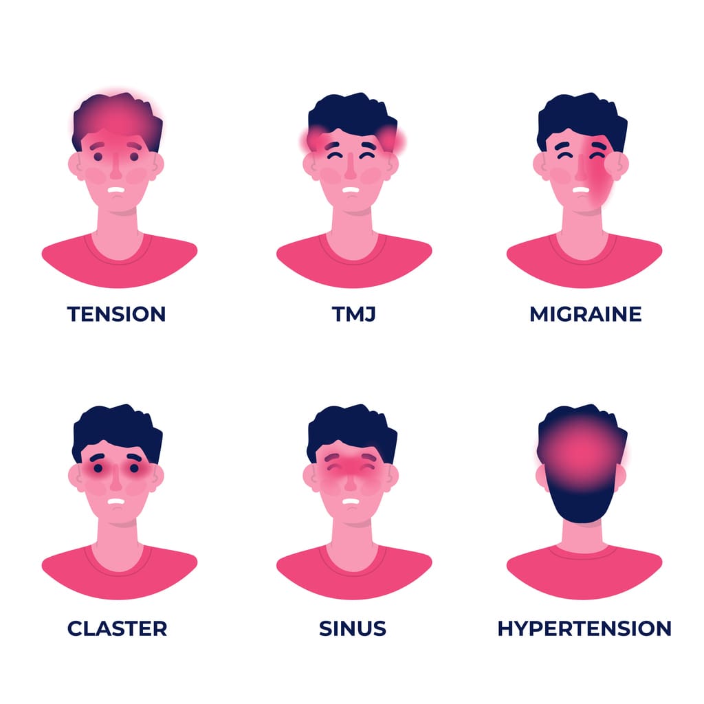
The Various Factors Correlating Headaches
Have you been experiencing tension around the back of your neck after a long day? Do you feel a dull ache after staring at the computer or phone screen? Or do you feel a pounding sensation that you must lie down for a few minutes? Many of these pain-like scenarios are associated with headaches that affect many individuals from time to time. Headaches are correlated with various biochemical and metabolic risk profiles or changes that cause central sensitization and neuronal dysfunction. (Walling, 2020) This causes many individuals to develop acute or chronic pain-like symptoms that affect their heads and various locations around the face and the neck area. Some of the multiple factors that can lead to the development of headaches include:
Stress
Allergies
Tension
Inability to sleep
Lack of water and food
Traumatic injuries
Bright strobing lights
Additionally, other factors like obesity can become a strong risk factor for secondary headaches like migraines to have symptoms of intracranial hypertension impact the body. (Fortini & Felsenfeld Junior, 2022) This could lead to the development of neck pain caused by headaches.
Headaches & Neck Pain
When it comes to headaches associated with neck pain, many individuals will experience tension and pain in the surrounding muscles and the ongoing symptoms. Neck pain can cause overlapping risk profiles to muscles, ligaments, facet joints, and visceral structures of the neck that can trigger the development of a headache or become a symptom that co-exists with a neck disorder. (Vicente et al., 2023) Additionally, neck pain and headaches are strongly associated as muscular pain plays a role in headache development as they provide negative consequences within their social lives. Headaches can hinder a person’s ability to concentrate, while neck pain causes limited mobility and stiffness. (Rodriguez-Almagro et al., 2020)
Tension Headaches Overview- Video
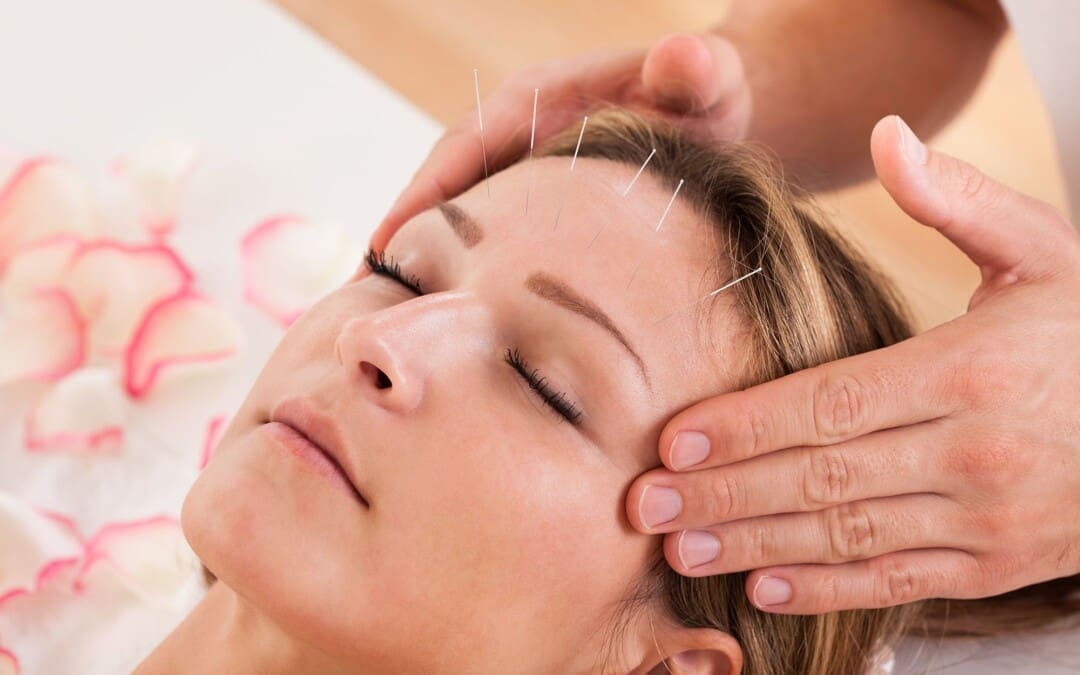
Acupuncture Reducing Headaches
When individuals are dealing with headaches, many will incorporate home remedies to reduce the tension they are experiencing from the various factors. This can provide temporary relief to mitigate the effects of the pain-like symptoms associated with headaches. However, when the pain from headaches becomes unbearable with neck pain in the mix, that is where non-surgical treatments could be the answer. Non-surgical treatments are effective on pain caused by headaches and customized to the person’s pain. For example, acupuncture could help with headaches and neck pain. Acupuncture is one of the oldest forms of non-surgical treatments; highly trained professionals use solid thin needles to be placed in various acupoints in the body to restore energy flow and reducing pain associated with headaches. (Turkistani et al., 2021)
Acupuncture can even help reduce the frequency and duration of headaches while disrupting the pain signals and help provide insight into the positive effects of pain reduction. (Li et al., 2020) When people start incorporating acupuncture as part of their health and wellness treatment plan, they will feel their headaches reduced and their neck mobility back to normal. Through consecutive treatment, they will feel much better and become more aware of the various factors pertaining to headache production while making small changes to reduce their chances of returning.
Li, Y. X., Xiao, X. L., Zhong, D. L., Luo, L. J., Yang, H., Zhou, J., He, M. X., Shi, L. H., Li, J., Zheng, H., & Jin, R. J. (2020). Effectiveness and Safety of Acupuncture for Migraine: An Overview of Systematic Reviews. Pain Res Manag, 2020, 3825617. https://doi.org/10.1155/2020/3825617
Rodriguez-Almagro, D., Achalandabaso-Ochoa, A., Molina-Ortega, F. J., Obrero-Gaitan, E., Ibanez-Vera, A. J., & Lomas-Vega, R. (2020). Neck Pain- and Unsteadiness-Inducing Activities and their Relationship to the Presence, Intensity, Frequency, and Disability of Headaches. Brain Sci, 10(7). https://doi.org/10.3390/brainsci10070425
Turkistani, A., Shah, A., Jose, A. M., Melo, J. P., Luenam, K., Ananias, P., Yaqub, S., & Mohammed, L. (2021). Effectiveness of Manual Therapy and Acupuncture in Tension-Type Headache: A Systematic Review. Cureus, 13(8), e17601. https://doi.org/10.7759/cureus.17601
Vicente, B. N., Oliveira, R., Martins, I. P., & Gil-Gouveia, R. (2023). Cranial Autonomic Symptoms and Neck Pain in Differential Diagnosis of Migraine. Diagnostics (Basel), 13(4). https://doi.org/10.3390/diagnostics13040590
Can nonsurgical therapeutic options help individuals with chronic low back pain find the relief they are looking for to restore body function?
Introduction
Between the upper, middle, and lower back portions of the musculoskeletal system, many individuals have succumbed to traumatic injuries, repetitive motions, and overlapping environmental risk profiles that cause pain and disability, thus affecting their everyday routine. As one of the most common work conditions, back pain can cause individuals to deal with socio-economic burdens and can range from acute to chronic, depending on the injuries and factors that correlate with this issue. As part of the musculoskeletal system, the back has various muscles in the three quadrants that support the upper and lower extremities and have an outstanding relationship with the spine as each muscle group surrounds the spine and protects the spinal cord. When environmental factors and traumatic injuries start to cause pain-like symptoms in the back, it can put a person in excruciating pain, hence why many seek non-surgical treatments to reduce the pain-like effects of back pain and find the relief they are seeking. Today’s article looks at the impact of chronic low back pain and how non-surgical treatments can positively affect individuals dealing with chronic low back pain. We talk with certified medical providers who consolidate our patients’ information to provide numerous non-surgical treatment options to minimize chronic lower back pain affecting their extremities. We also inform and guide patients on how various non-surgical treatments can benefit their health and wellness as they can help reduce musculoskeletal conditions like chronic back pain. We encourage our patients to ask their associated medical providers intricated and important questions about their chronic low back pain and what small changes they can incorporate to reduce its pain-like symptoms. Dr. Jimenez, D.C., includes this information as an academic service. Disclaimer.
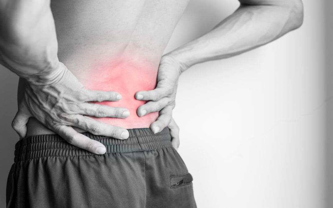
The Impact Of Chronic Low Back Pain
Do you constantly feel severe muscle aches or pains in your back after an excruciating long workday? Do you experience muscle tiredness from your back to your legs after carrying a heavy object? Or have you noticed that twisting or turning motions temporarily relieve your lower back, only to worsen after a while? Often, many of these pain-like scenarios are correlated with chronic low back pain, and it can be due to the various factors that correlate with this common musculoskeletal condition. When it comes to musculoskeletal conditions associated with chronic low back pain, they are prevalent while their impact is pervasive. To that point, they affect many individuals as they are the number one most common cause of severe long-term pain and physical disability. (Woolf & Pfleger, 2003) Since back pain can be either acute or chronic, it can become multifactorial as many other pain symptoms tend to cause overlapping risk profiles in the body. The impact of chronic low back pain has underlying pathological causes that are not well-defined but can be related to psychosocial dysfunction. (Andersson, 1999)
Additionally, degenerative changes within the spine can also cause an impact on the development of chronic lower back pain. The risk factors that cause overlapping risk profiles can range from smoking and obesity to various occupations that require excessive motions. (Atkinson, 2004) When that happens, it causes people to have unnecessary stress that impacts their lives and causes them to be miserable. This is where many individuals start seeking treatment to reduce the effects of chronic lower back pain and reduce the chances of seeking surgical intervention.
The Role Of Chiropractic Care On Improving Your Health- Video
Non-Surgical Treatments For Chronic Back Pain
When people deal with chronic lower back pain, many often don’t realize that various motions, ages, and pathologies can modify the spine, causing the spinal discs to go through degenerative changes that correspond to the development of chronic lower back pain. (Benoist, 2003) When degenerative changes start to cause pain-like symptoms in the back, many will begin looking for affordable and effective treatments. Hence, this is why non-surgical treatments can help reduce the pain-like symptoms of chronic lower back pain and help restore body mobility. Non-surgical treatments are personalized to the person’s pain and range from acupuncture to massage therapy and spinal decompression. Non-surgical treatments are also affordable and help reduce the overlapping risk profiles of chronic low back pain while reducing its associated conditions.
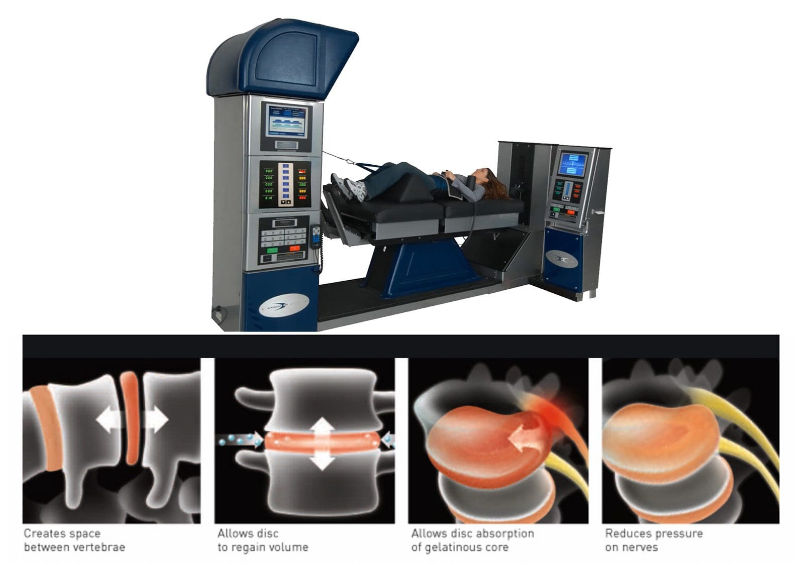
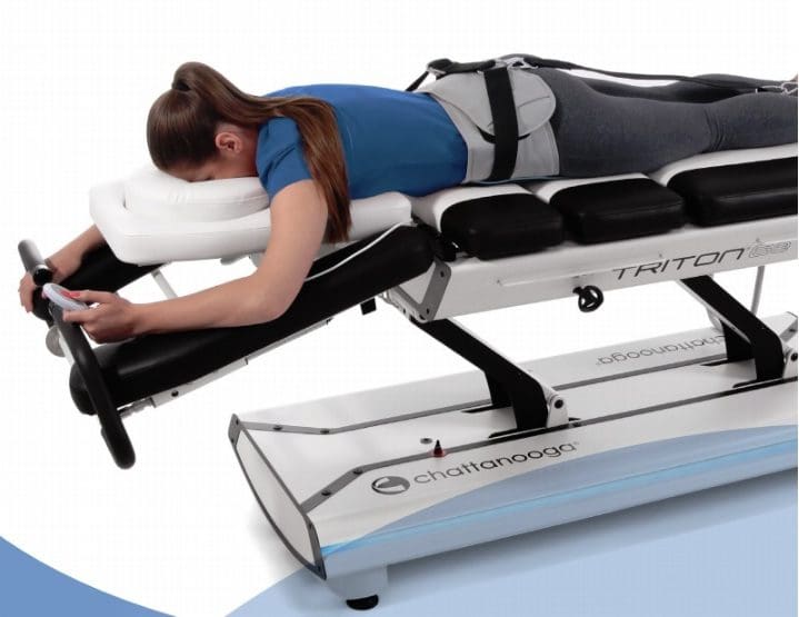
Spinal Decompression Effects On Chronic Low Back Pain
Spinal decompression, as stated before, is a form of non-surgical treatment that incorporates mechanical gentle traction on the spine to alleviate chronic low back pain and can reduce the pain-like symptoms associated with it. Spinal decompression helps reduce the friction of the lumbar muscles, affecting the lumbar spine but also provides pain relief and body function. (Choi et al., 2022) Spinal decompression is safe while being gentle on the spine, combined with stabilization exercises to enhance intra-abdominal pressure and spinal ability to the lumbar. (Hlaing et al., 2021) When a person incorporates spinal decompression as part of their health and wellness journey, their pain and disability will lower over time while strengthening weakened muscles that were affected by chronic lower back pain. Incorporating these non-surgical treatments can help a person be more mindful of the environmental impact they are inflicting on their backs and live a better and healthier life.
Choi, E., Gil, H. Y., Ju, J., Han, W. K., Nahm, F. S., & Lee, P. B. (2022). Effect of Nonsurgical Spinal Decompression on Intensity of Pain and Herniated Disc Volume in Subacute Lumbar Herniated Disc. International Journal of Clinical Practice, 2022, 6343837. https://doi.org/10.1155/2022/6343837
Hlaing, S. S., Puntumetakul, R., Khine, E. E., & Boucaut, R. (2021). Effects of core stabilization exercise and strengthening exercise on proprioception, balance, muscle thickness and pain related outcomes in patients with subacute nonspecific low back pain: a randomized controlled trial. BMC Musculoskelet Disord, 22(1), 998. https://doi.org/10.1186/s12891-021-04858-6
For individuals dealing with musculoskeletal pain, can incorporating acupuncture and dry needling therapy improve functionality?
Introduction
All around the world, many individuals have experienced musculoskeletal pain at one point in their lives. Musculoskeletal pain can range from acute to chronic, depending on how severe the affected muscles are in pain. Sometimes, when a person is dealing with pain in one location and feels it in a different body location, that is known as referred pain, and it can lead to overlapping risk profiles. Additionally, many individuals dealing with musculoskeletal pain are often associated with various environmental factors that contribute to its development or have traumatic injuries that causes the spine to be out of alignment with the body. When it comes to treating musculoskeletal pain, many individuals will do home remedies that can provide temporary relief until the pain flares up again. When that happens, many will seek treatment to reduce the pain-like effects and find the relief they are looking for. Today’s article examines two treatments, their benefits, and how they can reduce musculoskeletal pain. We talk with certified medical providers who consolidate our patients’ information to provide numerous treatments to minimize musculoskeletal pain affecting their bodies. We also inform and guide patients on how various treatments can be beneficial to their health and wellness as they can help reduce musculoskeletal pain. We encourage our patients to ask their associated medical providers intricated and important questions about the referred pain-like symptoms they are experiencing from musculoskeletal pain that is causing them pain and discomfort. Dr. Jimenez, D.C., incorporates this information as an academic service. Disclaimer.
What Is Acupuncture?
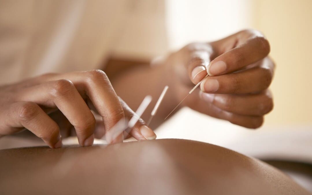
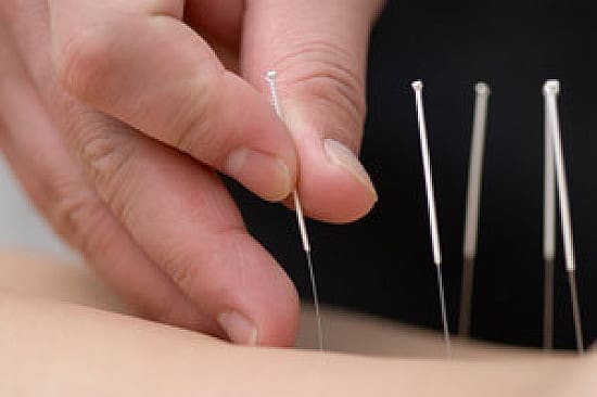
Do you wake up in the morning feeling general aches and pains in various muscle locations? Do you feel muscle stiffness in your neck, shoulders, or back after a long, hard workday? Or have you experienced pain-like symptoms like numbness or radiating burning sensations in your upper or lower body extremities? In these pain-like scenarios, many individuals are experiencing musculoskeletal pain once in their lives. This usually happens when environmental factors or traumatic injuries occur in the musculoskeletal system, causing the surrounding muscles, ligaments, and tissue to be overstretched, tight, or weakened, depending on the severity. When a person is dealing with musculoskeletal pain, they will seek treatments to reduce the musculoskeletal pain and be affordable and customizable to the individual’s pain, hence why non-surgical therapies can benefit the person dealing with musculoskeletal pain.
One of the oldest forms of non-surgical treatment is acupuncture, which can help reduce musculoskeletal pain. Acupuncture has been practiced in China for over two thousand years by modulating the body’s physiology by stimulating specific body regions or acupoints. (Wang et al., 2023) Acupuncture incorporates thin, solid needles used by highly trained medical professionals to restore the balance of qi or energy flowing through the body while positively affecting the central and peripheral nervous system. To that point, by changing pain perception, acupuncture can help reduce the inflammatory cytokines associated with musculoskeletal pain. (Kelly & Willis, 2019)
Acupuncture Benefits
Some of the benefits that acupuncture can help a person dealing with musculoskeletal pain include:
Release tight muscles.
Inflammation reduction
Regulate gut activity associated with musculoskeletal pain.
Improve pain and disability.
Since pain is a common indication for acupuncture, acupuncture for individuals experiencing musculoskeletal pain can help enhance the descending inhibitory effects while modulating the feelings of pain, which, to this point, modify central sensitization. (Zhu et al., 2021) This, in turn, helps many individuals experience positive effects from musculoskeletal pain reduction in their bodies.
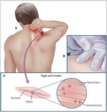
What Is Dry Needling?
Dry needling is a different form of acupuncture that combines TCM (traditional Chinese medicine) and structural manipulation of the affected muscles experiencing pain. Dry needling is safe and, like acupuncture, cost-effective. It reduces pain and improves fascial and scar tissue mobility back to the muscle. (Munoz et al., 2022) At the same time, dry needling is used by highly trained professionals to treat soft tissues and neurovascular bundles correlated with numerous neuromusculoskeletal pain syndromes by inserting fine monofilament needles and stimulating specific reactions in the targeted tissue. (Lara-Palomo et al., 2022)
Dry Needling Benefits
Dry needling can help reduce musculoskeletal pain symptoms associated with myofascial pain syndrome by mechanically disrupting the trigger points to elicit a local twitch response. (Lew et al., 2021) Some of the benefits that dry needling provides include:
Decrease muscle tightness.
Pain reduction
Joint and muscle mobility
Increase blood flow.
How Acupuncture & Dry Needling Help With Pain?
Depending on how severe the individual’s pain is affecting their daily life, they can choose either acupuncture or dry needling and combine them with other non-surgical therapies to reduce the chances of musculoskeletal pain from causing overlapping risk profiles that can make a person’s life miserable. Both non-surgical techniques can be effective with patients dealing with musculoskeletal pain, improve stiffness and fatigue, and enhance quality of life. (Valera-Calero et al., 2022) Incorporating these non-surgical treatments to reduce musculoskeletal pain with healthy habits can give helpful results to the individual by making small changes in their everyday lives and being mindful of their bodies. This allows them to reduce the chances of musculoskeletal pain and its associated factors from recurring again in the future.
Lara-Palomo, I. C., Gil-Martinez, E., Antequera-Soler, E., Castro-Sanchez, A. M., Fernandez-Sanchez, M., & Garcia-Lopez, H. (2022). Electrical dry needling versus conventional physiotherapy in the treatment of active and latent myofascial trigger points in patients with nonspecific chronic low back pain. Trials, 23(1), 238. https://doi.org/10.1186/s13063-022-06179-y
Lew, J., Kim, J., & Nair, P. (2021). Comparison of dry needling and trigger point manual therapy in patients with neck and upper back myofascial pain syndrome: a systematic review and meta-analysis. J Man Manip Ther, 29(3), 136-146. https://doi.org/10.1080/10669817.2020.1822618
Munoz, M., Dommerholt, J., Perez-Palomares, S., Herrero, P., & Calvo, S. (2022). Dry Needling and Antithrombotic Drugs. Pain Res Manag, 2022, 1363477. https://doi.org/10.1155/2022/1363477
Valera-Calero, J. A., Fernandez-de-Las-Penas, C., Navarro-Santana, M. J., & Plaza-Manzano, G. (2022). Efficacy of Dry Needling and Acupuncture in Patients with Fibromyalgia: A Systematic Review and Meta-Analysis. Int J Environ Res Public Health, 19(16). https://doi.org/10.3390/ijerph19169904
Wang, M., Liu, W., Ge, J., & Liu, S. (2023). The immunomodulatory mechanisms for acupuncture practice. Front Immunol, 14, 1147718. https://doi.org/10.3389/fimmu.2023.1147718
Zhu, J., Li, J., Yang, L., & Liu, S. (2021). Acupuncture, from the ancient to the current. Anat Rec (Hoboken), 304(11), 2365-2371. https://doi.org/10.1002/ar.24625
Can individuals with leg and back pain find the relief by incorporating decompression to reduce pain-like associated symptoms?
Introduction
The lower extremities help stabilize the upper body’s weight and provide movement to the individual. The lower body portions include the lower back, pelvis, hips, thighs, legs, and feet, as they all have specific jobs to do and have an outstanding relationship with each other. However, their lower back and legs are susceptible to injuries. When environmental factors or injuries start to cause issues in the musculoskeletal system, it can lead to referred pain and overlapping risk profiles that can cause a person to have mobility and stability issues. The affected muscles, tissues, ligaments, and nerve roots can become irritated, weak, and tight when environmental factors start to compress the spine and lead to pain over time. Today’s article looks at how the back and legs work together in the body, how they are impacted by pain from environmental factors, and how spinal decompression can reduce leg and back pain. We talk with certified medical providers who consolidate our patients’ information to provide numerous treatments to minimize back and leg pain affecting their mobility. We also inform and guide patients on how treatments like decompression can help reduce pain-like symptoms within the legs and back. We encourage our patients to ask their associated medical providers intricated and important questions about the referred pain-like symptoms they are experiencing from their legs and since that is disrupting their daily routine. Dr. Jimenez, D.C., incorporates this information as an academic service. Disclaimer.
How The Back & Legs Work Together?
Do you feel radiating pain in your back that is affecting your ability to walk? Do you experience muscle aches or tiredness in your legs after a long workday? Or do you feel stiffness in your back and legs after waking up? Many of these scenarios are correlated with leg and back pain that can impact a person’s gait and lead to associated pain-like symptoms. The back and leg muscles work together through the sciatic nerve, a long nerve from the lumbar spinal region, past the gluteal muscles, traveling down the back of the legs and stopping at the knees. The back consists of the core muscles and the lumbar spinal region, allowing the person to bend, twist, and extend.
Meanwhile, the leg muscles help a person become mobile while stabilizing the person’s weight. These two muscle groups have an outstanding relationship in the lower extremities, as people need to be mobile when doing activities. However, they can also become vulnerable to injuries and pain that can cause disability issues.
How Pain Is Associated With The Back & Legs?
When it comes to the lower back and the legs, environmental factors and traumatic injuries can affect the surrounding muscles, tendons, ligaments, and nerve roots. For example, when working individuals routinely lift heavy objects, it can increase the risk of developing lower back pain while causing whole-body vibrations in the legs. (Becker & Childress, 2019) This is because what the heavy loading object does to the lower back is that it causes the spine to be compressed and contract the surrounding muscle. When it is repeated constantly, it can cause the spinal disc to herniate and aggravate the nerve roots. When these nerve roots become aggravated, it can lead to nerve entrapment and inflammation, thus causing individuals to experience chronic leg pain, foot drop, or ankle stability that affects their mobility. (Fortier et al., 2021)
Additionally, back and leg pain can even happen when the spine starts to experience degeneration, a natural process when the spinal disc shrinks over time. When the spinal disc in the lumbar spinal region degenerates over time, the nutrient supplies and changes in the extracellular composition cause the discs to be less capable of maintaining their load distribution function in the lower extremities. (Kim et al., 2020) However, many people who are experiencing leg and back pain can seek treatment to reduce the pain-like symptoms.
Chiropractic Care For Leg Instability- Video
Spinal Decompression Reducing Pain On The Legs & Back
When it comes to treating leg and back pain, many individuals will start to seek affordable treatment that can reduce pain-like symptoms. Many non-surgical treatments like spinal decompression are excellent for reducing pain that is affecting the back and legs. Spinal decompression uses a traction machine that can help stretch out the tight muscles from the lower back and provide negative pressure to the affected disc by increasing the blood nutrient flow back to the disc while reducing pressure off the aggravated nerve root. (Choi et al., 2022) Spinal decompression can be combined with core stabilizing exercises that can help reduce pain and disability and improve stability in the legs and lower extremities. (Hlaing et al., 2021) With spinal decompression to reduce back and leg pain, many individuals can notice positive results after consecutive treatment, and their mobility is improved. (Vanti et al., 2021) When individuals who are experiencing leg and back pain and are looking for treatment can find the benefits of spinal decompression to be incorporated into their daily routine since it can be customizable and help them be more mindful of what movements and environmental factors are causing them pain. Making these small changes over time can improve their health and help them live healthier lives.
References
Becker, B. A., & Childress, M. A. (2019). Nonspecific Low Back Pain and Return To Work. American Family Physician, 100(11), 697-703. https://www.ncbi.nlm.nih.gov/pubmed/31790184
Choi, E., Gil, H. Y., Ju, J., Han, W. K., Nahm, F. S., & Lee, P. B. (2022). Effect of Nonsurgical Spinal Decompression on Intensity of Pain and Herniated Disc Volume in Subacute Lumbar Herniated Disc. International Journal of Clinical Practice, 2022, 6343837. https://doi.org/10.1155/2022/6343837
Fortier, L. M., Markel, M., Thomas, B. G., Sherman, W. F., Thomas, B. H., & Kaye, A. D. (2021). An Update on Peroneal Nerve Entrapment and Neuropathy. Orthop Rev (Pavia), 13(2), 24937. https://doi.org/10.52965/001c.24937
Hlaing, S. S., Puntumetakul, R., Khine, E. E., & Boucaut, R. (2021). Effects of core stabilization exercise and strengthening exercise on proprioception, balance, muscle thickness and pain related outcomes in patients with subacute nonspecific low back pain: a randomized controlled trial. BMC Musculoskelet Disord, 22(1), 998. https://doi.org/10.1186/s12891-021-04858-6
Kim, H. S., Wu, P. H., & Jang, I. T. (2020). Lumbar Degenerative Disease Part 1: Anatomy and Pathophysiology of Intervertebral Discogenic Pain and Radiofrequency Ablation of Basivertebral and Sinuvertebral Nerve Treatment for Chronic Discogenic Back Pain: A Prospective Case Series and Review of Literature. Int J Mol Sci, 21(4). https://doi.org/10.3390/ijms21041483
Vanti, C., Turone, L., Panizzolo, A., Guccione, A. A., Bertozzi, L., & Pillastrini, P. (2021). Vertical traction for lumbar radiculopathy: a systematic review. Arch Physiother, 11(1), 7. https://doi.org/10.1186/s40945-021-00102-5
IFM's Find A Practitioner tool is the largest referral network in Functional Medicine, created to help patients locate Functional Medicine practitioners anywhere in the world. IFM Certified Practitioners are listed first in the search results, given their extensive education in Functional Medicine