Individuals can develop a herniated, slipped or bulging disc in the neck or back.�Too much stress on the disc/s whether from poor posture, being overweight, injury, aging, and an unhealthy lifestyle can increase the risk for disc problems. Herniation can be caused by a combination of factors or physical injury. Several common questions about disc problems are answered.
Can Discs Slip
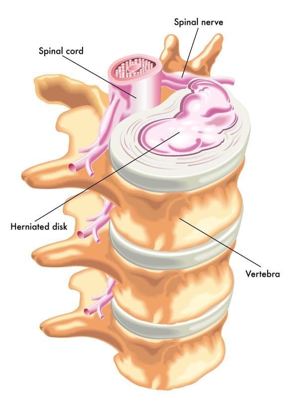
A slipped disc can mean a ruptured or herniated disc. We use the term slipped disc, however, the discs do not slip. Each disc is sandwiched between two vertebrae that are supported by a system of ligaments that hold the spine together. A bulging or herniated disc is the proper term.
Difference Between a Bulging and Herniated Disc
Disc disorders are categorized as contained or non-contained. Bulging disc is an example of a contained disc disorder.
Bulging
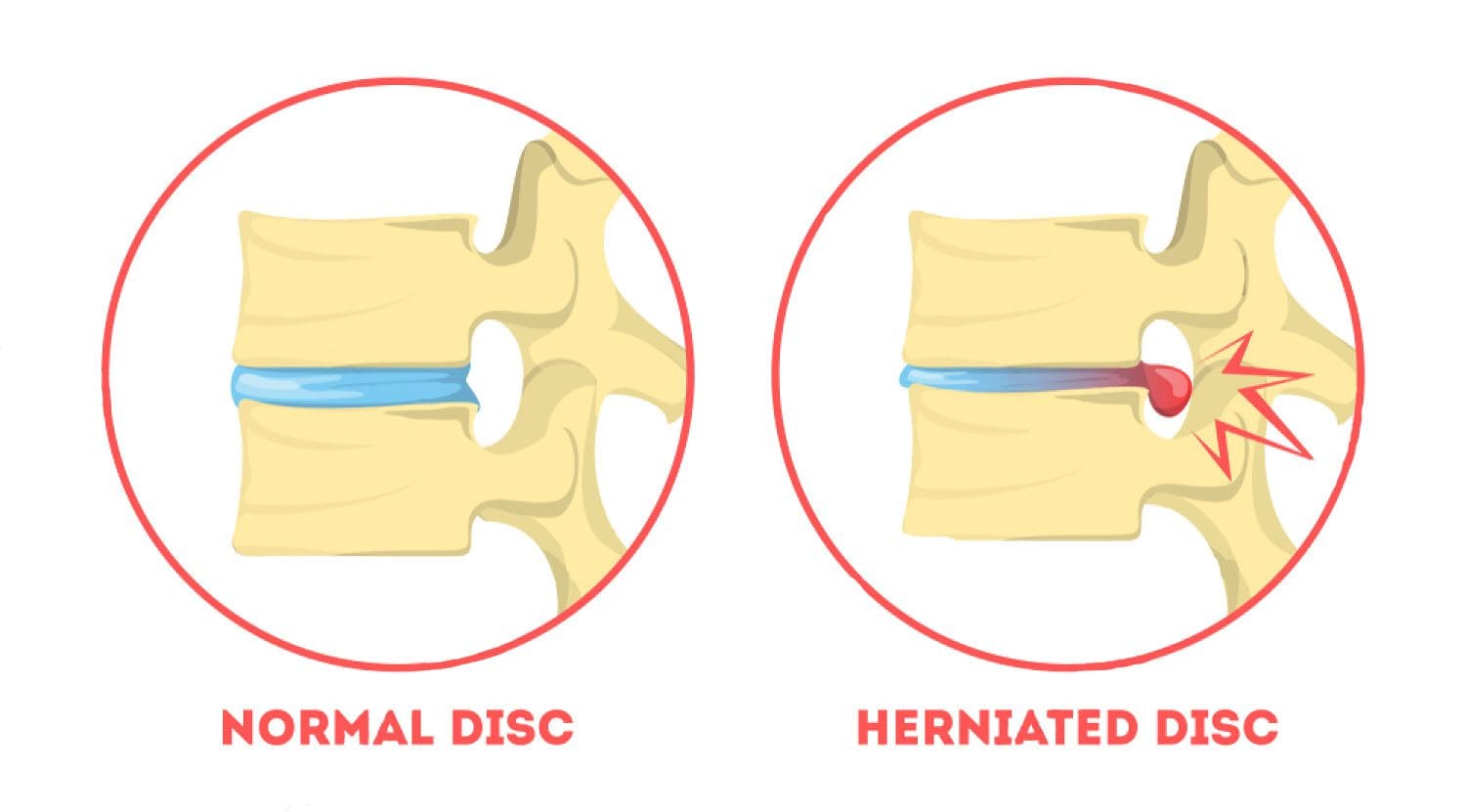
A bulging disc has not broken open meaning the nucleus is still contained inside the annulus fibrosus. The disc could protrude into the spinal canal without breaking open. The gel, the jelly interior does not leak out. The disc stays intact except a small bubble pops out.
Herniated/Ruptured
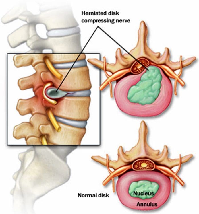
A non-contained disc has either partially or completely broken open, and that is a herniated/ruptured disc. Imagine a closed tube�being squeezed placed under pressure, which causes the contents to move wherever they can. If a portion of the tube is weak or there is too much pressure, the contents could leak or burst out. When a disc herniates the gel-like contents could spread out to the spinal cord and nerves.
Herniation Can Cause Pain
The disc’s gel-like nucleus has a chemical that irritates the nerves and causes them to become inflamed and swell. The chemical stays and continues to press on the irritated nerves. Sometimes fragments from the disc wall or the tube break off from the disc and drift into the spinal canal causing other nerves to inflame and swell. Based on the type of injury and condition of the discs, more than one disc could herniate, rupture, or bulge. Sometimes injury causes a combination of disc disorders.
Symptoms
Symptoms of a herniated disc can include:
Dull
Shooting pain
Muscle spasms
Cramping
Weakness
Tingling
Referred or radiating/traveling pain
Sometimes, however, a herniated disc does not cause any symptoms at all. When this happens it is called an asymptomatic herniated disc. Disc/s could be bulging or herniated, but if it or they are not applying pressure on the spinal nerve/sor the cord, symptoms like pain may not present. This makes a point about herniated disc symptoms that they are dependent on where you have a herniated disc.
Cervical Herniation Symptoms
With a herniated or bulging disc in the neck, then you could experience:
Neck soreness/pain
Muscle tightness
Cramping in the neck
Pain that travels down the arm/s
Tingling in the arm/s or hand/s
Weakness in the arm/s or hand/s
Lumbar Herniation Symptoms
With a herniated disc in the low back the following symptoms could happen:
Low back pain
Muscle tightness
Cramping in and around the low back
Radiating pain that travels down the leg/s
Tingling in your leg/s or foot/feet
Weakness in the leg/s or foot/feet
Referred Pain
Referred pain means that you have pain in another part of the body from the disc problem. An individual could have a bulging or herniated slipped disc in the low back and have pain in the leg. This is lumbar radiculopathy or sciatica. Usually, just one leg is affected. If you have a herniated disc in the neck, there could be referred pain going down the arm and into the hand.
Chiropractic Cares
A chiropractor can help relieve back pain and other herniated disc symptoms. A chiropractor will go through your medical history, do a physical exam, and perform orthopedic and neurological tests. They are looking at several things. Orthopedic and neurological exams can help the chiropractor figure out what’s going on.
Are reflexes functioning properly?�Meaning are your nerves sending messages correctly. An example is a reflex test is when a doctor taps the knee with a hammer and the leg kicks.
Is there a loss of muscle strength?
Signs of muscle/s wasting away?
Is there a loss of sensation, tingling or numbness along the nerve/s path?
They will carefully look at posture, and will probably order an X-ray or MRI to help with the diagnosis.
Chiropractors evaluate the entire spine. Even if you only have lower back pain, your chiropractor will examine your neck, too. They want to see how well your spine is functioning overall. Remember what happens to one area of your spine can influence another part of the spine and/or body.
Pain from a herniated disc can make it difficult to enjoy daily life. Walking, sitting, and sleeping normally/comfortably can become a nightmare. You should make an appointment with a doctor or chiropractor if your herniated slipped disc symptoms last for more than two weeks.
Herniated Disc Treatment
NCBI Resources
In the United States alone, acute cases of lower back pain are the fifth most frequent reason for doctor visits and cause 40% of missed days off work. What�s more, it is the leading cause of disability worldwide.
According to healthcare professionals, metabolic syndrome is a collection of conditions or disorders that can increase the risk of developing a variety of health issues, including diabetes, stroke, and heart disease. A combination of several of these risk factors, such as high blood pressure, high blood sugar levels, and excess waist fat, can ultimately increase the risk of a patient being diagnosed with metabolic syndrome and its associated health issues.
Metabolic syndrome affects approximately 23 percent of adults and the most common underlying causes of this collection of conditions or disorders include, excess weight and obesity, sedentary lifestyle, genetic factors, and age. As previously mentioned above, having 3 or more metabolic syndrome risk factors can increase the risk of developing a variety of other health issues, including diabetes, stroke, and heart disease, among other health issues.
Metabolic syndrome is characterized by three or more of the following metabolic measurements, including:
Excess waist fat (> 40 inches in men, and > 35 inches in women)
High blood pressure (130/85 mm Hg)
Hight blood sugar or glucose levels (100 mg/dL or greater)
High triglyceride levels (150 mg/dL or greater)
Low HDL cholesterol (< 40 mg/dL in men or < 50 mg/dL in women)
Diet and lifestyle modifications can help prevent, manage, or even reverse metabolic syndrome. Metabolic syndrome can increase the risk of developing various health issues, including diabetes, stroke, and heart disease if left untreated. Below, are important facts to know about metabolic syndrome.
Genetics Cause Metabolic Syndrome
If a close family member has diabetes or heart disease, you may already be genetically predisposed to metabolic syndrome. Getting a complete family health record generally includes information from three generations of relatives. Although it may seem challenging to collect this much information from your family, knowing what genetic predisposition you may have for developing metabolic syndrome and its associated health issues is important.
Body-Shape Influences Metabolic Syndrome
People with apple body-shapes have a much higher chance of developing metabolic syndrome than people with pear body-shapes. “Reducing your waist circumference can help prevent and manage health issues more than drugs and/or medication,� stated Erin Palinski-Wade, RD, CDE, author of Belly Fat Diet for Dummies. Excess waist fat is a key risk factor for the development of metabolic syndrome, diabetes, stroke, and heart disease, as well as cancer.
Plant-Based Diets Control Metabolic Syndrome
The current dietary guidelines for adults in the United States encourage plant-based diets. Julie Upton, RD, of San Francisco and co-founder of Appetite for Health, encourages following a Mediterranean diet. The Mediterranean diet includes eating more fruits, vegetables, legumes, whole grains, and seafood but involves eating less cheese, meat, and sweets. Plant-based diets and the Mediterranean diet can help lower the risks of developing metabolic syndrome.
Fiber Lowers Risk of Metabolic Syndrome
Include more foods that are rich in soluble fiber, such as beans and oats, into your diet. Insoluble fibers, such as whole grains, help transport foods through your gastrointestinal (GI) tract while keeping you feeling more satisfied. Fill at least half your plate with fruits and vegetables as well as whole-grain carbohydrates to make less room for less-beneficial food choices. Eating more fiber can ultimately help reduce the risk of developing metabolic syndrome.
Beverages & Drinks Also Affect Metabolic Syndrome
Several drinks and beverages, such as fruit juices and sodas, can increase blood sugar and triglyceride levels. Alcoholic beverages and drinks may also cause hypoglycemia and an initial drop in blood sugar. Water is recommended for healthy hydration. According to healthcare professionals, other healthy alternatives which provide water or hydration without extra calories can also include tea, coffee, skim or low-fat milk, fruits, and vegetables.
Joey Gochnour, RDN and exercise physiologist in Austin, Texas discusses that moderate aerobic exercise can improve cholesterol levels. He recommends exercising regularly, preferably at least 30 minutes a day, five days a week to help combat metabolic syndrome. According to Gochnour, �Strength training and intense aerobic exercise may improve your blood sugar and insulin sensitivity.� Exercise boosts metabolism and burns calories for weight loss.
Sitting Increases Risk of Metabolic Syndrome
According to several research studies, sitting is associated with the increased risk of developing metabolic syndrome even when you include moderate amounts of regular exercise and physical activity into your day. One research study published in June 2015 in Diabetologia closely associated sitting time with an increased risk of developing diabetes, demonstrated that for every hour of daily TV viewing, increased a person�s risk for diabetes by 3.4 percent.
Test Fasting Insulin Levels to Evaluate Risk for Metabolic Syndrome
A test for fasting insulin levels determines the risk of developing metabolic syndrome. Insulin plays a key role in metabolism. High insulin levels promote obesity, stimulate hunger, and increase fat storage. Sugary foods increase blood sugar and cause the pancreas to release insulin. But if the body is continuously exposed to high levels of insulin, the cells become resistant to the effects of insulin. Insulin resistance ultimately promotes high blood pressure, high blood sugar, and high cholesterol which are associated with metabolic syndrome, also known as insulin resistance syndrome.
Approximately 23 percent of adults in the United States have metabolic syndrome. Although the risk factors for developing the cluster of conditions or diseases are significant, there are good news. Many of the risk factors associated with metabolic syndrome can be addressed through diet and lifestyle modifications, such exercise and physical activity. By making these changes, people can ultimately significantly reduce their risks of developing a variety of other health issues, including diabetes, stroke, and heart disease. Although metabolic syndrome can be a serious health issue, people can significantly reduce their risks by reducing their weight; increasing exercise and physical activity; eating a heart-healthy diet that’s rich in fruits, vegetables, whole grains, and fish; as well as working with a healthcare professional to regulate blood pressure, blood sugar, blood cholesterol. In the following article, we will discuss several important facts to know about metabolic syndrome. – Dr. Alex Jimenez D.C., C.C.S.T. Insight
According to healthcare professionals, metabolic syndrome is a collection of conditions or disorders that can increase the risk of developing a variety of health issues, including diabetes, stroke, and heart disease. A combination of several of these risk factors, such as high blood pressure, high blood sugar levels, and excess waist fat, can ultimately increase the risk of a patient being diagnosed with metabolic syndrome and its associated health issues.
The scope of our information is limited to chiropractic, musculoskeletal, and nervous health issues or functional medicine articles, topics, and discussions. We use functional health protocols to treat injuries or disorders of the musculoskeletal system. Our office has made a reasonable attempt to provide supportive citations and has identified the relevant research study or studies supporting our posts. We also make copies of supporting research studies available to the board and or the public upon request. To further discuss the subject matter above, please feel free to ask Dr. Alex Jimenez or contact us at 915-850-0900.�
Heart Staff. �Your Risk for Metabolic Syndrome.� Www.heart.org, Heart Media, 31 July 2016, www.heart.org/en/health-topics/metabolic-syndrome/your-risk-for-metabolic-syndrome.
Heart Staff. �Symptoms and Diagnosis of Metabolic Syndrome.� Www.heart.org, Heart Media, 31 July 2016, www.heart.org/en/health-topics/metabolic-syndrome/symptoms-and-diagnosis-of-metabolic-syndrome.
Heart Staff. �Prevention and Treatment of Metabolic Syndrome.� Www.heart.org, Heart Media, 31 July 2016, www.heart.org/en/health-topics/metabolic-syndrome/prevention-and-treatment-of-metabolic-syndrome.
Taub-Dix, Bonnie. �Metabolic Syndrome: 10 Things You Need to Know About Your Risk: Everyday Health.� Everyday Health, Everyday Health Media, 31 Oct. 2017, www.everydayhealth.com/news/10-things-your-doctor-wont-tell-you-about-metabolic-syndrome/.
Dr. Alex Jimenez Podcast: Metabolic Syndrome
Metabolic syndrome is a cluster of risk factors that can ultimately increase the risk of developing a variety of health issues, including heart disease, stroke, and diabetes, among other problems. Central obesity, high blood pressure, high blood sugar, high triglycerides, and low HDL or good cholesterol levels are the 5 risk factors associated with metabolic syndrome. Having at least three of the five risk factors may suggest the presence of metabolic syndrome. Dr. Alex Jimenez, Alexander Jimenez, Truide Torres, Kenna Vaughn, and Astrid Ornelas explain the 5 risk factors associated with metabolic syndrome, in further detail, as they recommend diet and lifestyle modification advice and guidelines, such as the ketogenic diet or the keto diet, as well as demonstrate the biochemical and chemical pathways that the body goes through during ketosis to help people with metabolic syndrome improve their overall health and wellness. From eating good fats and staying hydrated to exercise and better sleep, Dr. Alex Jimenez, Alexander Jimenez, Truide Torres, Kenna Vaughn, and Astrid Ornelas discuss how diet and lifestyle modifications, such as the ketogenic diet or keto diet, can help improve the 5 risk factors associated with metabolic syndrome to prevent the risk of developing a variety of other health issues, including heart disease, stroke, and diabetes. – Podcast Insight
Neural Zoomer Plus for Neurological Disease
Dr. Alex Jimenez utilizes a series of tests to help evaluate neurological diseases. The Neural ZoomerTM Plus is an array of neurological autoantibodies which offers specific antibody-to-antigen recognition. The Vibrant Neural ZoomerTM Plus is designed to assess an individual�s reactivity to 48 neurological antigens with connections to a variety of neurologically related diseases. The Vibrant Neural ZoomerTM Plus aims to reduce neurological conditions by empowering patients and physicians with a vital resource for early risk detection and an enhanced focus on personalized primary prevention.
Food Sensitivity for the IgG & IgA Immune Response
Dr. Alex Jimenez utilizes a series of tests to help evaluate health issues associated with a variety of food sensitivities and intolerances. The Food Sensitivity ZoomerTM is an array of 180 commonly consumed food antigens that offers very specific antibody-to-antigen recognition. This panel measures an individual�s IgG and IgA sensitivity to food antigens. Being able to test IgA antibodies provides additional information to foods that may be causing mucosal damage. Additionally, this test is ideal for patients who might be suffering from delayed reactions to certain foods. Utilizing an antibody-based food sensitivity test can help prioritize the necessary foods to eliminate and create a customized diet plan around the patient�s specific needs.
Gut Zoomer for Small Intestinal Bacterial Overgrowth (SIBO)
Dr. Alex Jimenez utilizes a series of tests to help evaluate gut health associated with small intestinal bacterial overgrowth (SIBO). The Vibrant Gut ZoomerTM offers a report that includes dietary recommendations and other natural supplementation like prebiotics, probiotics, and polyphenols. The gut microbiome is mainly found in the large intestine and it has more than 1000 species of bacteria that play a fundamental role in the human body, from shaping the immune system and affecting the metabolism of nutrients to strengthening the intestinal mucosal barrier (gut-barrier). It is essential to understand how the number of bacteria that symbiotically live in the human gastrointestinal (GI) tract influences gut health because imbalances in the gut microbiome may ultimately lead to gastrointestinal (GI) tract symptoms, skin conditions, autoimmune disorders, immune system imbalances, and multiple inflammatory disorders.
Formulas for Methylation Support
XYMOGEN�s Exclusive Professional Formulas are available through select licensed health care professionals. The internet sale and discounting of XYMOGEN formulas are strictly prohibited.
Proudly,�Dr. Alexander Jimenez makes XYMOGEN formulas available only to patients under our care.
Please call our office in order for us to assign a doctor consultation for immediate access.
If you are a patient of Injury Medical & Chiropractic�Clinic, you may inquire about XYMOGEN by calling 915-850-0900.
For your convenience and review of the XYMOGEN products please review the following link. *XYMOGEN-Catalog-Download
* All of the above XYMOGEN policies remain strictly in force.
Modern Integrated Medicine
The National University of Health Sciences is an institution that offers a variety of rewarding professions to attendees. Students can practice their passion for helping other people achieve overall health and wellness through the institution’s mission. The National University of Health Sciences prepares students to become leaders in the forefront of modern integrated medicine, including chiropractic care. Students have an opportunity to gain unparalleled experience at the National University of Health Sciences to help restore the natural integrity of the patient and define the future of modern integrated medicine.
Industrialization/modernization has impacted our food, the way we eat and our weight. Processed fast food can be purchased pretty much anywhere. We no longer expend physical energy to hunt and forage for food. Because of this back pain is probably the most common and most troublesome condition that individuals experience. Eight out of 10 will struggle with back pain at some point in their life. Low chronic back pain can become aggravated by a variety of triggers.
Mechanical stress
Excessive strain
Muscle weakness
Poor sleep position
No exercise
Excessive weight
These can all contribute to making back pain even worse. Obesity is defined as a disease. It is a serious disorder that affects adults and children. Being overweight can contribute to the development of coronary heart disease, diabetes, high blood pressure, and colon cancer. And it can also contribute to symptoms associated with:
Osteoporosis
Osteoarthritis
Rheumatoid arthritis
Degenerative disc disease
Spinal stenosis
Spondylolisthesis
Osteoporosis coupled with a sedentary lifestyle and an unhealthy diet can affect the density and the strength of the bones. When the structural integrity of the spine is compromised there is a higher risk for fracture. Vertebral fractures can be painful and disabling. Those diagnosed with osteoporosis probably have lost between 25% to 30% of bone density.
Being Overweight Affects the Spine
The spine carries the body’s weight and distributes the weight in an equal manner when resting and during activity. When there is excess weight the spine is forced to compromise which can lead to injuries and structural damage. The area of the spine that is the most vulnerable is the low back or lumbar spine.
Exercise is Important
When we don’t exercise it leads to:
Poor flexibility
Weak back muscles
Weak pelvis
Weak thighs
This can increase the lower back’s curve, which causes the pelvis to tilt forward in an unhealthy manner. This is detrimental to proper posture and when our posture weakens, the rest of the body follows.
Age Relation
These disorders of the spine might be thought of as the normal aging process. This is true that as we age, the body tissues begin to change and the spine’s disc wear down. But, if you are overweight, chances are you have, or will have pain in the back.
Weight Loss Reduces Back Pain
Weight loss can contribute to a partial or complete reduction in the back pain symptoms. The research between weight loss and back pain is still ongoing but numerous practitioners report cases of patients experiencing a serious reduction in pain after losing the excess weight. Because the extra weight is taken off the spine further stress is avoided. Especially when a chiropractor realigns the vertebral column. According to the American Spine Society, individuals that stay around 10 pounds of their proper weight are those least likely to experience back problems.
Back Health and Physical Activity
Chiropractic is one of the most popular treatments for back pain. With chiropractic adjustments, pain is alleviated but also the root cause of the problems is corrected. Spinal adjustments can help even more when combined with a proper diet and weight loss treatment program.
Besides weight loss, exercising and strengthening the core muscles can guarantee the proper distribution of the body�s weight. Stronger muscles, less weight, and better posture will provide amazing long-term benefits. If you want to know how to incorporate weight loss and exercise in your daily routine, speak to your chiropractor. They are ready to guide you along the way. If you aren�t currently seeing a chiropractor, give us a call. We�re here to help!
Chiropractic Weight Loss
NCBI Resources
While chiropractic adjustments and associated therapies can ease back pain symptoms and aid in healing, lifestyle changes like a healthy diet are just as important. The more a patient can get to their ideal weight, the easier it becomes to treat and eliminate the back pain.
According to healthcare professionals, metabolic syndrome is a collection of conditions or disorders that can increase the risk of developing a variety of health issues, including diabetes, stroke, and heart disease. A combination of several of these risk factors, such as high blood pressure, high blood sugar levels, and excess waist fat, can ultimately increase the risk of a patient being diagnosed with metabolic syndrome and its associated health issues.
What is Metabolic Syndrome?
Metabolic syndrome affects approximately 23 percent of adults and the most common underlying causes of this collection of conditions or disorders include, excess weight and obesity, sedentary lifestyle, genetic factors, and age. As previously mentioned above, having 3 or more metabolic syndrome risk factors can increase the risk of developing a variety of other health issues, including diabetes, stroke, and heart disease, among other health issues.
Metabolic syndrome is characterized by three or more of the following metabolic measurements, including:
Excess waist fat (> 40 inches in men, and > 35 inches in women)
High blood pressure (130/85 mm Hg)
Hight blood sugar or glucose levels (100 mg/dL or greater)
High triglyceride levels (150 mg/dL or greater)
Low HDL cholesterol (< 40 mg/dL in men or < 50 mg/dL in women)
The Importance of Understanding Metabolic Syndrome
Healthcare professionals diagnose metabolic syndrome when a patient has a collection of conditions or diseases that can increase the risk of developing diabetes, stroke, and heart health. Moreover, research studies found that people with metabolic syndrome have a much higher chance of developing these health issues compared with individuals who do not have metabolic syndrome. Chances increase when more of these risk factors are present.
Metabolic syndrome has become increasingly common in the United States. Several factors increase the likelihood of acquiring metabolic syndrome:
Excess weight/obesity. These are critical and potential causes of metabolic syndrome. Too much fat in the abdomen is most commonly associated with metabolic syndrome. The reasons why excess weight/obesity and metabolic syndrome seem to be linked are complex and misunderstood.
Insulin resistance: This has been closely associated with metabolic syndrome. Several people are genetically predisposed to insulin resistance.
Race and gender: People of certain races, as are men more than women, ultimately have�a greater risk of developing metabolic syndrome.
As previously mentioned above, people with metabolic syndrome have an increased risk of developing the following health issues, including:
Cardiovascular disease and heart attacks. If the arteries that supply blood to the heart are narrowed or blocked by fatty deposits, known as plaque, they decrease the amount of blood and oxygen reaching the heart, which can cause chest pain (angina) or a heart attack.
Type 2 diabetes. Diabetes is another well-known health issue that happens when the body can’t produce enough insulin or is unable to utilize insulin properly. This causes sugar to build-up in the blood and increases the risk of developing kidney failure and heart disease.
Metabolic syndrome is also commonly associated with another well-known health issue, known as insulin resistance. People with insulin resistance experience problems where their own body prevents itself from utilizing insulin efficiently. Healthcare professionals and researchers alike sometimes refer to metabolic syndrome as insulin resistance syndrome. Furthermore, high blood sugar and insulin resistance are associated with diabetes.
Diagnosis and Treatment of Metabolic Syndrome
Healthcare professionals will look for the presence of three or more of the following risk factors to diagnose metabolic syndrome, including:
Excess waist fat (> 40 inches in men, and > 35 inches in women)
High blood pressure (130/85 mm Hg)
Hight blood sugar or glucose levels (100 mg/dL or greater)
High triglyceride levels (150 mg/dL or greater)
Low HDL cholesterol (< 40 mg/dL in men or < 50 mg/dL in women)
Metabolic syndrome treatment requires addressing several risk factors that can increase the risk of developing a variety of health issues, including:
Eating better. Follow a diet rich in fruits, vegetables, whole grains, lean meats, skinless poultry, and non-fried fish as well as low-fat or fat-free dairy products. Avoid processed foods, which are often high in saturated and trans fats, sodium and added sugar, among other processed foods.
Get active. Add at least 150 minutes of exercise and physical activity to a regular routine. Walking is the easiest place to start but you may want to find something else that gets your heart rate up. If necessary, break your exercise and physical activity into several short, sessions throughout the day.
Lose weight. Learn your recommended calorie intake, the number of food calories you’re consuming, and the energy calories you’re burning off with different levels of exercise and physical activity. Balance healthy eating with a healthy level of exercise and physical activity to lose weight.
If diet and lifestyle modifications alone do not control the conditions or diseases associated with metabolic syndrome, a healthcare professional may ultimately prescribe drugs/medications to help control blood pressure, cholesterol, and other symptoms. Carefully following your healthcare professional’s instructions can help prevent long term effects of metabolic syndrome. Your hard work and attention will make a difference in overall health and wellness.
Approximately 23 percent of adults in the United States have metabolic syndrome. Although the risk factors for developing the cluster of conditions or diseases are significant, there are good news. Many of the risk factors associated with metabolic syndrome can be addressed through diet and lifestyle modifications, such exercise and physical activity. By making these changes, people can ultimately significantly reduce their risks of developing a variety of other health issues, including diabetes, stroke, and heart disease. Although metabolic syndrome can be a serious health issue, people can significantly reduce their risks by reducing their weight; increasing exercise and physical activity; eating a heart-healthy diet that’s rich in fruits, vegetables, whole grains, and fish; as well as working with a healthcare professional to regulate blood pressure, blood sugar, blood cholesterol.
Dr. Alex Jimenez D.C., C.C.S.T. Insight
According to healthcare professionals, metabolic syndrome is a collection of conditions or disorders that can increase the risk of developing a variety of health issues, including diabetes, stroke, and heart disease. A combination of several of these risk factors, such as high blood pressure, high blood sugar levels, and excess waist fat, can ultimately increase the risk of a patient being diagnosed with metabolic syndrome and its associated health issues.
The scope of our information is limited to chiropractic, musculoskeletal, and nervous health issues or functional medicine articles, topics, and discussions. We use functional health protocols to treat injuries or disorders of the musculoskeletal system. Our office has made a reasonable attempt to provide supportive citations and has identified the relevant research study or studies supporting our posts. We also make copies of supporting research studies available to the board and or the public upon request. To further discuss the subject matter above, please feel free to ask Dr. Alex Jimenez or contact us at 915-850-0900.�
Heart Staff. �Your Risk for Metabolic Syndrome.� Www.heart.org, Heart Media, 31 July 2016, www.heart.org/en/health-topics/metabolic-syndrome/your-risk-for-metabolic-syndrome.
Heart Staff. �Symptoms and Diagnosis of Metabolic Syndrome.� Www.heart.org, Heart Media, 31 July 2016, www.heart.org/en/health-topics/metabolic-syndrome/symptoms-and-diagnosis-of-metabolic-syndrome.
Heart Staff. �Prevention and Treatment of Metabolic Syndrome.� Www.heart.org, Heart Media, 31 July 2016, www.heart.org/en/health-topics/metabolic-syndrome/prevention-and-treatment-of-metabolic-syndrome.
Dr. Alex Jimenez Podcast: Metabolic Syndrome
Metabolic syndrome is a cluster of risk factors that can ultimately increase the risk of developing a variety of health issues, including heart disease, stroke, and diabetes, among other problems. Central obesity, high blood pressure, high blood sugar, high triglycerides, and low HDL or good cholesterol levels are the 5 risk factors associated with metabolic syndrome. Having at least three of the five risk factors may suggest the presence of metabolic syndrome. Dr. Alex Jimenez, Alexander Jimenez, Truide Torres, Kenna Vaughn, and Astrid Ornelas explain the 5 risk factors associated with metabolic syndrome, in further detail, as they recommend diet and lifestyle modification advice and guidelines, such as the ketogenic diet or the keto diet, as well as demonstrate the biochemical and chemical pathways that the body goes through during ketosis to help people with metabolic syndrome improve their overall health and wellness. From eating good fats and staying hydrated to exercise and better sleep, Dr. Alex Jimenez, Alexander Jimenez, Truide Torres, Kenna Vaughn, and Astrid Ornelas discuss how diet and lifestyle modifications, such as the ketogenic diet or keto diet, can help improve the 5 risk factors associated with metabolic syndrome to prevent the risk of developing a variety of other health issues, including heart disease, stroke, and diabetes. – Podcast Insight
Neural Zoomer Plus for Neurological Disease
Dr. Alex Jimenez utilizes a series of tests to help evaluate neurological diseases. The Neural ZoomerTM Plus is an array of neurological autoantibodies which offers specific antibody-to-antigen recognition. The Vibrant Neural ZoomerTM Plus is designed to assess an individual�s reactivity to 48 neurological antigens with connections to a variety of neurologically related diseases. The Vibrant Neural ZoomerTM Plus aims to reduce neurological conditions by empowering patients and physicians with a vital resource for early risk detection and an enhanced focus on personalized primary prevention.
Food Sensitivity for the IgG & IgA Immune Response
Dr. Alex Jimenez utilizes a series of tests to help evaluate health issues associated with a variety of food sensitivities and intolerances. The Food Sensitivity ZoomerTM is an array of 180 commonly consumed food antigens that offers very specific antibody-to-antigen recognition. This panel measures an individual�s IgG and IgA sensitivity to food antigens. Being able to test IgA antibodies provides additional information to foods that may be causing mucosal damage. Additionally, this test is ideal for patients who might be suffering from delayed reactions to certain foods. Utilizing an antibody-based food sensitivity test can help prioritize the necessary foods to eliminate and create a customized diet plan around the patient�s specific needs.
Gut Zoomer for Small Intestinal Bacterial Overgrowth (SIBO)
Dr. Alex Jimenez utilizes a series of tests to help evaluate gut health associated with small intestinal bacterial overgrowth (SIBO). The Vibrant Gut ZoomerTM offers a report that includes dietary recommendations and other natural supplementation like prebiotics, probiotics, and polyphenols. The gut microbiome is mainly found in the large intestine and it has more than 1000 species of bacteria that play a fundamental role in the human body, from shaping the immune system and affecting the metabolism of nutrients to strengthening the intestinal mucosal barrier (gut-barrier). It is essential to understand how the number of bacteria that symbiotically live in the human gastrointestinal (GI) tract influences gut health because imbalances in the gut microbiome may ultimately lead to gastrointestinal (GI) tract symptoms, skin conditions, autoimmune disorders, immune system imbalances, and multiple inflammatory disorders.
Formulas for Methylation Support
XYMOGEN�s Exclusive Professional Formulas are available through select licensed health care professionals. The internet sale and discounting of XYMOGEN formulas are strictly prohibited.
Proudly,�Dr. Alexander Jimenez makes XYMOGEN formulas available only to patients under our care.
Please call our office in order for us to assign a doctor consultation for immediate access.
If you are a patient of Injury Medical & Chiropractic�Clinic, you may inquire about XYMOGEN by calling 915-850-0900.
For your convenience and review of the XYMOGEN products please review the following link. *XYMOGEN-Catalog-Download
* All of the above XYMOGEN policies remain strictly in force.
Modern Integrated Medicine
The National University of Health Sciences is an institution that offers a variety of rewarding professions to attendees. Students can practice their passion for helping other people achieve overall health and wellness through the institution’s mission. The National University of Health Sciences prepares students to become leaders in the forefront of modern integrated medicine, including chiropractic care. Students have an opportunity to gain unparalleled experience at the National University of Health Sciences to help restore the natural integrity of the patient and define the future of modern integrated medicine.
There are different kinds of head and upper cervical disorders that are also known as:
Upper neck disorders
Craniovertebral junction – CVJ – abnormality
Craniocervical disorders
Some are present at birth while others are developed later in life. Whether congenital or acquired, they share many of the same symptoms.
Neck Pain/Headache
Where your skull and spine meet is where upper cervical disorders take place. Neck pain along with headaches along the back of the head are the most common symptoms. People with craniocervical disorders often experience neck pain and headache in combination and symptoms worsen with head and neck movement. Even just a sneeze or cough can cause pain. Neck pain can also spread to the arms if spinal nerves are compressed.
Spinal Cord Compression
The more complex upper cervical disorders involve spinal cord compression. When the spinal cord gets compressed, nerve problems can happen that make daily function a challenge.
With spinal cord compression, neurological symptoms can present like:
Weakness in the arms, legs or both
Loss of sensation and awareness of the limbs called positional sense
There can be an electrical/shooting pain or tingling running down the spine and into the legs when pitching the neck forward called Lhermitte sign
Hot and cold sensational loss in hands, feet or both
All of these can cause malformation and physical changes in the neck. The neck may appear short, webbed, or twisted. There can also be a limited ability to move the neck.
Symptoms can also be caused by brain and cranial nerve pressure
Conditions like platybasia, basilar invagination, and craniocervical tumors can place pressure on the brain stem and the cranial nerves around it. If this happens the following symptoms can occur:
Eye issues like abnormal eye movements and double vision
Throat and speech problems like hoarseness, slurred speech, and swallowing problems
Loss of coordination
Sleep problems like sleep apnea
Vertigo Faint Feeling
Some individuals could experience a set of symptoms that don’t occur too often called vertebrobasilar ischemia.
This happens when the head position changes placing pressure on the cranial arteries and cutting off the blood supply.
The result is:
Weakness
Confusion
Light-headedness
Feeling faint
Vertigo or a dizzy spinning sensation
Syringomyelia
Syringomyelia is a fluid-filled cyst known as a syrinx that forms inside the spinal cord. If the cyst grows, it can damage the spinal cord causing painful neurological symptoms throughout the body that include weakness, and numbness. In extreme cases loss of ability to feel heat or cold in the neck, hands, and possibly throughout the spine. The hand muscles can become paralyzed.
Quality of Life with Upper Cervical Disorder
While some individuals have no symptoms associated with a craniocervical disorder, others experience intense pain and neurological dysfunction. There are treatment options that can help you successfully manage and eliminate symptoms.
Chiropractic
When headaches present it usually means there is a misalignment in the cervical spine. This is often combined with tight muscles in the neck, shoulders, and surrounding area. Pressure on the nerves can cause sharp shooting pain along with a continual dull throbbing sensation.
A chiropractor will assess the area and then move forward with an adjustment to relieve the pressure and pain. Chiropractic medicine does not just focus on relieving symptoms but finding the root cause and correcting the problem. In addition, the chiropractor will take the time to educate you on the importance of diet, exercise, stretching, and making healthy lifestyle choices.
Whiplash Massage Therapy
NCBI
The effectiveness of chiropractic care for headaches has been proven by many research studies. Chiropractic is not only excellent for treating headaches, but is gentle, non-invasive, does not require medications, and is known for being free of side effects.
You don’t have to be highly athletic to do back extensions. They’re common in yoga, Pilates, and other stretching and strengthening exercises. Health and fitness begin with the desire to improve oneself. When desire and belief are strong enough, it leads to action. When done properly and consistently, success follows. Considering all that the back goes through in a typical day, it isn’t much of a stretch to think that pain could present at times. This is especially true if the back is not properly aligned.
Strong back muscles can reduce injuries and improve posture. There are some muscles along the length of the spine that support the spine. One of the most important�deep muscle groups in your back is the erector spinae. If this muscle group becomes weak, back pain can start to present.
Back Extension
Back extensions can be done:
Using a machine
With an exercise ball
No equipment or objects at all described below
When beginning it’s a good idea to consult a certified personal trainer that has experience rehabilitating people with back pain. They will also teach proper form, body mechanics and alignment. Before doing any exercises consult your chiropractor or primary doctor.
Three You Can Do
The Cow Pose
Begin on hands and knees.
Hands are shoulder-distance apart and knees are hip-distance apart.
Inhale and arch your spine by rocking your pelvis toward the floor.
Look up toward the ceiling.
As you exhale, move back to your starting neutral back position.
Inhale, and repeat. Link your inhales and exhales with your movement.
Do the cow stretch 5 to 10 times once a day.
Upper Back Extension
Begin on your stomach with a small pillow or�rolled�towel under your hips.
Arms should be along your sides.
Slowly lift your upper body off the floor and contract your low back muscles.
Hold 3 seconds before lowering back to the floor.
Repeat 10 times.
Opposite Arm/Leg Extension
Start on your stomach with legs together and arms stretched in front.
Breathe in and tighten your tailbone as you lift your arms and legs off the floor (about hip height and if you can higher).
Pump the right arm down and left leg down so they just graze the ground, and then bring them back to hip height and pump the left arm and right leg down.
Alternating right arm/left leg and left arm/right leg in small movements. Keep your eyes toward the mat.
Repeat 3 times every day.
These are just a few back extensions that can help you build strong back muscles. Do these exercises regularly to decrease and prevent back pain.
Belief Action Success
All of us want to be living healthy lives.
When realistic, specific and measurable goals are expertly set-up in a health program what really keeps us from getting there is a break in the link that falls between belief and the action. And that is the excuses that seem to pop up at those moments.
Every time I try to exercise or eat healthy, something comes up
Someone or something else is responsible for the situation.
We know what we need to do but just don’t want to do it. We believe that something out of our control prevents us.
Be Proactive
It means more than just taking initiative. It means a responsibility or you can think of it as a response and the ability for our own lives. People that become proactive do not blame circumstances, conditions, etc for their behavior or lack of behavior. It is a product of their own conscious choice. Once you realize the power you have, obstacles become welcome challenges to be overcome. With just a little success, confidence grows and commitment to health and fitness goals becomes a priority.
Back Pain Specialist
NCBI Resources
Dealing with joint or muscle pain can be a daunting experience. It�s important to work on maintaining�mobility and flexibility. The more flexible, the less likely to be injured. One of the best ways to improve flexibility is through stretching. A�chiropractor�can recommend stretches, or you can use some of these basic techniques
Metabolic syndrome is a cluster of risk factors that can ultimately increase the risk of developing a variety of health issues, including heart disease, stroke, and diabetes, among other problems. Central obesity, high blood pressure, high blood sugar, high triglycerides, and low HDL or good cholesterol levels are the 5 risk factors associated with metabolic syndrome. Having at least three of the five risk factors may suggest the presence of metabolic syndrome. Dr. Alex Jimenez, Alexander Jimenez, Truide Torres, Kenna Vaughn, and Astrid Ornelas explain the 5 risk factors associated with metabolic syndrome, in further detail, as they recommend diet and lifestyle modification advice and guidelines, such as the ketogenic diet or the keto diet, as well as demonstrate the biochemical and chemical pathways that the body goes through during ketosis to help people with metabolic syndrome improve their overall health and wellness. From eating good fats and staying hydrated to exercise and better sleep, Dr. Alex Jimenez, Alexander Jimenez, Truide Torres, Kenna Vaughn, and Astrid Ornelas discuss how diet and lifestyle modifications, such as the ketogenic diet or keto diet, can help improve the 5 risk factors associated with metabolic syndrome to prevent the risk of developing a variety of other health issues, including heart disease, stroke, and diabetes. – Podcast Insight
[00:00:14] All right, guys, we’ve come to another podcast. And welcome to Dr. Jimenez and crew podcast. Welcome. And you have a family here.
[00:00:23] We’re gonna go over metabolic syndrome today. Metabolic syndrome is a disorder that ultimately affects a whole lot of people. What happens is it actually affects one of the largest populations in El Paso, pretty much in this region. And what we have is, it’s not a disease. OK. First of all, it’s a combination of presentations that medical doctors and the World Health Organization have determined that high-risk factors in order to have a stroke, kidney disorders and even problems with dementia. But overall, it’s pretty much if you have metabolic syndrome, you really feel crummy. So today what we’re going to do is we’re gonna discuss the issues and we’d like to at least present it to you so that it becomes useful for you. And the information provided by us is going to be helpful for you or a family member. So if you have the opportunity and it’s something that you enjoy, please go ahead and at the bottom area, there’s a little bell to subscribe and a little belt in markets so that you could be the very first person to get information in the future when we ever post it. And it also gives you the opportunity to present or ask us for things that are important to you in the health-related realm. And now what we’re going to do today, my name is Dr. Alex Jimenez, I have my entire staff here. We’re gonna go ahead and we’re going to present each one of them in different moments. And we’re gonna do some really interesting dynamics. We also have our resident biochemist at the National University of Health Science who’s actually going to chime in, who’s gonna give us a little bit of a foundation about chemistry. This information is gonna be helpful. We’re gonna try to make it as simple but as useful as possible. Now, bear in mind everything that we’re gonna be talking about today revolves around the metabolic syndrome. Metabolic syndrome is what the health care organizations have determined as well as the cardiac departments have determined, as five major symptoms, now, you have to have three of them at least in order to be classified as metabolic syndrome. Now, the first thing is to ask, what do you feel? Pretty much you feel like crap. And it’s not really a good feeling to feel this way, but you’ll see that if you have some of these presentations, you’re gonna notice that your doctor may give you a diagnosis of metabolic syndrome. Now, the first thing that happens is you have usually a little bit of belly fat. Now, the belly fat that people have, people measure it. Now, for men, it’s a belly, kind of like the lonja, the belly that actually hangs over. And it’s about a good I’d say about 40 inches or greater in the male, in women it’s 35 inches or more. Now, that’s one of the first presentations. Now, the other presentation is high blood pressure. Now the high blood pressure that they use is 135 milligrams over deciliter. Oh, sorry. Yeah. Miller Mercury’s millimeters or Mercury or the slaters over to determine exactly the diastolic and the systolic. So the systolic is gonna be 135. The diastolic is going to be over 85. Now that doesn’t, again, you’re gonna notice something. These aren’t really extreme ranges. Okay. Now metabolic syndrome has high triglycerides. Now the high triglycerides are going to be noted in the blood. Okay. Now one of the things that can be determined early on is high blood pressure which is also a study associated with metabolic syndrome. So the other final one is the elevation of or decrease actually of HDL or the good fragments of cholesterol. Alexander is going to be a resident biochemist, is going to talk to us a little bit more about that in the latter part of the show. Now, bear in mind, I’ve given five things A, fat, B, high blood pressure, C, the blood glucose levels and also the triglycerides along with the lowering of the HDL. The question is, how are we going to be able to control this? Now, I want to give you some real good basic ways that you can actually control metabolic syndrome. And by the time we’re done today, we’re going to be able to assess the situation. And even if you have it, you basically will be able to control it. There are rare diseases that you can actually have. And again, this is not a disease. It’s a combination of syndromes or symptoms, collectively called a syndrome. So metabolic syndrome is one that can be misconstrued. Now, you’ll notice that the level of blood glucose is going to be elevated, usually over a hundred. Now, these are really relatively normal numbers that people have. But if they’re higher than that, they do create issues. Now, also, when you have the belly fat 40, that much. A lot of people have it. People have also blood glucose levels that are higher than 5.6 on your blood glucose when we would see now these numbers, along with the 150 milligrams per deciliter of triglycerides, they’re all normal. But in combination together, they do ultimately create a scenario that is not favorable to a cardiac issue. Cardiovascular issues do present as a result. So what we’re gonna try to do is try to bring down and control these issues. Now, what are the things that cause metabolic syndrome? Well, one of the things is stress, smoking, a sedentary lifestyle, and also even sleep problems and disturbances. Each one of these we’re going to be elaborating in the future podcasts but we’re gonna be able to tell exactly what’s actually going on in a better way. We also have issues of inflammation and processed foods. Now, at the core, metabolic syndrome, the main issue is insulin sensitivity issues along with high blood pressure issues and inflammation. So what are we going to do to control that? I want you to know that every single one of these five issues, whether its blood glucose, high triglycerides, low HDL counts or blood glucose, they’re all relatable to one disorder. It’s insulin sensitivity, insulin sensitivity controls every one of these factors from raising high blood pressure, the kidneys actually are controlled by the insulin causing their increase in blood pressure. We’ll discuss that issue and the correlation of it so we can bring ourselves to control the blood glucose. We ultimately have the fastest and the surest way to provide the fastest route to heal and to fix an individual with metabolic syndrome. So let’s go ahead and talk about the issues that are going to be resulting from that. Now, as I’ve got this, we’re gonna notice that if over a period of time you continue to have a lifestyle that has high levels of these particular five factors, you’re going to notice that you’re going to tend to have high cardiac risks. Now we have a team here and I want to introduce each one. We have Kenna Vaughn, who is our health coach. Our health coach is the one that’s going to be the one that explains to our patients what is going on. I’ll bring her in. We also have the clinical liaison, which is Truide Torres, the individual that is going to be able to bring out and ask the questions and determine what kind of issues and treatments are appropriate for you. So we’ll be discussing those. And we have our resident chief editor Astrid Ornelas, who’s gonna be the one that also explains the studies on it. All the way from Illinois. We also have Alexander, which we have here. Right. We added the backway. You can’t see him, but he’s presenting in. Say Hello, Alexander. You got him there. Hello. All right. So he’s out there. He’s going to be able to discuss the issues on the biochemistry side of things. And we’re looking forward to being able to explain those issues. Now, one of the things we have to do is go back to the issue of insulin sensitivity. Insulin sensitivity is at the root of all these issues. So what we’re gonna do is we’re going to discuss exactly how insulin can actually be controlled. But what we’ve learned through these studies and I’m going to bring in Mrs. Ornelas here to discuss the studies that we have pertaining to how to control blood glucose and blood sensitivity study. What did you find out recently that actually shows the proof and actually presents the easiest way to control blood, insulin and elevate HDL?
[00:08:08] OK. Well, first of all, just as you mentioned, metabolic syndrome, it’s a collection of health issues that can increase the risk of developing heart disease, stroke, and diabetes. Basically like, you know, it can affect our overall health and wellness.
[00:08:29] And I’ve done quite some research. And I’ve found through the National Center of Biotechnology Information, the NCBI, there’s a variety of research which basically states that metabolic syndrome or people with metabolic syndrome, one of the easiest, you know, quote-on-quote, easiest. Or one of the best ways maybe out there that can be used to help.
[00:09:04] Restore it, yeah, to help restore or reverse your metabolic syndrome would be through the ketogenic diet, or the Keto diet, as a lot of people know it best by. It is a low carbohydrate, high-fat diet, which according to research studies, offers many benefits to people with metabolic syndrome.
[00:09:28] It can help improve or promote weight loss. And it can help reduce diabetes.
[00:09:38] Basically, you know what I mentioned right there?
[00:09:42] I have found nothing faster to lower blood glucose and actually reverse triglycerides issues in HDL issues. Than the ketogenic diet. So in essence, if you want to do it fast, it’s amazing the speed at which it restores the body back. What else is there?
[00:10:00] Yeah. Yeah. Yeah. So basically the human body normally uses glucose or sugar. It is supposed to be our main source of fuel. Our main source of energy. But for people that have metabolic syndrome. People who have obesity. Insulin resistance. Diabetes or an increased risk of diabetes. The ketogenic diet can be very beneficial towards that because the ketogenic diet, first of all, it is a low carbohydrate diet. Carbohydrates essentially turn into sugar or glucose and we don’t want that.
[00:10:41] Like if people have metabolic syndrome, they have, you know, diabetes and insulin resistance. You don’t want sugar in their bodies because they produce too much of it. They have too much blood sugar.
[00:10:53] But by increasing the number of fats that you eat and then decreasing the number of carbohydrates, you actually keep a low amount.
[00:11:05] If you keep insulin low by eating more fats, you basically make the body go into a state of ketosis.
[00:11:18] You know what? Let me ask you something. I’m going to feed this over to right now to Kenna. And I’m gonna ask Kenna. Kenna, in your experiences with the blood sugar issues, how is it that we contain and we learn to be able to manage someone’s blood sugar the quickest, the fastest? What is it that you do in terms of coaching individuals, helping them back by coaching individuals?
[00:11:41] I definitely always evaluate their diet. And the main thing I like to focus on is education because so many people are not educated about, as she was saying, carbs and how they actually feed your body. A Big Mac might have 54 carbs and a sweet potato might have 30 carbs. And people don’t really realize that they’re that different. They only see 20 points or something like that. But the way that the carbohydrate breaks down in the body is huge. And that’s why the ketogenic diet works so well because you’re using those good carbs that are going to actually contain protein as well. And so it’s going to help to break it down slower versus a Big Mac, which is just going to spike your insulin way out.
[00:12:23] And what part of the Big Mac is the thing that spikes the sugar? I mean, in terms of.
[00:12:26] Right. So the bread, the carbs in the bread, it actually breaks down differently in the body than a sweet potato would. And so that’s what’s going to give you that high glucose level. And then after that, you’re gonna have the fall of the glucose level, which just your blood sugar going up and down does not feel great.
[00:12:43] So it’s not good. Of course, you’re paying for the sugars. When you ask the types of sugars that you have. You just mentioned right now that the quality of the type of carbohydrate matters.
[00:12:52] Yes, a little bit about the quality. Like I was saying, sweet potatoes, avocados, things like that. They’re going to have the carbohydrates that are better for you, meaning you break them down differently than you would faster sugar like sucrose and things like that.
[00:13:12] So simple sugars are out, basically, which is the reason that, first of all, metabolic syndrome did not even exist prior to the advent of refined foods. So refined sugars have caused this problem. So what we want to do is, sugar leads to inflammation. Sugar leads to triglyceride issues, sugar or basically insulin sensitivity issues are the things that are the basis of this process. All roads lead to insulin sensitivity in this process and in the organ that provides us with insulin. The greatest amount is in the pancreas. The pancreas is nonstop. And depending on how the pancreas responds to this blood sugar drama, it really determines the fate of the individual. It will alter the triglycerides. They will alter the blood pressure by having a direct effect of holding sodium in the kidneys. The body prepares, it retains the sodium, and by nature of sodium, the blood pressure soars, so the fastest way to lower your blood pressure is a ketogenic diet. And this is amazing because it really is simple. It’s not that complex. We can go extreme. And I know that our state really had a good research document on that. Tell me a bit about what you noticed.
[00:14:24] Yeah, basically, like what I was saying before. A lot of people don’t know the difference between what type of carbohydrates they want to eat. Like, for example, as you said, you know, a lot of people will eat a Big Mac and don’t eat that sweet potato. And they don’t know the difference between a good carbohydrate. Basically, we want to eat what you call complex carbohydrates. We want to eat whole wheat or we want to eat good starches because the body breaks those down into glucose, into sugar. But they’re used much more slowly. The body won’t immediately use them. And then you’ll get that crash, that sugar crash, because of the insulin spike.
[00:15:11] Right. So that controls the spike. You know what? I want to bring in our resident biochemist here. OK, so our biochemist is Alexander. He’s got a presentation here, actually, if I can see it there and see if it pops up here. Let me see it. And there he is. Alex, can you tell us a bit about what you’re trying to explain here on the biochemistry side of things?
[00:15:30] So as you guys were mentioning, just in general. Glucose is the main energy source in the way that we use it for the breakdown. Its breakdown of energy consumption is called glycolysis. So without getting too much into it, our end goal here is pyruvate, which then goes into the citric acid cycle to be turned into an acetyl-CoA in normal conditions. It’s good to have a carbohydrate meal, but in excess, you produce too much acetyl-CoA. When too many acetyl-CoA is used? You end up inducing fatty acid synthesis which is induced by large levels of insulin. So by doing so, you have an acetyl-CoA that ends up turning into palpitate. And one thing that I was mentioning is that not all foods are of equal quality. So here we can kind of see all the different types of fatty acids. So without going too much into the biochemistry, but kind of just giving you an idea of what’s going on here, these numbers on the left side represent the number of carbons in a row and then the numbers to the right of the semicolon are the number of double bonds. And normally double bonds will play a large role until you get into the effect of digestion in the way the body uses these. So by having more double bonds, it’s more fluid. So you notice the difference between a piece of lard and olive oil. What’s the difference? The only difference really is the number of carbons and the number of double ones. So here we have olive oil and then we have some sort of saturated fat. We can see that the difference is large in the number of carbons as well as double bonds. Double bonds allow for a lower melting point. That’s why olive oil is a liquid at room temperature vs. fatty acids and this plays a large role when it comes into how the body uses these types of things.
[00:17:26] Alex, are you saying that, obviously we all know that the good work of olive oil and avocado oil and coconut oil are the best thing. Is this the reason why this happens?
[00:17:35] Exactly. So the more double bonds they have, the more fluid it is going to be within the body and allow for the body to use those fats in a timely manner versus clogging up arteries and creating plaques within those arteries?
[00:17:48] Excellent. You know what? One of the things that insulin does? It packs away carbohydrates and energy in the cell. If you do that, what happens with this blood sugar? Eventually, insulin spikes it and puts it in the cells. Eventually, the cell grows. Hence the belly fat. That is ultimately what happens to the belly, it starts gaining fat cells and they start getting bigger, bigger, and bigger because they get injected in there. That stuff starts seeping out and once it can’t go anymore, it ends up in places like the pancreas. It ends up in places like the liver. It ends up in the intramuscular, in the muscular tissue. And that’s why we have the accumulation. And when you have a big belly, that’s what tips off the doctor, not only with the triglycerides and the blood glucose levels but also the belly fat. And that’s one of the things we have to kind of assess. So is this.
[00:18:36] Now, these fatty acids, which are fatty acids used for, almost everything within the body, especially for energy consumption. It’s like saying, would you rather be able to go five miles or 10 miles a week? Or go 10 miles. Right. And so gram for gram fat as an energy source is much more fuel-efficient and glucose or carbs.
[00:19:04] So carbs provide four grams of calories per gram and fats are around nine. So it’s almost more than double, the amount of energy that you’re producing from these sites, from these fatty acids. The difficult part is just knowing which ones are good. So kind of going into the good fatty acids which are going to be the ones with the double bonds. So I mean, any plant oils, animal fats, depending on which ones, we tend to want to stay away from large amounts of acid that tend to cause inflammation responses through the inflammation path. But the rest of these are really, really good, especially EPA and DHA. So DHA is actually used within the nervous system. It’s turned into neurotic acid any day as well. So getting these marine oils are really, really going to be good for your system just in general.
[00:19:55] You know what, as I understand these processes and I start realizing the biochemistry behind it, bring it home to this process, down to the cellular component it honors and it shows appreciation in terms of what creates the fatty acid excess. Now, again, what happens as a result of too much of these fatty acids or carbohydrates in the bloodstream? The body tries to store it in the form of fat and it’s shoved into the pancreas. So you get this fat inside the pancreas. If it can’t do it there, it eventually puts it in the liver. And like we mentioned, it gets it in the stomach or that’s when we see it as a final thing. I’d like to take the explanation and break apart one other point. The high blood pressure component. Insulin has a direct effect on the kidneys. Insulin tells the kidneys.
[00:20:43] Look, we need to pack this stuff into the fat and without getting beyond too much of the chemistry dynamics. You can see that what’s going to occur is that the kidneys are going to be commanded to hold more sodium. What we learned in chemistry and in biochemistry and in clinical sciences that the more sodium we retain, the blood pressure rises. In essence, that’s how quick the blood pressure goes. So you do that for a period of time and then you force the collection of atherosclerotic plaques because of the fat that is in there and it can’t go anywhere. You’re going to have a problem in the long-term future. So speaking about the oils, as Alexander just did, one of the things we ask, well, what oils do we not want to use? Canola oil, corn oil, sesame seed oil. I love sesame seeds. But the problem is that sesame seed oil causes inflammation, as Alex said, with acids. So what we got to do is we got to figure out exactly what types of oils we can do. And avocados, as Kenna had mentioned, are a great source of fats that we can use and make things more able to be processed. Our bodies and the old pyramid of diet is really bad because it’s heavy on carbohydrates. So one of the things that we look at is maintaining all those components. So we talked about the triglycerides, right? We talked about the belly fat, how it’s put together. And in each one of these, I wanted to point this out again. Each one of these, the high blood pressure, which is 135, high blood pressure is not considered high. High blood pressure at 135 usually it’s at 140, OK. So, why are we using triglycerides at 150 are not considered excessive. You know, HDL is lower than 50 are not considered horrible, but in combination together, if you have one at all, these three of these components out of the five, that’s what leads to a preposition of being sick and feeling crummy, let alone any prolonged period of this will end up leading to metabolic disorders, heart problems, stroke problems, dementias that actually occur as a result of prolonged metabolic syndrome, states that are within the individual. I know Alexander when I ask Alexander, he’s got some really interesting dynamics as I want to present right now. I’m going to show his screen right here because he’s got some interesting components on what also affects metabolic syndrome.
[00:23:00] Alexander. So kind of going into what it is, I guess, ketosis, because everyone wonders kind of what goes on. So I kind of got this diagram here to draw for you guys. We’re going to ignore the Federer pathway over here, but just in general. So what’s going to happen first is you’re going to deplete any glucose that you have. So the body normally stores around 100 grams of glucose in the liver and around 400 grams within the muscle components of the entire body. So if you times, 500 times four, that’s about 2000 calories, which is your daily limit. So you’ve got about a day worth of glucose always stored within your body. But once you deplete that, your body’s gonna start looking for other things. In the meantime, it takes a few days for your body to switch over from burning sugar, which is glucose, to burning ketone bodies from fat. So what’s going to happen? Your, first of all, your adrenals are releasing epinephrine, its precursor is norepinephrine. And it causes a couple of different things?
[00:23:56] You’re gonna get a little jittery at first and you can feel really bad for the first couple days. But then your body is gonna start switching over as your brain starts to start using these ketone bodies for its energy source. So as you’re producing norepinephrine, these are just like this is the cell surface here. These are just different precursor markers. So we have B1, B2, B3 and eight. So by doing these, they’re going to mark and signal to the G.S. protein, which is going to allow for adenosine class to activate ATP into cyclic AMP. Now, cyclic AMP is a very important component of degradation of fatty acids and the cool part is it’s actually inhibited by phosphodiesterases. So when people come in and say, why is caffeine a good fat burner? The main reason why is because caffeine actually inhibits phosphodiesterase to a certain extent. You don’t want to go too crazy with the caffeine and start doing lots of cups.
[00:24:52] Alex should I have like 8 glasses of coffee or how many?
[00:24:56] I think one glass of coffee is definitely more than enough. So by having cyclic AMP more active, you end up activating this thing called protein kinase which activates ATP and then it activates hormone-sensitive light base, once hormone-sensitive light base is activated. It begins degradation against a breakdown, fatty acids. Once these fatty acids enter and are broken down, they then become they then enter into the mitochondria and the mitochondria will then end up producing heat from this. So that’s why people who are in ketosis are always really warm. So what do I recommend when people are starting Keto. Ketosis Diet, Water, Keto diet. Definitely water. And as well as I would say L-carnitine. So as we’re looking at L-carnitine here, we could see that during fatty acid degradation you use L-carnitine as a main transporter between the outer mitochondrial membrane and the inner mitochondrial membrane. So by using fatty acids here, fatty acetyl-CoA after we’ve broken down these fatty acids. It’s going to enter CBT-1 which is carnitine. Poly transferase one. It’s going to enter and interact with carnitine and then it’s gonna turn it to acetylcarnitine. Once acetylcarnitine turns into it that it can enter the inner mitochondrial membrane through these two enzymes translocated and CPT-2 to be broken down back into acetyl-CoA, which is the same biproduct as glucose eventually.
[00:26:29] So then your mitochondria can use these in beta-oxidation. One thing to note is you have to drink a lot of water because people who are going through ketosis are going to be up-regulating the urea cycle. So you need to make sure that you drink a lot of water throughout the day. I would say anyone who’s doing it could drink a minimum of a gallon of water throughout the day. Not all at once. Throughout the day.
[00:26:51] It’s amazing, Alex, that you put that together because that makes perfect sense to me. And also explains why people do say when we put them on the ketogenic diet that they do increase body temperature and the water obviously helps you kind of keep the whole system pumping because that’s what we’re pretty much made of. And also the pathways that you indicated, the hydrogen in the water are necessary for the process to occur.
[00:27:15] Yes. Yes. There are certain aspects within each of these that they fuel each other, it’s all an interconnected pathway. But you will up-regulate the urea cycle during ketosis much more than when you’re not. For example, cats are notoriously known for having a rancid urine smell.
[00:27:37] And we have to take a look at that from the reason why. Right. So, in general, in humans, the urea contents in our urine is 3 percent. In cats, on the other hand, it’s anywhere between 6 to 9 percent. You have to think about it. What is the only mammal on the planet that is a carnivorous animal that only eats meat? The feline family.
[00:27:57] So since they only eat meat, they upregulate the urea cycle, thus having more urea in their urine. So if you’re only a meat-eater, you’re gonna have more urea. Thus you need to drink more water to flush it out through your kidneys.
[00:28:09] That’s amazing because it explains why we make sure that everybody drinks a lot. And then they feel better. And I guess if we don’t monitor it. Right, if we don’t do it right. We get that thing called the ketogenic flu. Right. And then the body just feels kind of crummy until it restores and it stabilizes the blood glucose through ketones. Now, the body can use ketones for sugar as it’s known. So one of the things that we do is we teach the people exactly how to go through the process. And I know we got some research articles here. And Astrid wants to discuss a little bit about that.
[00:28:42] So basically, as Alex mentioned, when people start following the ketogenic diet.
[00:28:49] You know, as he said, we want to make sure that they stay hydrated. But more so than that.
[00:28:56] I guess another thing that we want to educate people on is that not a lot of people know that we need to store up the body with good fats so that as the body adjusts, it starts up basically burning fat as a fuel rather than sugar or glucose. So we want to teach people what are the good fats that we want them to basically eat because like we need to store up fat in these parts so the body can go into ketosis and it can go through the whole process that Alex just explained.
[00:29:39] You know what? I’d like to bring Truide here because she’s the one that actually connects with the patients at the moment. We do assess someone to have metabolic syndrome in terms of the resources. How do we? How do you go through the process of presenting? Hello, Truide. And what we’re gonna do there is going to ask you, how do you bring this? Because she’s our clinical liaison, our wellness liaison. And she’s the one that basically is going to give us the information that helps the patient in the right direction.
[00:30:05] Well, hello. You know, it is all great information. And, you know, which is amazing that we are able to provide this to the public. And I know this can be very overwhelming for people that don’t have this information. So that’s where I come in. When people come, you know, either call us or come in inquiring about different symptoms that they have. They don’t necessarily know that they’re experiencing metabolic syndrome. But, you know, one of the main concerns is their weight gain based on their concerns. I connect them either to their primaries, which is Kenna, and they go ahead and say, OK, well, what is it that, what are the steps that we have to take and Kenna certainly educates them as far as ok this is your lab work, we’re going to have to take, we connect them with Dr. Jimenez. After we know exactly what is their main concern. And we’re going to start kind of peeling things apart like an onion to get to the bottom of things and get them feeling better. They’re not only going to walk away with the specific results, but they’re also going to walk away with like what Astrid was saying, what are the good fats to have? What should I be eating? They’re going to be walking away with a lot of information, but also structure. Another thing that we’re offering is that Kenna is always going to be there to answer any questions. And also, Dr. Jimenez. So they don’t have to feel overwhelmed with the processes they’re going through for a better, healthy lifestyle.
[00:31:27] You know what? That’s one of the things is it’s, there’s a lot of confusion out there. And I got to be honest with you, there’s a lot of misinformation out there. This misinformation can be categorized as intentional or kind of old or not. It’s just not up to date with these five elements and an individual having three of them. It’s very important to make it repeat. Exactly how to fix this issue with the individual and how to change your lives, because there’s nothing quicker to change the body than the ketogenic diet. We also have to monitor the individuals with the monitor them through the process. Now we have Kenna Vaughn that she’s got some methods that we use, that we employ in the office and are useful for that. Doctors do this around the country, but it’s very helpful in helping guide and allow for interaction and communication between us, the providers and the patient. What kind of things do we offer?
[00:32:18] We have one on one coaching, which is great for when you’re just starting something out like they were talking about the ketogenic diet. You might be confused and there is misinformation. So what this one on one coaching, it’s great because we can connect through an app that we have and you just pull out your phone. You can send a quick text message, hey, I saw that I could one website said that I could eat this, but another said this. Can I have this? Things like that. We can clear up that confusion fast, which can really keep you on track rather than doing that guessing game. We also have scales that connect to this app which allows us to monitor the water weight that they have as well as the fat that they have. And we can also monitor their activity through a wristband that we can constantly check the steps that they’re taking. Make sure that they’re doing exercise because exercise is also great to help lower blood glucose level.
[00:33:08] You know, you mentioned that about the monitoring. We do that in the office along where we send the patients home with actual scales that actually are BIAs. Many BIAs along with their hand and wrist. We can pretty much for those patients that want to have a connection with our office. We directly can get the information downloaded and we can actually see their BIAs changing. We also use the InBody system, which we do a deep analysis of the basal metabolic rate along with other factors that we’ve discussed on prior podcasts. This allows us to put together a quantifiable method to assess how the body is changing and a very quick way of restoring the body back to or away from metabolic syndrome episodes. It’s a very uncomfortable feeling. It really can. There’s nothing that destroys the body as these combination of issues at one time. However, it’s easy to see that the body does everything really quickly and it fixes, a ketogenic diet removes body weight. It decreases the fat in the liver, it decreases the intramuscular fat, it restores the blood sugar. It gets the mind working better. It actually helps the HDL through some studies. And I know that Astrid knows there’s a study out there that actually pulls the information up on the on how the HDL is actually are actually elevated with and with a ketogenic diet. We have a study here actually presented put it on the screen right there that I think you found that actually shows that the HDL. Am I correct? And the apple protein, the lipid part of the HDL also is raised and activates the genetic component. Tell me about that.
[00:34:53] So basically something that a lot of researchers, something that a lot of health care professionals out there, doctors, they often say is that people have high cholesterol.
[00:35:07] You know we’re usually talking about the bad cholesterol and they have high-bat cholesterol or the LDL. It’s generally associated with the genetic predisposition, like according to several research articles out there. If your parents, if your grandparents had high cholesterol, there is also like an increased risk of you having a genetic predisposition to already having high cholesterol plus like add that like your diet. And if you have if you follow a sedentary lifestyle and you don’t do it enough exercise or physical activity, you do have an increased risk of having higher bad cholesterol.
[00:35:59] You know, I’m going to pull the information from my notice that Alexander is pulling something information up here on the screen. And he’s actually presenting the monitor where you can actually see his blood glucose and the screens that he’s going. Go ahead and put that up there for him. There you go. Alex, tell me which you’re talking about right there. I see that you’re talking about the apoprotein and the lipoproteins and the HDL fragments there.
[00:36:23] So I’m kind of going into a little bit of everything here. So what happens when you eat something that is going to cause an increase in cholesterol? So first of all, is you have these genes called kilomicrons that are within the intestinal lumen or within your GI tract. And they have apoprotein B-48. The reason they have 48 percent of apoprotein B 100. So it’s just a little different variation. These kilomicrons are actually going to bring these in through the body and transfer it into the capillaries using apoprotein C2 and apoprotein E once they enter the capillaries. They are going to degrade and allow for different aspects of the body to use them. So I have three tissues. We have adipose tissue, cardiac tissue, and skeletal muscle.
[00:37:14] So cardiac tissue has the lowest KM and adipose tissue has the highest KM. So what is KM? KM is just a measurement of the way that the enzymes use. So a low KM means a high specificity for binding to these fatty acids and a high KM means that it has a low specificity for them. So what are the three parts of the body they use the most energy?
[00:37:40] It’s the brain, the heart, and the kidneys. Those are the most caloric consumption parts of the body. Just to stay alive. So first of all, the heart relies large amounts on these fatty acids here.
[00:37:52] And transferring them to the heart uses mostly fatty acids. I think it’s about 80 percent, 70 to 80 percent of its fuel comes from fatty acids. And to deliver these, your body uses these KM. So once the KMs exits the capillaries. It’s already as an LDL. So it needs to have it has two choices. The LDL. It can be taken back to the liver or it can switch its contents with HDL and the deals can deliver them correctly to the proper places.
[00:38:25] So that’s why HDL is so important because they deliver them to the proper places in the event that these� kilomicrons or these LDL aren’t transferred correctly back to the liver. So why are LDL so detrimental to the system of our body? So here’s a couple of reasons why. So as an LDL is scavenged throughout the body, they are seen as a foreign object by our macrophages or macrophages are our cells that are used for immune response. So macrophages end up engulfing these LDLs and they turn it into these things called foam cells. Foam cells become plaques eventually. But what they do is they actually embed themselves within or under the surface of the epithelial lining, causing a buildup of these foam cells here and eventually blocking the pathways, causing a plaque. So by eating better fats, having a higher amount of HDL as you can get these plaques and avoid plaques which are clogging up your arteries.
[00:39:27] You know, actually the link between sclerotic plaques and metabolic syndrome, you’ve made it very, very clear at this point, and that is the reason why prolonged states of metabolic syndrome do create these disorders. I want to take a moment to thank the entire crew here because what we’re doing is we’re bringing in a lot of information and a lot of teams. And if someone has an issue, I want them to meet the face that they’re going to see when they walk in the office. So, Truide, tell them how we greet them and what we do with them when they walk in if they feel that they may be a victim of metabolic syndrome.
[00:40:01] Well, we’re very blessed to have a very exciting and energized office. You’re always going to feel at home. You’re always going to if we don’t have the right answer at that moment, we’re certainly going to research. We’re not going to toss you aside we’re always going to get back with you. Everybody gets treated as an individual. You know, each vessel that we have, it’s unique in its own way. So we certainly don’t create a cookie-cutter approach. We’re always going to make sure that, as I said, you walk away with the most valuable informed option for yourself. We’re just a phone call away. We’re just a click away. And you don’t ever feel that there’s not a reasonable question. We always want to make sure that all the questions and concerns that you have they always get the best answer possible.
[00:40:50] Guys, I want to tell you. Thank you. And I want to also share with you that we happen to be in one of the most amazing facilities when we do, there’s exercise involved with returning the body back to a normal state. We function out of the PUSH Fitness Center. We’re actually actually. Doing the podcast from the fitness center. And you can actually see the information here with Danny Alvarado. And he’s the one that or Daniel Alvarado, who is the director of Push Fitness, who we work with, a bunch of therapists and physical therapists to help you restore your body back to where it should be. We look forward to coming back. And like I say, if you appreciate or you like what we have here, you reach down on the little bottom, hit the little button and hit subscribe and then make sure you hit the bell so you can be the first to hear what we got to go on. OK. Thank you, guys. And we welcome you again. And God bless. Have a good one.
The following Neurotransmitter Assessment Form can be filled out and presented to Dr. Alex Jimenez. The following symptoms listed on this form are not intended to be utilized as a diagnosis of any type of disease, condition, or any other type of health issue.
Podcast: Metabolic Syndrome Explained
Metabolic syndrome is a collection of risk factors that can ultimately increase the risk of developing a variety of health issues, including heart disease, stroke, and diabetes, among other problems. Central obesity, high blood pressure, high blood sugar, high triglycerides, and low HDL are the 5 risk factors associated with metabolic syndrome. Having at least three of the five risk factors may suggest the presence of metabolic syndrome. Dr. Alex Jimenez and Dr. Mario Ruja explain the 5 risk factors associated with metabolic syndrome, in further detail, as they recommend diet and lifestyle modification advice and guidelines to help people with metabolic syndrome improve their overall health and wellness. From eating fiber and staying hydrated to exercise and better sleep, Dr. Alex Jimenez and Dr. Mario Ruja discuss how diet and lifestyle modifications can help improve the 5 risk factors associated with metabolic syndrome to ultimately prevent the risk of developing a variety of other health issues, including heart disease, stroke, and diabetes. – Podcast Insight
Neural Zoomer Plus for Neurological Disease
Dr. Alex Jimenez utilizes a series of tests to help evaluate neurological diseases. The Neural ZoomerTM Plus is an array of neurological autoantibodies which offers specific antibody-to-antigen recognition. The Vibrant Neural ZoomerTM Plus is designed to assess an individual�s reactivity to 48 neurological antigens with connections to a variety of neurologically related diseases. The Vibrant Neural ZoomerTM Plus aims to reduce neurological conditions by empowering patients and physicians with a vital resource for early risk detection and an enhanced focus on personalized primary prevention.
Food Sensitivity for the IgG & IgA Immune Response
Dr. Alex Jimenez utilizes a series of tests to help evaluate health issues associated with a variety of food sensitivities and intolerances. The Food Sensitivity ZoomerTM is an array of 180 commonly consumed food antigens that offers very specific antibody-to-antigen recognition. This panel measures an individual�s IgG and IgA sensitivity to food antigens. Being able to test IgA antibodies provides additional information to foods that may be causing mucosal damage. Additionally, this test is ideal for patients who might be suffering from delayed reactions to certain foods. Utilizing an antibody-based food sensitivity test can help prioritize the necessary foods to eliminate and create a customized diet plan around the patient�s specific needs.
Gut Zoomer for Small Intestinal Bacterial Overgrowth (SIBO)
Dr. Alex Jimenez utilizes a series of tests to help evaluate gut health associated with small intestinal bacterial overgrowth (SIBO). The Vibrant Gut ZoomerTM offers a report that includes dietary recommendations and other natural supplementation like prebiotics, probiotics, and polyphenols. The gut microbiome is mainly found in the large intestine and it has more than 1000 species of bacteria that play a fundamental role in the human body, from shaping the immune system and affecting the metabolism of nutrients to strengthening the intestinal mucosal barrier (gut-barrier). It is essential to understand how the number of bacteria that symbiotically live in the human gastrointestinal (GI) tract influences gut health because imbalances in the gut microbiome may ultimately lead to gastrointestinal (GI) tract symptoms, skin conditions, autoimmune disorders, immune system imbalances, and multiple inflammatory disorders.
Formulas for Methylation Support
XYMOGEN�s Exclusive Professional Formulas are available through select licensed health care professionals. The internet sale and discounting of XYMOGEN formulas are strictly prohibited.
Proudly,�Dr. Alexander Jimenez makes XYMOGEN formulas available only to patients under our care.
Please call our office in order for us to assign a doctor consultation for immediate access.
If you are a patient of Injury Medical & Chiropractic�Clinic, you may inquire about XYMOGEN by calling 915-850-0900.
For your convenience and review of the XYMOGEN products please review the following link. *XYMOGEN-Catalog-Download
* All of the above XYMOGEN policies remain strictly in force.
Modern Integrated Medicine
The National University of Health Sciences is an institution that offers a variety of rewarding professions to attendees. Students can practice their passion for helping other people achieve overall health and wellness through the institution’s mission. The National University of Health Sciences prepares students to become leaders in the forefront of modern integrated medicine, including chiropractic care. Students have an opportunity to gain unparalleled experience at the National University of Health Sciences to help restore the natural integrity of the patient and define the future of modern integrated medicine.
IFM's Find A Practitioner tool is the largest referral network in Functional Medicine, created to help patients locate Functional Medicine practitioners anywhere in the world. IFM Certified Practitioners are listed first in the search results, given their extensive education in Functional Medicine