Evidence-Based Integrative Chiropractic Care for Hip Impingement and Hypermobility in Dancers: Ultrasound-Guided PRP, Rehabilitation, and Stability Strategies
Abstract
In this educational post, I present a comprehensive, step-by-step look at how integrative chiropractic care and targeted physical therapy support dancers with hip impingement, instability, and hypermobility. Using a real-world case of a young dancer with end-range pain and clicking, I explain the role of high-concentration platelet-rich plasma (PRP) delivered under ultrasound guidance to the intra-articular hip, and anchor it within a modern, multimodal care plan: precise manual therapy, neuromuscular control training, kinetic chain strengthening, and load-management strategies. I discuss why hip joints tolerate low-volume biologic injections, how labral irritation differs from labral tears, and why stabilizing the capsule, labrum, and deep rotators is essential for long-term outcomes. Throughout, I synthesize the latest evidence from leading researchers while sharing observations from my clinical practice at El Paso Back Clinic to help athletes return to pain-free performance with durable stability.
Introduction: Framing Hip Impingement and Hypermobility in Dancers
As Dr. Alexander Jimenez, DC, APRN, FNP-BC, CFMP, IFMCP, ATN, CCST, I routinely evaluate dancers and artistic athletes who present with hip impingement, hypermobility, end-range pain, and mechanical clicking. These individuals often possess an extraordinary range of motion, but their joint stability and neuromuscular control can lag behind their flexibility. In this post, I will:
Clarify the anatomy and pathophysiology of femoroacetabular impingement (FAI), hip instability, and labral irritation.
Explain why careful, low-volume PRP can be helpful in certain intra-articular hip cases and how ultrasound guidance improves accuracy and safety.
Detail how integrative chiropractic care and physical therapy anchor recovery through manual therapy, corrective exercise, motor control retraining, and graded load management.
Present a clear, staged plan for returning a dancer to durable performance while protecting the labrum and capsule.
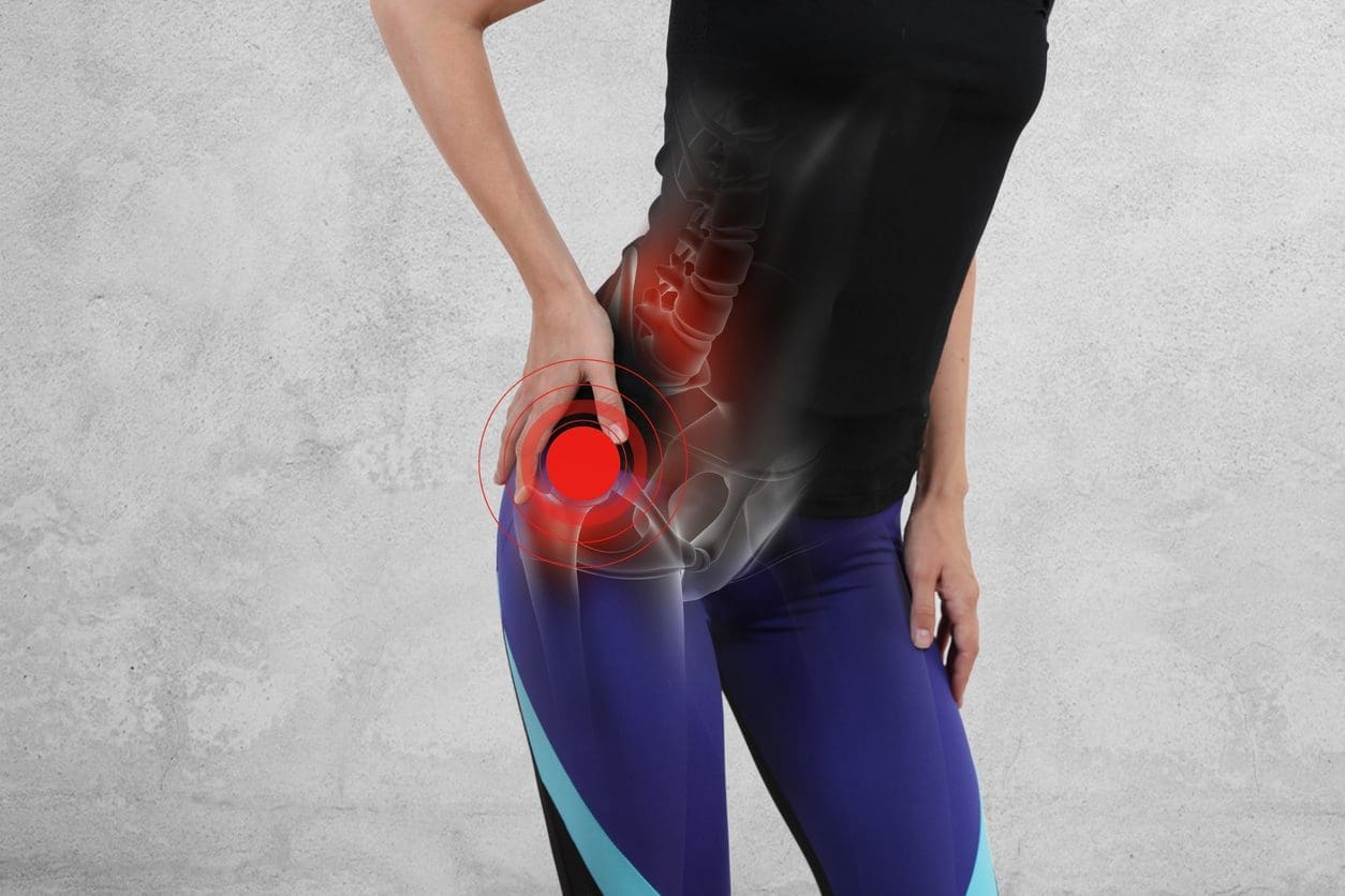
Clinical Context: A Dancer with Hip Impingement and Hypermobility
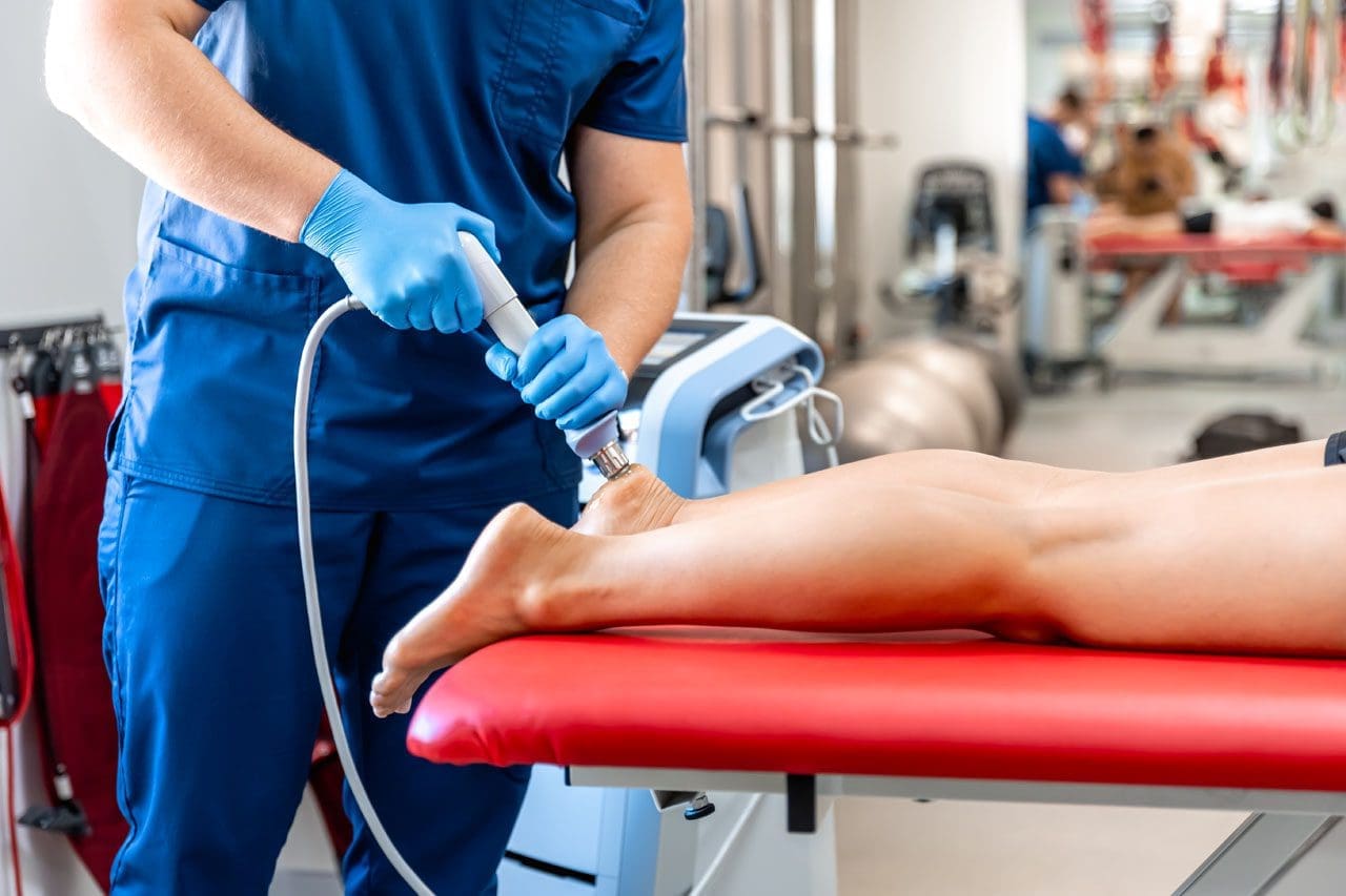
The case involves a young dancer with hip impingement, clicking, and pain at end range. She has a history of hypermobility—meaning her passive tissue elasticity and joint laxity are high, but her dynamic control may be insufficient under load or at extreme positions. Ultrasound imaging shows the femoral head centrally, the acetabulum superior-lateral, and the triangular acetabular labrum hugging the joint margin. We have identified irritation and instability without a large labral tear.
Why this matters: Dancers often drive the hip into extremes of flexion, abduction, and external rotation. In FAI, bony morphology (cam or pincer) plus capsulolabral stress can irritate the labrum and capsule. In hypermobile athletes, the capsule may be lax, and repetitive end-range positions can produce shearing and clicking. The labrum acts as a suction seal and stabilizer; when irritated, it can become symptomatic even without a discrete tear.
Key Pathophysiology: Stability, Labrum, and the Capsule
The acetabular labrum increases the depth of the socket and contributes to joint pressurization—maintaining a negative intra-articular pressure for a “seal” that stabilizes the hip during rotational movements (Nepple et al., 2015).
The capsule (with ligaments like the iliofemoral ligament) provides passive restraint, especially in extension and external rotation. Hyperlaxity or micro-failure of capsular fibers can allow excessive translation, increasing labral stress (Domb et al., 2013).
The deep hip rotators (quadratus femoris, gemelli, obturator internus/externus) and gluteus medius/minimus provide dynamic stability, controlling femoral head position during motion. Weakness or delayed activation can lead to excessive femoral internal rotation and adduction, increasing anterosuperior labral load (Lewis & Sahrmann, 2006).
In FAI, altered bony contours cause abnormal contact between the femoral head-neck junction and the acetabular rim, particularly in flexion with internal rotation. Dancers with hypermobility may paradoxically experience impingement because lax passive structures permit unsafe end-range positioning.
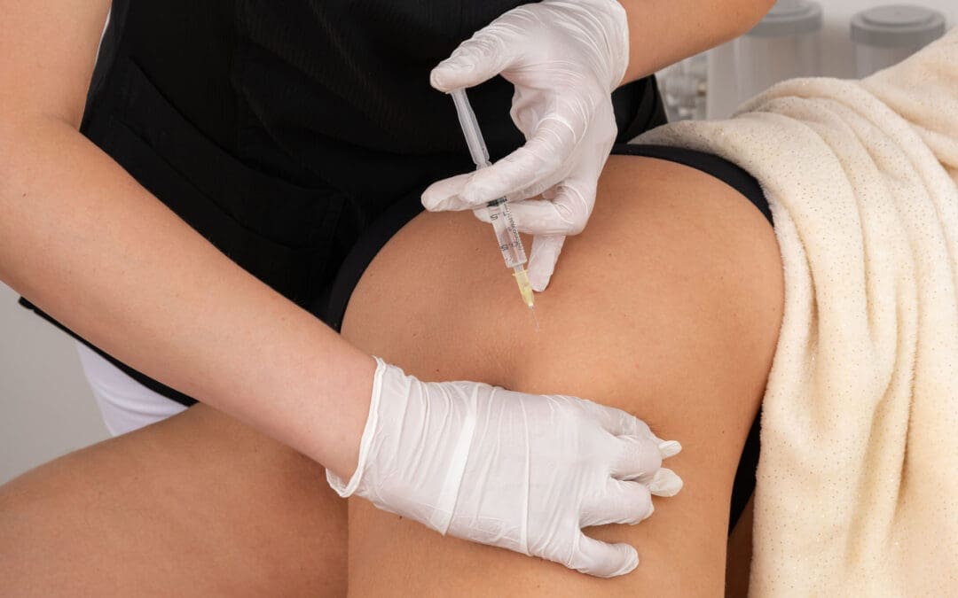
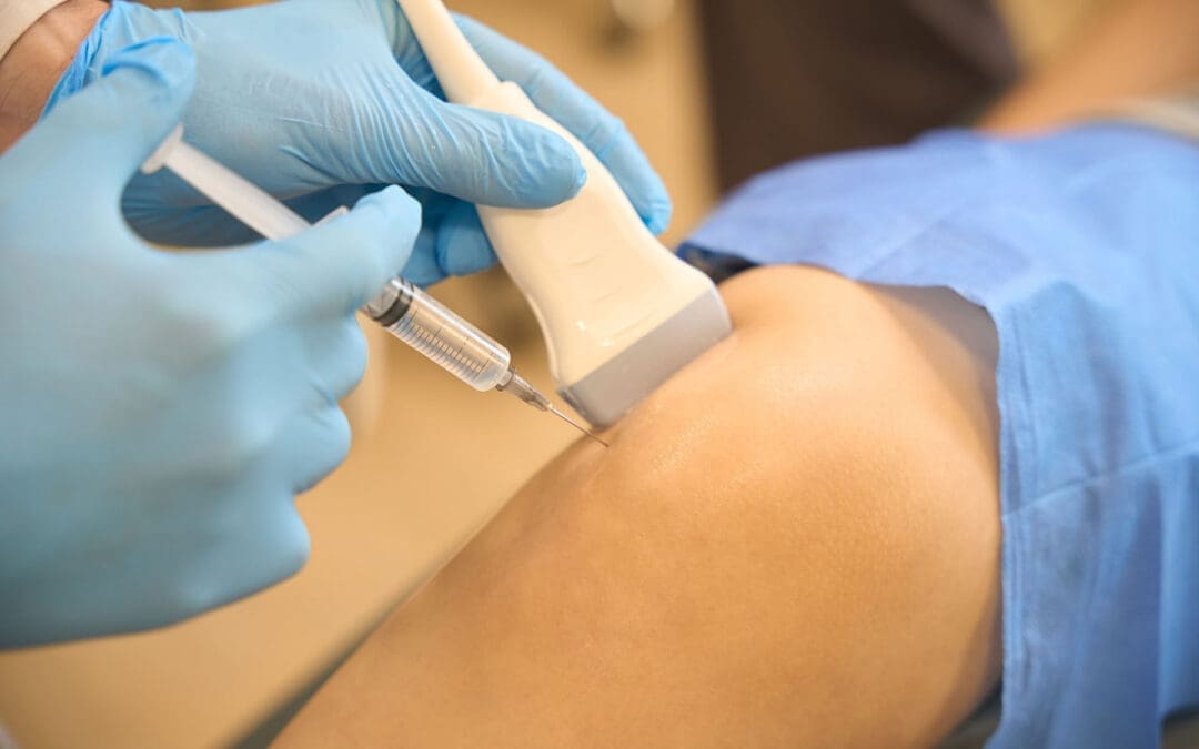
Ultrasound-Guided PRP: Rationale, Technique, and Safety
For this dancer, we delivered a high-concentration PRP solution into the intra-articular space under ultrasound guidance. We used approximately 4 cc of concentrated PRP plus 2 cc of plasma protein concentrate to limit volume while maintaining bioactive content. Hips tolerate less injection volume than knees due to smaller capsular capacity and pressure sensitivity.
Why PRP in this setting:
Biologic modulation: PRP contains growth factors (e.g., PDGF, TGF-β, VEGF) that may promote healing responses, reduce synovial inflammation, and support matrix homeostasis in the labrum and capsule (Mautner et al., 2015; Fitzpatrick et al., 2017).
Symptom relief and function: Evidence suggests PRP can reduce pain and improve function in certain chronic tendinopathies and intra-articular conditions; in hips, results are mixed but promising in selected patients, especially when combined with a structured rehab plan (Smith, 2016).
Stability support: For irritative labral conditions without large tears, PRP may help calm the joint environment, enabling focused rehabilitation on motor control without persistent synovial irritation.
Technique principles emphasized in the procedure:
Use ultrasound to identify the femoral head, acetabulum, and labrum while avoiding neurovascular structures, such as the femoral artery, medially.
Maintain visualization of the needle at all times to confirm intra-articular positioning. If injection becomes painful and resistant, reassess to ensure you are not in soft tissue.
Employ an appropriate needle gauge (e.g., 23-gauge with PRP admixture; 21-gauge for more viscous concentrates) and thoroughly purge air to avoid echogenic artifacts and ensure smooth delivery.
Limit volume to protect capsular compliance and avoid pressure pain; hips typically do not tolerate large volumes well.
Importantly, PRP is an adjunct—not a stand-alone fix. The outcomes depend heavily on the quality of post-injection rehabilitation focused on stability and movement control.
Integrative Chiropractic Care: Building the Foundation for Hip Stability
At El Paso Back Clinic, our integrative approach blends chiropractic precision with physical therapy and sports rehabilitation. The goals are to:
Restore optimal joint centration and reduce aberrant motion.
Enhance neuromuscular control of the pelvis and hip through targeted activation.
Address regional interdependence—how spine, pelvis, foot, and thorax mechanics influence the hip.
Clinical observations from my practice:
Dancers with hypermobility often present with rib cage flare, anterior pelvic tilt, and lumbar extension bias. This pattern increases anterior hip joint load and narrows the clearance for hip flexion, exacerbating impingement.
Correcting breathing mechanics and pelvic positioning reduces hip flexor tone, improves diaphragmatic control, and normalizes intra-abdominal pressure, which stabilizes the lumbopelvic complex.
Manual Therapy: When, Why, and How
Manual therapy in hypermobile hips requires finesse: the aim is not to “loosen” lax joints but to normalize soft-tissue tone, improve joint mechanics, and facilitate motor learning.
Soft-tissue release for overactive muscles (iliopsoas, TFL, adductors): Reduces anterior shear and internal rotation bias, allowing the deep rotators to engage effectively. We use instrument-assisted techniques and targeted myofascial release to reduce nociceptive drive and guarding (Littlewood et al., 2013).
Joint mobilization: Low-amplitude, directional-specific mobilizations to improve posterior glide during flexion and enhance congruency without overstressing the capsule. In hypermobility, we avoid high-velocity thrusts directed at already lax segments and prioritize stabilization-oriented mobilizations (Kaltenborn, 2003).
Pelvic and lumbar adjustments: When segmental restrictions in the SI joint or lumbar spine increase compensatory hip motion, gentle, well-placed adjustments can restore symmetry. We carefully monitor for hypermobility and follow adjustments with stability drills to lock in motor control.
Why this matters physiologically:
Reducing myofascial tone can decrease abnormal compressive loads and nociceptive input, thereby improving the motor recruitment of stabilizers.
Improving arthrokinematics supports the labral seal by encouraging even femoral head loading rather than asymmetric rim stress.
Neuromuscular Control: Teaching the Hip to Stabilize
Rehabilitation for dancers hinges on motor control, not just strength. Our plan typically includes:
Deep rotator activation: Quadratus femoris and obturators provide transverse plane control, limiting excessive femoral internal rotation during flexion. Drills: prone hip external rotation isometrics, sidelying ER pulses with minimal ROM, and short-lever resisted ER in neutral. Rationale: These muscles act as local stabilizers, centering the femoral head and decreasing labral shear (Lewis & Sahrmann, 2006).
Gluteus medius/minimus re-education: These muscles resist pelvic drop and control frontal plane motion. Drills: lateral band walks with a neutral pelvis, isometric wall abductions emphasizing trunk stacking. Rationale: Better pelvis-on-femur control reduces end-range compensation and impingement mechanics (Semciw et al., 2013).
Adductor co-contraction: Balanced adductor activation with gluteals improves pelvic stability in turnout positions common in dance. Rationale: Adductors contribute to hip joint compression and stability when coordinated properly; imbalance leads to anterior shear.
Core sequencing and breathing: Diaphragm-first breathing with lateral rib expansion, followed by gentle pelvic floor and deep abdominal engagement. Rationale: Appropriate intra-abdominal pressure and rib-pelvis alignment stabilize the lumbopelvic complex, reducing hip overuse.
Programming details:
Early-phase isometrics minimize joint shear while enhancing proprioception.
Progress to short-range controlled articular rotations (CARs) in pain-free arcs to improve capsulolabral nutrition and synovial flow without end-range irritation.
Integrate perturbation training (elastic band pulls, multi-planar micro-perturbations) to build reflexive co-contraction.
Load Management: Protecting the Labrum While Building Resilience
We work closely with dancers and coaches to calibrate training loads:
Volume and intensity caps post-PRP: Initially reduce deep flexion and turnout volume; avoid prolonged end-range splits and extreme external rotation while the joint environment normalizes.
Temporal spacing of rehearsals: Micro-dosing technique works across the week rather than clustering high-intensity sessions. Rationale: Cartilage and labral tissue require time to recover; high-frequency end-range exposure elevates synovial irritation.
Landing mechanics: Soft landings with a neutral pelvis and stacked rib cage; reduce knee valgus and excessive hip internal rotation during jumps. Rationale: Limits combined shear-compression forces on the anterosuperior labrum.
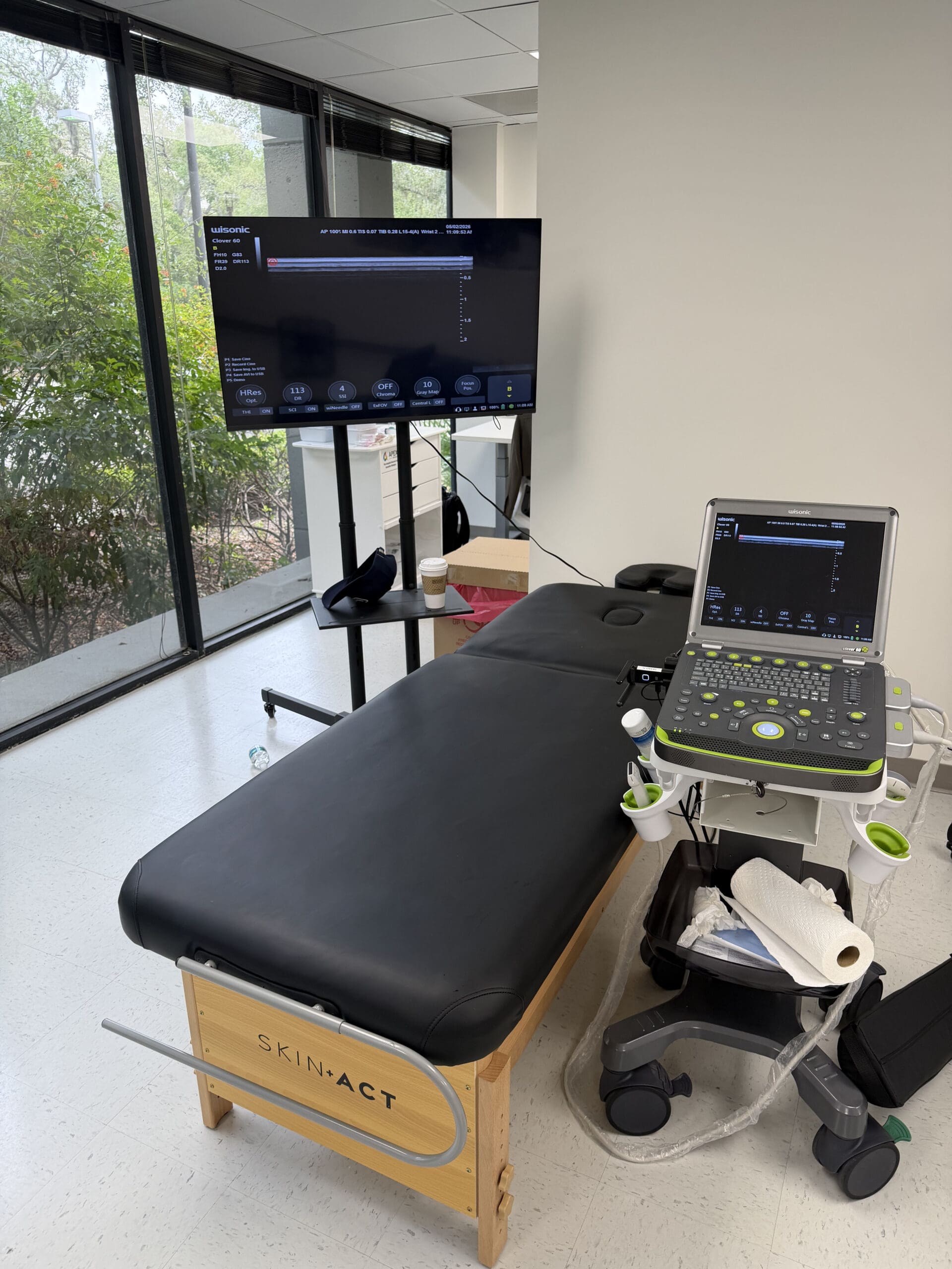
Ultrasound Guidance: Visualizing Safety and Accuracy
In the procedure, we identified the femoral artery medially to avoid vascular puncture, then positioned the ultrasound to obtain a crisp, perpendicular view of the femoral head and joint space. As the needle advanced, we maintained visualization to confirm intra-articular placement. If injection caused disproportionate pain and resistance, we reassessed needle location to avoid extra-articular soft-tissue expansion.
Why ultrasound:
Real-time visualization improves accuracy of intra-articular delivery and reduces complications.
Dynamic scanning lets us confirm landmarks and adjust needle angle to achieve the safest trajectory.
For the hips, where depth and proximity to adjacent neurovascular structures increase risk, ultrasound offers a high-safety profile.
Rehabilitation Timeline: From PRP to Performance
While exact timelines vary, our structured approach commonly follows these phases:
Phase 1: Acute modulation (Weeks 0–2)
Goals: Calm irritation, protect the labrum, initiate motor control.
Actions: Relative rest from extremes; isometric deep rotator and gluteal activation; diaphragmatic breathing; gentle posterior chain mobility; low-load blood flow restriction (BFR) as appropriate to maintain conditioning while minimizing joint stress (Hughes et al., 2017).
Rationale: Minimize synovial irritation post-PRP; build a foundation for stability.
Phase 2: Controlled mobility and strength (Weeks 2–6)
Goals: Restore controlled ROM, increase strength without compromising stability.
Actions: Short-range CARs, band-resisted ER/abduction, controlled hinge patterns, foot tripod training to improve lower-chain mechanics.
Rationale: Gradual load promotes collagen remodeling and neuromuscular integration.
Phase 3: Dynamic control and return-to-technique (Weeks 6–12)
Goals: Build tolerance to dance-specific positions.
Actions: Turnout drills with strict pelvic control, landing pattern coaching, tempo progressions for leaps, proprioceptive perturbations.
Rationale: Bridge clinic gains to stage performance, ensuring capacity before exposure to extremes.
Rationale: Maintain the labral seal and capsular integrity under real-world demands.
Integrative Chiropractic and Physical Therapy Synergy
Our emphasis at El Paso Back Clinic is the synergy of manual care and movement retraining:
Chiropractic care targets alignment and segmental mobility that influence hip mechanics—especially in the lumbopelvic region. We emphasize precision adjustments when necessary, followed by stabilization drills to retain improved mechanics.
Physical therapy builds durable control and strength in the hip girdle through progressive overload, task-specific cues, and feedback-rich training environments.
Education ensures that athletes understand how habits such as deep lumbar extension and anterior pelvic tilt can compromise hip space. We coach sustainable alignment strategies for practice and performance.
Clinical Pearls from My Practice
In hypermobile dancers, prioritize strength and control over flexibility. A more passive range is rarely the answer; better control of the existing range is.
Pain during injection that is sharp and pressure-resistant often indicates extra-articular placement or capsular over-distension; reassess under ultrasound to confirm needle position.
Persistent clicking without a discrete tear may indicate a labral suction seal disruption. Focus on deep rotator activation and pelvic control to restore functional sealing.
Measuring progress: Use outcomes such as the Hip Outcome Score (HOS), return-to-technique benchmarks, and movement-quality metrics during controlled tasks.
When Surgery Is Considered—and Often Avoided
While hip arthroscopy for labral tears and FAI morphology can be beneficial in select cases, many dancers without large tears respond well to conservative care. If structural impingement is severe, surgical consultation may be warranted; however, careful rehab, load management, and biologic adjuncts like PRP can often provide significant relief and allow continued performance (Griffin et al., 2016).
Keeping Hormones and Medications in the Background
We maintain a primarily chiropractic and rehabilitation-centered approach. Hormonal factors, systemic inflammation, and medication considerations are reviewed as part of whole-person care, but they remain secondary to hands-on, movement-based strategies that directly influence hip stability and mechanics for dancers.
Putting It All Together: A Practical Plan for Dancers
Assess thoroughly with imaging and functional testing to differentiate between irritation and tear and to identify instability patterns.
Use ultrasound-guided PRP judiciously to modulate symptoms and support tissue healing in selected cases.
Apply manual therapy to normalize tone and mechanics—avoid overstretching lax joints.
Drive neuromuscular control of deep rotators, gluteals, and core with progressive, feedback-rich drills.
Implement load management and technique coaching to prevent end-range overuse.
Track objective outcomes and adjust the plan in response to functional and performance demands.
Conclusion: Durable Stability for High-Performance Hips
For dancers, the pathway back to pain-free, confident movement runs through stability, control, and smart loading. Biologic adjuncts like PRP, delivered safely under ultrasound guidance, can help create the conditions for successful rehabilitation. The heart of the solution, however, lies in integrative chiropractic care and physical therapy—precise manual techniques paired with targeted neuromuscular retraining, all tuned to the demands of dance. With this approach, many dancers move beyond pain and clicking to sustained performance, preserving the labral seal and protecting the capsule over the long term.
PRP Therapy for Sports Injuries: How It May Speed Healing Without Surgery
Sports injuries can slow life down fast. A sore tendon, a strained ligament, or a muscle tear can make it difficult to train, work, sleep, or even walk comfortably. That is one reason Platelet-Rich Plasma, or PRP, has gained attention in sports medicine. PRP is made from a patient’s own blood and then injected into an injured area to support healing. Medical centers such as Yale Medicine, Penn Medicine, Johns Hopkins Medicine, and Temple Health describe PRP as a biologic or regenerative treatment that may help repair tissue, lower pain, and improve function in certain musculoskeletal injuries. It is often used for tendon, ligament, muscle, cartilage, and joint problems, including some cases of osteoarthritis. (Johns Hopkins Medicine, n.d.; Penn Medicine, 2025; Yale Medicine, n.d.).
PRP is appealing because it is non-surgical and uses the body’s own healing tools. Still, it is not a miracle fix for every athlete or every injury. Research shows promising results in many cases, but outcomes can vary depending on the tissue involved, how long the injury has been present, how the PRP is prepared, and whether the person also follows a successful rehab plan. In other words, PRP works best as part of a comprehensive care strategy rather than a stand-alone shot. (Saini et al., 2021; Jimenez, n.d.).
What PRP Therapy Is
PRP stands for Platelet-Rich Plasma. Plasma is the liquid part of blood, and platelets are blood components best known for their role in clotting. However, platelets also carry growth factors and signaling molecules that help tissue repair. To make PRP, a clinician draws a small amount of blood, spins it in a centrifuge, and separates out a platelet-rich portion. That concentrated solution is then placed into the injured area. The goal is to increase healing signals directly at the site of tissue damage. (Johns Hopkins Medicine, n.d.; Yale Medicine, n.d.; HSS, n.d.; Penn Medicine, 2025).
A simple way to think about PRP is this: it does not just try to numb pain. It tries to support the body’s repair response. Hospital for Special Surgery describes PRP as a form of regenerative medicine that amplifies natural growth factors in blood cells to help damaged tissue heal. Johns Hopkins Medicine similarly explains that the concentrated growth factors in PRP may stimulate tissue regeneration and speed healing in the treated area. (HSS, n.d.; Johns Hopkins Medicine, n.d.).
What the procedure usually includes
A small blood draw from the patient
Processing the sample in a centrifuge
Preparing the platelet-rich portion
Injecting the PRP into the injured tissue
In some cases, using ultrasound to guide the injection
A visit that often takes less than an hour
This basic process is described by major medical centers, including Penn Medicine, Yale Medicine, and Johns Hopkins Medicine. (Johns Hopkins Medicine, n.d.; Penn Medicine, 2025; Yale Medicine, n.d.).
How PRP May Help Sports Injuries Heal
When tissue is injured, the body sends platelets to the area early in the healing process. Temple Health explains that platelets contain growth factors that help promote cell growth, repair tissue, and reduce inflammation. Yale Medicine notes that PRP contains concentrated platelets, cytokines, and growth factors with anti-inflammatory properties. This is why PRP is often used for injuries that have been slow to heal on their own. (Temple Health, 2021; Yale Medicine, n.d.).
PRP may be especially useful in tissues that do not receive a strong blood supply. The 2021 review in the Indian Journal of Orthopaedics notes that tendons heal more slowly than many other tissues because of their poor vascularity. That same review also explains that PRP has been studied in tendon disorders such as Achilles tendinopathy, rotator cuff tendinitis, and epicondylitis, as well as in muscle strains and osteoarthritis. (Saini et al., 2021).
For athletes, this matters because many sports injuries are overuse or repetitive-stress injuries. If a tendon stays irritated for months, or a ligament strain never fully calms down, the body may need extra support to restart a healthier repair process. Some research suggests earlier PRP use in select injuries may help guide inflammation toward recovery and restore tissue balance. Even so, researchers also note there is no universal PRP formula or perfect protocol yet, so treatment must be individualized. (Saini et al., 2021).
Common Sports Injuries PRP Is Used For
Medical centers and sports medicine sources commonly describe PRP for the following problems:
Chronic tendinitis or tendinopathy
Tennis elbow
Patellar tendinopathy or “jumper’s knee”
Achilles tendon problems
Ligament strains
Muscle strains and some muscle tears
Cartilage irritation
Osteoarthritis in active adults
These uses are repeatedly listed by Penn Medicine, Yale Medicine, Temple Health, and HSS. (Penn Medicine, 2025; Temple Health, 2021; Yale Medicine, n.d.; HSS, n.d.).
Temple Health highlights tennis elbow and jumper’s knee as common orthopedic conditions that may benefit from PRP. In its overview, Penn Medicine also lists structures such as the Achilles tendon, ACL, hamstring, patellar tendon, and cartilage as areas in sports medicine where PRP is used. Yale Medicine adds tendon, ligament, and muscle conditions, as well as degenerative joint conditions, to that list. (Penn Medicine, 2025; Temple Health, 2021; Yale Medicine, n.d.).
There is also supportive evidence for muscle injury care when injections are placed carefully. A 2014 study in Blood Transfusion reported that athletes with grade II muscle lesions who received ultrasound-guided PRP showed full healing on ultrasound, pain resolution, and return to sport, with only one relapse reported a year later. That does not prove PRP is right for every muscle injury, but it does show why sports clinicians remain interested in it. (Borrione et al., 2014).
What Recovery Feels Like After PRP
One important point for patients is that PRP can cause short-term soreness. Yale Medicine says the most common side effects are discomfort, pain, and stiffness at the injection site. Penn Medicine also notes that mild soreness, swelling, or stiffness is common for the first few days. Johns Hopkins Medicine adds that some people notice soreness and bruising after the procedure. In most cases, these effects are temporary. (Johns Hopkins Medicine, n.d.; Penn Medicine, 2025; Yale Medicine, n.d.).
Patients also need realistic expectations. PRP is not usually an instant pain reliever. Penn Medicine says improvement may take a few weeks to become noticeable, with fuller benefits developing over months. Yale Medicine reports that some people notice pain improvement in four to six weeks, with continued progress for up to a year. (Penn Medicine, 2025; Yale Medicine, n.d.).
Aftercare often includes
Resting the area for a short time
Avoiding hard exercise right away
Using a guided rehab plan
Following instructions about pain control
Avoiding some anti-inflammatory medicines when advised
Penn Medicine and HSS both note that anti-inflammatory medicines may interfere with the early healing response that PRP is meant to support, so patients should follow their treating clinician’s advice. (HSS, n.d.; Penn Medicine, 2025).
Why Ultrasound-Guided PRP Matters
Not every injection needs the same level of precision, but many sports injuries benefit from careful image guidance. Both Johns Hopkins Medicine and Yale Medicine acknowledge the use of ultrasound during PRP procedures. Research in athletes also supports this approach. The 2014 study on muscle injuries emphasized that ultrasound was important for both locating the lesion and guiding the needle accurately into it. The 2021 sports injury review similarly reported that ultrasound-guided injections improve accuracy, particularly for musculoskeletal conditions. (Johns Hopkins Medicine, n.d.; Yale Medicine, n.d.; Borrione et al., 2014; Saini et al., 2021).
On Dr. Alexander Jimenez’s public clinical website, one recent educational article describes ultrasound-guided intra-articular hip PRP as a precision-focused procedure in which ultrasound helps the clinician visualize anatomy, confirm correct placement, and improve safety. That same article stresses that biologic injections work best when they are combined with rehabilitation and movement-based recovery rather than used alone. (Jimenez, n.d.).
Dr. Alexander Jimenez’s Clinical Observations and the Value of Integrated Care
Dr. Alexander Jimenez, DC, APRN, FNP-BC, describes his El Paso practice as a multidisciplinary and integrative model that combines chiropractic care, functional medicine thinking, sports medicine principles, rehabilitation, and regenerative strategies. His website presents regenerative medicine as a natural, non-surgical option designed not only to reduce pain but also to improve structure, movement, and function. (Jimenez, n.d.).
That point matters in sports injury care. A tendon or muscle may not stay healthy if the athlete still has poor joint mechanics, weak stabilizers, incorrect loading patterns, or nutrition and recovery habits that slow healing. Dr. Jimenez’s site repeatedly frames recovery as a full process that includes a detailed history, physical evaluation, attention to biomechanics, regenerative options when appropriate, chiropractic care to improve motion, rehab planning, and follow-up focused on function. (Jimenez, n.d.).
In a comprehensive clinic model, that means PRP can be paired with structural care, progressive rehabilitation, and functional medicine support. The injection may help the tissue biologically, while rehab helps the athlete move better and reduce repeated stress on the injured area. This combined approach aligns with the broader message from both sports medicine research and Dr. Jimenez’s clinical content: better recovery usually comes from treating the tissue and the movement pattern together. (Borrione et al., 2014; Jimenez, n.d.; Saini et al., 2021).
Benefits and Limits of PRP
Possible benefits
Uses the patient’s own blood
Minimally invasive
May reduce pain and improve function
May help some chronic tendon, ligament, muscle, and joint problems
Can be part of a non-surgical recovery plan
Can be combined with rehab and other supportive care
These benefits are commonly described by Yale Medicine, Penn Medicine, Johns Hopkins Medicine, and HSS. (HSS, n.d.; Johns Hopkins Medicine, n.d.; Penn Medicine, 2025; Yale Medicine, n.d.).
Important limits
Results vary from person to person
Some injuries still need surgery or other procedures
Relief may take weeks or months, not days
PRP preparation methods are not fully standardized
Some tissues have stronger evidence than others
Those limits are important because proper medicine depends on the right treatment for the right injury at the right time. PRP may be a strong option, but it should be chosen carefully after a full exam and diagnosis. (Saini et al., 2021; Penn Medicine, 2025).
Final Thoughts
PRP therapy offers a promising non-surgical option for sports injuries because it delivers a concentrated dose of the patient’s own platelets to damaged tissue, where growth factors may support repair, reduce inflammation, and improve recovery. It is commonly used for chronic tendinopathy, ligament strain, muscle injury, and some joint conditions. Short-term soreness at the injection site can happen, but serious side effects are uncommon. The best results usually come when PRP is matched to the right injury and combined with smart rehabilitation, movement correction, and careful follow-up. (Johns Hopkins Medicine, n.d.; Penn Medicine, 2025; Yale Medicine, n.d.; Jimenez, n.d.).
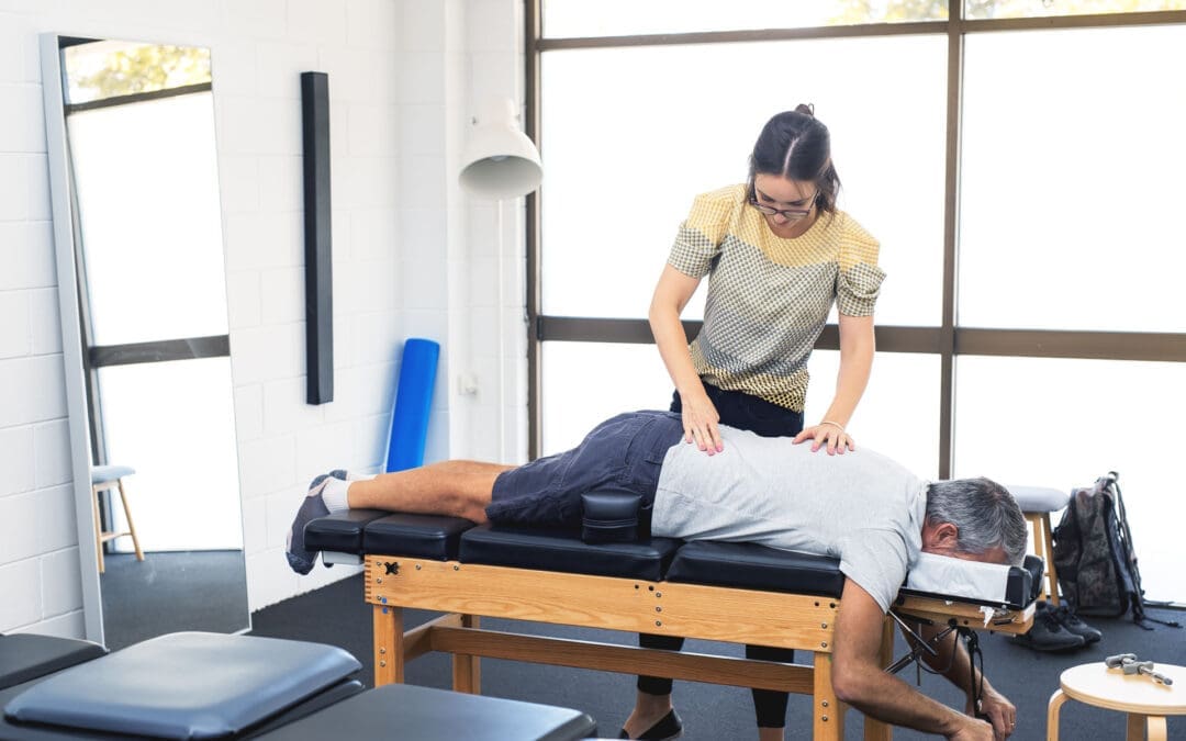
Restore Flexibility and Mobility with Integrative Chiropractic Care and Shockwave Therapy at El Paso Back Clinic
Many El Paso residents wake up with stiff joints or tight muscles, making simple daily tasks feel hard. Reaching overhead, bending down, or walking for long stretches can become painful or limited. At El Paso Back Clinic, integrative chiropractic care combined with Extracorporeal Shockwave Therapy (ESWT) offers a natural solution. This approach restores proper joint alignment, reduces muscle tension, and resolves soft-tissue restrictions, allowing patients to move freely again. Led by Dr. Alexander Jimenez, DC, APRN, FNP-BC, the clinic’s team uses gentle adjustments, stretching, exercises, and advanced shockwave treatments to help people regain flexibility and enjoy life in El Paso.
What Integrative Chiropractic Care Does for Flexibility at El Paso Back Clinic
Integrative chiropractic care at El Paso Back Clinic treats the whole body instead of just one problem area. It corrects small misalignments, called subluxations, in the spine and joints. These misalignments put pressure on nerves and tighten muscles. Regular adjustments gently move everything back into place. This restores proper joint alignment, eases tension, and lets the nervous system send clearer signals to the muscles.
When joints line up correctly, range of motion improves right away. Stiffness fades, and daily movements become smoother and more efficient. Patients at the clinic often say they feel looser and more energetic after just a few visits. (Gentle Chiro, n.d.) The care also includes stretching and therapeutic exercises to maintain gains over time. Muscles and joints start working together as a team, building resilience that lasts.
How Chiropractic Adjustments Restore Joint Alignment and Reduce Stiffness
Adjustments form the core of care at El Paso Back Clinic. The team uses precise, gentle pressure to correct subluxations. This simple step brings clear benefits that patients notice quickly:
Better range of motion, so joints glide freely without catching
Less muscle tension around the back, neck, and limbs
Improved nervous system function for better balance and coordination
Smoother daily activities like turning your head while driving or reaching for groceries
Lower risk of future stiffness because proper alignment trains the body to stay balanced
Many people in El Paso report that these changes make physical activities feel easier and less tiring. (Rodgers Stein Chiropractic, n.d.) The adjustments help the body move more efficiently without pain, supporting an active lifestyle.
Adding Stretching and Therapeutic Exercises for Long-Term Results
Adjustments open the door to better movement, but stretching and exercises keep it open. At El Paso Back Clinic, the rehabilitation team creates simple home programs that match each patient’s needs. Dynamic stretches warm up the body before activity. Static stretches hold the new mobility after adjustments. Therapeutic exercises strengthen the muscles that support the joints.
These steps build endurance and agility. Patients find they can stay active longer without soreness. The clinic’s sports medicine approach helps people return to hiking in the Franklin Mountains, playing with family, or working without the same old limitations. (Chiropractic Fitness, n.d.) Consistent practice turns short-term gains into lasting flexibility.
Introducing Extracorporeal Shockwave Therapy (ESWT) at El Paso Back Clinic
ESWT uses focused sound waves to reach deep into muscles, tendons, and ligaments. The waves create tiny pulses that restart healing in areas stuck with scar tissue or chronic tightness. This noninvasive treatment increases blood flow, breaks down old buildup, and reduces inflammation. At El Paso Back Clinic, ESWT is available as a key component of advanced care plans for patients who need additional support for soft tissue problems.
Why Combining Chiropractic Care and ESWT Delivers Stronger Flexibility Gains
The real power at El Paso Back Clinic comes from pairing chiropractic adjustments with ESWT. Adjustments fix the mechanical side—joint position and nerve signals—while ESWT handles the soft-tissue side—scar tissue, poor circulation, and stubborn tension. Together, they create faster, longer-lasting results than either method alone.
This dual approach works in several key ways:
Chiropractic restores spinal and joint mobility
ESWT breaks down scar tissue and releases tight fascia
The pair reduces inflammation and collagen cross-linking that causes stiffness
Blood flow improves, helping muscles and tendons heal
Patients regain a greater range of motion because both structure and tissue health get better at once
Clinic reports show that this combination can significantly improve outcomes compared with standard care. Many El Paso patients with ongoing tightness notice a real return of freedom of movement.
Common Conditions That Benefit from This Integrated Approach
El Paso Back Clinic uses this combined approach to treat several conditions that rob people of flexibility. Here are some of the most common:
Frozen shoulder – Adjustments free stuck joints while ESWT dissolves scar tissue and calcium deposits. Patients often regain full arm motion without pain.
Achilles tendinopathy – Chiropractic realigns the lower body to ease strain. Shockwave therapy stimulates the growth of new blood vessels and clears chronic buildup, so walking and running feel normal again.
General chronic muscle tension – Tightness in the back, neck, or legs from stress, work, or old injuries—responds well. The therapies release trigger points and restore smooth movement.
Post-injury stiffness from car accidents or sports – The clinic specializes in personal injury care. The combination speeds recovery and safely rebuilds mobility.
Other issues, such as plantar fasciitis and tennis elbow, also improve because the care addresses both alignment and tissue damage. (Bend Total Body Chiropractic, n.d.)
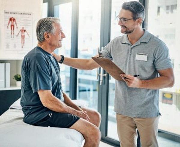
Clinical Insights from Dr. Alexander Jimenez at El Paso Back Clinic
Dr. Alexander Jimenez, DC, APRN, FNP-BC, leads El Paso Back Clinic with more than 30 years of experience. As both a Doctor of Chiropractic and a board-certified Family Nurse Practitioner, he brings a unique integrative perspective to every patient. In his clinical work in El Paso, Dr. Jimenez sees how chiropractic adjustments correct subluxations and improve nervous system function, thereby boosting flexibility and range of motion. When combined with ESWT, the results are even stronger for soft tissue injuries from accidents or overuse.
Dr. Jimenez often notes that this teamwork helps patients break down scar tissue, reduce inflammation, and restore proper movement patterns faster than traditional methods alone. His approach includes personalized functional medicine, nutritional support, and rehabilitation exercises to help patients build lasting resilience. At the clinic’s convenient El Paso locations, patients receive complete care that addresses the root causes of stiffness and helps them return to daily life and favorite activities with confidence.
Tips to Get the Most from Care at El Paso Back Clinic
Start with a full evaluation so the team can build a plan that fits your body and lifestyle. Attend regular adjustments and ESWT sessions as recommended. Follow the simple stretching and exercise routine at home every day. Support your progress with good posture, daily walks, proper hydration, and enough rest. The friendly staff at El Paso Back Clinic makes the process easy and supportive. Many patients see big improvements in flexibility within just a few weeks when they stay consistent.
A Natural Path to a More Flexible, Resilient Life in El Paso
Integrative chiropractic care and ESWT at El Paso Back Clinic offer a powerful, drug-free way to fight stiffness and reclaim natural movement. By correcting joint alignment, releasing muscle tension, and healing soft tissues, this approach makes daily life and physical activity feel effortless again. Muscles and joints work in harmony, the nervous system functions smoothly, and the body stays strong through the years.
Whether you deal with occasional tightness or a specific injury, the experienced team at El Paso Back Clinic can help. Contact the clinic today to schedule an evaluation and discover how these natural tools can work for you. With the right plan, better flexibility and mobility are well within reach for El Paso residents.
That “Reset Pain” After You Sit or Hold a Weird Position: What It Is and How El Paso Back Clinic Approaches It
Have you ever held your body in an awkward position—like slouching on a couch, twisting in a chair, leaning on one hip, or sleeping with your neck turned—then you stand up and feel a sharp ache, tightness, or a “catch”? Sometimes it feels like a joint or muscle has to “reset” before you feel normal again. You might even feel clumsy for a minute, then things settle down.
At El Paso Back Clinic, this pattern is commonly discussed as a mix of postural strain, muscle guarding, myofascial tightness (trigger points), and sometimes joint restriction—especially when movement has been limited for too long or posture has been stressing the same tissues over and over.
This article explains what that “reset” feeling usually means, why it happens, and how integrative chiropractic care—like the approach described at El Paso Back Clinic—can help restore smoother motion and reduce the chances of it happening again.
What Do You Call This “Reset” Feeling?
There isn’t one single official name that covers every case, because different tissues can create the same sensation. But the most common clinical labels include:
Postural strain (tissues overloaded by a sustained position)
Muscle stiffness (tightness and reduced ease of motion)
Muscle guarding (protective tension driven by the nervous system)
Myofascial trigger points (irritable “knots” in muscle/fascia)
Joint restriction / joint dysfunction (a joint that temporarily doesn’t glide well)
Many people casually call it a “stuck joint” or “something out of place.” In reality, it’s often less dramatic than it feels—more like a temporary movement problem plus a protective muscle response.
Why It Often Hurts When You Return to Neutral (Not While You’re Sitting)
This surprises many people: “If the posture was the problem, why didn’t it hurt until I moved?”
Because your body adapts to the position you hold. While you’re still:
Your muscles settle into a holding pattern
Your joints move less
Your fascia (connective tissue) can get less “slippery” with inactivity or repeated stress
Your nervous system may “turn down” certain signals until movement starts again
Then you stand, rotate, or straighten up—and your tissues have to slide, load, and coordinate again. That’s when you feel the catch, the sting, or the awkward “reset” moment.
What’s Actually Happening: 5 Common Mechanisms Behind the “Reset”
Most cases are a combo, not just one thing.
Postural Strain: You Overloaded a Region
When you hold a position that isn’t friendly to your body—like forward head posture, slumped sitting, or a rotated spine—you can stress:
muscles
ligaments
joint capsules
fascia
Over time, those tissues complain when you ask them to move again. El Paso Back Clinic describes how repetitive positions and mechanical issues can contribute to stiffness and restriction patterns.
Muscle Guarding: Your System “Braces” for Safety
Muscle guarding is your nervous system’s way of saying, “I’m not sure this movement is safe, so I’m going to tighten things up.” It can feel like:
locked
braced
hard to relax
stiff even when you try to stretch
El Paso Back Clinic notes that pain patterns can keep muscles guarded and that stiffness may involve more than “tight muscles.”
Trigger Points: The “Knot” That Bites When You Move
Trigger points are sensitive spots in tight muscle bands. When you change position, those fibers stretch and can cause sharp, deep, or referred pain.
Fascia health is closely tied to this, because fascia surrounds muscle and helps movement feel smooth. Johns Hopkins Medicine explains that fascia can become “gummy,” stiff, and painful with limited movement, repetitive movement, or trauma.
Fascial Stiffness: The “Gummy Tissue” Effect
Fascia is like a body-wide web. When you don’t move much or repeat the same posture all day, fascia can get less elastic and less hydrated. That can make motion feel “sticky.”
Johns Hopkins Medicine specifically lists limited activity, repetitive movement, and trauma as factors that can contribute to fascia adhesions and stiffness.
Joint Cavitation: The Pop or Release
Sometimes the reset comes with a pop. A well-known imaging study found evidence that joint cracking is linked to cavity formation in the joint fluid (not bones grinding).
A pop isn’t automatically “good” or “bad.” What matters more is:
Do you move more easily afterward?
Does pain decrease?
Or does pain increase and function drop?
Why You Feel Awkward for a Bit After the “Reset”
That lingering weirdness—seconds to minutes—is often your body downshifting from protection back into normal movement.
Common reasons include:
muscles slowly letting go of guarding
irritated tissue calming down
fascia rehydrating and sliding better with movement
your brain re-mapping posture and balance (proprioception “recalibration”)
This is one reason many people feel better after a short walk post-sitting.
A Quick Self-Check: Is This Normal Stiffness or Something More?
Muscle stiffness is common and often improves with gentle movement and better posture habits. The Cleveland Clinic notes that stiffness often improves without medical treatment, but it should be taken more seriously if it comes with concerning symptoms such as fever, weakness, swelling, or persistent worsening.
Consider getting evaluated if you notice:
pain that’s getting worse over days/weeks
tingling, numbness, or weakness
pain that wakes you up repeatedly
symptoms after a significant fall or crash
the “reset pain” keeps happening in the exact same spot
What You Can Do Right Away (Safe, Simple, and Usually Helpful)
The 2–3 minute “reset without forcing it”
Stand up and walk 30–90 seconds
Do small, slow movements in a pain-free range
Try a long exhale breathing pattern (relaxes guarding)
Use gentle heat if it helps you relax
Simple posture habits that reduce repeat episodes
Change position every 30–60 minutes
Avoid “camping” in end-range posture (deep slouch, deep twist)
Use a supportive setup for workstations when possible
Build basic endurance in the muscles that hold posture (core, glutes, upper back)
How El Paso Back Clinic Approaches This Pattern (Integrative Chiropractic Style)
El Paso Back Clinic describes an integrative model that blends chiropractic care with rehab-style strategies and multidisciplinary support for spine and soft tissue problems.
Identify what’s actually driving the “reset”
Sometimes stiffness isn’t just “tight muscles.” It may involve:
joint restrictions
spine or pelvis mechanics
inflammation around a joint
pain patterns that keep muscles guarded
nerve-related problems
That’s why an exam matters—so the plan matches the cause.
Restore motion with chiropractic adjustments or mobilization
A chiropractic adjustment is a controlled force applied to a spinal joint to improve motion and movement ability.
When a joint isn’t moving well, nearby muscles often overwork and tighten. Improving joint motion can reduce the need for your body to “force” a painful reset.
Address myofascial tightness (muscle + fascia)
Because fascia can become stiff due to limited movement or repetitive strain, integrative care often includes hands-on work and guided movement to improve tissue glide.
Stabilize the area so it doesn’t keep “getting stuck”
If a joint repeatedly feels like it “locks,” the missing piece is often:
strength
endurance
timing/control
movement habits
El Paso Back Clinic frequently emphasizes rehabilitation and conditioning alongside chiropractic care to restore normal function after spine and soft-tissue issues.
A “Stop the Reset Cycle” Plan (2–3 Weeks)
These are general strategies that many patients tolerate well. Keep it gentle and pain-free.
Daily (2–5 minutes, 1–2 times/day)
1 minute easy walking
5 slow neck turns each side (easy range)
8 shoulder blade squeezes (2–3 sec hold)
8 hip hinges (small, smooth)
3 slow breaths with long exhale
During the day (30–60 seconds every hour)
stand up
10–20 steps
reset your sitting position (hips back, chest relaxed, neck tall)
3 days/week (10–15 minutes)
core stability (dead bug / modified plank)
glute strength (bridges / step-ups)
upper back endurance (band rows)
If stretching makes symptoms worse, or if stiffness keeps returning the same way, that’s a good reason to get assessed—El Paso Back Clinic even notes that persistent stiffness may signal joint restrictions or mechanics issues beyond “tight muscles.”
When to Reach Out to El Paso Back Clinic
If your “reset pain” is frequent, sharp, or starting to change your daily routine, it’s reasonable to get an evaluation—especially if you suspect joint restriction, posture-related mechanics, or muscle guarding patterns.
El Paso Back Clinic lists multiple El Paso locations and a main phone line for help and questions.
Phone: (915) 850-0900
Location (example listing): 11860 Vista Del Sol, Ste 128, El Paso, TX 79936
Key Takeaway
The experience of “I held a posture → now it hurts → then it resets” usually indicates that your body is showing a predictable pattern:
posture overloads tissues
fascia and muscle tension increase
a joint may move less smoothly
the nervous system guards
returning to neutral triggers a brief recalibration
The goal isn’t to chase pops or force releases. The goal is to restore smooth motion + stable control, so your body doesn’t keep needing that painful “reset.”
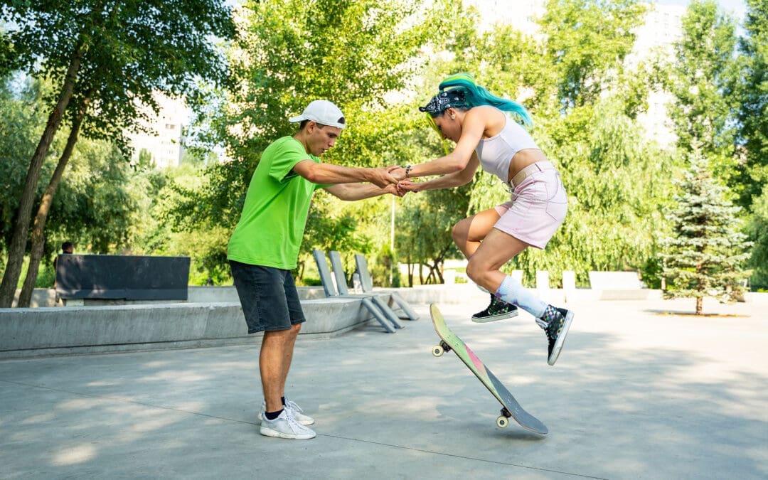
Skateboarding Training Essentials: Strength, Balance, and Injury Prevention with Chiropractic Support at El Paso Back Clinic
Skateboarding is an exciting sport that mixes skill, speed, and style. It began as a land-based surf practice but has grown into a worldwide hobby for many. To excel in skateboarding, you need targeted training that strengthens your core and legs, improves balance, and teaches safe falling to prevent harm. This training uses repetitive drills, explosive jumps, and endurance workouts to create automatic responses and lasting energy. It also includes mental prep like imagining tricks and steady practice routines.
The sport’s demands, such as repeated one-sided pushes and hard landings, can strain your body. That’s where integrative chiropractic care shines. At El Paso Back Clinic in El Paso, Texas, this approach improves joint mobility, corrects imbalances from skateboarding habits, and accelerates healing after impacts. It improves balance, body sync, and bendiness while offering diet and safety tips to reduce injury risk. Led by Dr. Alex Jimenez, DC, APRN, FNP-BC, the clinic offers tailored care for skateboarders and athletes, blending chiropractic care with rehab and nutrition to support top performance.
This article covers skate training basics and how chiropractic at El Paso Back Clinic supports it. For beginners or pros, these insights can help you advance safely. Visit https://elpasobackclinic.com/ to learn more about their services.
Core Elements of Skateboarding Training
Skateboarding success starts with body and mind prep. Training goes beyond board time—it’s about a solid base for tricks and endurance. Prioritize core and leg power, as these drive your actions (Austin Simply Fit, n.d.). Muscles like abs, lower back, quads, hamstrings, glutes, and calves handle shifts from an upright to a low position in moves like ollies.
Core Workouts: Try planks by holding a straight body pose for 30 seconds. Side versions hit obliques for twist stability.
Leg Boosters: Squats mimic board crouches—lower then rise for three sets of 10 reps.
Importance: Strong cores prevent shakes during jumps, lowering fall risks.
Balance is vital in skating. Poor balance leads to wipeouts on basic maneuvers. Newbies should pick a stance: regular (left-forward) or goofy (right-forward). Place the feet over the truck bolts for maximum stability (Skateboard GB, n.d.).
Balance Practices: Stand on one foot and draw letters with the other toe. Switch sides for ankle strength.
Next Level: Manuals lift the front wheels, balancing on the rear for ramp preparation.
Routine: Dedicate 10 minutes daily to weight shifts on your board for a natural feel.
Safe falling is key to injury avoidance. Falls are part of skating, but proper methods reduce severe damage. Roll instead of bracing with arms to protect wrists (Healthcare.utah.edu, 2024).
Fall Methods: Tuck chin and roll to distribute force. Aim for protected spots like padded knees.
Gear Essentials: Helmets, wrist, knee, and elbow pads absorb shocks.
Safe Start: Use grass or mats for low-risk practice.
Repetitive training builds muscle memory. Repeat actions until they’re instinctive, like pushing and halting (Braille Skateboarding, n.d.). This aids tricks such as frontside kickturns and backwheel pivots (How to Skate, 2018).
Drill Reps: Push 10 times, stop, and redo for fluid flow.
Trick Steps: Divide into parts, like board pop, then foot flick for kickflips.
Side Hops: Mimic skating with 30-second lateral jumps.
Gains: Higher leaps and fast reflexes elevate skills.
Cardio keeps you going strong. Skating provides some, but extras build heart health (Skateboard GB, n.d.).
Rope Skipping: 30 seconds on, rest, three rounds for calf power and breath control.
Crawls: Bear walk forward and back 10 meters.
Cardio Value: Longer sessions with quicker recovery.
Mental training tackles fear. Visualize wins before attempts (Florida Atlantic University, n.d.). Commitment means regular sessions despite setbacks.
Imagery: Eyes shut, see perfect landings.
Fear Busting: Small steps build confidence.
Drive: Love for skating fuels persistence.
Follow principles such as targeted work, gradual increases, and variety to ensure safe progress (The Daily Push, n.d.). Skate-specific drills, slight pushes, and mixes prevent plateaus.
This foundation makes skating enjoyable, but one-sided strains need expert help, like at El Paso Back Clinic.
Integrative Chiropractic Care for Skateboarders at El Paso Back Clinic
At El Paso Back Clinic, integrative chiropractic merges adjustments with therapies for whole-body health. For skaters, it enhances joint flow in hips, knees, and ankles, easing restrictions from twists (Push as RX, n.d.). The clinic’s team uses advanced tools for custom plans.
Adjustments: Hands-on fixes realign for better motion.
Skating often causes imbalances—one leg pushes more, enlarging muscles unevenly (Instagram Reel, n.d.). This risks pain or bad posture.
Balance Fixes: Single-side workouts like one-leg squats.
Clinic Approach: Exams spot issues, then adjustments and drills even out.
Prevention: Avoids strains from overuse.
Falls bring impacts, but clinic care hastens recovery by reducing inflammation (Injury 2 Wellness, n.d.). For sprains, they combine rest and rehab.
Healing Tools: Ice, wraps, and elevations cut swelling. Adjustments aid nerves.
Rehab: Planks and stretches rebuild strength.
Quick Return: Less time off the board.
The clinic boosts balance, sync, and flexibility. Core support from deep muscles aids control (Robins, n.d.). Alignment improves awareness.
Balance Enhancers: Fixes heightened position sense.
Sync Training: Patterns restored post-injury.
Flex Moves: Stretches like yoga poses loosen spines.
Nutrition and prevention advice lowers risks. Proteins and veggies aid repair; warm-ups are key (Thompson, n.d.). Clinic experts guide anti-inflammation diets.
Food Advice: Fruits and healthy fats for recovery.
Safety Steps: Check-ups catch problems early; use gear.
Habits: Stay hydrated, foam roll to loosen up.
Dr. Alex Jimenez, a clinic leader with 30+ years, notes that integrative methods prevent injuries by addressing root causes such as imbalances (Jimenez, n.d.). He blends functional medicine, nutrition, and rehab for skateboarders. LinkedIn shares tips on sciatica and balanced routines (Jimenez, n.d.). For skate injuries like ankles or wrists, assessments lead to adjustments and strengthening (Jimenez, n.d.). Teamwork with therapies ensures full recovery.
Chiropractic at the clinic elevates performance, keeping bodies primed (Dallas Thrive, n.d.). Their sports focus includes strength, flexibility, and proprioception for athletes.
Conclusion
Pair skate training with the chiropractic services at El Paso Back Clinic for strength, balance, and safety. Build habits through drills and mental work. Let experts fix strains, speed healing, and advise prevention. Consistency pays off—practice wisely. For personalized care in El Paso, check https://elpasobackclinic.com/.
Understanding Chiropractic Wedges: Their Role in Pain Relief and Spinal Health
Chiropractic care helps people feel better by fixing problems in the spine and body without surgery or strong medicines. One tool that chiropractors often use is called a wedge. These are simple, triangle-shaped blocks made from foam or other firm materials. They are placed on parts of the body, such as the neck, hips, or feet. The idea is to use gravity—the Earth’s natural pull—to gently stretch and align the body. This can help correct spinal curves, ease pain, and improve overall body function (Diamond State Chiropractic, n.d.).
Wedges are not like hard adjustments where the chiropractor pushes on the spine. Instead, they let the body relax and correct itself slowly. Patients lie on them for a few minutes, and gravity does the work. This makes them good for people who want gentle care, such as older adults or pregnant individuals. They can help with back pain, neck strain, and even headaches by improving the body’s alignment (Tiger Lily Chiropractic, n.d.).
In this article, we’ll look at how these wedges work, the different types, and why they fit into a bigger picture of health care. We’ll also discuss how clinics that combine different treatments can improve patient outcomes.
What Are Chiropractic Wedges, and How Do They Work?
Chiropractic wedges are basic tools that look like small ramps. They come in different sizes and shapes, but most are firm enough to support the body’s weight. When a person lies on one side, the wedge lifts a specific area, such as the neck or pelvis. This creates a gentle pull that stretches tight muscles and helps bones return to their proper positions.
The main goal is to restore the spine’s natural curves. The spine isn’t straight; it has gentle bends that help us stand tall and move easily. If these curves become flat or twisted due to poor posture, injuries, or daily stress, it can lead to pain. Wedges use the body’s own weight to fix this over time (Core Chiropractic, n.d.).
Here’s how they typically work:
Placement: The chiropractor places the wedge at the right spot based on the body’s needs.
Time: Patients relax on it for 5 to 10 minutes, sometimes longer, as they get used to it.
Gravity’s Role: No pushing or twisting—just letting gravity pull things into alignment.
Safety: Always start slow to avoid strain, and stop if it hurts (Pure Health, n.d.).
This passive method means no sudden moves, making it comfortable for most people. It’s often part of a plan that includes other care, such as exercises or advice on sitting better.
Types of Chiropractic Wedges
There are a few main kinds of wedges, each for a different part of the body. They target specific issues but can help the whole body feel better.
Neck Wedges (Cervical Wedges)
These are for the upper spine, which includes the neck. Many people lose the natural curve in their neck from looking down at phones or computers all day. This is called forward head posture, and it puts extra pressure on the neck and shoulders.
To use a neck wedge:
Lie on your back on a flat surface.
Place the wedge so the flat side is against your shoulders, and your head rests on the sloped part.
Relax for 5-10 minutes, letting gravity stretch the neck.
Start with short times and build up (YouTube – Cordova & Siegmund, n.d.).
Benefits include less neck pain, fewer headaches, and better posture. It can even help with things like dizziness or tingling in the arms by taking stress off nerves (Pure Health, n.d.). One clinic notes that consistent use, along with adjustments, helps the curve come back and makes changes last longer (Chiropractic First, n.d.).
Pelvic Wedges or SOT Blocks
These are used in the Sacro Occipital Technique (SOT). They go under the hips or pelvis while the person lies face down. The wedges act like a see-saw, using gravity to balance the lower spine and hips.
How they’re placed:
Two wedges under the hips, angled to fix tilts or twists.
The patient lies still, and gravity corrects imbalances.
They are beneficial for conditions such as low back pain, sciatica, or uneven hips (Tiger Lily Chiropractic, n.d.).
They help with conditions like scoliosis or coccydynia (tailbone pain) by aligning the pelvis without hard thrusts. This is ideal for people who can’t tolerate stronger adjustments, such as those with acute pain or older individuals (Walkley Chiropractic Group, n.d.). Dr. Alexander Jimenez, a chiropractor with over 30 years of experience, notes that misaligned hips can cause pain that spreads to the back, legs, and even the knees. He uses non-invasive methods, such as decompression, to fix this, which pairs well with wedge techniques (Jimenez, n.d.a; Jimenez, n.d.b).
Foot Wedges
These smaller wedges go under the feet or in shoes. They fix problems with how the feet roll in or out, called pronation or supination. Bad foot mechanics can affect the knees, hips, and spine.
Uses include:
Placing them to encourage better foot movement.
Helping with pain in the feet, ankles, or higher up the body.
Unlike stiff inserts, they promote natural motion (PhysioFlexx Ayrshire, n.d.).
They can ease nagging aches or prevent injuries by improving the body’s overall movement. For example, if one foot turns in too much, it might tilt the pelvis and cause back issues (Boroondara Osteopathy, n.d.).
Benefits of Using Wedges in Chiropractic Care
Wedges offer many advantages because they’re simple and effective. They don’t require fancy equipment, and patients can often use them at home after learning how to use them.
Key benefits:
Pain Relief: They reduce pressure on nerves and joints, helping with back, neck, and hip pain (Diamond State Chiropractic, n.d.).
Better Alignment: Restore natural spine curves to improve posture and reduce strain (Core Chiropractic, n.d.).
Gentle for Everyone: Safe for pregnant people, older individuals, or those recovering from injuries (Walkley Chiropractic Group, n.d.).
No Side Effects: Unlike pills, they work naturally without risks (National Center for Complementary and Integrative Health [NCCIH], n.d.).
Long-Term Help: When used regularly, they help adjustments last and prevent problems from recurring (Pure Health, n.d.).
Studies show that about 11% of U.S. adults used chiropractic care in 2022, often for pain, and tools like wedges play a big role (NCCIH, n.d.).
Conditions Treated with Wedges
Wedges aren’t a cure-all, but they help with many common issues. Chiropractors check the body first to see if they’re right for you.
Common conditions:
Neck and Shoulder Pain: From poor posture or stress (YouTube – Cordova & Siegmund, n.d.).
Low Back Pain and Sciatica: By balancing the pelvis (Tiger Lily Chiropractic, n.d.).
Scoliosis: Gentle corrections to ease curves (Diamond State Chiropractic, n.d.).
Coccydynia (Tailbone Pain): Using cushions or wedges to reduce pressure while sitting or lying (El Paso Chiropractor Blog, 2019).
Headaches: Less tension in the neck means fewer migraines (Integrated Chiropractic of Boca, n.d.).
Hip Misalignment: Fixes uneven hips that cause limping or leg pain (Jimenez, n.d.a).
Dr. Jimenez notes that hip issues often stem from daily habits, such as carrying heavy bags on one side. He combines alignments with lifestyle changes for better results (Jimenez, n.d.b).
Integrative Clinics and Holistic Approaches
Many chiropractic clinics now take a holistic view, meaning they look at the whole person—not just the spine. This includes mixing wedges with other treatments for better healing.
In an integrative clinic, highly trained experts work together. They might use:
Manual adjustments to move bones.
Physical therapy for strength and flexibility.
Acupuncture to ease pain and inflammation.
Nutritional advice to support the body’s repair (Involve Health, n.d.).
This team approach helps mobility, reduces pain, and boosts quality of life. It’s like what the NCCIH describes: care that combines different methods for overall wellness (NCCIH, n.d.; All Cure Spine and Sports, n.d.).
For example, a patient with back pain might get wedge sessions, then exercises, and tips on eating anti-inflammatory foods. Clinics like Nexus Chiropractic even offer seat wedges for better sitting posture, helping people who work at desks (Nexus Chiropractic, n.d.).
Dr. Jimenez’s practice in El Paso, Texas, shows this well. As a DC, APRN, and FNP-BC, he blends chiropractic with functional medicine. He looks at factors such as diet, stress, and genes to address root causes. For sciatica, he uses adjustments and self-massage tools, including wedge-like supports. His patients report less pain and better movement after integrative plans (Jimenez, n.d.a; Jimenez, n.d.b).
Other benefits of multidisciplinary care:
Faster Healing: Combining therapies speeds up recovery (Dallas Accident and Injury Rehab, n.d.).
Less Medication: Natural methods cut down on pills, including opioids (All Cure Spine and Sports, n.d.).
Personalized Plans: Care fits your life, like adding positive psychology for stress (Involve Health, n.d.).
Prevention: Learn habits to stay healthy in the long term (Poets Corner Medical Centre, n.d.).
Medical doctors often see chiropractors as helpful partners. They value how chiropractic restores movement without surgery (AICA, n.d.).
How to Use Wedges Safely at Home
Some chiropractors teach patients to use wedges at home. Videos show simple steps, like for lumbar or neck stretches (Facebook – West Chiropractic, n.d.; YouTube – Pelvic Wedges, n.d.).
Tips:
Always get checked by a pro first.
Start with 1-2 minutes and add time slowly.
Use on a firm surface, not a soft bed.
Relax fully—don’t tense up.
Stop if you feel pain and talk to your doctor (Pure Health, n.d.).
Consistency matters. Using them daily, along with healthy habits, leads to big changes.
Clinical Observations from Dr. Alexander Jimenez
Dr. Alexander Jimenez has seen thousands of patients over 30 years. He notes that many pains start with small imbalances, such as in the hips or spine. In his clinic, he uses digital X-rays to spot issues, then non-invasive fixes like decompression. While he doesn’t always mention wedges, his focus on gentle alignment aligns with their use. For example, in treating sciatica, he combines adjustments with home tools like foam rollers, which are similar to wedges for pressure relief (Jimenez, n.d.b).
He stresses integrative care: “Addressing the whole person—body, nutrition, and mind—leads to lasting health.” His work with veterans and athletes shows how these methods improve life without drugs (Jimenez, n.d.a).
Conclusion
Chiropractic wedges are a smart, gentle way to support the body’s healing. They fix alignments, ease pain, and fit into bigger health plans. Whether for neck curves, pelvic balance, or foot mechanics, they offer real benefits. In integrative clinics, like Dr. Jimenez’s, they team up with other therapies for the best results. If you’re dealing with pain, talk to a chiropractor—they can show if wedges are right for you.
Common Fastpitch Softball Injuries and How El Paso Back Clinic’s Integrative Chiropractic Care Can Help
Fastpitch softball is a tough sport that asks a lot from players. Pitchers use the underhand windmill throw frequently, and everyone must move quickly and change direction quickly. This leads to pain in muscles and bones. The most common are overuse problems in the shoulder and elbow, like rotator cuff strains and UCL tears from all that pitching. Then there are sudden hurts, such as ACL tears in the knee, ankle sprains, and breaks from sliding, diving, or running into others. Players also deal with finger and hand issues, lower back pain, and concussions. At El Paso Back Clinic in El Paso, TX, they use integrative chiropractic care. This is a gentle, whole-body approach that includes spinal adjustments, muscle therapy, and rehab exercises. It addresses both acute injuries and the root causes of overuse. This care helps softball players heal faster, get stronger, and prevent re-injury. Led by Dr. Alexander Jimenez, DC, APRN, FNP-BC, the clinic focuses on athletes with personalized plans.
Common Injuries in Fastpitch Softball
Fastpitch softball can cause injuries due to its speed and repeated moves. Pitchers throw hard and often, putting stress on their arms. Other players dive, slide, and run, which can twist joints or cause impacts. Research shows shoulder and elbow overuse is the top issue for pitchers because of the windmill pitch (Rothman Orthopaedics, n.d.; Andrews Sports Medicine, n.d.). Lower-body problems result from quick stops and turns (Sports Medicine Clinics, 2025). Head injuries come from hits or crashes (Children’s Health, n.d.).
Here are some main overuse injuries:
Rotator cuff strains: Repeated throwing inflames the shoulder muscles, causing pain. This hits pitchers and throwers hard (Share UPMC, 2020; HDP Chiro, n.d.).
UCL tears: The elbow ligament gets stretched or torn due to the pitching force. Young players who overdo it are at risk (UC Health, n.d.; North Central Surgical, n.d.).
Sudden, acute injuries include:
ACL tears: Knee ligament rips during fast changes in direction. It can keep players out for months (Andrews Sports Medicine, n.d.; PubMed, n.d.).
Ankle sprains: Ankles twist while running or sliding into bases (Rock Valley PT, n.d.; Children’s Hospital, 2022).
Fractures: Breaks in fingers, hands, or wrists from dives or ball hits (Summit Orthopedics, 2022; Therapy Partners Group, n.d.).
Other common problems are:
Finger and hand injuries: From catching or batting (UC Health, n.d.).
Lower back pain: Caused by twisting or bad pitching form (North Central Surgical, n.d.; Share UPMC, 2020).
Concussions: Brain injuries from collisions or head hits (Children’s Health, n.d.; YouTube, n.d.).
These often stem from excessive play without breaks (PubMed, n.d.; PMC, n.d.). Strains and sprains are frequent in arms and legs (PMC, n.d.). To prevent them, use warm-ups, good technique, rest, and pitch limits (Rothman Orthopaedics, n.d.; UC Health, n.d.; NCYS, 2022).
Integrative Chiropractic Care at El Paso Back Clinic
At El Paso Back Clinic, integrative chiropractic care treats the whole body without surgery or meds. It’s holistic, meaning it looks at everything that affects health. The clinic combines chiropractic care with functional medicine and sports rehabilitation to address injuries and their causes (El Paso Back Clinic, n.d.; Integrative Chiro Center, n.d.). Dr. Alexander Jimenez and his team use evidence-based ways to help athletes.
Key parts of their care:
Spinal adjustments: These correct spinal misalignments to reduce pain, improve mobility, and support nerve function (Injury2Wellness, n.d.; SCUHS, n.d.).
Soft tissue therapy: Techniques such as massage reduce swelling and promote muscle healing (SCUHS, n.d.; Peoria Spine and Sport, n.d.).
Functional rehabilitation: Exercises build strength, balance, and flexibility to prevent re-injury (Push as RX, n.d.; Dallas Accident and Injury Rehab, n.d.).
The clinic also offers nutrition, stress management, and lifestyle tips to support full recovery (El Paso Back Clinic, n.d.). This differs from basic care by addressing root causes of softball injuries, such as poor posture or weak muscles (Chiropractic Sports Care, n.d.; El Paso Back Clinic, n.d.).
Benefits for Softball Players at El Paso Back Clinic
El Paso Back Clinic helps softball players recover quickly, play better, and avoid injuries. Their care corrects alignment and reduces inflammation to promote faster healing (SCUHS, n.d.). Players gain more power from balanced bodies, leading to stronger pitches and quicker moves (Dallas Accident and Injury Rehab, n.d.). Prevention is key—they spot problems early (Push as RX, n.d.; El Paso Back Clinic, n.d.).
Dr. Alexander Jimenez shares from his work: Overuse in softball causes inflammation and nerve issues. His methods, such as adjustments and nutrition, can help without surgery (Dr. Alexander Jimenez, n.d.; Dr. Alexander Jimenez LinkedIn, n.d.). He treats shoulders, knees, and backs with movement checks to stop repeats. This fits softball, where arm strain is common.
Benefits include:
Quicker recovery: Adjustments reduce pain and swelling so players return soon (Injury2Wellness, n.d.; SCUHS, n.d.).
Better performance: Stronger muscles and joints mean harder throws and faster runs (Dallas Accident and Injury Rehab, n.d.).
Injury prevention: Regular visits address imbalances, reducing overuse risk (El Paso Back Clinic, n.d.; Push as RX, n.d.).
Studies and videos support this. One shows that therapy for softball injuries is beneficial (YouTube, n.d.). At the clinic, athletes receive custom plans that include rehabilitation and education (El Paso Back Clinic, n.d.).
If you’re in El Paso or nearby, like Horizon City, contact El Paso Back Clinic today. Call +1-915-850-0900 or schedule an appointment. Locations include 11860 Vista Del Sol, Ste 128. Discover how Dr. Jimenez can help your game.
In the end, fastpitch softball risks injuries, but El Paso Back Clinic’s integrative care offers real help. It heals holistically and builds strength. Players stay on the field longer and stronger.
IFM's Find A Practitioner tool is the largest referral network in Functional Medicine, created to help patients locate Functional Medicine practitioners anywhere in the world. IFM Certified Practitioners are listed first in the search results, given their extensive education in Functional Medicine