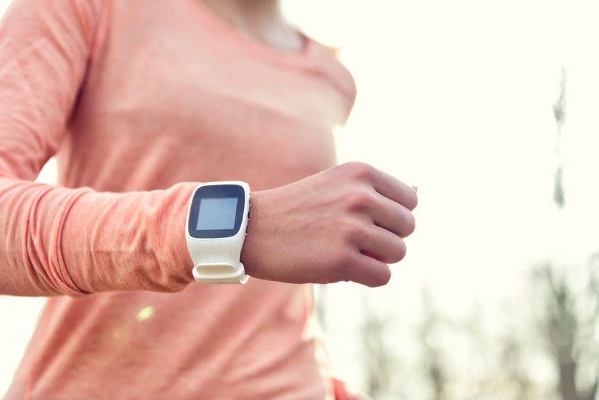
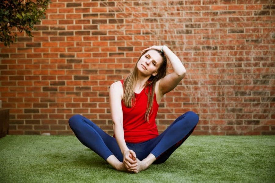
Being physically fit does not mean training for a triathlon. Regular light exercise/activity is all that is needed. Just going for a 30-minute walk around the neighborhood or playing a 20-minute game regularly is highly beneficial to your health. And the more active you are the better for your health.
Skating
Bicycling
Jogging
Swimming
Walking
Playing
Regular Activity
Whatever the activity, so long as you get at least 20 minutes of exercise a day will go along way in the future. Regular activity/exercise can help prevent diseases and injuries, which include osteoporosis.
Nothing crazy, just begin to work some activity little by little into a routine. One way is after some sitting work/schoolwork once the brain has had enough is the perfect time to go outside and move around. Do some chores that require physical movement, like vacuuming, sweeping, hanging laundry, etc and turn it into a workout.� A daily routine of light to moderate physical activity strengthens and maintains the body by helping to:
Build healthy bones, muscles, and joints
Control weight
Build lean muscle
Reduce overall body fat
Prevent the development of high blood pressure hypertension��
Here are a few suggestions on how to get 20-30 minutes of daily exercise/activity.
Try an online fitness class.
Check out your local gym for online to see what classes are available.
Family time can become a fun activity/exercise time.
Take a walk with the family, as many are already doing, play basketball, soccer, or other favorite sport together.
Invite friends to be physically active online, maybe playing a workout video game and workout together.
If regular physical activity is difficult or you have a medical condition,�consult your doctor to recommend the appropriate amount of physical activity and exercises that are safe�to perform. But if you are a healthy person, but have not exercised for a while then try for 30 minutes of physical activity a day to keep you healthy and strong.
Core Exercises That Help With Back Pain
Here are some examples of abdominal exercises that can help develop strong abs and help with back pain prevention. These exercises and the number of repetitions are only suggestions. Talk to your doctor before trying these exercises, and remember to listen to your body. If it doesn’t feel right, stop right away.
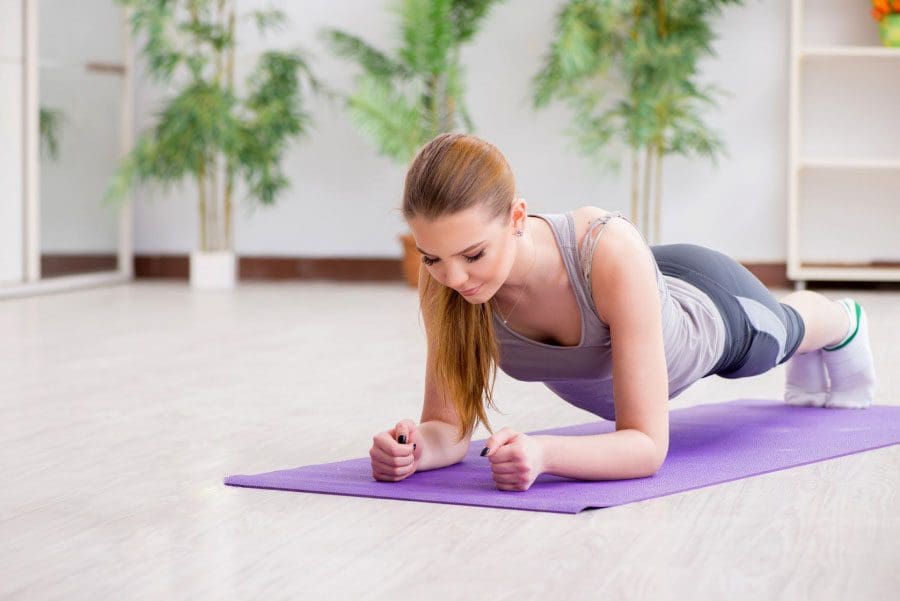
Elbow Planks
Lie down on your stomach with your body straight.
Elbows should be at 90-degrees and close to the body’s sides.
Rest the forearms on the floor and interlace the fingers.
Gently push your body up using the forearms.
Don’t’ let the back fall/drop.
Stay straight.
Engage the core muscles during the entire movement.
Hold this position for 30 seconds, release, and repeat 3 times.
Do this once a day.
Crunches
Lie on your back with the knees bent and the feet flat on the floor, about hip-distance.
Interlace the fingers of your hands behind your head with the elbows out wide.
Inhale and then as you exhale, use the abdominal muscles and not the neck muscles to slowly raise the head, neck, and back off the floor.
Inhale and slowly lower the upper body back to the floor, and repeat.
Try for 3 sets of 10 crunches every day.
Push-ups
Lie down on the stomach so your body is straight.
Place the hands on the floor a little higher/further than the shoulders.
The hands should be wider than the shoulders.
Lift your body so that you’re balanced on the hands and toes.
Maintain a straight back, lower your body to the floor, and slowly bend your elbows until at 90 degrees.
Push back up using arm strength, upper back, and chest muscles, and repeat.
Try for 3 sets of 10 every day.
Once the body becomes stronger, you can go for more reps.
Doing these along with other core exercises you will notice your core strength leading to overall and optimal body strength. Other exercise forms that can help develop core strength while keeping the spine safe are yoga and Pilates. A good idea is to work with a physical therapist/chiropractor that can create a specifically targeted exercise plan that involves core strengthening and flexibility exercises to keep the spine healthy and help maintain proper posture.
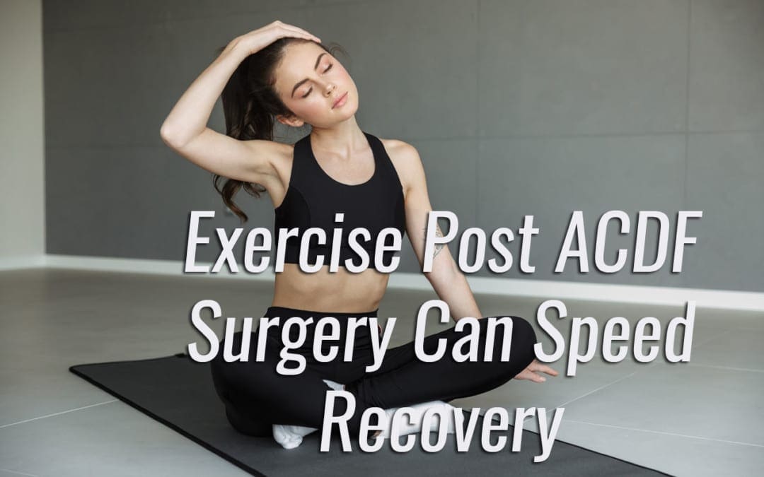
Anterior cervical discectomy and fusion (ACDF)�is a procedure that treats chronic neck conditions and is the most common spinal surgery performed in the U.S.�30 percent of Americans a year experience neck pain, chronic neck pain and radiculopathy or pain that spreads out and radiates to other parts of the body, in this case down the arms.
It�s a procedure that can work wonders, but as many as two-thirds of patients continue to manage neck pain and dysfunction after ACDF. One of the best ways of managing neck pain and dysfunction is to exercise. Research shows that a prescribed exercise program right after surgery can help lessen the pain and create less dependence on medications. A 2020 study published in SPINE suggests that patients that began therapeutic exercises right away had better results than individuals that started an exercise program after the six-week checkup/examination.
What Anterior Cervical Discectomy Fusion Treats
The procedure is performed on individuals with degenerative disc disease or a bulging or herniated disc. These conditions can cause the spinal disc to place pressure on the spinal cord and nerve roots that branch out, creating:
Numbness
Tingling
Pain
Weakness in one or both arms
Most individuals that are recommended to undergo the surgery experience symptoms that don�t respond to non-surgical therapies or medication/s. A significant symptom is hand/arm weakness and arm pain that�s worse than the neck pain.
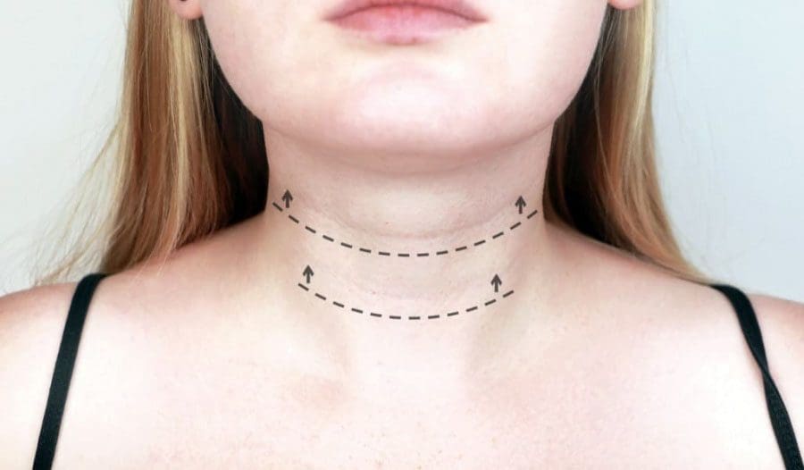
ACDF Surgery
The ACDF procedure is broken into two parts: a discectomyand a fusion. Anterior means in front and in this case it’s the front of the neck that the surgeon accesses the damaged discs.�In a discectomy, the surgeon removes a portion/s or all of an intervertebral disc/s to release the pressure on the nerves. The fusion part fuses the two vertebrae together. This eliminates the painful movements. A bone graft is inserted between the vertebrae at the spot where the disc was removed. The bone graft serves as a structural scaffold that the body uses to build new tissue and cause the vertebrae to grow together.
The graft can come from three sources:
Your own bone called an autograft, this is usually a piece of bone from the pelvis just above where the front jean pocket would be.
Bone donation called an allograft that comes from a cadaver from a bone donor bank.
Substitute material/s like man-made plastic, ceramic, or bioresorbable compounds.
Home Exercise Program
There are those that are of the opinion to hold off on physical therapy or exercise until about six weeks post-ACDF when recovery is established. However, the study suggests it is more effective to begin a home exercise program (HEP) as soon as possible. At this time telemedicine is the replacement for in-person physical therapy sessions but works just as well. The study examined 28 individuals’ outcomes over 12-months post-operation.
The participants were divided into two groups:� Standard care and Early HEP. Both groups went through the standard postoperative care, along with medication, a cervical collar or neck brace, and restrictions of certain activities. The standard care group received physical therapy referral six weeks after surgery while the early HEP group was given a home exercise program to perform during the postoperative six-weeks.
This included walking and sleeping instructions and a range of motion/strengthening exercises. A cognitive-behavioral strategy was also given to help relax. The participants would phone-conference with a physical therapist on a weekly basis. Exercises increased in difficulty every two weeks, based on the therapists’ judgment. When compared to the standard care group, the early HEP group reported a reduction in short-term neck pain and were less likely to be using pain meds/opioids twelve months after their surgery.
Recovery Tips
Recovery time after ACDF surgery typically lasts about four to six weeks. If the bone graft was from the pelvis, there could be pain, soreness, and stiffness. To minimize discomfort, try not to sit or lie down for long periods. And remember to change position or take a quick short walk down the hall, to the kitchen, etc. every 20-30 minutes. If you are referred to physical therapy combine it with the home exercise program. The therapist will teach you exercises and proper form. Exercising ten minutes every day is far more effective than doing 45 minutes once a week.
Resuming normal activities gradually is crucial. No pain, no gain does not apply when recovering from ACDF surgery. Recovery exercises can feel great. However, pain and fatigue can sneak up the next day or the following week. Gradually easing back into daily activities can help avoid major setbacks. Work, shopping, house chores, childcare, and lawn care all fall into the activity category.
A recurrence of neck pain after recovery is normal. However,�discomfort can be minimized by taking a few precautions.
Use proper form when lifting
Keep objects close to the body
Keep the back flat as you lift
Maintain neutral neck position
Be aware of your posture when sitting, standing, walking, and sleeping
Gradually increase the exercise program
Don’t overdo it
Address underlying fear/anxiety
Fear of generating pain can cause tension and exacerbate or even create new pain symptoms
Stop smoking, as it has shown to impede the fusion and heighten the risk for complications
ACDF surgery can improve quality of life. Your surgeon is responsible for performing the procedure, it�s up to the individual to follow through with an exercise program and proper ergonomics for optimal results.
Methods in treating inflammation are the focus of this video. Dr. Alex Jimenez presents a discussion of natural ways to treat inflammatory cascades. Knowing what are the best supplements are presented. This is a safe alternative that is well researched.
If you have enjoyed this video and/or we have helped you in any way
please feel free to subscribe and share us.
Thank You & God Bless.
Dr. Alex Jimenez RN, DC, MSACP, CCST
PODCAST: Daniel Alvarado of Push Fitness Center and Dr. Alex Jimenez discuss the complications of losing weight. How to keep a focus and how to work our to the sticking point. An Ernest Discussion. Dr. Alex Jimenez, a chiropractor in El Paso, TX, and Daniel (Danny) Alvarado, owner of the PUSH Fitness Center, continues to discuss the importance of weight loss and how people and athletes can continue the effort to stay healthy. Metabolic syndrome is characterized by 5 risk factors, including excess waist fat, high blood pressure, high blood glucose or sugar, high triglycerides, and low HDL or good cholesterol levels. Diet and lifestyle modifications, such as participating and engaging in exercise and physical activity, can ultimately help improve the 5 risk factors associated with metabolic syndrome and a variety of health issues, including diabetes, stroke, and heart disease. Dr. Alex Jimenez and Daniel (Danny) Alvarado discuss how motivation is one of the most fundamental elements in the continued effort to stay healthy. Following a diet and lifestyle modifications that are unique to each individual can also help promote overall well-being. – Podcast Insight
If you have enjoyed this video and/or we have helped you in any way please feel free to subscribe and share us.
Thank You & God Bless.
Dr. Alex Jimenez RN, DC, MSACP, CCST
Subscribe: http://bit.ly/drjyt
Facebook Fitness Center Page: https://www.facebook.com/PUSHftinessathletictraining/
Yelp: El Paso Rehabilitation Center: http://goo.gl/pwY2n2
Yelp: El Paso Clinical Center: Treatment: https://goo.gl/r2QPuZ
PODCAST: Dr Alex Jimenez, chiropractor in El Paso, TX, Kenna Vaughn, health coach, Truide Torres, Alexander Jimenez, and Astrid Ornelas discuss metabolic syndrome. The following podcast will focus on a deeper look at understanding metabolic syndrome. Metabolic syndrome is a collection of conditions which can increase the risk of developing a variety of health issues, including diabetes, stroke, and heart disease. Moreover, risk factors such as excess waist fat, high blood sugar, high blood pressure, high triglycerides, and low HDL levels. Diet and lifestyle modifications can ultimately help promote weight loss which can help improve metabolic syndrome and its associated health issues. Several different types of nutraceuticals, including Niacin or vitamin B3, vitamin D, DHEA, Nrf2, and green tea, among others. Weight loss is important to help improve metabolic syndrome. – Podcast Insight
If you have enjoyed this video and/or we have helped you in any way please feel free to subscribe and share us.
Thank You & God Bless.
Dr. Alex Jimenez RN, DC, MSACP, CCST
PODCAST: Dr. Alex Jimenez and his crew focus on making several facts about the COVID-19 pandemic clear. Currently, there is no definitive cure for COVID-19. But what can we do then you may ask? Dr. Alex Jimenez, chiropractor in El Paso, TX, and his crew presents Antiviral Strategies that have scientific substantiations from healthcare professionals around the world. In a moment where information is spread without scientific support, we must look into the research studies that have sound science evidence in order to understand non-fiction from what the true science has presented. These are difficult times and very scary times. We present a body of information present from the works of Dr. Alex Vasquez. His information at time cryptic and found in his writings do shed some light and direction which many of us can take in order to prepare for optimal support from our own immunity. The scientific community is clear. Again, at this time there is no cure for COVID-19. This does not mean we must not look at a way to optimize our immunity. Specifically, since there is much that has been studied in the ways the Antiviral Stategies can prepare our bodies. Dr. Alex Jimenez and his crew continue to discuss Antiviral Strategies and how these can improve our immune system. – Podcast Insight
If you have enjoyed this video and/or we have helped you in any way please feel free to subscribe and share us.
Thank You & God Bless.
Dr. Alex Jimenez RN, DC, MSACP, CCST
PODCAST: Dr. Alex Jimenez, chiropractor in El Paso, TX, and Dr. Marius Ruja, chiropractor in El Paso, TX, ultimately discuss the reasons why choosing a functional medicine approach can safely and effectively improve overall health and wellness. The world has shifted in health care. There’s no time more than now which has started to look to the cause of disease as the present day focused on functional medicine approaches, methods and protocols. We discuss the “why” to choose the option of functional medicine in the present day health care system. Functional medicine focuses on natural treatment approaches to promote the healing of the human body. In this day and age, functional medicine may be essential to help improve our immune system. – Podcast Insight
If you have enjoyed this video and/or we have helped you in any way please feel free to subscribe and share with us.
Thank You & God Bless.
Dr. Alex Jimenez RN, DC, MSACP, CCST
IFM's Find A Practitioner tool is the largest referral network in Functional Medicine, created to help patients locate Functional Medicine practitioners anywhere in the world. IFM Certified Practitioners are listed first in the search results, given their extensive education in Functional Medicine