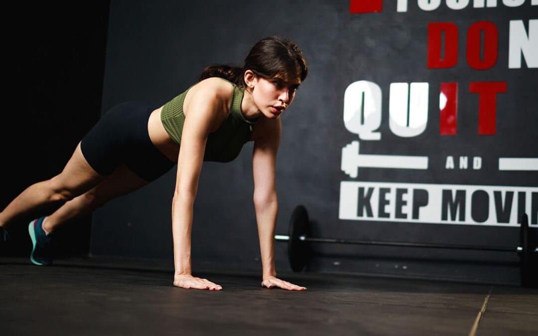
Motivation That Lasts: Fun, Low-Impact Workouts and SMART Goal Strategies
Losing weight does not have to feel impossible, even if back pain, low energy, or busy days get in the way. Many people in El Paso start with easy exercises like short walks or gentle stretches, but staying motivated is what brings real results. The good news is that small, smart steps, plus help from a local expert team, can make all the difference. At El Paso Back Clinic, patients discover how chiropractic care and functional medicine remove roadblocks so basic weight-loss exercises feel safe, doable, and even enjoyable. This guide shares straightforward ways to set goals, track progress, choose fun movement, and get professional support right here in El Paso. You will learn practical tips that fit real life and see how the clinic’s team, led by Dr. Alexander Jimenez, helps turn “I can’t” into steady success.
Basic weight-loss exercises like walking, light yoga, or dancing burn calories without stressing your joints. When your body feels better and pain drops, motivation stays strong. El Paso Back Clinic combines chiropractic adjustments, personalized rehab, and health coaching to make these simple moves part of your everyday routine.
Setting Attainable SMART Objectives for Steady Progress
SMART goals keep your weight-loss journey clear and reachable. SMART means Specific, Measurable, Achievable, Relevant, and Time-bound. Instead of saying “I need to lose weight,” try “I will walk for 15 minutes after dinner, five days this week.” This type of goal is easy to follow and gives quick wins. (Hey Life Training, n.d.; El Paso Back Clinic, n.d.-b)
Here are SMART goal examples perfect for basic weight-loss exercises:
Walk briskly for 15 minutes, five days a week, starting this Monday.
Do gentle yoga stretches for 10 minutes each morning for the next two weeks.
Dance to favorite music for 15 minutes, three evenings a week.
Swim or walk in water for 15 minutes twice a week at a local pool.
Take the stairs instead of the elevator at least five times daily this week.
Start small, so you build confidence fast
At El Paso Back Clinic, health coaches help patients turn these goals into custom plans that match their energy and schedule.
Monitoring progress keeps motivation alive. Use a simple notebook or phone app to log your walks, steps, or how your back feels after movement. Seeing checkmarks add up or a line on a graph climb feels rewarding. Patients at the clinic often say watching their own improvements beats staring at the scale. (Zen Habits, n.d.)
To avoid burnout, pick fun, low-impact activities. Yoga, swimming, and walking ease joints and lift mood through natural feel-good chemicals. These basic exercises become something you look forward to instead of dread. (HelpGuide.org, n.d.)
Find accountability with a workout buddy or the clinic’s support network. Many patients walk with family or join gentle group sessions. Reward small wins with non-food treats like new walking shoes or a relaxing evening. Remember your “why”—more energy for family, better sleep, or less back pain. Read it daily on tough days. (Planet Fitness, n.d.-a)
Easy, Efficient Strategies to Stay Motivated Every Day
Consistency beats intensity when building habits. Here are proven strategies that work well with basic weight-loss exercises:
Start small for lasting consistency: Begin with just 10–15 minutes of movement. This avoids burnout and makes exercise a normal part of your day. (Reddit community insights, 2024)
Track your development: Write down workouts, steps, or how clothes fit. Graphs show real progress and keep you excited. (Zen Habits, n.d.)
Make it fun: Choose dancing, swimming, cycling, or active games. Fun turns movement into “me time.” (HelpGuide.org, n.d.)
Reward yourself: After five good days, celebrate with new socks, a movie, or a quiet bath. (Modern Image Aesthetics, n.d.)
Build accountability: Walk with a friend, pet, or join a beginner class. The clinic’s health coaches provide extra check-ins. (Healthline, n.d.)
Recall your “why”: Focus on deeper reasons like steady energy or pride in your posture. (Planet Fitness, n.d.-b)
Prepare for low-energy days: Have a backup like 10 minutes of gentle stretches at home. (Cleveland Clinic, n.d.)
These steps fit real El Paso life—hot days, long work hours, and family needs. Short walks during lunch or evening strolls add up fast.
Walking Your Way to Better Results: Clinic-Approved Tips
Walking is one of the easiest basic weight-loss exercises, and El Paso Back Clinic shares clear ways to burn more fat while protecting your back. Start with 15 minutes daily, five days a week, then add five minutes each week. Walk at a brisk pace faster than normal, swing your arms, and keep a healthy posture. Add short speed bursts or gentle hills for extra calorie burn without hurting knees. Wear supportive shoes and breathe steadily. (El Paso Back Clinic, n.d.-c)
Benefits include stronger bones, less joint pain, better mood, and reduced belly fat linked to heart health. Even short 15-minute walks several times a day work when time is tight. Patients at the clinic combine walking with chiropractic care for faster mobility gains and steady motivation.
Making Fitness Enjoyable and Part of Your Routine
Pick activities you actually like. If running hurts, try dancing at home, water walking, or bike rides on flat paths. Listen to music or podcasts while moving. Many patients discover they enjoy low-impact options once pain eases. (Medical Beauty and Weight Loss, n.d.)
Social support helps too. Walk with neighbors or join light classes. At El Paso Back Clinic, personalized rehab programs make movement feel safe again, so you stay consistent longer.
How El Paso Back Clinic Boosts Motivation Through Integrative Care
Back pain or low energy often stops people from exercising. El Paso Back Clinic, led by Dr. Alexander Jimenez, DC, APRN, FNP-BC, removes these barriers with chiropractic and functional medicine. Their approach helps thousands of El Paso patients move more freely and lose weight sustainably.
Chiropractic adjustments reduce chronic back, hip, and joint pain, so walking or yoga no longer hurts. Better spinal alignment improves nervous system signals that control metabolism and fat burning. When the body works more smoothly, energy rises, and motivation follows naturally. (El Paso Back Clinic, n.d.-a; Adjusted Life Chiropractic, n.d.)
Dr. Alexander Jimenez has observed over 30 years that fixing spinal misalignments breaks the pain-obesity cycle. Pain leads to less movement and comfort eating; extra weight adds more pain. His team uses gentle adjustments, advanced imaging, and lab tests to address root causes such as inflammation, hormonal imbalances, and gut issues. Patients report less pain, better sleep, steadier moods, and fewer cravings. (Jimenez, n.d.; El Paso Back Clinic, n.d.-a)
Custom low-impact exercise plans are a clinic specialty. Instead of heavy gym work, they recommend practical moves: walking programs, water exercises, light resistance bands, and core stretches that fit daily life. These plans build confidence fast because they feel safe. The clinic’s rehabilitation centers offer guided sessions with trainers who understand back issues. (Robinhood Integrative Health, n.d.; El Paso Back Clinic, n.d.-c)
Functional medicine digs deeper. The team checks for slow metabolism, insulin resistance, or stress hormones that block weight loss. Personalized nutrition advice, supplements, and lifestyle tips clear these hurdles. Health coaches then create step-by-step plans with SMART-style process goals—like “walk three to four times this week”—so patients focus on what they can control. (El Paso Back Clinic, n.d.-b, n.d.-d)
Stress management is built in
High stress raises cortisol and belly fat while lowering motivation. Chiropractic care relaxes tight muscles and calms the nervous system. Many patients report feeling more positive and ready to move on after visits. (Dr. P Chiro, n.d.)
Personalized accountability keeps progress on track. Regular check-ins, body scans, and plan updates show results beyond the scale. Improved posture from adjustments makes patients stand taller and feel stronger—boosting confidence to keep going. (Obesity Action Coalition, n.d.; Westport Chiropractic, n.d.)
Dr. Jimenez often reminds patients that big changes start with small, consistent steps. His team at El Paso Back Clinic offers multiple convenient locations across El Paso, including rehab and fitness centers with 24/7 access. Military discounts, virtual coaching options, and meal-prep support make healthy living easier. Patients with past injuries or long-term back pain often return to activities they once avoided, creating a positive cycle of more movement and faster weight-loss results.
By reducing pain, improving mobility, addressing metabolic issues, and providing expert coaching, El Paso Back Clinic turns basic weight-loss exercises into something patients actually enjoy and stick with long-term.
Putting It All Together for Real, Lasting Success
Begin today with one small change. Choose a SMART goal, schedule a 15-minute walk, and note your “why.” Add music or a friend for fun. If back pain or low energy holds you back, contact El Paso Back Clinic for a personalized evaluation. Dr. Alexander Jimenez and his multidisciplinary team combine chiropractic care, functional medicine, and health coaching to support your goals safely.
Motivation comes and goes—some days feel easier than others, and that is normal. The strategies here—SMART goals, tracking, fun movement, rewards, accountability, and professional help—help you bounce back quickly. Over weeks and months, these habits create real momentum.
Basic weight-loss exercises like daily walking or gentle yoga do more than burn calories. They improve heart health, lift mood, strengthen muscles, ease back pain, and raise self-esteem. With support from El Paso Back Clinic, you gain energy for work, family, and life. Celebrate every step, every stretch, and every healthy choice. You have local experts ready to help—one simple, consistent day at a time.
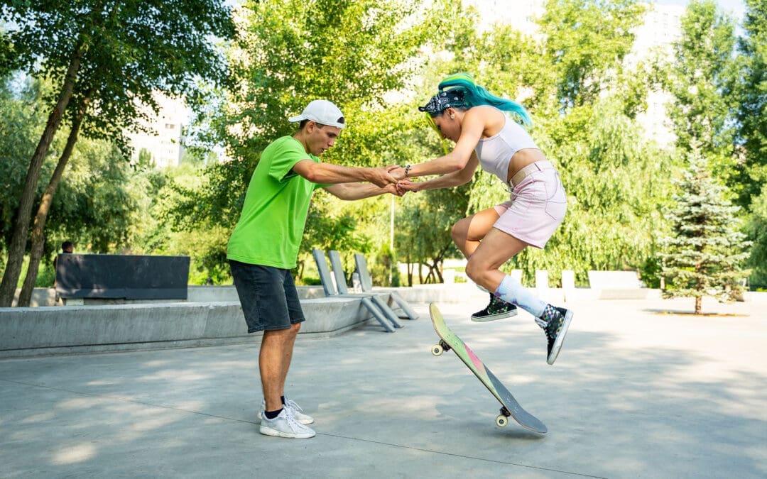
Skateboarding Training Essentials: Strength, Balance, and Injury Prevention with Chiropractic Support at El Paso Back Clinic
Skateboarding is an exciting sport that mixes skill, speed, and style. It began as a land-based surf practice but has grown into a worldwide hobby for many. To excel in skateboarding, you need targeted training that strengthens your core and legs, improves balance, and teaches safe falling to prevent harm. This training uses repetitive drills, explosive jumps, and endurance workouts to create automatic responses and lasting energy. It also includes mental prep like imagining tricks and steady practice routines.
The sport’s demands, such as repeated one-sided pushes and hard landings, can strain your body. That’s where integrative chiropractic care shines. At El Paso Back Clinic in El Paso, Texas, this approach improves joint mobility, corrects imbalances from skateboarding habits, and accelerates healing after impacts. It improves balance, body sync, and bendiness while offering diet and safety tips to reduce injury risk. Led by Dr. Alex Jimenez, DC, APRN, FNP-BC, the clinic offers tailored care for skateboarders and athletes, blending chiropractic care with rehab and nutrition to support top performance.
This article covers skate training basics and how chiropractic at El Paso Back Clinic supports it. For beginners or pros, these insights can help you advance safely. Visit https://elpasobackclinic.com/ to learn more about their services.
Core Elements of Skateboarding Training
Skateboarding success starts with body and mind prep. Training goes beyond board time—it’s about a solid base for tricks and endurance. Prioritize core and leg power, as these drive your actions (Austin Simply Fit, n.d.). Muscles like abs, lower back, quads, hamstrings, glutes, and calves handle shifts from an upright to a low position in moves like ollies.
Core Workouts: Try planks by holding a straight body pose for 30 seconds. Side versions hit obliques for twist stability.
Leg Boosters: Squats mimic board crouches—lower then rise for three sets of 10 reps.
Importance: Strong cores prevent shakes during jumps, lowering fall risks.
Balance is vital in skating. Poor balance leads to wipeouts on basic maneuvers. Newbies should pick a stance: regular (left-forward) or goofy (right-forward). Place the feet over the truck bolts for maximum stability (Skateboard GB, n.d.).
Balance Practices: Stand on one foot and draw letters with the other toe. Switch sides for ankle strength.
Next Level: Manuals lift the front wheels, balancing on the rear for ramp preparation.
Routine: Dedicate 10 minutes daily to weight shifts on your board for a natural feel.
Safe falling is key to injury avoidance. Falls are part of skating, but proper methods reduce severe damage. Roll instead of bracing with arms to protect wrists (Healthcare.utah.edu, 2024).
Fall Methods: Tuck chin and roll to distribute force. Aim for protected spots like padded knees.
Gear Essentials: Helmets, wrist, knee, and elbow pads absorb shocks.
Safe Start: Use grass or mats for low-risk practice.
Repetitive training builds muscle memory. Repeat actions until they’re instinctive, like pushing and halting (Braille Skateboarding, n.d.). This aids tricks such as frontside kickturns and backwheel pivots (How to Skate, 2018).
Drill Reps: Push 10 times, stop, and redo for fluid flow.
Trick Steps: Divide into parts, like board pop, then foot flick for kickflips.
Side Hops: Mimic skating with 30-second lateral jumps.
Gains: Higher leaps and fast reflexes elevate skills.
Cardio keeps you going strong. Skating provides some, but extras build heart health (Skateboard GB, n.d.).
Rope Skipping: 30 seconds on, rest, three rounds for calf power and breath control.
Crawls: Bear walk forward and back 10 meters.
Cardio Value: Longer sessions with quicker recovery.
Mental training tackles fear. Visualize wins before attempts (Florida Atlantic University, n.d.). Commitment means regular sessions despite setbacks.
Imagery: Eyes shut, see perfect landings.
Fear Busting: Small steps build confidence.
Drive: Love for skating fuels persistence.
Follow principles such as targeted work, gradual increases, and variety to ensure safe progress (The Daily Push, n.d.). Skate-specific drills, slight pushes, and mixes prevent plateaus.
This foundation makes skating enjoyable, but one-sided strains need expert help, like at El Paso Back Clinic.
Integrative Chiropractic Care for Skateboarders at El Paso Back Clinic
At El Paso Back Clinic, integrative chiropractic merges adjustments with therapies for whole-body health. For skaters, it enhances joint flow in hips, knees, and ankles, easing restrictions from twists (Push as RX, n.d.). The clinic’s team uses advanced tools for custom plans.
Adjustments: Hands-on fixes realign for better motion.
Skating often causes imbalances—one leg pushes more, enlarging muscles unevenly (Instagram Reel, n.d.). This risks pain or bad posture.
Balance Fixes: Single-side workouts like one-leg squats.
Clinic Approach: Exams spot issues, then adjustments and drills even out.
Prevention: Avoids strains from overuse.
Falls bring impacts, but clinic care hastens recovery by reducing inflammation (Injury 2 Wellness, n.d.). For sprains, they combine rest and rehab.
Healing Tools: Ice, wraps, and elevations cut swelling. Adjustments aid nerves.
Rehab: Planks and stretches rebuild strength.
Quick Return: Less time off the board.
The clinic boosts balance, sync, and flexibility. Core support from deep muscles aids control (Robins, n.d.). Alignment improves awareness.
Balance Enhancers: Fixes heightened position sense.
Sync Training: Patterns restored post-injury.
Flex Moves: Stretches like yoga poses loosen spines.
Nutrition and prevention advice lowers risks. Proteins and veggies aid repair; warm-ups are key (Thompson, n.d.). Clinic experts guide anti-inflammation diets.
Food Advice: Fruits and healthy fats for recovery.
Safety Steps: Check-ups catch problems early; use gear.
Habits: Stay hydrated, foam roll to loosen up.
Dr. Alex Jimenez, a clinic leader with 30+ years, notes that integrative methods prevent injuries by addressing root causes such as imbalances (Jimenez, n.d.). He blends functional medicine, nutrition, and rehab for skateboarders. LinkedIn shares tips on sciatica and balanced routines (Jimenez, n.d.). For skate injuries like ankles or wrists, assessments lead to adjustments and strengthening (Jimenez, n.d.). Teamwork with therapies ensures full recovery.
Chiropractic at the clinic elevates performance, keeping bodies primed (Dallas Thrive, n.d.). Their sports focus includes strength, flexibility, and proprioception for athletes.
Conclusion
Pair skate training with the chiropractic services at El Paso Back Clinic for strength, balance, and safety. Build habits through drills and mental work. Let experts fix strains, speed healing, and advise prevention. Consistency pays off—practice wisely. For personalized care in El Paso, check https://elpasobackclinic.com/.
Fitness Optimization in El Paso, TX: How to Organize a Weekly Workout Plan With Warm-Ups, Cool-Downs, and Integrative Chiropractic Support
A woman doing her weekly workout
A weekly workout plan should do two things at the same time:
Help you get stronger, fitter, and more mobile
Help you stay consistent without getting hurt or burned out
That balance matters even more in El Paso, Texas, where heat, dry air, and busy schedules can make training feel harder than expected. A smart plan incorporates strength training, cardio, mobility, and recovery—and includes warm-ups and cool-downs in every session.
This guide is written for real life. It is geared to the El Paso Back Clinic approach: improving movement quality, addressing posture and joint mechanics, and supporting safer training through an integrative model that blends chiropractic and clinical assessment. ()
Why most people struggle with weekly workout planning
Many people start with motivation, then hit one of these problems:
They do too much too fast (and flare up pain)
They skip warm-ups and feel stiff or strained
They train hard but don’t recover well
They repeat the same muscle groups without enough rest
They don’t have a simple weekly structure that they can repeat
A better plan is not “perfect.” It is repeatable.
A common starting target for beginners and intermediate exercisers is 3–5 workout days per week, depending on schedule, recovery, and current fitness level. (Mayo Clinic, 2023; EōS Fitness, 2024) ()
What a balanced weekly workout plan includes
A strong weekly plan usually includes these building blocks:
Strength training (2–3 days/week)
Cardio (2–3 days/week)
Mobility (most days, even 5–10 minutes helps)
Recovery (at least 1 full rest day, plus lighter days)
Many gyms and fitness instructors recommend alternating training styles throughout the week—such as upper body, lower body, and cardio—to give muscles time to recover while you stay active. (Grinder Gym, 2025; ISSA, 2022)
El Paso-specific training: heat, hydration, and timing
El Paso’s climate can change how workouts feel, especially if you train outdoors. Dry air can increase fluid loss, and heat can accelerate fatigue.
Simple El Paso-friendly adjustments:
Train early morning or later evening outdoors when possible
Build hydration into your plan, not as an afterthought
Hydration tip: If you sweat heavily or train longer, you may need electrolytes—especially during hot weather—based on your personal needs and health status. (American College of Sports Medicine, 2007)
Warm-ups and cool-downs: the 5–10 minute habit that protects progress
If you only change one thing in your training week, make it this:
Warm up for 5–10 minutes (dynamic movement)
Cool down for 5–10 minutes (gradual slowdown + stretching/breathing)
Why warm-ups matter
Warm-ups help your body transition from rest to work. Mayo Clinic explains that warm-ups prepare the cardiovascular system, raise temperature, increase blood flow to muscles, and may lower injury risk. (Mayo Clinic, 2023) ()
Why cool-downs matter
Cooling down helps your body transition back toward rest. Mayo Clinic Press emphasizes that cooldown supports recovery and helps the body transition out of high-intensity exercise more smoothly. (Mayo Clinic Press, 2025) ()
A simple warm-up you can reuse for almost any workout (5–10 minutes)
Keep it easy. The goal is to feel warmer, looser, and more “ready,” not exhausted.
Warm-up (choose this as your default):
2 minutes of easy movement
brisk walk, light bike, easy row
2 minutes dynamic mobility (pick 3–4)
arm circles
hip circles
ankle rocks
thoracic (upper back) rotations
2–4 minutes workout-specific prep
strength day: 1–2 lighter sets of your first lift
cardio day: start slower and gradually build pace
Mayo Clinic Press notes that warm-up duration depends on intensity, but 5–10 minutes is a solid baseline for many people, with longer warm-ups for higher-intensity work. (Mayo Clinic Press, 2025) ()
A simple cool-down you can reuse (5–10 minutes)
Cool-downs work best when they are consistent.
Cool-down template:
3–5 minutes gradual slowdown
walk slowly, easy cycling, gentle movement
2–5 minutes stretching + breathing
hamstrings
hip flexors
calves
chest/shoulders
gentle low back rotation (if comfortable)
Mayo Clinic explains that warm-ups and cool-downs are often the same activity, performed at a lower intensity before and after the workout. (Mayo Clinic, 2023) ()
The best weekly workout schedules for beginners and intermediates (3–5 days/week)
Below are three schedules you can choose from. Pick the one you can follow most weeks.
Option A: 3-day plan (simple and sustainable)
This is perfect if you are starting again, staying consistent, or managing pain flare-ups.
Day 1 (Mon): Full-body strength + short walk
Day 2 (Wed): Cardio + mobility
Day 3 (Fri): Full-body strength + core
Weekend: 1 light activity day + 1 full rest day
Many weekly workout guides recommend 2–3 strength sessions and at least one rest day for recovery. (Health, n.d.) ()
Option B: 4-day plan (upper/lower split + cardio)
This is a popular plan for steady progress.
Mon: Lower body strength
Tue: Upper body strength
Thu: Lower body strength + core
Sat: Cardio + mobility (or a class)
Splitting upper/lower body helps prevent repeating the same muscle groups on back-to-back days and makes recovery easier to manage. (ISSA, 2022; Grinder Gym, 2025) ()
Option C: 5-day plan (shorter sessions, more frequency)
This works well if you like shorter workouts and a daily structure.
Mon: Strength (full body)
Tue: Cardio
Wed: Strength (upper)
Thu: Mobility + easy cardio
Fri: Strength (lower)
Sat: Optional class or easy walk
Sun: Rest
EōS Fitness emphasizes building a weekly plan based on your goals and starting level, often incorporating strength, cardio, and recovery. (EōS Fitness, 2024) ()
What to do inside each strength workout (so it’s organized)
A clean structure keeps you from wandering around the gym and doing random exercises.
Strength session structure (45–60 minutes):
Warm-up: 5–10 minutes
Main lift: 10–15 minutes
Assistance work: 15–25 minutes
Core: 5–10 minutes
Cool-down: 5–10 minutes
Main lift examples:
squat pattern (leg press or squat)
hinge pattern (deadlift variation or hip hinge)
press (dumbbell press)
pull (row or pulldown)
Assistance work examples:
glute bridges or hip thrusts
split squats or step-ups
face pulls or band work for shoulders
hamstring curls
carries (farmer carry)
This aligns with structuring training days around major patterns (push/pull/lower) to build balanced strength and avoid overuse. (Grinder Gym, 2025; ISSA, 2022) ()
Cardio planning: simple is better than perfect
Cardio should support your life, not crush you.
Great El Paso-friendly cardio options:
incline treadmill walking (easy on joints)
stationary bike
rowing machine
brisk outdoor walking (timing matters in heat)
Easy weekly cardio goals:
2 days of steady cardio (20–40 minutes)
1 optional interval day (shorter, only if you tolerate it)
Health.com outlines weekly schedules that combine strength and cardio while protecting recovery. (Health, n.d.) ()
Mobility and recovery: the glue that holds the week together
Recovery is not “doing nothing.” It is training your body to stay ready for the next workout.
Recovery habits that work:
sleep consistency
hydration plan
protein and balanced meals
walking on rest days
mobility work for hips, ankles, upper back, and shoulders
Simple mobility “micro-dose” (5 minutes):
1 minute hip flexor stretch (each side)
1 minute calf stretch (each side)
1 minute thoracic rotations
1 minute shoulder mobility
This kind of daily movement keeps joints from stiffening, especially if you sit a lot.
How integrative chiropractic supports routine optimization
Many people don’t need more willpower. They need:
better joint motion
better movement patterns
better recovery
fewer flare-ups
The El Paso Back Clinic approach: integrative care and movement-focused support
The El Paso Back Clinic describes an integrated model led by Dr. Alex Jimenez, DC, APRN, FNP-BC, combining chiropractic care and clinical assessment within a multidisciplinary setting. (El Paso Back Clinic, n.d.)
From a routine-optimization standpoint, that integrative approach can help people who struggle with:
recurring neck or low back tightness during training
posture-related strain (desk work, long driving, “tech neck”)
limited hip or shoulder mobility
compensation patterns (one side always “takes over”)
The clinic also discusses advanced collaboration and diagnostics, including imaging relationships when needed for complex cases—especially when symptoms do not match what someone expects from “normal soreness.” (El Paso Back Clinic, n.d.) ()
Clinical observations from Dr. Jimenez (fitness-focused takeaways)
Across the clinic’s educational content, Dr. Jimenez emphasizes:
improving posture and movement quality to reduce repeated strain patterns (El Paso Back Clinic, n.d.) ()
using mobility and functional training to build resilience and prevent re-injury (El Paso Back Clinic, n.d.) ()
integrating training structure with recovery so people can stay consistent long-term (El Paso Back Clinic, n.d.) ()
In simple terms: train with a plan, move better, recover better.
A weekly “checklist” you can follow
Use this to keep your week on track:
✅ 3–5 workouts completed (based on your plan)
✅ Warm-up done every workout (5–10 minutes) (Mayo Clinic, 2023)
pain that worsens with training, even after deloading
trouble figuring out what movements are safe for your body
If you want clinic support, El Paso Back Clinic provides contact and appointment options, including online scheduling information listed on their site. (El Paso Back Clinic, n.d.) ()
Beginner Gym Workout Routine: Build Strength, Flexibility, and Avoid Injuries
Young hispanic man does a beginner gym workout with weights.
Starting a workout at a sports training gym can feel exciting but also a bit scary if you are new to it. A good beginner routine helps build strength in all parts of your body. It uses big movements that work many muscles at once. These are called compound exercises. Things like squats, lunges, push-ups, rows, and planks are key. Do this routine three times a week. Each exercise should have three sets of eight to twelve reps. This builds a strong base without too much strain (Planet Fitness, n.d.a).
The goal is to mix full-body strength training with some easy cardio. Low-impact cardio means activities that do not jar your joints too much, such as walking on a treadmill or using an elliptical. This helps you get fit without overdoing it. Adding chiropractic care can make it even better. It helps with movement, cuts injury risk, and speeds up recovery. Let’s break this down step by step.
Why Start with a Balanced Routine?
A good starting plan focuses on functional strength. This means exercises that help with everyday activities, like picking things up or climbing stairs. For beginners, full-body workouts are best. They work all major muscle groups without splitting days for arms or legs only. This way, you recover faster and see progress soon (Mikolo, 2024).
Experts say beginners should aim for consistency over intensity. Start slow to learn proper form. Bad form can lead to hurts. A routine with strength and cardio boosts heart health, muscle tone, and energy. It also helps with weight control and mood. But without good recovery, you might get sore or injured. That’s where things like stretching and chiropractic come in.
Key Exercises for Beginners
Here are some top exercises for a sports training gym. They build strength, flexibility, and stability. Most use bodyweight or simple machines. Do them in order for a full workout.
Squats: Stand with feet shoulder-width apart. Bend your knees and lower yourself as if you were sitting in a chair. Keep your chest up and knees over toes. Push back up. This works legs, glutes, and core (Refinery29, 2020).
Lunges: Step forward with one foot. Lower until both knees are bent at 90 degrees. Push back to start. Alternate legs. This exercise is beneficial for enhancing balance and building leg strength (Kong, 2024).
Push-ups: Start on your hands and toes or on your knees. Lower your chest to the ground, then push up. This hits the chest, arms, and shoulders. Modify by using a wall if needed (Magnus Method, 2023).
Rows: Use a machine or dumbbells. Pull weights toward your body, squeezing your shoulder blades. This exercise enhances back strength and improves posture (Planet Fitness, n.d.b).
Planks: Hold a push-up position on forearms. Keep your body straight. Hold for 20-30 seconds. Strengthens core for stability (Quora, n.d.).
Do three sets of 8-12 reps for each, except planks, which are timed. Rest 60 seconds between sets. Warm up with 5 minutes of light walking first.
Sample Weekly Routine
A three-day plan works well for beginners. Space days out, like Monday, Wednesday, and Friday. This gives time to rest. Each session lasts 30-45 minutes.
Day 1: Full Body Strength Focus
Warm-up: 5 min treadmill walk.
Squats: 3 sets of 10 reps.
Push-ups: 3 sets of 8 reps.
Rows: 3 sets of 10 reps.
Planks: 3 holds of 30 seconds.
Cool-down: Stretch legs and arms.
Day 2: Rest or Light Cardio
Walk or bike for 20 minutes at an easy pace.
Day 3: Lower Body Emphasis
Warm-up: 5 min elliptical.
Lunges: 3 sets of 10 per leg.
Glute bridges: 3 sets of 12.
Calf raises: 3 sets of 15.
Planks: 3 holds of 30 seconds.
This builds on basics. As you get better, add weights (Under Armour, n.d.). Track your progress in a notebook.
Adding Cardio for Endurance
Cardio is key for heart health and stamina. For beginners, start low-impact. Use machines like a treadmill or a rower. Aim for 15–20 minutes after strength training. Walk at a 5-8% incline on a treadmill to build legs without running (Kong, 2024). This burns calories and boosts recovery.
Mix it in: Do cardio on off days or at the end of your workout. Things like jumping jacks or brisk walking work too. Cardio helps with overall fitness, but do not overdo it. Too much can tire you out.
The Role of Integrative Chiropractic Care
Integrative chiropractic care is more than just spinal cracks. It looks at the whole body. Dr. Alexander Jimenez, a chiropractor with over 30 years of experience, notes it helps with injury prevention and better movement (Jimenez, n.d.a). He combines adjustments with exercises and nutrition.
For beginners, it identifies hidden issues such as muscle imbalances. These can lead to injuries if ignored. Adjustments fix joint problems, improving the range of motion. This lets you do exercises with better form (Pushasrx, n.d.).
Dr. Jimenez observes that chiropractic boosts nerve function. This helps muscles adapt faster and cuts pain. In his clinic, he uses functional assessments to identify weaknesses early (Jimenez, n.d.b). For sports training, it keeps you going without breaks.
Benefits of Chiropractic for Gym Beginners
Chiropractic makes starting safer. Here are key perks:
Injury Prevention: Spots imbalances before they hurt. Fixes tight muscles or stiff joints (Atlas Total Health, 2022).
Better Mobility: Improves joint range. It helps with squats or lunges without causing strain (Elevate to Life, 2023).
Faster Recovery: Uses soft-tissue work and exercises to help you heal more quickly. It also helps reduce soreness after workouts (Team Elite Chiropractic, 2022).
Dr. Jimenez stresses holistic care. He integrates chiropractic care with fitness, such as HIIT, to build strength. This prevents chronic issues and boosts performance (Jimenez, n.d.a).
When to Get Chiropractic Adjustments
Timing matters. Get adjusted before workouts to optimize nerve and muscle function. This prevents strain. After workouts, it aids recovery by reducing inflammation (Atlas Total Health, 2022). Dr. Jimenez recommends regular visits for long-term health.
Do at-home exercises too. Things like glute bridges or cat-cow stretches support treatment (Elevate to Life, 2023). These speed healing and keep balance.
Recovery Tips to Stay Injury-Free
Recovery is as important as working out. Add these to your routine:
Stretching: Do dynamic stretches before and static stretches after. This practice enhances your flexibility, according to 10 Fitness (n.d.).
Rest Days: Allow muscles to grow. Walk lightly if active rest.
Corrective Exercises: Fix imbalances. Hip openers or spine mobilizations prevent injury (Asheville Medical Massage, 2025).
Nutrition and Sleep: Eat protein-rich foods. Sleep 7-9 hours for repair (Squatwolf, n.d.).
If injured, stay fit with low-impact activities like swimming. Balance activity to heal (RP3 Rowing, n.d.). Chiropractic helps here, too, per Dr. Jimenez.
Putting It All Together
A good beginner workout at a sports training gym mixes strength, cardio, and care. Start with compounds three times a week. Add chiropractic for safety. Dr. Jimenez’s work shows this approach builds a strong, injury-free base (Jimenez, n.d.b). Stay consistent, listen to your body, and progress slowly. This makes fitness fun and lasting.
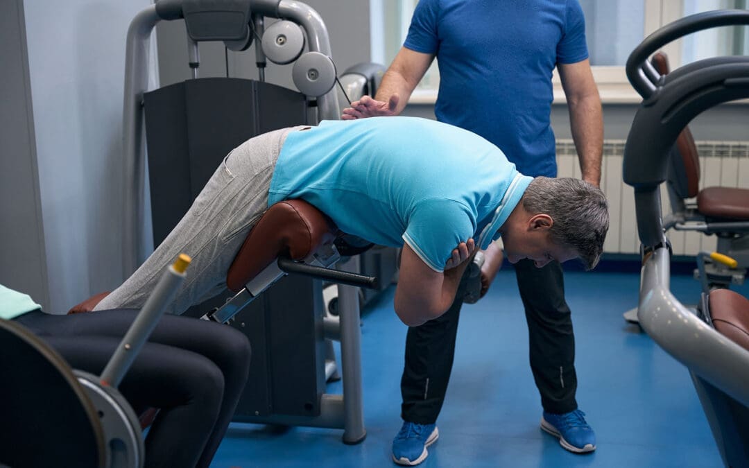
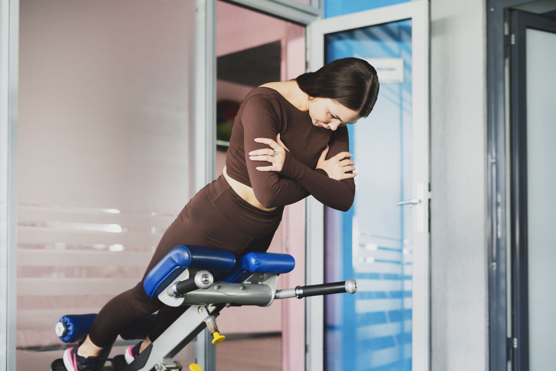
Back Extension Machine (Roman Chair) Training for a Stronger Back
A woman engages in back extension exercises to strengthen back muscles, improve core stability, and relieve chronic back pain.
A practical, El Paso Back Clinic–style guide to core stability, safer form, and pain prevention
If you’ve ever used a back extension machine—also called a hyperextension bench or Roman chair—you already know it looks simple. You lock your feet, rest your hips on the pad, and hinge forward and back up.
But the best results come from how you do it.
At El Paso Back Clinic, the goal is not just “stronger muscles.” It’s a smarter plan that supports spine stability, hip power, and better movement habits—especially for people who deal with recurring low back tightness, desk-related stiffness, or training-related flare-ups. Dr. Alexander Jimenez, DC, APRN, FNP-BC, often emphasizes that many back problems improve when you combine movement quality, targeted strengthening, and a whole-person plan (Jimenez, n.d.-a; Jimenez, n.d.-b).
This article explains:
what the back extension machine actually trains,
how to set it up correctly,
how to avoid the common mistakes that irritate backs,
and how integrative care (chiropractic + NP-style whole-body support) fits into a complete plan.
What the Back Extension Machine Trains (and Why It Matters)
Back extensions are a posterior chain exercise. That means they train the muscles on the back side of your body, including:
Erector spinae (spinal extensor muscles that help you stay upright) (MasterClass, 2021).
Glutes (hip extension power and pelvic support) (MasterClass, 2021).
Hamstrings (help control the lowering phase and assist hip extension) (MasterClass, 2021).
Deep core stabilizers (the “bracing” muscles that keep the spine steady while the hips move) (WebMD, 2024).
This is important because many people think “core” means only the abs. In real life, core stability is about the ability to resist unwanted motion and control the spine while the hips move.
A back extension machine helps train that pattern if you do it as a hip hinge, not as a “low back bend.” (More on that below.)
Roman Chair vs. Back Extension Machine: Same Goal, Different Feel
You’ll see a few styles:
45-degree hyperextension bench (most common “Roman chair” style)
90-degree Roman chair (more upright)
Seated back extension machine (you sit and extend backward against resistance)
Verywell Fit notes that these machines are often grouped together because they train similar movement patterns and posterior chain muscles, even though the setup and feel can differ (Verywell Fit, 2025).
If you’re choosing equipment for home or clinic use, adjustability matters. Many benches are built to adjust pad position and angle so different body types can hinge correctly (Valor Fitness, n.d.).
Step 1: Set Up the Machine Correctly (This Is Where Most People Go Wrong)
Before you do a single rep, take 30 seconds to set it up.
The best setup checkpoints
Hip pad position: The pad should sit around your hip crease (where your hips fold). If it’s too high, you can’t hinge well. If it’s too low, you may feel unstable (WebMD, 2024).
Feet locked in: Your heels and feet should feel secure in the restraints (WebMD, 2024).
Top position posture: At the top, you want a straight line from head to hips—not a “lean back” pose (MasterClass, 2021).
Quick self-test
If you feel the movement mostly in your low back joints (pinchy or compressed) rather than in your glutes/hamstrings, your setup or technique needs adjustment.
Step 2: Use the Right Form (Neutral Spine + Hip Hinge)
A safer back extension is controlled and clean. The spine stays neutral, and the movement comes mostly from the hips.
How to do it (simple steps)
Brace first: Take a breath and tighten your midsection like you’re preparing to be lightly bumped.
Hinge down: Push your hips back and lower your chest slowly. Keep your neck neutral.
Drive up: Squeeze glutes and hamstrings to lift your torso back up.
Stop at neutral: Finish tall and braced. Do not crank into hyperextension (MasterClass, 2021; WebMD, 2024).
Good cues that help
“Hips back, not ribs up.”
“Move like a hinge, not a bendy straw.”
“Glutes finish the rep.”
Chuze Fitness also describes back extensions as a way to work against gravity and build strength in a simple, repeatable pattern, with the option to progress by adding load later (Chuze Fitness, n.d.-a).
The #1 Mistake: Hyperextending at the Top
One of the biggest errors is leaning back too far at the top. People do it to “feel” the lower back more, but it often adds compression where you don’t want it.
What you want instead: a neutral, stacked finish.
Ribs down
Glutes tight
Spine tall
No “backward bend” finish (MasterClass, 2021).
If you can’t stop at neutral, reduce the range of motion and slow the tempo.
Another Common Mistake: Turning It Into a Low-Back Exercise Only
Back extensions are often taught as if they only train the lower back. In reality, they work best when the hips do the job and the trunk stays braced.
A helpful way to think:
The hips create motion
The spine controls motion
That is a big reason back extensions can be useful for stability—when done correctly (WebMD, 2024).
Reps and Sets: Simple Programming That Works
The “right” plan depends on your goal and your history.
Beginner (control first)
2–3 sets of 8–12 reps
Bodyweight only
Slow lowering (2–3 seconds down)
General strength and pain prevention
3 sets of 10–15 reps
Add light load only if form stays clean (Chuze Fitness, n.d.-a).
Stronger posterior chain (experienced lifters)
3–5 sets of 6–10 reps
More rest
Still stop at neutral (no hyperextension)
Rule: load is earned by control.
Verywell Fit’s equipment review also highlights that comfort, stability, and fit matter for consistent training—especially for people using these tools as part of a back-strengthening routine (Verywell Fit, 2025).
Safer Progressions (If Your Back Is Sensitive)
If your back flares easily, you can still train the posterior chain—you just need smarter progressions.
Options that tend to be more back-friendly:
Shorter-range back extensions (only move where you can stay neutral)
Isometric holds at neutral (hold 10–20 seconds)
Lower load, slower tempo
Add glute-focused assistance work (like bridges) alongside back extensions
At El Paso Back Clinic, Dr. Jimenez often frames strengthening as part of a bigger plan: improve mechanics, build tolerance, and progress gradually based on the person’s symptoms and daily demands (Jimenez, n.d.-a; Jimenez, n.d.-c).
When to Pause and Get Checked (Red Flags)
Back extension training should feel like muscular effort, not nerve pain.
Stop and seek professional guidance if you have:
Pain shooting down the leg
Numbness or tingling
Weakness in the foot/leg
Pain that worsens over time with extension-based movements
WebMD also encourages careful form and smart choices when using back extensions, especially when they’re used for “back health” rather than just bodybuilding (WebMD, 2024).
How This Fits the El Paso Back Clinic Approach: Strength + Mobility + Whole-Person Support
Many people try one thing:
“I’ll just strengthen my back.”
Or:
“I’ll just stretch more.”
Or:
“I’ll just get adjusted.”
But most lasting results come from combining the right tools in the right order.
Chiropractic care to improve mechanics
Chiropractic-focused care often aims to:
improve joint motion where stiffness limits your hinge,
reduce irritation that changes how you move,
and help you restore better spinal and pelvic mechanics.
El Paso Back Clinic content emphasizes a whole-body view of pain and function, including movement habits and multi-step plans (Jimenez, n.d.-c).
Exercise to build stability and strength
Once movement is cleaner, exercises like the Roman chair can help you:
reinforce a strong hinge,
strengthen posterior chain muscles,
and build stability that carries into work, lifting, and sports (MasterClass, 2021).
Nurse practitioner support to address barriers to recovery
NP-style integrative support often helps by addressing factors that keep people “stuck,” such as:
sleep quality,
stress load,
inflammation drivers,
safe pain management planning (when appropriate),
and screening for problems that need further testing or referral.
In short: your back isn’t separate from the rest of you.
A Simple 3-Phase Plan You Can Follow
Here is a practical approach that matches how many integrative clinics structure back-pain recovery and performance.
Phase 1: Calm things down and restore motion (1–2 weeks)
Gentle mobility (hips + mid-back)
Light back extensions with short range
Walk daily if tolerated
Focus on bracing and hinge control
Phase 2: Build capacity (3–6 weeks)
Back extensions: 2–3 days/week
Add glute and hamstring work
Add core stability work
Slowly add reps before adding load
Phase 3: Build real-world resilience (ongoing)
Add load gradually (only if neutral form is automatic)
Transfer strength into squats, hinges, and carries
Keep a weekly routine of mobility + stability work
This kind of integrated plan—adjustments plus exercise and habit change—is also described in chiropractic-focused integration articles discussing the value of combining care approaches to improve outcomes (OPTMZ State, 2026).
Key Takeaways
The back extension machine is best used as a hip-hinge strength tool, not a “bend your spine” tool (MasterClass, 2021).
Proper setup (hip pad alignment + stable feet) helps you move safely (WebMD, 2024).
Avoid the big mistake: hyperextending at the top. Stop at neutral.
Strong results often come from a full plan: chiropractic mechanics + targeted exercise + whole-person support, a theme repeated across El Paso Back Clinic education from Dr. Jimenez (Jimenez, n.d.-a; Jimenez, n.d.-c).
Fun Ways to Stay Active: Alternatives to Boring Workouts for Better Health
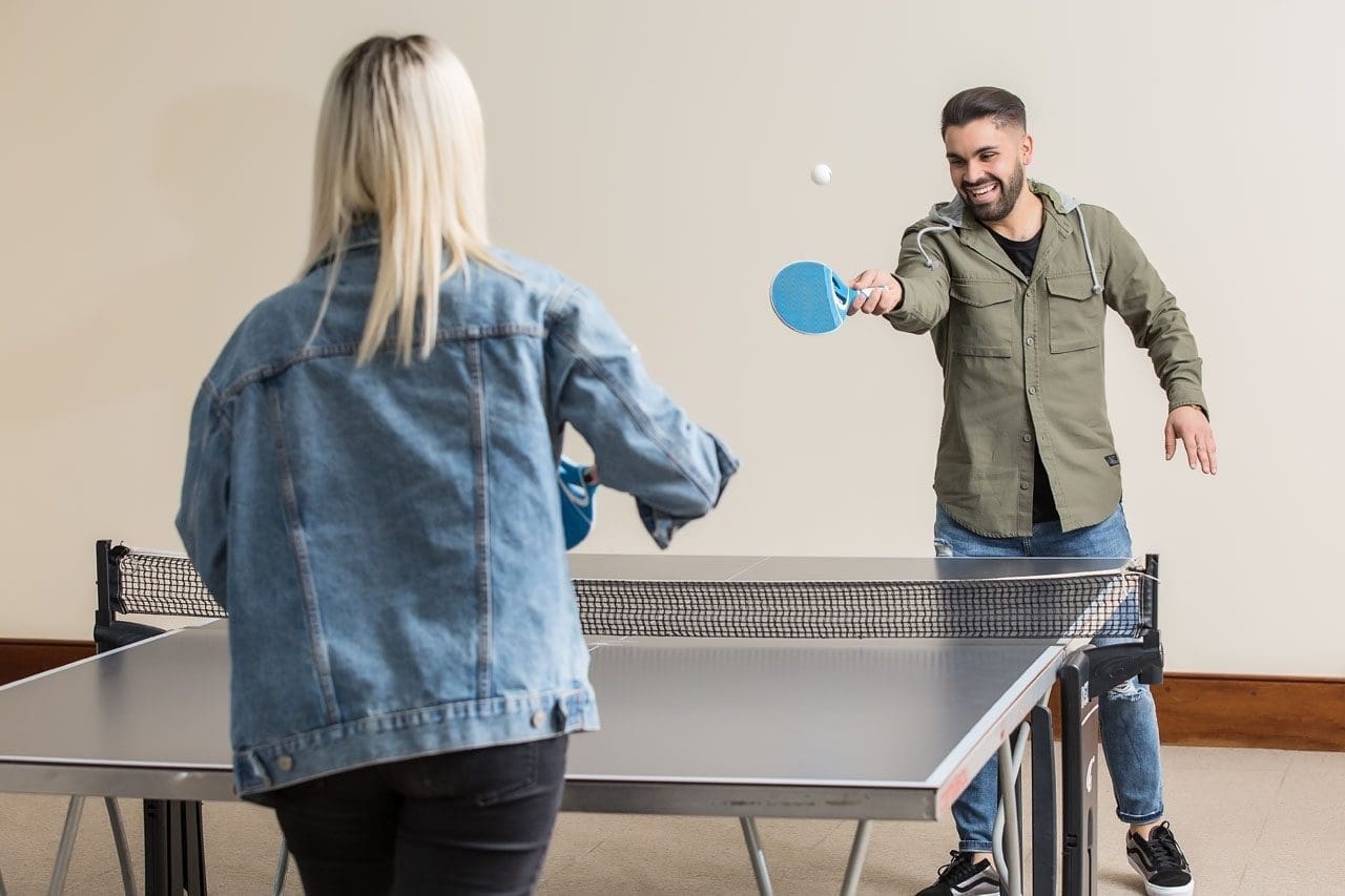
Friends play table tennis as a way to start making fitness fun and as a doable health goal.
Many people start the new year with big fitness goals. They promise to hit the gym every day or run miles each week. But often, these plans fall apart quickly. Life gets busy, motivation fades, and suddenly, exercise feels like a chore. If this sounds like you, don’t worry. Giving up on strict resolutions doesn’t mean giving up on health. Instead, shift to activities that feel more like play than work. Fun sports and easy movements can keep you moving without the dread of traditional workouts. This approach makes staying active sustainable and enjoyable, leading to better long-term habits (Bayou Bend Health System, n.d.).
Research shows that making physical activity fun boosts your chances of sticking with it. For example, choosing things you enjoy turns exercise into a hobby. This can improve your mood, reduce stress, and even help with weight management. Health experts recommend at least 150 minutes of moderate activity per week for adults, but it doesn’t have to be in a gym (NHS, n.d.). Simple swaps like walking in nature or dancing to music can meet these goals while feeling effortless.
In this article, we’ll explore ways to restart your fitness journey with joy. We’ll cover fun sports, social options, and relaxing practices. We’ll also discuss low-impact choices for those who struggle with standard routines. Plus, learn how professionals like integrative chiropractors and nurse practitioners can guide you. Drawing from expert insights, including clinical observations from Dr. Alexander Jimenez, DC, APRN, FNP-BC, this guide offers practical tips to get back on track.
Restarting After a Failed New Year’s Resolution
If your resolution crashed early, it’s time for a fresh start. The key is picking activities that excite you. Fun, easy sports like hiking, dancing, swimming, or biking can make movement feel rewarding. These options build fitness without the pressure of sets and reps.
Hiking: Head to a trail for fresh air and views. It’s a great way to explore while getting your heart rate up. Start with short paths and build up. Hiking strengthens legs and improves balance, all while enjoying nature (MultiCare Clinic, n.d.).
Dancing: Put on your favorite tunes and move freely. Whether alone or in a class, dancing boosts cardio and coordination. It’s low-pressure and can burn calories without feeling like exercise (Whispering Oaks Senior Living, n.d.).
Swimming: Water supports your body, making it gentle on joints. Swim laps or just splash around for fun. It’s ideal for all ages and helps with endurance (Vista Springs Living, n.d.).
Biking: Ride a bike around your neighborhood or on paths. It’s easy to adjust speed and distance. Biking tones muscles and can be a social outing (Blue Cross NC, n.d.).
These activities trick your brain into thinking you’re playing, not working out. Studies support this: enjoyable exercise leads to better adherence and health outcomes (Exercise is Medicine, n.d.).
Beyond solo sports, join social activities to add fun. Pickleball, tennis, or team sports bring people together, making commitment easier.
Pickleball: A mix of tennis and ping-pong, it’s easy to learn and play. Courts are popping up everywhere, and it’s great for quick games with friends (Nerd Fitness, n.d.).
Tennis: Hit the court for rallies that improve agility. Doubles makes it less intense and more chatty (Athlean-X, n.d.).
Team Sports: Join a recreational league for soccer, basketball, or volleyball. The group vibe keeps you motivated, and games feel like events, not drills (Quora, n.d.).
Social exercise can reduce feelings of isolation while building strength. One study notes that group activities enhance mental health alongside physical benefits (Reddit, n.d.).
For a calmer approach, try mind-body practices like yoga or Tai Chi. These are low-impact and focus on relaxation.
Yoga: Gentle poses improve flexibility and reduce stress. Start with beginner videos at home. It helps with breathing and mindfulness (Piedmont Wellness Center, n.d.).
Tai Chi: Slow, flowing movements build balance and calm the mind. It’s perfect for easing into activity without strain (Care Insurance, n.d.).
These practices are adaptable for any fitness level. They promote relaxation, which can lower blood pressure and improve sleep (NHLBI, n.d.).
To build habits, start small. Aim for 10–15 minute sessions a few times a week. Gradually increase as you gain confidence. This prevents burnout and lets your body adjust (Bayou Bend Health System, n.d.). Track progress in a journal to see improvements, like feeling more energetic.
Options for Those Who Dislike Traditional Workouts
Not everyone loves the gym or running. If weights and treadmills bore you, low-impact or sociable sports offer alternatives. These keep you active without the monotony, focusing on enjoyment and variety.
Swimming and biking stand out as low-impact favorites. Swimming provides a full-body workout in a supportive environment, reducing joint stress (Seniors Helping Seniors, n.d.). Biking lets you control the pace, making it accessible for beginners (MultiCare Clinic, n.d.).
Hiking and dancing add adventure. Hiking varies with terrain, keeping things interesting, while dancing lets you express yourself creatively (Blue Cross NC, n.d.; Whispering Oaks Senior Living, n.d.).
For a challenge, try rock climbing. It’s low-impact but builds strength and problem-solving skills. You can start indoors at a gym with easy walls (The Telegraph, n.d.).
Joining a recreational sports league brings community. Options like softball or ultimate frisbee emphasize fun over competition (Nerd Fitness, n.d.).
Benefits of These Activities:
More engaging than repetitive workouts.
Build social connections.
Adaptable to your energy level.
Improve mood through endorphins (Sanguina, n.d.).
These choices make the activity feel natural. For instance, walking briskly counts as exercise and can be done anywhere (Quora, n.d.). Or jump rope for short bursts—it’s simple and effective for cardio (MCU, n.d.).
If mobility is an issue, modify exercises. Chair-based routines or water aerobics allow movement without strain (ParentGiving, n.d.; Care.com, n.d.). The goal is consistency over intensity.
Experts agree: low-impact options like these support heart health and flexibility, especially for those with limits (Gaddis Premier, n.d.; Prairie Hills at Independence, n.d.).
How Integrative Professionals Can Help
When starting or restarting activity, professional guidance ensures safety. Integrative chiropractors and nurse practitioners offer tailored care, especially if you have physical limits.
Integrative chiropractors focus on the whole body. They use adjustments to align the spine, reducing pain and improving movement. This holistic approach addresses root causes rather than just symptoms (Integral Chiropractic, n.d.; Impastato Chiropractic, n.d.).
For example, if joint pain stops you from hiking, a chiropractor can ease stiffness through manipulations and exercises (Elysian Wellness Centre, n.d.; De Integrative Healthcare, n.d.). They often include nutrition and lifestyle advice for better results (AFP Fitness, n.d.; Together4Health Wellness, n.d.).
Nurse practitioners add medical expertise. They assess your health and create plans that address limits, such as suggesting low-impact swimming for arthritis (Buckner Parkway Place, n.d.; Cor Health Ontario, n.d.).
Together, these pros provide personalized care. They work with your abilities to help you enjoy activities again (Wellness Center FW, n.d.; Fortitude Health, n.d.).
Dr. Alexander Jimenez, DC, APRN, FNP-BC, embodies this integrated approach. With over 30 years in practice, he combines chiropractic and nursing for comprehensive care. His clinical observations highlight non-invasive methods for pain management and mobility.
In his work, Dr. Jimenez notes that tailored programs, like resistance band exercises, strengthen muscles without high impact. This helps people with injuries return to fun activities like biking or dancing. He emphasizes flexibility for joint health, noting that restricted movement can lead to pain, but gentle practices like yoga can restore it.
On LinkedIn, Dr. Jimenez shares insights on sciatica and back pain, recommending core exercises like modified squats for those with limitations. He advocates stretching to prevent stiffness, noting, “If you don’t stretch, your body ‘pays interest'” in reduced mobility.
His practice includes functional medicine, addressing nutrition and the environment for wellness. For example, he uses assessments to create plans that fit patients’ lifestyles, helping them stay active despite chronic conditions (All Injury Rehab, n.d.; Motus Integrative Health, n.d.).
How They Help:
Assess limits and set realistic goals.
Provide exercises like water aerobics for joint relief (Activ Therapy, n.d.).
Offer advice on enjoyable activities to build habits (Nepute Wellness Center, n.d.).
Monitor progress to adjust plans.
This support makes returning to movement less daunting. Integrative care focuses on harmony in physical, mental, and emotional health (Wellness Center FW, n.d.).
Wrapping Up: Make Movement Joyful
Staying active doesn’t require grueling workouts. By choosing fun options like hiking or yoga and seeking professional help when needed, you can rebuild habits. Remember Dr. Jimenez’s observation: personalized, holistic care unlocks better mobility. Start small, stay consistent, and enjoy the process. Your health will thank you.
Relieve Lower Back and Hip Pain with Squats, Core Exercises, and Chiropractic Care at El Paso Back Clinic®
Many people in El Paso suffer from lower back pain and hip discomfort due to daily activities, work demands, injuries, or long-term issues. These problems often stem from muscle strains, poor posture, tight hips or glutes, and weak supporting muscles. At El Paso Back Clinic® in El Paso, TX, we specialize in helping patients overcome these challenges through personalized chiropractic care, rehabilitation, and safe exercises.
Squats and core exercises, performed correctly, strengthen the muscles that support the spine, improve alignment, and enhance hip mobility. This reduces stress on the back during movement. They are effective for chronic low back pain, mild sciatica, and general aches from weak muscles. Proper form is essential—sharp pain, numbness, or weakness means you should seek professional evaluation first.
Strong Core + Chiropractic for Lower Back and Hip Pain Relief
The lower back and hips are closely connected through shared muscles, joints, and nerves. Tight hips or glutes can tug on the back, leading to strain. Weak core muscles cause spinal instability and poor posture, leading to chronic pain.
Muscle imbalances force the back to overcompensate in everyday tasks.
Reduced hip mobility leads to excessive forward leaning, stressing the lower back.
Problems in ankle or upper back mobility contribute further.
These factors can result in lumbar instability or pain radiating from the hips to the back.
How Squats Benefit Lower Back and Hip Conditions
Squats strengthen the legs, glutes, and core. With proper technique, they relieve pressure from the lower back.
Proper squats maintain a neutral spine and engaged core, providing stability and minimizing lumbar strain. Activating core and hip muscles during squats supports the spine, preventing excessive arching or rounding.
Squats also increase hip mobility. Tight hip flexors are a common cause of back pain during deeper squats. Improved flexibility allows the hips to function better, sparing the back from overload.
Builds glutes and legs for stronger spinal support.
Enhances blood flow and reduces inflammation in the area.
Aids mild pain that improves with gentle activity.
Research supports that the correct form reduces risks associated with squats.
Core Exercises: A Key to Back and Hip Relief
Core exercises focus on deep muscles in the abdomen, back, and pelvis, acting as a natural spinal brace.
Strong core muscles enhance posture and balance, easing the load on spinal discs and preventing persistent pain from inadequate support. Studies show core stability exercises effectively reduce non-specific low back pain and improve function.
Core training also supports hip pain by stabilizing the pelvis, which is beneficial for conditions like arthritis or glute tightness.
Planks and bird-dogs develop endurance in stabilizing muscles.
Pelvic tilts and bridges safely activate deep muscles.
Standing core activities help relieve pain from prolonged sitting.
Evidence indicates that core exercises often outperform general workouts in reducing pain.
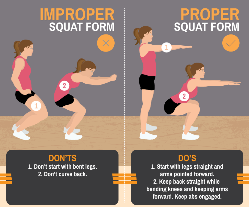
Mastering Proper Form for Safe Squats and Core Work
Incorrect squat form is a leading cause of lower back pain. Frequent mistakes include back rounding, knee collapse, or excessive weight.
Safe squat guidelines:
Position feet shoulder-width apart, toes slightly turned out.
Engage your core as if bracing for impact.
Hinge at the hips, keep the chest high, and descend until the thighs are parallel to the ground.
Drive up through heels, maintaining a neutral spine.
For core exercises, prioritize controlled movement. Hold planks straight with tight abs—avoid dipping or arching.
Begin with bodyweight versions and always warm up to boost circulation and lower injury risk.
Pain during squats typically indicates a weak core, tight hips, or mobility deficits. Address these with targeted stretches and progressive loading.
When Exercises Are Helpful and When to Get Professional Care
Squats and core exercises support:
Chronic low back pain from muscle weakness.
Mild sciatica by decreasing nerve pressure.
Hip tightness referring pain to the back.
Posture-related daily discomfort.
They foster long-term resilience and prevent compensatory back strain. Halt immediately if experiencing severe pain, numbness, weakness, or loss of balance—these may indicate serious conditions such as a disc herniation.
Consult a provider before beginning, especially if you have pre-existing injuries.
Integrative Care at El Paso Back Clinic®
At El Paso Back Clinic®, Dr. Alexander Jimenez, DC, APRN, FNP-BC, leads a team that delivers comprehensive, integrative chiropractic and wellness care for lower back and hip pain. Our approach combines squats and core exercises with chiropractic adjustments, spinal decompression, physical therapy, functional medicine, and rehabilitation programs.
Chiropractic adjustments correct misalignments and joint dysfunctions. A reinforced core helps maintain these corrections by enhancing spinal stability.
Dr. Jimenez creates tailored plans that address root causes through evidence-based protocols, drawing on over 30 years of experience in complex injuries, sciatica, and chronic pain. This multidisciplinary method often yields superior, sustained results compared to isolated treatments.
Visit our main location at 11860 Vista Del Sol, Suite 128, El Paso, TX 79936, or call (915) 850-0900 to schedule your consultation.
Beginner Exercises to Try Under Guidance
Start with these fundamentals, supervised by our team:
Bodyweight Squats: 3 sets of 10-15 repetitions, emphasizing technique.
Glute Bridges: Lie on your back, and elevate your hips by engaging your glutes.
Bird-Dog: On hands and knees, extend opposite arm and leg while bracing core.
Planks: Maintain position for 20-30 seconds, gradually increasing duration.
Pelvic Tilts: On the back, press the lower back into the floor via a pelvic tilt.
Incorporate 2-3 sessions weekly. Include hip mobility work and advance gradually.
Regain Comfort and Mobility Today
At El Paso Back Clinic®, squats and core exercises form integral components of our rehabilitation strategies for lower back and hip pain. They fortify stabilizing muscles, correct alignment, and promote mobility to manage strains, poor posture, instability, and tightness.
Combined with expert chiropractic and integrative care under Dr. Alexander Jimenez, they deliver lasting strength and relief.
Reach out to El Paso Back Clinic® today. Our team will assess your needs and develop a customized plan for optimal recovery.
IFM's Find A Practitioner tool is the largest referral network in Functional Medicine, created to help patients locate Functional Medicine practitioners anywhere in the world. IFM Certified Practitioners are listed first in the search results, given their extensive education in Functional Medicine