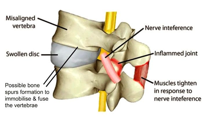
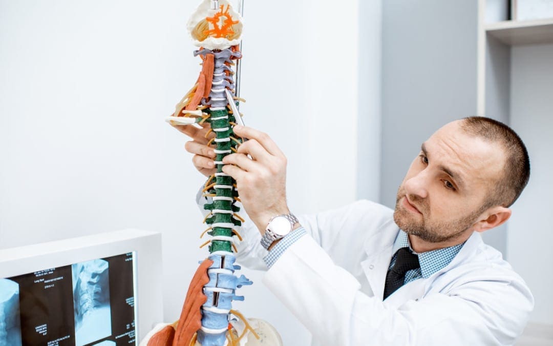
Individuals typically don’t think about their spine as they go through their everyday lives, bending, reaching, twisting, lifting, etc. However, it is through all of these movements/motions that spinal misalignments can occur causing discomfort, pain, and health problems. This happens gradually, with individuals learning how to live with the misalignment, accepting it as a normal part of life. Spinal misalignments, known as subluxations, are common but can be resolved through chiropractic treatment.
Spinal Misalignment
The spine needs to stay strong, stable, and able to support the musculoskeletal system to keep the body upright and protect the central nervous system. When the spine is properly aligned the body stays strong and mobile. When the spine is misaligned, it can cause dysfunction of the central nervous system. Chiropractic is highly effective and a non-invasive way to improve the body’s health. An individual will want to know if their spine is out of alignment to know what to do and what to avoid when back pain presents. Symptoms that the spine is out of alignment includes:
Headaches, Back, and/or Joint Pain
Headaches, joint pain, and backaches can be misalignment symptoms and are the most common. Many individuals learn to live with chronic headaches and migraines but do not realize that chiropractic treatment can help to reduce, or in a case like this, can eliminate them. Back pain, especially low back pain is another symptom of misalignment. However, there can be a variety of causes, like a herniated disc, degenerative disc disease, a chiropractor will alleviate the pain without medication or surgery. A chiropractor will find the root cause and correct any misalignments.
The Heels of Shoes Wear Out Unevenly
This is a symptom that can come from cheap shoes, but often it is from a misaligned pelvis/pelvic tilt. What happens is the hips are shifted out of their proper position, which causes the feet to land on the ground unevenly.
Stiffness and/or Inability To Turn The Head or Hips
If the neck is stiff or there is difficultness, pain when turning, or you can hear the neck crack when turning, this is a sure symptom of cervical misalignment. The same is true with the hips. A subluxation can also cause muscles to tighten or damage the connective tissues, like the ligaments or tendons. Individuals can get used to feeling this way, but it can lead to losing the full range of motion along with pain, stiffness, and inability to perform certain tasks.
Joint Stiffness, Aches, and Pains
Trying to push through back stiffness, along with aches and pains is not healthy. Improper ergonomics and postures could be caused by spinal misalignment or could worsen a subluxation causing further injury. A chiropractor will:
Educate on proper ergonomics
Show how to improve posture
Perform adjustments
Recommend anti-inflammatory supplements
Perform physical therapeutic massage to eliminate stiffness, alleviate pain, and relieve stress.
Numbness or Tingling in Hands or Feet
This is a sure symptom that of a misaligned spine. Numbness and/or tingling sensations often indicate pinched/compressed nerves. The compression or irritation can result in pain or the aforementioned sensations. A chiropractor will ease the pressure on the nerves, allowing for proper circulation, bringing back the full sensation to the affected area/s.
Body Composition
Fiber Health Benefits
Fiber health benefits include lowering the risk of developing diseases like diabetes, obesity, hypertension, and more. It is also beneficial for those who have diabetes to improve insulin sensitivity. Adding more fiber to your diet can enhance weight loss goals. Fruits and vegetables are the highest-fiber foods available.
Fiber and Gut Health
The beneficial bacteria that live in the gut thrive from fruit and vegetable fiber. Because the body does not absorb fiber, the bacteria ferment the fiber. Anti-inflammatory fatty acids are released as a by-product of this process and help to protect the health of the gut. They can also help with appetite regulation.
The information herein is not intended to replace a one-on-one relationship with a qualified health care professional, licensed physician, and is not medical advice. We encourage you to make your own health care decisions based on your research and partnership with a qualified health care professional. Our information scope is limited to chiropractic, musculoskeletal, physical medicines, wellness, sensitive health issues, functional medicine articles, topics, and discussions. We provide and present clinical collaboration with specialists from a wide array of disciplines. Each specialist is governed by their professional scope of practice and their jurisdiction of licensure. We use functional health & wellness protocols to treat and support care for the musculoskeletal system’s injuries or disorders. Our videos, posts, topics, subjects, and insights cover clinical matters, issues, and topics that relate to and support, directly or indirectly, our clinical scope of practice.* Our office has made a reasonable attempt to provide supportive citations and has identified the relevant research study or studies supporting our posts. We provide copies of supporting research studies available to regulatory boards and the public upon request. We understand that we cover matters that require an additional explanation of how it may assist in a particular care plan or treatment protocol; therefore, to further discuss the subject matter above, please feel free to ask Dr. Alex Jimenez or contact us at 915-850-0900.
Dr. Alex Jimenez DC, MSACP, CCST, IFMCP*, CIFM*, CTG*
email: [email protected]
phone: 915-850-0900
Licensed in Texas & New Mexico
References
Czaprowski, Dariusz et al. “Non-structural misalignments of body posture in the sagittal plane.” Scoliosis and spinal disorders vol. 13 6. 5 Mar. 2018, doi:10.1186/s13013-018-0151-5
Formica, M et al. “ALIF in the correction of spinal sagittal misalignment. A systematic review of the literature.” The European spine journal: official publication of the European Spine Society, the European Spinal Deformity Society, and the European Section of the Cervical Spine Research Society vol. 30,1 (2021): 50-62. doi:10.1007/s00586-020-06598-y
Granacher, Urs et al. “Effects of core instability strength training on trunk muscle strength, spinal mobility, dynamic balance and functional mobility in older adults.” Gerontology vol. 59,2 (2013): 105-13. doi:10.1159/000343152
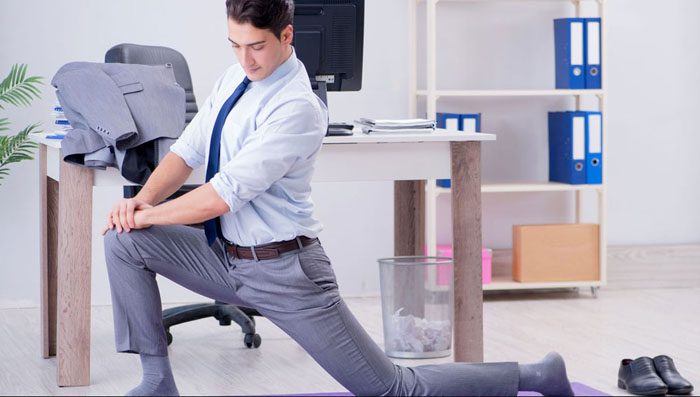
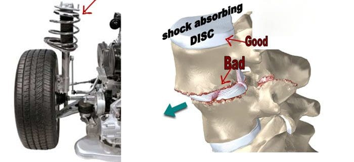
Here are a few recommended stretches and exercises for relieving herniated disc symptoms. The vertebrae are the small bones that make up the spine. They have cushion discs between each one. These are the intervertebral discs and are the body’s shock absorbers. The discs can be thought of as small balloons that are filled with an elastic gel-type material. There are twenty-three of these cushions.
Functioning as the body’s shock absorbers transferring various forces, weight, and stress from vertebra to vertebra, so that no one is overburdened taking on all the impact the body goes through. But like any machine, the discs can wear down over time, and sustain injury. When this happens the cushioning gel can leak out and press on the nerve roots emerging from the spine. This type of injury is a herniated disc.
Herniated Disc Treatment
A herniated disc can lose its height because of fluid and water loss.
This loss affects the bone structures bringing them closer together affecting the ligaments that connect each segment. The ligaments become loose and do not provide the same stability. Ligaments cannot be strengthened with exercise making it more important to strengthen the muscles around the spine to make up for this stability loss. Depending on the severity of the injury, the displaced disc can cause pressure to build upon the nerves, resulting in pain and other discomforts. This comes from the loss of the disc’s cushion causing the vertebrae to rub against each other. Stretches and exercises designed for herniated discs can work in conjunction with conservative treatment to relieve the pain and discomfort.
Stretches and Exercises for Pain Relief
Consult a medical spine specialist/chiropractor before beginning a stretch and exercise regimen. This is because the herniation can become worse or additional injury/s can occur without proper instruction. Once the injury and clinical considerations have been addressed, gentle stretches and exercises can help reduce the pain and other symptoms. Strengthening the back and hamstring muscles reduces pressure on the spinal column helping to prevent pain and promotes healing by:
Increasing blood flow to the spine
Building strength to support the spinal muscles
Decreases stress on the spine
Helps relieve the pain
Improves abnormal postures and awkward body positions
Equipment is not necessary but there are few items that can help the process.
A herniated disc in the neck is usually caused by a forward head posture and a swayback or excessive curvature of the spine.
Isometric hold
Sit straight
Relax the shoulders
Place one hand on the forehead
Push head into the hand without moving the head
Hold for 5 to 15 seconds.
Repeat 15 times
Chin tuck
Lie on your back on a flat surface
Place arms at sides
Tuck the chin in and down toward the chest until a stretch is felt
Hold for 5 to 10 seconds
Repeat 15 to 20 times
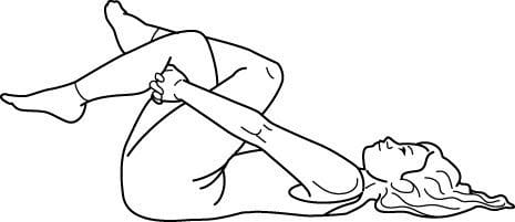
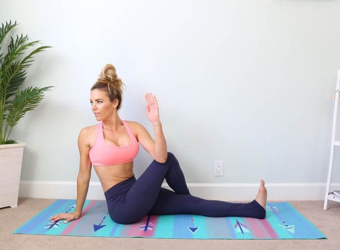
Lumbar/Low Back Stretches and Exercises
Back flexion stretch
This stretch extends the back muscles to relieve low back pain.
Lie flat on your back
Pull the knees toward the chest and wrap your arms around the knees
Lift head straight up off the floor until there is a stretch across the mid and low back
Hold for 10 seconds
Repeat 5 to 10 times
Piriformis stretch
This stretches the small muscle in the buttocks helping to relieve low back pain and helps with sciatica.
Lie flat on your back on the floor or yoga mat
Bend the knees
Plant feet on the floor
Pick up one leg and rest the ankle on the other leg’s bent knee
Reach one arm through the leg and use both hands to grasp the bent leg
Pull the leg toward the chest until there is a stretch in the buttock
Hold for 30 seconds
Repeat on the other leg
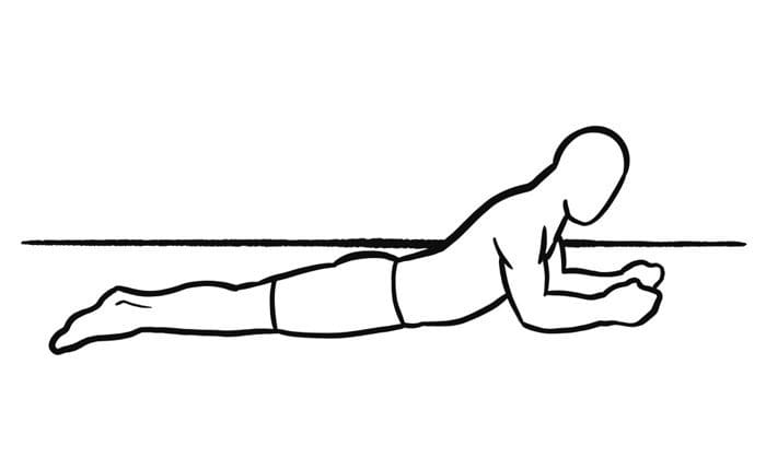
Prone extension stretch
This stretch helps reposition the disc back to its proper position, expediting the healing process. Start slowly and if pain presents, stop immediately.
Lie face down on the floor or yoga mat
Place the forearms on the floor next to the body
The elbows should be at a 45- degree angle
Slowly prop the body up, being sure to keep the hips on the floor
Keep pressing upward until the elbows are at a 90-degree angle
Hold the position for 10-15 seconds
Return to starting position
Repeat the stretch 10 times
Gradually increase the upward position hold time until it can be maintained for 30 seconds
Performing these stretches and exercises or similar types will help with herniated injury recovery and prevent worsening or creating new injuries.
Body Composition
Benefits of yoga
Yoga benefits mental and physical health. Yoga helps improve individual physical health. Specific poses can help:
Improve balance
Flexibility
Build/Tone muscle
Prevent injury
Improve sense of well-being
Yoga stretches the muscles while relieving physical and emotional stress. Practicing yoga regularly can prevent obesity, and reduce the risk of developing metabolic syndrome. Yoga can help decrease leptin which is a hormone that helps control appetite. This is important for individuals going through chronic stress who are twice as likely to develop metabolic syndrome.
Disclaimer
The information herein is not intended to replace a one-on-one relationship with a qualified health care professional, licensed physician, and is not medical advice. We encourage you to make your own health care decisions based on your research and partnership with a qualified health care professional. Our information scope is limited to chiropractic, musculoskeletal, physical medicines, wellness, sensitive health issues, functional medicine articles, topics, and discussions. We provide and present clinical collaboration with specialists from a wide array of disciplines. Each specialist is governed by their professional scope of practice and their jurisdiction of licensure. We use functional health & wellness protocols to treat and support care for the musculoskeletal system’s injuries or disorders. Our videos, posts, topics, subjects, and insights cover clinical matters, issues, and topics that relate to and support, directly or indirectly, our clinical scope of practice.* Our office has made a reasonable attempt to provide supportive citations and has identified the relevant research study or studies supporting our posts. We provide copies of supporting research studies available to regulatory boards and the public upon request. We understand that we cover matters that require an additional explanation of how it may assist in a particular care plan or treatment protocol; therefore, to further discuss the subject matter above, please feel free to ask Dr. Alex Jimenez or contact us at 915-850-0900.
Dr. Alex Jimenez DC, MSACP, CCST, IFMCP*, CIFM*, CTG*
email: [email protected]
phone: 915-850-0900
Licensed in Texas & New Mexico
References
Court C, Mansour E, Bouthors C. Thoracic disc herniation: Surgical treatment, Orthopaedics & Traumatology: Surgery & Research, 104(1)S31-@40, 2018, https://www.sciencedirect.com/science/article/pii/S1877056817303419.
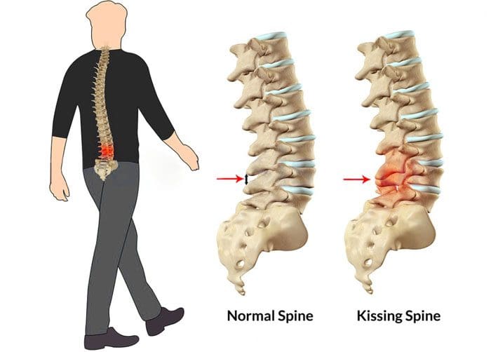
Baastrup’s syndrome is named after Christian Ingerslev Baastrup. He discovered and described the condition in 1933. In this syndrome, pain and inflammation are triggered when the spinous processes of two adjacent vertebrae begin to touch each other. This is where the term kissing spine came from. Most back and neck pain is attributed to inflammation or degeneration of the spinal vertebrae, discs, muscles, and nerves. This is a spinal condition that can cause problems with age. If experiencing pain that worsens when arching the back, consult a professional chiropractor. A physical examination and imaging could reveal the spine is going through this underdiagnosed condition.
Kissing Spine
Spine problems mostly involve the vertebrae and the discs. However, the spine has other components, which include spinous processes. These are thin segments of bone that protrude off the back of each vertebra. Kissing spine syndrome, also known as Baastrup’s disease, or interspinous bursitis, happens when these spinous processes begin to move close together and touch/kiss. Pain and inflammation can be triggered by this.
It is believed to develop as a result of degeneration in the spine that comes with age. As vertebral discs break down from all the wear and tear of life, this can cause the spinous processes to move closer together and touch. This typically develops in the lumbar spine/lower back, but can also affect the cervical spine/neck. The most common symptom of kissing spine syndrome is back pain that worsens when touched or arching the back. For some individuals slumping forward or rounding the back, can help diminish the pain.
When the spinous processes touch, they begin abrasively rubbing against each other. This wears them down and can lead to other types of spinal degeneration. Over time secondary problems can begin to present including neurological conditions caused by compressed nerves. The condition is common in older adults from the natural wear and tear on their spines. But young individuals specifically athletes, can develop the syndrome.
Poor posture
Obesity
Spinal injury/s are additional risk factors.
Diagnosing the condition involves a physical exam and imaging scans to confirm that the spinous processes are in fact touching.
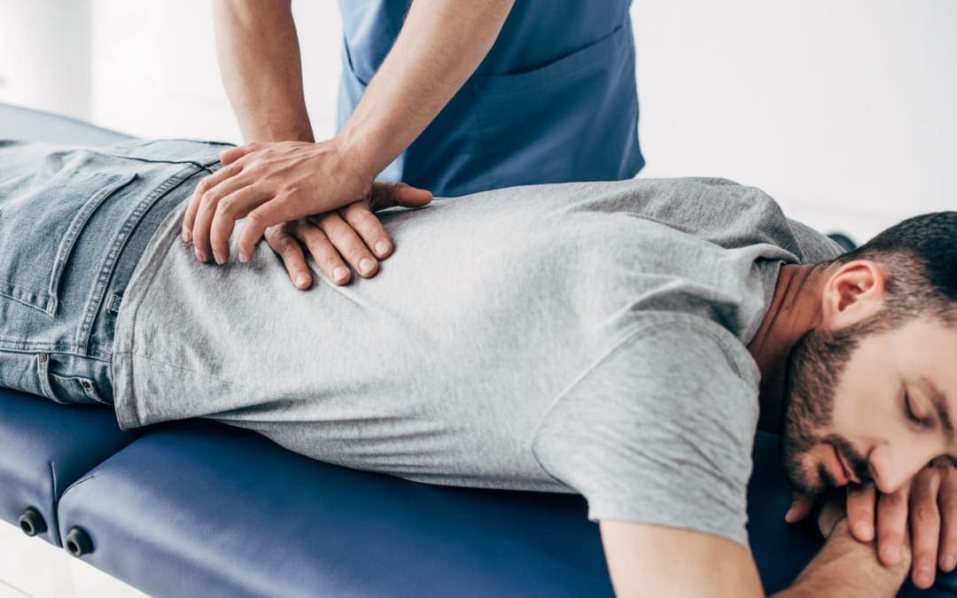
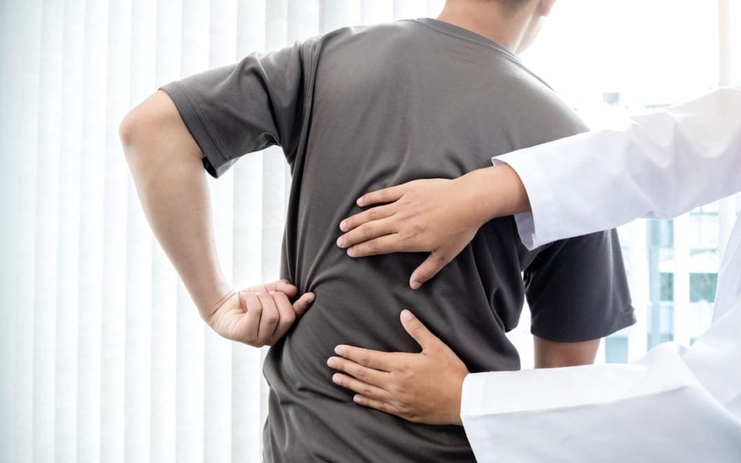
Chiropractic Care
A chiropractor can help manage the pain caused by degenerative disc disease and kissing spine syndrome. Treatment protocols for kissing spine syndrome include:
Spinal adjustments
Physical therapy massage
Spinal manipulation
Stretches
Exercises
Anti-inflammatory diet
Spinal decompression techniques can open the spinal segments so that the spinous processes don’t touch. Manipulations can facilitate proper joint mobility and alleviate inflammation. Physical therapy exercises and stretches will help stretch the spine and the supportive tissues. If experiencing neck or low back pain, contact Injury Medical Chiropractic and Functional Medicine Clinic. Our spine specialists will listen, discuss, and develop a personalized treatment plan. We provide non-invasive approaches for long-term pain management and spinal correction for lasting comfort.
Body Composition
The Paleo Diet
The Paleo diet consists of eating foods that would have been available to humans before modern agriculture was established. If the food was not available to these human ancestors and they did not eat it, then it is not part of the Paleo diet. This includes eating:
Lean meats
Fish
Vegetables
Fruits
Eggs
Nuts
The Paleo cuts out foods like:
Grains
Legumes
Dairy
Sugars
Processed oils
One study published in the American Journal of Clinical Nutrition compared the Paleo diet to other control diets based on United States nutritional guidelines. The researchers found that the Paleo diet generated improvements in waist circumference, triglyceride levels, and blood pressure.
Disclaimer
The information herein is not intended to replace a one-on-one relationship with a qualified health care professional, licensed physician, and is not medical advice. We encourage you to make your own health care decisions based on your research and partnership with a qualified health care professional. Our information scope is limited to chiropractic, musculoskeletal, physical medicines, wellness, sensitive health issues, functional medicine articles, topics, and discussions. We provide and present clinical collaboration with specialists from a wide array of disciplines. Each specialist is governed by their professional scope of practice and their jurisdiction of licensure. We use functional health & wellness protocols to treat and support care for the musculoskeletal system’s injuries or disorders. Our videos, posts, topics, subjects, and insights cover clinical matters, issues, and topics that relate to and support, directly or indirectly, our clinical scope of practice.* Our office has made a reasonable attempt to provide supportive citations and has identified the relevant research study or studies supporting our posts. We provide copies of supporting research studies available to regulatory boards and the public upon request. We understand that we cover matters that require an additional explanation of how it may assist in a particular care plan or treatment protocol; therefore, to further discuss the subject matter above, please feel free to ask Dr. Alex Jimenez or contact us at 915-850-0900.
Dr. Alex Jimenez DC, MSACP, CCST, IFMCP*, CIFM*, CTG*
email: [email protected]
phone: 915-850-0900
Licensed in Texas & New Mexico
References
Philipp LR, Baum GR, Grossberg JA, Ahmad FU. Baastrup’s Disease: An Often Missed Etiology for Back Pain. Cureus 8(1): e465. Published January 22, 2016. https://www.cureus.com/articles/3982-baastrups-disease-an-often-missed-etiology-for-back-pain. Accessed December 20, 2018.
Filippiadis DK, Mazioti A, Argentos S, et al. Baastrup’s disease (kissing spines syndrome): a pictorial review. Insights Imaging. 2015 Feb; 6(1): 123–128. Published online January 13, 2015. https://link.springer.com/article/10.1007/s13244-014-0376-7 Accessed December 20, 2018.
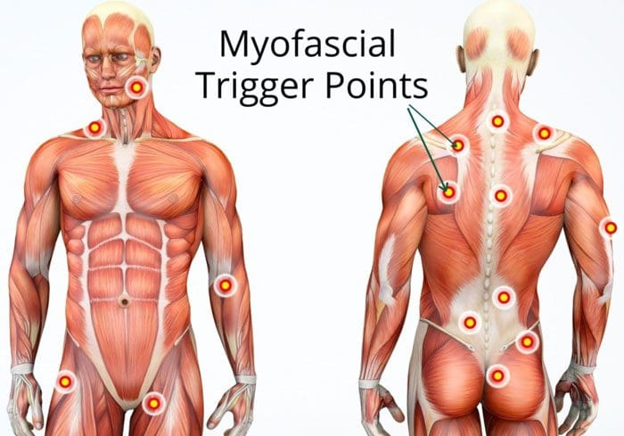
Muscle knots are common and can occur anywhere on the body. They can cause aching and pain in the muscles and joints. When examining a muscle knot also known as myofascial trigger points, it can feel swollen, tense, or like a bump. A particular area where these knots present is in the low back. This happens from excessive wear-and-tear on the lower spine from work, school, daily tasks, and chores. This causes the muscle fibers to tear, and with restricted or no time to rest the area and let it heal properly leads to the fibers bunching/clumping together forming a painful knot.
Muscle Knots In The Lower Back
A muscle knot in the lower back causes aching, soreness, and full-on pain. They tighten and contract even when the muscle is resting. The affected area often becomes inflamed or swollen causing pain and aches to radiate/spread to the gluteal muscles, as well.
Development
These knots develop when the tissue fibers pull apart and bunch up together. They start to stick together and with time the area becomes thicker. This results in the muscle knot. They can be caused by:
Stress
Tension
Poor posture
Muscle overuse
Muscle strain
Sedentary habits
Body dehydration and an unhealthy diet can also contribute to muscle knots. They look like a small bump under the skin. The bump can be red and is usually tender/sore when touched. However, not all muscle knots are visible, but when touched there is soreness and/or pain.
Do They Go Away?
They can go away on their own, but this comes from proper rest and recovery time. However, muscle knots should not be ignored, as even the smallest knot can compress surrounding nerves and muscle tissues. This can cause irritation and weakness. Larger muscle knots could cause movement/mobility issues.
Therapies
Stretching
Stretching will help stretch out and release tight muscle knots. Stretching loosens the muscle fibers and prevents them from becoming attached. Stretches to release a muscle knot include:
Start with these simple stretches/exercises and slowly work up to more vigorous ones.
Chiropractic Care
Chiropractic care can break down muscle knots through various adjustments. They are experts on the musculoskeletal system and understand where the problem is occurring along with the connected muscles.
A chiropractor will palpate the spot where the most pain presents and the surrounding area.
They will begin with a soft massage. This warms up the area getting the blood circulating. The blood circulation helps prevent pain making the adjustment/s far more effective.
Then pressing on nearby joints that the muscle knot is connected to breaks up the tight fibers.
Then the section/area is stretched out. This extends the fibers and prevents them from winding back into a knot.
They will recommend stretches and exercises
Therapeutic Massage
A massage helps to release tension and encourages muscle knots to loosen up and break down. A massage therapist will perform a deep tissue massage or a Swedish massage. Massage helps to release endorphins, which are the body’s natural painkiller. These calm the body and reduce pain. They will also recommend simple massages at home. These can include:
Rolling a massage ball/roller on the muscle knot
Self-massage using the fingers in circular motions on the affected area
Heat and Ice
Hot and cold therapy can calm and prevent inflammation. Heating pads are best if the area has stiffness or is painful. The heat relaxes tight muscles and increases blood flow. Cold therapy stops the swelling. If the muscle knot gets bigger or begins turning red, icing the area is recommended. Alternating between the two can eliminate symptoms and assist with quicker healing.
Body Composition
Building Functional Strength
There are exercises to improve functional strength. Functional training targets specific areas:
Then lift the outside leg up as high as possible and hold for 10 seconds
Repeat on the other side
Do 6-10 repetitions on each side
This exercise builds shoulder, arm, and hip strength. It engages the core and abdominal muscles and improves flexibility in the shoulders, back, and hips.
Disclaimer
The information herein is not intended to replace a one-on-one relationship with a qualified health care professional, licensed physician, and is not medical advice. We encourage you to make your own health care decisions based on your research and partnership with a qualified health care professional. Our information scope is limited to chiropractic, musculoskeletal, physical medicines, wellness, sensitive health issues, functional medicine articles, topics, and discussions. We provide and present clinical collaboration with specialists from a wide array of disciplines. Each specialist is governed by their professional scope of practice and their jurisdiction of licensure. We use functional health & wellness protocols to treat and support care for the musculoskeletal system’s injuries or disorders. Our videos, posts, topics, subjects, and insights cover clinical matters, issues, and topics that relate to and support, directly or indirectly, our clinical scope of practice.* Our office has made a reasonable attempt to provide supportive citations and has identified the relevant research study or studies supporting our posts. We provide copies of supporting research studies available to regulatory boards and the public upon request. We understand that we cover matters that require an additional explanation of how it may assist in a particular care plan or treatment protocol; therefore, to further discuss the subject matter above, please feel free to ask Dr. Alex Jimenez or contact us at 915-850-0900.
Dr. Alex Jimenez DC, MSACP, CCST, IFMCP*, CIFM*, CTG*
email: [email protected]
phone: 915-850-0900
Licensed in Texas & New Mexico
References
Cramer, Holger et al. “Postural awareness and its relation to pain: validation of an innovative instrument measuring awareness of body posture in patients with chronic pain.” BMC musculoskeletal disorders vol. 19,1 109. 6 Apr. 2018, doi:10.1186/s12891-018-2031-9
Malanga, Gerard A et al. “Mechanisms and efficacy of heat and cold therapies for musculoskeletal injury.” Postgraduate medicine vol. 127,1 (2015): 57-65. doi:10.1080/00325481.2015.992719
Women are familiar with abdominal cramps, Pre Menstrual Syndrome, and headaches that accompany their menstrual cycle. However not as many are aware of backache to throbbing back pain sometimes before and/or after a monthly cycle. Many women go to over-the-counter pain medication like ibuprofen. In a study, the regular use of NSAIDs found that it can lead to:
Stomach problems
Bleeding ulcers
Fluid retention
High blood pressure
Kidney
Heart problems
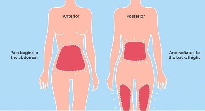
Why Back Pain Presents During Menstrual Cycle
When the uterus is in a contracting state, the nerves around the pelvis feel the sensations. The uterus only contracts for a few seconds, but repeatedly for hours. Sometimes, the uterus compresses blood vessels in the region. This can limit or completely block the blood vessels supplying the muscles around the pelvis. This is a major contributor to back pain during a period. This is known as referred pain, which means the body feels the pain in one area, in this case, the lower back. But the pain is caused by another area of the body, the uterus. This can cause cramping and low back pain before, during, and after a period. If cramps and back pain become debilitating or worsen over time, it could indicate:
If fever is present along with back pain seek professional help as soon as possible.
Here are a few ways that can help bring relief from back pain during the monthly cycle.
Heat Therapy
Heat generates increased blood circulation, specifically where it is applied. Therefore any blood vessels that are blocked by the uterus will have improved circulation to the muscles surrounding the uterus, allowing them to relax. This could be the use of:
Heating pads
Hot water bottle
Warm bath or shower
If at work, many pharmacy stores and regular stores sell heat patches that are applied with adhesive tape. These can be used on the lower abdomen or lower back, providing soothing heat.
Light Exercises
Most doctors refer to exercise throughout the month, just not during the period. As staying in shape will maintain the body’s proper circulation and keep the muscles strong. However, some women can perform light exercises like yoga or swimming. This helps decrease back pain even on the first or second day of a menstrual cycle which for many women is the heaviest and most painful.
Meditate
Meditation can help gain control and insight from feelings about life situations. It takes practice, but once an individual gets the hang of it they are amazed at how much pain can be reduced with a 15-minute meditation session.
Supplemental Support
Taking omega 3’s and magnesium supplements can help with the pain. Omega 3s reduce blood clotting and improve circulation. They are natural anti-inflammatories that decrease prostaglandin, which is associated with backaches and cramps. Magnesium supplements, especially those that contain vitamin B6, can help relieve back pain before and after a period. Magnesium can also be found in:
Beans
Beets
Salmon
Shrimp
Chiropractic Care
The uterus, like every organ in the body, sends and receives nerve signals, from the brain to the uterus. The menstrual cycle has a close relationship with the spine because of its location. Regular chiropractic adjustments can maintain proper communication between the brain and uterus. Chiropractic realigns the entire spine back to its proper position. This relieves the pressure on the nerves of the reproductive organs. Seeing a chiropractor is the right step towards stopping the pain and healing the body naturally.
Positive body composition changes can be seen with higher load volume and less explosive tempo that is combined with shorter rest periods when weight or strength training.
When lifting weights or resistance training, it could be difficult to gain muscle mass if on birth control, near the perimenopausal stage, or officially on menopause.
One of the benefits of weight and strength training is that it can help an individual feel better about themselves. Weight training is associated with significant improvements in:
Body image
Quality of life
Physical activity behaviors
Overall satisfaction
Well being
Disclaimer
The information herein is not intended to replace a one-on-one relationship with a qualified health care professional, licensed physician, and is not medical advice. We encourage you to make your own health care decisions based on your research and partnership with a qualified health care professional. Our information scope is limited to chiropractic, musculoskeletal, physical medicines, wellness, sensitive health issues, functional medicine articles, topics, and discussions. We provide and present clinical collaboration with specialists from a wide array of disciplines. Each specialist is governed by their professional scope of practice and their jurisdiction of licensure. We use functional health & wellness protocols to treat and support care for the musculoskeletal system’s injuries or disorders. Our videos, posts, topics, subjects, and insights cover clinical matters, issues, and topics that relate to and support, directly or indirectly, our clinical scope of practice.* Our office has made a reasonable attempt to provide supportive citations and has identified the relevant research study or studies supporting our posts. We provide copies of supporting research studies available to regulatory boards and the public upon request. We understand that we cover matters that require an additional explanation of how it may assist in a particular care plan or treatment protocol; therefore, to further discuss the subject matter above, please feel free to ask Dr. Alex Jimenez or contact us at 915-850-0900.
Dr. Alex Jimenez DC, MSACP, CCST, IFMCP*, CIFM*, CTG*
email: [email protected]
phone: 915-850-0900
Licensed in Texas & New Mexico
References
Brynhildsen, J O et al. “Does the menstrual cycle and use of oral contraceptives influence the risk of low back pain? A prospective study among female soccer players.” Scandinavian journal of medicine & science in sports vol. 7,6 (1997): 348-53. doi:10.1111/j.1600-0838.1997.tb00165.x
Forozeshfard, Mohammad et al. “Short term effects of Kinesio taping on pain and functional disability in young females with menstrual low back pain: A randomized control trial study.” Journal of back and musculoskeletal rehabilitation vol. 29,4 (2016): 709-715. doi:10.3233/BMR-160673
Seguin, Rebecca A et al. “Strength Training Improves Body Image and Physical Activity Behaviors Among Midlife and Older Rural Women.” Journal of extension vol. 51,4 (2013): 4FEA2.
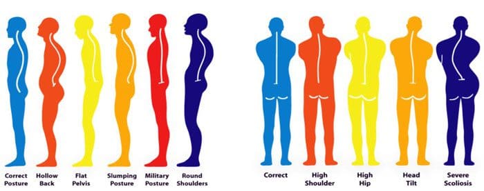
Improper posture affects the whole body and can lead to various pain issues throughout the body. Correcting posture is recommended before trying to correct it when pain begins to present. If pain is presenting, chiropractic treatment will bring relief, stabilize the spine, realign/balance the body, and educate the individual on maintaining proper posture through stretches, exercises, physical therapy, and lifestyle adjustments.
Improper posture symptoms
Neck Pain
Discomfort, stiffness, tightness, and pain are common when sitting at a workstation. This comes from a forward head/head jutting position. The head pushes forward and is not aligned with the shoulders. This means that the neck takes on a compromised position. The head forward position places significant strain on the neck muscles. Because of this neck discomfort and pain often occur later in the afternoon and evening. If not sure whether head jutting is taking place, try placing the chin to the chest. If not able or if there is discomfort/pain in the upper back, there is some forward head jutting.
Shoulder Discomfort and Pain
When we sit for extended periods, the body relaxes muscles that would normally be used if standing. One set of muscles is in the upper back. This causes slouching with a rounded upper back/shoulders. The more time the body stays in any one position, the more it begins to conform to the unhealthy position. This also causes pain in the upper, front part of the shoulders. The pain is noticeable when trying to bring the arm/s overhead or when trying to perform exercises like pushups or pullups.
Regular Headaches
Regular headaches are another symptom of improper posture. Forward head posture is usually a contributor combined with the long hours sitting or standing. However, headaches can be caused by a variety of causes that include:
Stress
Tension
Dehydration
Low Back, Tailbone Discomfort, and Pain
Lower back pain is a very common symptom of improper posture. For individuals under 40, pain and discomfort present because of improper posture while sitting or standing and a lack of stretching and exercise. Sitting for a long time causes the muscles that bring the thighs towards the chest, known as the hip flexors to be consistently flexed, with no relief. This causes the hip flexors to shorten and tighten. This pulls the pelvis out towards the front of the body, creating an exaggerated spinal curve.
Buttocks or Stomach Pushes Outward
Take a look at the body’s profile, does the butt and/or stomach stick out? If so this could be hyperlordosis also known as Donald duck posture. This can come from wearing high heels too much, the body having to carry extra weight in the stomach area, and sometimes this comes from pregnancy. Sometimes, this happens when individuals stand with their knees locked. This causes the rear end and/or belly to push out.
Correcting Improper Posture
The main problem with correcting posture is the ability to maintain proper posture. Many individuals go back to the unhealthy positioning without recognizing that they are doing it. There are devices to help correct poor posture habits. This could be a brace or harness that detects when the body is slouching and vibrates to let the individual return to a proper position.
Chiropractic Care and Physical Therapy
The most effective and thorough way to correct years of improper posture is to see a professional chiropractor. A complete diagnosis, inspection, and analysis of an individual’s posture when sitting, standing, walking, and running will be done. They will educate the individual on correct posture, how to achieve and maintain it. If pain is presenting, the chiropractor will take steps to correct any subluxations, misalignments, and develop a personalized treatment plan, to heal the body first. Treatment modalities can include:
Chiropractic adjustments
Physical therapy
Massage
Heat therapy
Infrared
Ultrasound
TENS device
Health coaching
Nutritional advice
Once the body has healed and is moving freely, the doctor will recommend exercises and stretching programs to do at home. This will improve and help maintain proper posture. An experienced musculoskeletal professional will keep the body balanced and prevent further injuries.
Body Composition
Hydrating the body with water or a sports drink
Many individuals prefer drinking sports drinks during and after physical activities, sports, and exercise. Many are opposed to water because of the lack of taste, while sports drinks have taste and added electrolytes. But many sports drinks have added ingredients and sugars. This makes them not the best choice for individuals trying to lose calories. Take a look at some of the additional ingredients:
Electrolytes
Minerals, like potassium, sodium, and magnesium, have an electric charge that helps maintain the body’s ionic balance. The body loses electrolytes when sweating. A sports drink can help replace the lost electrolytes.
Carbohydrates
Most of the carbohydrates come from sugars. Carbohydrates are one of the body’s energy sources and sports drinks are designed to refuel the body after hard physical activity.
Amino acids
These are protein building blocks. Drinking a sports drink after an intense workout can help the body recover quicker. Therefore, some of the additional ingredients in sports drinks offer hydration extras that water on its own cannot. However, water should always be the first drink of choice. But there are certain times when a sports drink is what the body needs.
When participating in high-intensity physical activities, workouts, sports that last longer than 45 minutes to an hour. Here a sports drinks can help replenish the body’s electrolytes better than water.
Individuals that sweat high levels of sodium (look for sweat stains/rings on skin or clothing) can benefit from re-hydrating with a sports drink.
Endurance athletes, triathletes, marathon runners, long-distance athletes, etc can also benefit from a sports drink, from the increased fluid loss.
In these activities, athletes should make sure the sports drink they are consuming contains carbohydrates and electrolytes.
Disclaimer
The information herein is not intended to replace a one-on-one relationship with a qualified health care professional, licensed physician, and is not medical advice. We encourage you to make your own health care decisions based on your research and partnership with a qualified health care professional. Our information scope is limited to chiropractic, musculoskeletal, physical medicines, wellness, sensitive health issues, functional medicine articles, topics, and discussions. We provide and present clinical collaboration with specialists from a wide array of disciplines. Each specialist is governed by their professional scope of practice and their jurisdiction of licensure. We use functional health & wellness protocols to treat and support care for the musculoskeletal system’s injuries or disorders. Our videos, posts, topics, subjects, and insights cover clinical matters, issues, and topics that relate to and support, directly or indirectly, our clinical scope of practice.* Our office has made a reasonable attempt to provide supportive citations and has identified the relevant research study or studies supporting our posts. We provide copies of supporting research studies available to regulatory boards and the public upon request. We understand that we cover matters that require an additional explanation of how it may assist in a particular care plan or treatment protocol; therefore, to further discuss the subject matter above, please feel free to ask Dr. Alex Jimenez or contact us at 915-850-0900.
Dr. Alex Jimenez DC, MSACP, CCST, IFMCP*, CIFM*, CTG*
email: [email protected]
phone: 915-850-0900
Licensed in Texas & New Mexico
References
Hao, Ning et al. “Enhancing creativity: Proper body posture meets proper emotion.” Acta Psychologica vol. 173 (2017): 32-40. doi:10.1016/j.actpsy.2016.12.005
Jaromi, Melinda et al. “Treatment and ergonomics training of work-related lower back pain and body posture problems for nurses.” Journal of clinical nursing vol. 21,11-12 (2012): 1776-84. doi:10.1111/j.1365-2702.2012.04089.x
O’Connor B. Sitting Disease: The New Health Epidemic. The Chopra Center Web site. http://www.chopra.com/articles/sitting-disease-the-new-health-epidemic. Accessed January 7, 2017.
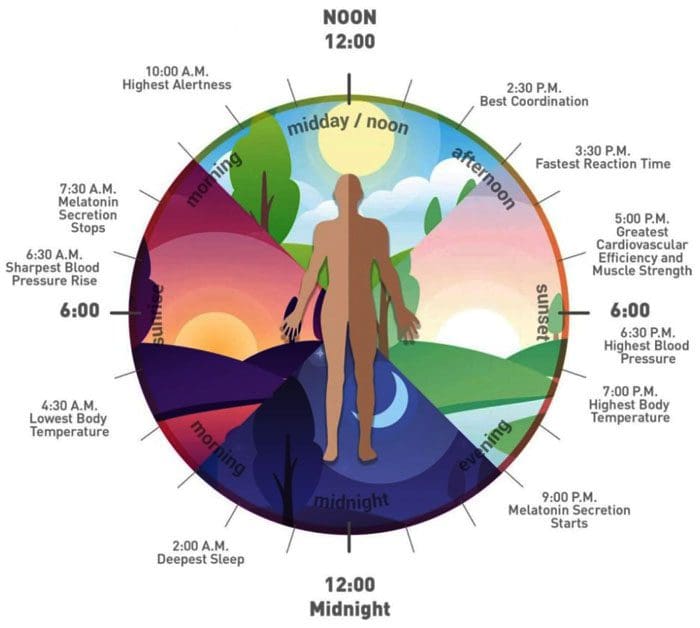
Learning how to manage and combat insomnia. Being wide awake early in the morning, trying hard to fall back to sleep before the alarm goes off. Individuals that have trouble falling asleep find that it usually happens right before a vacation. Everyone experiences an occasional sleepless night, but if insomnia continues on a regular basis it can lead to various health issues.
The average adult requires over eight hours of sleep for the body to function properly. But managing hectic lives means individuals end up going to bed later than sooner and not following the body’s natural biological rhythm. Remote and in-person learning, jobs, children, and other obligations require getting up with the birds with only 4-6 hours of sleep. A disruption to the body’s circadian rhythm that regulates:
Hormone production
Body temperature
Sleep
Can lead to insomnia.
Mind and Body Performance
The body needs adequate, restful sleep to perform its best. Insomnia that is prolonged can cause brain fog and interfere with performing daily activities. It also increases the risk for:
Depression
Headaches
Learning abilities
Accidents – auto, sports, work, personal
Can lead to sleep medication dependency.
Stress, anxiety, profound caffeine, and alcohol consumption can contribute to insomnia. Learning how to effectively manage stress is recommended to getting a proper night’s sleep. Making lifestyle adjustments can make a significant difference in the number of sleep hours. Here are a few strategies to try that could be effective:
Regular exercise/physical activity
Getting some physical activity before dinner can help put the body in a restful state before going to bed. However, do not exercise close to bedtime as this could make the body restless.
Getting out in the late evening sun as often as possible will help stimulate melatonin release. This will help reset the body’s circadian rhythm.
Stress-reduction
Stress-reduction techniques like yoga, meditation, and Tai Chi are recommended to help teach the mind and body to relax.
Caffeine, tobacco, and alcohol
These keep the body stimulated. Try to reduce/avoid from mid-afternoon until bedtime, and keep consumption of alcohol to a minimum.
Snacks
Have a small snack of protein with a complex carbohydrate just before bedtime. This could be peanut butter on a whole-grain cracker that can keep blood sugar from dropping, causing the individual to wake up.
Sleep cycle
Maintain the same sleep and wake schedule every day.
Do not alter by more than an hour on the weekends or on vacation.
Electronic devices
No television, computer, and phone use at least an hour before going to bed. This stimulates the brain, making it difficult to get to sleep.
Darkness
Keep the bedroom dark, quiet and cool.
If lying awake for more than 20 minutes, get up and sit in another dimly lit room until sleepiness starts to set in, then go back to bed.
Give these strategies a try and research others. They could help. For more information contact Injury Medical Chiropractic and Functional Medicine Clinic, to see how we can help.
Body Composition
Learning How To Incorporate Prebiotics
Incorporating more prebiotics into one’s diet is best done through nutrition. Prebiotic foods supply these nutrients directly to the colon, where they are broken down, fermented, and utilized. Prebiotic foods consist mainly of fruits, vegetables, grains, and beans.
However, cooking could alter the food’s fiber content, so look at recipes. Prebiotics also come in the form of supplements to make them easier to consume.
Disclaimer
The information herein is not intended to replace a one-on-one relationship with a qualified health care professional, licensed physician, and is not medical advice. We encourage you to make your own health care decisions based on your research and partnership with a qualified health care professional. Our information scope is limited to chiropractic, musculoskeletal, physical medicines, wellness, sensitive health issues, functional medicine articles, topics, and discussions. We provide and present clinical collaboration with specialists from a wide array of disciplines. Each specialist is governed by their professional scope of practice and their jurisdiction of licensure. We use functional health & wellness protocols to treat and support care for the musculoskeletal system’s injuries or disorders. Our videos, posts, topics, subjects, and insights cover clinical matters, issues, and topics that relate to and support, directly or indirectly, our clinical scope of practice.* Our office has made a reasonable attempt to provide supportive citations and has identified the relevant research study or studies supporting our posts. We provide copies of supporting research studies available to regulatory boards and the public upon request. We understand that we cover matters that require an additional explanation of how it may assist in a particular care plan or treatment protocol; therefore, to further discuss the subject matter above, please feel free to ask Dr. Alex Jimenez or contact us at 915-850-0900.
Dr. Alex Jimenez DC, MSACP, CCST, IFMCP*, CIFM*, CTG*
email: [email protected]
phone: 915-850-0900
Licensed in Texas & New Mexico
References
Goto, Viviane et al. “Chiropractic intervention in the treatment of postmenopausal climacteric symptoms and insomnia: A review.” Maturitas vol. 78,1 (2014): 3-7. doi:10.1016/j.maturitas.2014.02.004
Jamison, Jennifer R. “Insomnia: does chiropractic help?.” Journal of manipulative and physiological therapeutics vol. 28,3 (2005): 179-86. doi:10.1016/j.jmpt.2005.02.013
Kingston, Jana et al. “A review of the literature on chiropractic and insomnia.” Journal of chiropractic medicine vol. 9,3 (2010): 121-6. doi:10.1016/j.jcm.2010.03.003
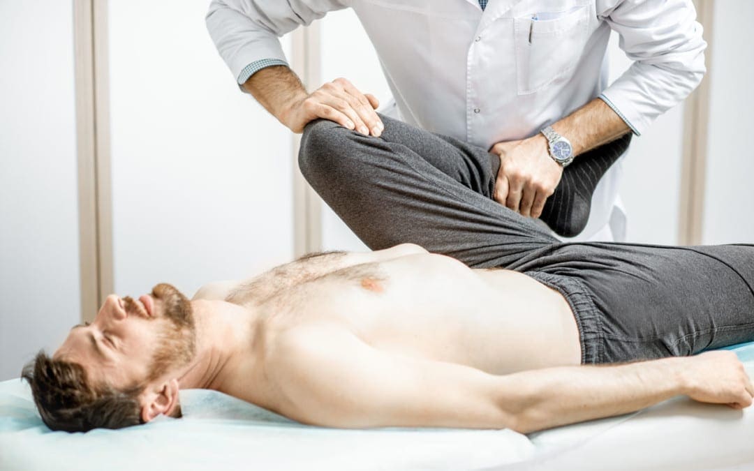
The spinal cord is the information pathway that transmits signals from the brain to the rest of the body. The body’s limbs and organs cannot function properly without regular communication flowing through the spinal cord. Understanding how the sciatic nerve responds to the other parts of the body can clarify how a doctor of chiropractic heals sciatica. When severe pain presents normal communications are overridden for the sake of body and health preservation. The sciatic nerve is an important part of the spinal cord and requires a trained professional to aid in proper and effective healing.
Basics On Sciatica
Sciatica happens when the nerves are compressed/pinched in some form. Lower back conditions can be the cause of such compression. Sciatica can be reduced and healed. Conditions that can lead to sciatic pain include:
Herniated discs
Subluxations
Disc Degeneration
Spinal Stenosis
Lower back disc bulge
Piriformis syndrome
Symptoms
Sciatica often includes:
Sharp pain
Numbness
Burning
Tingling sensation
Individuals can also experience weakness down the leg.
Normal activities can become strained as individuals experience these symptoms. Pressure on the nerve can be decreased and healed with professional chiropractic care. The human spine consists of 31 pairs of nerves. Five of these pairs are in the lumbar/lower back region and five are right below that area in the sacral region. The sciatic nerve starts in the lower back. It goes down through the hips, buttocks, thighs, knees, calves, and ends in the foot.
Causes could be something as simple as sitting on a bulging wallet or sitting for extended lengths of time can result in a decreased blood flow to the area. Also, direct trauma to the gluteal muscles from automobile accidents, sports, and work injuries can lead to sciatic nerve issues and pain.
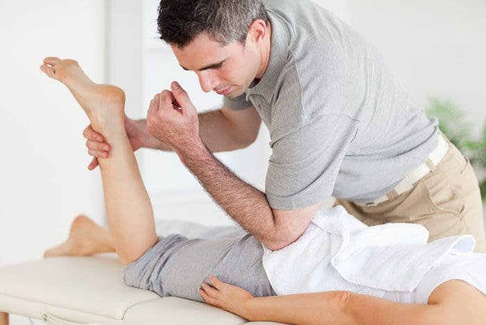
Treatment Options
Tests and examinations will be conducted to figure out the origin of the pain to develop a personalized treatment plan to expedite the healing process. Treatment can include:
Chiropractic treatment
Decreasing muscle tension
Core stabilization
Spinal decompression
Rehabilitation therapy
Chiropractic treatment is designed to help the body activate its own healing system. It is non-invasive and drug-free providing an organic alternative. Advanced cases of sciatica can result in muscle weakness or a loss of sensation in the legs, if not properly treated. Individual treatment plans vary depending on an individual’s specific needs.
Prevention
Prevention is important once the condition is healing to not cause flare-ups. Sciatica symptoms can return, especially if proper and continued care to the spine is not maintained. Preventative care and maintenance are essential for ensuring a healthy spine and body. These tips can help avoid back injuries that can lead to sciatica and help with reoccurrences:
Practice proper posture
Regular exercise/physical activity with safe movements
Bend at the knees, especially when lifting heavy objects
Follow specific instructions given by a chiropractor
Body Composition
Healthy Snacks
Eggs
Eggs make a great snack that will keep the body full and help to eat less. They are a great source of protein and fat and have a healthy variety of vitamins and minerals. Hard-boiled eggs are easy to make ahead of time and are portable.
Cheese
Cheese is a great snack for protein and fat. But it is important to watch the serving size if watching/limiting calories. Cottage cheese is highest in protein but cream cheese and cheese sticks are also good options. Cheese can be paired with a serving of fruit or vegetables like grape tomatoes, bell peppers, celery, apples, or pears.
Jerky
Jerky is convenient and portable and is a great way to pack in extra protein throughout the day. The best option is grass-fed. Remember that some jerky is high in sodium if limiting sodium.
Greek Yogurt
Natural Greek yogurt is a great option for a high-protein snack. However, many yogurts are high in sugar. Therefore choose yogurt with less than 10 grams of sugar per serving, or opt for plain yogurt to avoid the sugar altogether. The yogurt can be sweetened with honey, fresh fruit, or mixed into a smoothie.
Hummus and Guacamole Dips
Hummus is a great source of plant-based protein and is balanced with carbohydrates, fat, and fiber. It is the olive oil in the hummus that provides a healthy dose of heart-healthy polyunsaturated fats. Guacamole is a delicious source of healthy fats.Both dips are calorie-dense, therefore it is important to be aware of portion sizes. These can be paired with carrot sticks, bell peppers, or celery.
Disclaimer
The information herein is not intended to replace a one-on-one relationship with a qualified health care professional, licensed physician, and is not medical advice. We encourage you to make your own health care decisions based on your research and partnership with a qualified health care professional. Our information scope is limited to chiropractic, musculoskeletal, physical medicines, wellness, sensitive health issues, functional medicine articles, topics, and discussions. We provide and present clinical collaboration with specialists from a wide array of disciplines. Each specialist is governed by their professional scope of practice and their jurisdiction of licensure. We use functional health & wellness protocols to treat and support care for the musculoskeletal system’s injuries or disorders. Our videos, posts, topics, subjects, and insights cover clinical matters, issues, and topics that relate to and support, directly or indirectly, our clinical scope of practice.* Our office has made a reasonable attempt to provide supportive citations and has identified the relevant research study or studies supporting our posts. We provide copies of supporting research studies available to regulatory boards and the public upon request. We understand that we cover matters that require an additional explanation of how it may assist in a particular care plan or treatment protocol; therefore, to further discuss the subject matter above, please feel free to ask Dr. Alex Jimenez or contact us at 915-850-0900.
Dr. Alex Jimenez DC, MSACP, CCST, IFMCP*, CIFM*, CTG*
email: [email protected]
phone: 915-850-0900
Licensed in Texas & New Mexico
References
National Institutes of Health. (2019.) “Sciatica.” medlineplus.gov/sciatica.html.
North American Spine Society. (2012.) “Clinical Guidelines for Diagnosis and Treatment of Lumbar Disc Herniation with Radiculopathy.” www.spine.org/Portals/0/assets/downloads/ResearchClinicalCare/Guidelines/LumbarDiscHerniation.pdf
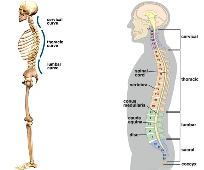
Individuals believe that a straight spine is a healthy spine. This is a misconception, a chiropractor knows that a healthy spine has the proper curvature, specifically an S curve from the top to the bottom. This curve allows/enables the spine to properly function for balancing and stabilizing the body. There are three primary curves that make up the S shape. These are:
Lordosis is defined as an inward curve, while kyphosis is an outward curve.
Curvature Significance
When the spine is properly curved and aligned there is flexibility and optimal support of the body. The curves of each section/area create a spring structure that enables the spine to perform as a natural shock-absorber. If the spine was straight, it would take a significant toll with all the wear and tear from constant vibration and shock. This would lead to fractures and other injuries. The curvature is important in aiding with weight distribution and the weight of gravity. Each curve works to distribute weight evenly throughout the spine so that the different muscle groups do not get overworked. This happens during dynamic movement.
Lifting
Bending
Reaching
Twisting
Turning
All require the spine to move harmoniously with the rest of the body, supporting the body’s weight and energy transfer.
The curves help reduce pressure between the individual vertebral discs. Because the discs are not stacked on top of each other, the weight from the above vertebra is not completely placed on the one below. So the lower vertebra takes only a portion of the weight, allowing the discs to perform as an absorbing cushion.
Maintaining The Curvature
An adult spine gradually becomes an S curve and is formed through development. Children are born with a C curve, with an outward curve until they begin to crawl. When the child begins to raise their heads up, the cervical portion of the proper S shape begins to take form. As the child learns to walk upright, the spine continues on its ultimate role of support. However, because spinal curvature is formed in the developmental stages, natural curvature formation has a limited time frame. Issues that happen at this time can disrupt the proper formation of the curves. This can result in malformations that could require long-term chiropractic treatment/rehabilitation to correct.
For adults that develop a normal S curve, spinal maintenance is critical. When subluxations/misalignments occur bulging, herniated discs, pinched nerves, and sciatica are sure to follow. Chiropractic adjustments will ensure the spine is reset and properly supported. As natural spinal development has passed maintenance is essential. Using precision tools and techniques, chiropractic treatment realigns the spine returning the natural S curve. Treatment plans and adjustments are customized to the individual and applied specifically to their unique spinal shape. This makes it possible to maintain a spine with precise curvature.
Body Composition
Fruit and Plant Antioxidants
Regular inclusion of antioxidant-rich foods in an individual’s diet will help reduce free radicals and prevent oxidative stress from accumulating. Fruits and veggies like:
Berries
Dark-colored grapes
Spinach
Kale
Sweet potatoes
Carrots
All are great sources of antioxidants.
Purple-colored fruits like blueberries and dark grapes contain an antioxidant called resveratrol that has therapeutic properties for heart disease. Beta-carotene is an orange-colored carotenoid with antioxidant properties that can also protect against heart disease. Orange, yellow-colored plant foods, and leafy greensare a solid source of the antioxidant carotenoid beta-carotene or pro-vitamin A. Minerals like zinc and selenium have antioxidants that can be found in vegan sources. And minerals found in fruits and veggies contain blood pressure-lowering potassium, magnesium, and calcium.
Disclaimer
The information herein is not intended to replace a one-on-one relationship with a qualified health care professional, licensed physician, and is not medical advice. We encourage you to make your own health care decisions based on your research and partnership with a qualified health care professional. Our information scope is limited to chiropractic, musculoskeletal, physical medicines, wellness, sensitive health issues, functional medicine articles, topics, and discussions. We provide and present clinical collaboration with specialists from a wide array of disciplines. Each specialist is governed by their professional scope of practice and their jurisdiction of licensure. We use functional health & wellness protocols to treat and support care for the musculoskeletal system’s injuries or disorders. Our videos, posts, topics, subjects, and insights cover clinical matters, issues, and topics that relate to and support, directly or indirectly, our clinical scope of practice.* Our office has made a reasonable attempt to provide supportive citations and has identified the relevant research study or studies supporting our posts. We provide copies of supporting research studies available to regulatory boards and the public upon request. We understand that we cover matters that require an additional explanation of how it may assist in a particular care plan or treatment protocol; therefore, to further discuss the subject matter above, please feel free to ask Dr. Alex Jimenez or contact us at 915-850-0900.
Dr. Alex Jimenez DC, MSACP, CCST, IFMCP*, CIFM*, CTG*
email: [email protected]
phone: 915-850-0900
Licensed in Texas & New Mexico
References
Yang, Feng et al. “Balance chiropractic therapy for cervical spondylotic radiculopathy: study protocol for a randomized controlled trial.” Trials vol. 17,1 513. 22 Oct. 2016, doi:10.1186/s13063-016-1644-2
Taylor, J A. “Full-spine radiography: a review.” Journal of manipulative and physiological therapeutics vol. 16,7 (1993): 460-74.
Fedorchuk, Curtis et al. “Improvements in Cervical Spinal Canal Diameter and Neck Disability Following Correction of Cervical Lordosis and Cervical Spondylolistheses Using Chiropractic BioPhysics Technique: A Case Series.” Journal of radiology case reports vol. 14,4 21-37. 30 Apr. 2020, doi:10.3941/jrcr.v14i4.3890
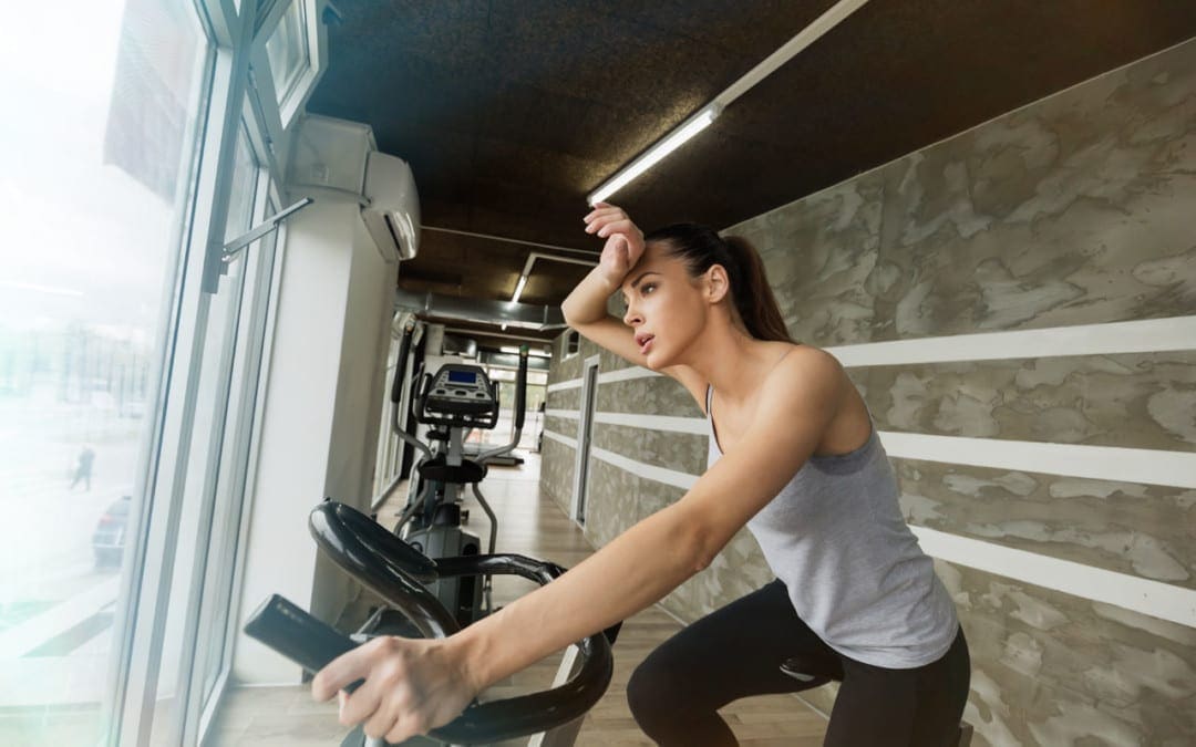
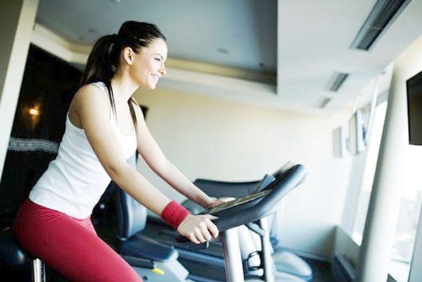
At home or a gym, working out with cardio exercise equipment can be a highly beneficial treatment for back discomfort, soreness, and pain. However, when checking out all the cardio machines it is recommended to use those that will help with back pain and not worsen or cause further injury. The same goes for purchasing cardio equipment to use at home. Research has found that exercise training could be highly effective in the treatment of back pain. A study on pain found that the endorphins generated from aerobic exercise help to lessen low back pain. The North American Spine Society announced evidence-based recommendations for the treatment of back pain, and one of the top recommendations was aerobic exercise. Individuals can always go outside to:
Walk
Hike
Run
Bike ride
But mixing it up with cardio equipment can have its own benefits for different reasons. It could be too hot, raining, sometimes individuals prefer a set workout program to reach calorie or distance goals, and it could be easier on the spine. Working out at home or at a gym, cardiovascular machines can help bring relief for back pain.
Cardiovascular Exercise Back Pain Treatment
Cardiovascular exercise is highly recommended for everyone. For individuals dealing with back pain, exercise combined with conservative therapy is usually part of a treatment plan. This includes:
Physical therapy
Chiropractic care
Health coaching
Diet
Aerobic exercise regimen
With an exercise program, experts recommend starting with moderate-intensity aerobic exercise. Moderate intensity workouts are meant to get an individual’s heart and blood pumping, sweating a little, and slightly deep breathing. These types of exercise include:
Power walking outside
Power walking on a treadmill
Stationary biking
As long as the physical activity gets the heart rate up, these exercises have been shown to decrease back pain, relieve stress, and elevate mood. 20 minutes of moderate-intensity exercise three to five times a week for six weeks is what is recommended. This will help the back become healthier, feel better, and is recommended by the American Heart Association.
Exercise Not For All Spine Conditions
However, not all spinal conditions benefit from regular exercising. Getting an evaluation from a doctor, spine specialist, or chiropractor is recommended for injuries, severe and/or persistent back pain before starting a cardiovascular exercise program. This could be a spinal fracture, or spinal condition that requires bracing, or intense physical therapy/rehabilitation. Individuals that do not exercise regularly or have a medical or heart condition/s definitely need to get a doctor’s clearance before beginning a cardiovascular workout regimen.
Top Cardiovascular Exercises and Equipment
Once a doctor clears the individual for aerobic exercise there is no cardio equipment that is off-limits. Elliptical machines and stationary bikes are the most well-tolerated by individuals with back problems/conditions. Because they are low impact. However, if it is tolerable using a jogging treadmill is beneficial as well. Listen to the body. If a workout on a treadmill causes back pain that is not just workout soreness, stop with that machine and try different cardiovascular equipment that is more low impact. Do not ignore back pain. If pain is continuous and exercising is not helping, stop and see a doctor, or chiropractor to evaluate and analyze the situation. Then they can adjust the exercise part of the treatment plan according to the presenting symptoms.
Body Composition
Concurrent Training
Concurrent training is the combination of aerobic and resistance exercises during the same workout session. Aerobic and resistance exercise impacts the body in different ways. The type of aerobic training determines how it interacts with resistance exercise. The order of the types of exercises like aerobic and resistance workouts can make a difference. Having an understanding of a few specifics about concurrent training will help to make decisions about an exercise program.
Aerobic/interval and resistance training does not seem to interfere with the others’ adaptations
However, gaining strength could be lowered by adding running to a resistance program
Whereas bicycling does not have the same effect.
Cycling and the ergonomics that go with it are similar to traditional lower-body resistance exercises. The muscle contractions that come with running result in muscle damage, while the contractions in cycling also cause muscle damage, it is not to the same extent. Pairing the exercise programs correctly is key, such as a running program in combination with an upper-body lifting exercise can be beneficial. While running and doing leg presses every day could interfere with each other and could cause injuries. Or if doing both aerobic and resistance exercises in the same session, or on the same day, consider the order of the exercises, depending on what the goal is.
Disclaimer
The information herein is not intended to replace a one-on-one relationship with a qualified health care professional, licensed physician, and is not medical advice. We encourage you to make your own health care decisions based on your research and partnership with a qualified health care professional. Our information scope is limited to chiropractic, musculoskeletal, physical medicines, wellness, sensitive health issues, functional medicine articles, topics, and discussions. We provide and present clinical collaboration with specialists from a wide array of disciplines. Each specialist is governed by their professional scope of practice and their jurisdiction of licensure. We use functional health & wellness protocols to treat and support care for the musculoskeletal system’s injuries or disorders. Our videos, posts, topics, subjects, and insights cover clinical matters, issues, and topics that relate to and support, directly or indirectly, our clinical scope of practice.* Our office has made a reasonable attempt to provide supportive citations and has identified the relevant research study or studies supporting our posts. We provide copies of supporting research studies available to regulatory boards and the public upon request. We understand that we cover matters that require an additional explanation of how it may assist in a particular care plan or treatment protocol; therefore, to further discuss the subject matter above, please feel free to ask Dr. Alex Jimenez or contact us at 915-850-0900.
Dr. Alex Jimenez DC, MSACP, CCST, IFMCP*, CIFM*, CTG*
email: [email protected]
phone: 915-850-0900
Licensed in Texas & New Mexico
References
British Journal of Sports Medicine. (November 2020) “Which specific modes of exercise training are most effective for treating low back pain? Network meta-analysis” https://bjsm.bmj.com/content/54/21/1279
Pain. (December 2020) “Are endogenous opioid mechanisms involved in the effects of aerobic exercise training on chronic low back pain? A randomized controlled trial” https://journals.lww.com/pain/Citation/2020/12000/Are_endogenous_opioid_mechanisms_involved_in_the.23.aspx
North American Spine Society. (2020) “Evidence-Based Clinical Guidelines for Multidisciplinary Spine Care” https://www.spine.org/Portals/0/assets/downloads/ResearchClinicalCare/Guidelines/LowBackPain.pdf
IFM's Find A Practitioner tool is the largest referral network in Functional Medicine, created to help patients locate Functional Medicine practitioners anywhere in the world. IFM Certified Practitioners are listed first in the search results, given their extensive education in Functional Medicine