Discover the impact of stretching & flexibility on joint pain relief. Incorporate these tips into your routine for optimal results.
Contents
Chiropractic Care and Stretching: A Holistic Approach to Joint Pain Relief
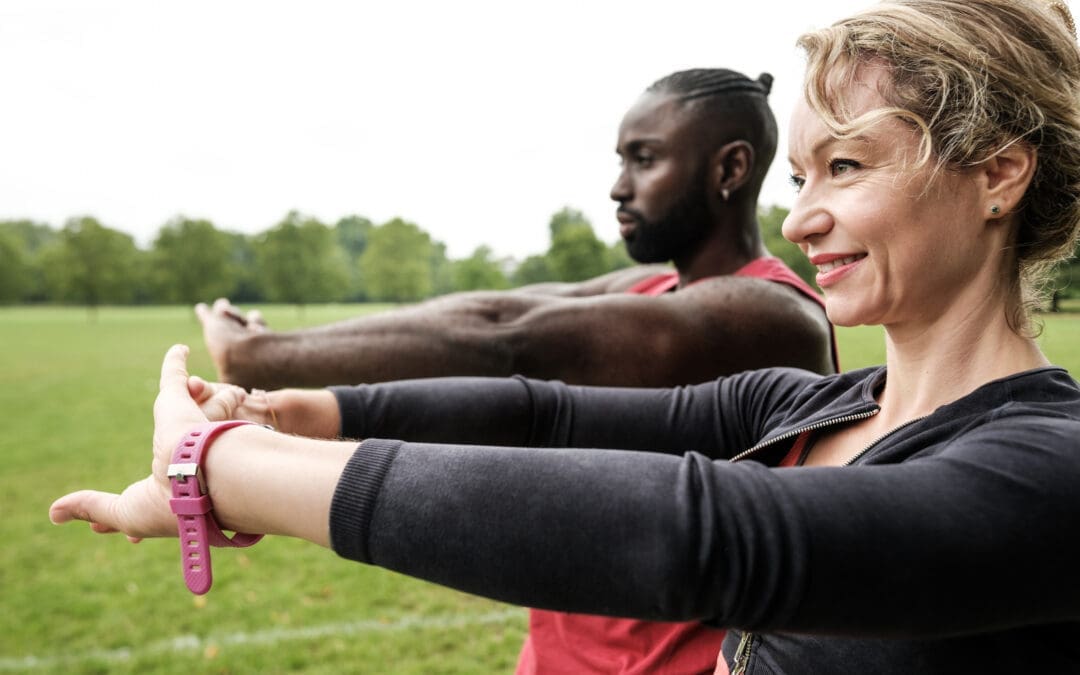
Joint pain can transform routine tasks like walking, lifting, or sitting into daunting challenges. Whether it’s a persistent ache in your knees, stiffness in your shoulders, or discomfort in your back, joint pain affects countless individuals globally. Fortunately, nonsurgical solutions such as chiropractic care, combined with stretching and flexibility exercises, provide a natural and effective way to manage pain, enhance mobility, and improve overall well-being. These methods address both symptoms and underlying causes, promoting long-term healing and a better quality of life.
This comprehensive guide explores the clinical rationale for integrating chiropractic care with stretching to alleviate joint pain. We’ll examine the causes and risk factors for joint pain in the upper and lower extremities, highlight the role of integrative therapies like massage and acupuncture, and provide practical stretching exercises suitable for home or gym settings. Supported by scientific research and expert insights, this article empowers you to take control of your joint health and recover from injuries, including those sustained in motor vehicle accidents (MVAs), bicycle collisions, or 18-wheeler crashes.
5 Things You Need to Know About
Ligamentous Injuries Before They Get Worse-Video
Understanding Joint Pain: Causes and Risk Factors
Joint pain arises from a complex interplay of factors, from acute injuries to chronic conditions. Identifying these causes is crucial for developing an effective treatment plan. Below are the primary contributors to joint pain in both upper and lower extremities:
1. Mechanical Factors
Joint Misalignment: Misaligned joints in the spine, shoulders, or knees can create uneven stress on surrounding muscles, tendons, and ligaments, leading to pain and inflammation. For instance, knee malalignment may contribute to patellofemoral pain syndrome, common among active individuals (Steinberg et al., 2021).
Overuse and Repetitive Stress: Repetitive motions from sports, work, or daily activities can strain joints, leading to conditions such as shoulder impingement syndrome or tennis elbow. Overhead athletes, such as cyclists or swimmers, often experience shoulder pain due to repetitive stress (Tauqeer et al., 2024).
Trauma or Injury: Acute injuries, such as sprains, fractures, or dislocations from MVAs or bicycle accidents, can damage joint structures, causing pain and reduced mobility. For example, anterior cruciate ligament (ACL) injuries are prevalent in athletes and can lead to significant knee pain and instability (Hurley, 1997).
2. Degenerative Conditions
Osteoarthritis: A leading cause of joint pain, osteoarthritis involves the breakdown of cartilage in joints such as the knees, hips, and hands, resulting in pain, stiffness, and a limited range of motion (Luan et al., 2022).
Rheumatoid Arthritis: This autoimmune condition causes inflammation in the synovial lining of joints, leading to tenderness, swelling, and potential joint damage (Dumoulin et al., 2023).
3. Generalized Joint Hypermobility (GJH)
Some individuals have naturally flexible joints, a condition known as generalized joint hypermobility (GJH). While advantageous for activities like dance, it increases the risk of joint instability and pain, particularly in the upper cervical spine or knees (Russek et al., 2023; Steinberg et al., 2021).
4. Inflammation and Systemic Factors
Inflammatory Conditions: Diseases like rheumatoid arthritis or psoriatic arthritis drive joint inflammation, exacerbating pain. Subclinical inflammation can cause tenderness in joints, such as the metacarpophalangeal (MCP) joints, even without a formal diagnosis (Dumoulin et al., 2023).
Muscle Imbalances and Poor Posture: Weak core muscles or poor posture can increase stress on joints, particularly in the spine, hips, and shoulders, leading to pain and dysfunction.
5. Lifestyle and Environmental Factors
Sedentary Lifestyle: A lack of movement can cause muscle stiffness and reduce joint lubrication, thereby increasing the risk of pain.
Obesity: Excess body weight places additional stress on weight-bearing joints, such as the knees and hips, accelerating cartilage wear (Luan et al., 2022).
Poor Ergonomics: Improper workstation setups or repetitive tasks, such as typing or lifting, can strain upper extremity joints, contributing to conditions like carpal tunnel syndrome.
Overlapping Risk Profiles
These factors often overlap, creating a complex risk profile for joint pain. For example, an individual with GJH may have weak supporting muscles, increasing the risk of joint instability. Similarly, someone with osteoarthritis might experience worsened symptoms due to repetitive stress or poor posture. Chiropractic care and stretching target these overlapping risks by improving joint alignment, enhancing muscle function, reducing inflammation, and promoting stability, offering a holistic approach to pain management and recovery from injuries like those sustained in MVAs or bicycle collisions.
The Clinical Rationale for Chiropractic Care and Stretching
Chiropractic care, paired with stretching and flexibility exercises, addresses the root causes of joint pain, offering a nonsurgical alternative to pain management. This integrative approach restores joint function, enhances muscle performance, and promotes the body’s natural healing processes, particularly for injuries from MVAs, 18-wheeler crashes, or bicycle accidents. Below is the clinical rationale for combining these modalities:
1. Restoring Joint Alignment and Function
Chiropractic Adjustments: Chiropractic adjustments, or thrust joint manipulations, involve applying controlled force to misaligned joints to restore proper alignment. This reduces stress on surrounding tissues, improves mobility, and alleviates pain. For example, spinal adjustments can help relieve low back pain associated with MVAs by correcting subluxations that irritate nerves (Rhyu et al., 2015).
Reducing Joint Stress: Misaligned joints lead to compensatory muscle tightness and inflammation. Adjustments redistribute forces across joints, reducing wear and tear, particularly in degenerative conditions such as osteoarthritis (Luan et al., 2022).
Evidence: Research shows thrust joint manipulation is effective for improving joint function and reducing pain in the lumbar and thoracic spine, with high confidence in its safety for these regions (Puentedura et al., 2017).
2. Enhancing Muscle Function and Proprioception
Muscle Activation: Joint damage from accidents or osteoarthritis can reduce voluntary muscle activation, resulting in weakness and muscle atrophy. Chiropractic care, combined with targeted exercises, helps restore muscle function by enhancing neural signaling (Hurley, 1997).
Proprioception: Injuries, particularly from MVAs or bicycle collisions, can impair proprioception, increasing the risk of further injury. Stretching and strengthening exercises enhance proprioceptive feedback, improving joint stability (Steinberg et al., 2021).
Evidence: Isometric exercises, often prescribed alongside chiropractic care, increase muscle activity and reduce pain in patients with low back pain from accidents (Rhyu et al., 2015).
3. Reducing Inflammation and Pain
Anti-Inflammatory Effects: Chiropractic adjustments and stretching improve joint mobility and blood flow, reducing inflammation. This is particularly effective for inflammatory conditions like rheumatoid arthritis or whiplash-associated disorders (WAD) from MVAs (Dumoulin et al., 2023).
Pain Modulation: Stretching exercises, particularly when combined with manual therapy, have been shown to significantly reduce pain in conditions such as knee osteoarthritis and shoulder impingement syndrome (Luan et al., 2022; Tauqeer et al., 2024).
Evidence: A meta-analysis found that stretching exercises alone resulted in a clinically meaningful reduction in knee osteoarthritis pain, with enhanced benefits when combined with other therapies (Luan et al., 2022).
4. Preventing Long-Term Complications
Joint Stability: For individuals with GJH or scoliosis, chiropractic care and targeted exercises strengthen supporting muscles, reducing the risk of joint instability and related injuries (Russek et al., 2023; Steinberg et al., 2021).
Holistic Healing: By addressing biomechanical, muscular, and neurological factors, chiropractic care promotes long-term joint health, preventing chronic pain and disability from accident-related injuries.
Evidence Suggests That Rehabilitation programs incorporating stretching and strengthening exercises improve outcomes in patients with joint hypermobility, scoliosis, or post-accident trauma, thereby reducing the risk of patellofemoral pain (Steinberg et al., 2021).
5. Complementary Therapies
Massage Therapy: Massage reduces muscle tension, improves circulation, and prepares tissues for chiropractic adjustments. It is particularly effective for shoulder impingement and whiplash injuries, enhancing range of motion and functional capacity (Tauqeer et al., 2024).
Acupuncture: Acupuncture stimulates endorphin release, reduces inflammation, and improves neural signaling, making it a valuable adjunct for managing pain from osteoarthritis, low back pain, or MVA injuries.
Integrative Medicine: An integrative approach combining chiropractic adjustments, stretching, strengthening, massage, and acupuncture addresses the multifaceted nature of joint pain, promoting natural healing (El Paso Back Clinic, n.d.).
Evidence: Manual therapies, including massage, significantly reduce pain and improve function in patients with chronic shoulder conditions and post-accident injuries (Tauqeer et al., 2024).
6. Patient-Centered Care
Clear communication ensures tailored treatment plans that address individual needs, whether recovering from an 18-wheeler crash or managing chronic arthritis. Patient education enables individuals to perform home exercises that maintain progress (El Paso Back Clinic, n.d.).
Evidence Suggests That Patient education and active participation in rehabilitation programs enhance adherence and outcomes in musculoskeletal care (Jimenez, 2016).
By targeting overlapping risk factors—misalignment, muscle weakness, inflammation, and instability—chiropractic care and stretching provide a comprehensive solution for joint pain relief and recovery from accident-related injuries.
Stretching and Flexibility Exercises for Joint Pain Relief
Stretching and flexibility exercises are essential for maintaining joint health, improving range of motion, and reducing pain, especially after MVAs or bicycle accidents. Below are practical exercises suitable for home or gym settings, supported by research. Consult a healthcare provider before starting, particularly if you have injuries or conditions like GJH or scoliosis.
1. Cat-Cow Stretch (Spinal Flexibility)
Purpose: Enhances spinal flexibility, reduces low back pain, and improves core stability, ideal for MVA recovery.
How to Perform:
Position yourself on your hands and knees, with your hands under your shoulders and your knees under your hips.
Inhale, letting your abdomen drop toward the floor while gently arching your back (Cow Pose).
Exhale, arching your back upward like a cat, tucking your chin to your chest (Cat Pose).
Repeat 3–5 times, moving slowly.
Benefits: Increases spinal mobility and reduces tension in back muscles (Jimenez, 2016).
Frequency: Perform daily, morning and evening, for 5–10 minutes.
Tip: Move smoothly to avoid straining the spine.
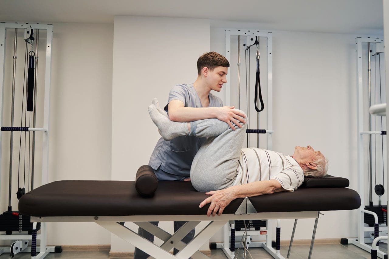
2. Knee-to-Chest Stretch (Lower Back and Hip Flexibility)
Purpose: Relieves tension in the lower back and hips, beneficial for low back pain and sciatica from MVAs.
How to Perform:
Lie on your back with knees bent and feet flat.
Grasp one knee with both hands and pull it toward your chest.
Hold for 30 seconds, then return to the starting position.
Repeat with the other knee or both knees together.
Benefits: Improves lumbar flexibility and reduces pain (Jimenez, 2016).
Frequency: Perform 2–3 times per leg, twice daily.
Tip: Keep your lower back pressed against the floor.
3. Scorpion Stretch (Lower Back and Core)
Purpose: Stretches the lower back and strengthens core muscles, ideal for chronic back pain post-accident.
How to Perform:
Lie face down with arms extended out to the sides.
Lift your right leg and move it toward your left arm, keeping your torso stable.
Hold for 10 seconds, then return to the starting position.
Repeat with the left leg toward the right arm.
Benefits: Enhances lumbar flexibility and core strength (Jimenez, 2016).
Frequency: Perform 2–3 repetitions per side, once daily.
Purpose: Enhances scapular mobility and reduces shoulder impingement pain, common in bicycle accidents.
How to Perform:
Sit or stand with arms relaxed at your sides.
Squeeze your shoulder blades together, as if holding a pencil between them.
Hold for 5–10 seconds, then release.
Benefits: Improves scapular range of motion and reduces shoulder pain (Tauqeer et al., 2024).
Frequency: Perform 10–15 repetitions, 2–3 times daily.
Tip: Keep your shoulders relaxed to avoid shrugging.
6. Standing Quadriceps Stretch (Knee and Hip Flexibility)
Purpose: Stretches the quadriceps to reduce knee pain and improve mobility.
How to Perform:
Stand near a wall for balance, holding one ankle with the same-side hand.
Pull your ankle toward your buttocks, keeping your knees aligned.
Hold for 20–30 seconds, then switch legs.
Benefits: Enhances knee flexibility and reduces patellofemoral pain (Steinberg et al., 2021).
Frequency: Perform 2–3 times per leg, daily.
Tip: Tuck your pelvis to avoid arching your lower back.
7. Neck Rotation Stretch (Cervical Flexibility)
Purpose: Reduces neck stiffness and improves cervical mobility, especially for GJH or whiplash from MVAs.
How to Perform:
Sit or stand with your back straight.
Turn your head to the right, looking over your shoulder, and hold for 15–20 seconds.
Return to the center and repeat on the left.
Benefits: Improves cervical range of motion and reduces symptoms of instability (Russek et al., 2023).
Frequency: Perform 3–5 repetitions per side, twice daily.
Tip: Move within your comfortable range to avoid strain.
Tips for Safe Stretching
Warm Up First: Engage in 5–10 minutes of light activity, such as walking, to prepare your muscles and joints (Jimenez, 2016).
Avoid Overstretching: Stretch to mild tension, not to the point of pain, to prevent injury.
Breathe Deeply: Inhale and exhale slowly to enhance relaxation and muscle lengthening.
Consult a Professional: Work with a chiropractor or physical therapist to ensure proper technique, especially for post-accident recovery or conditions like GJH or scoliosis.
Integrative Therapies for Enhanced Joint Pain Relief
Integrative therapies, such as massage and acupuncture, complement chiropractic care and stretching, addressing muscle tension, inflammation, and neurological factors, particularly in cases related to accidents.
1. Massage Therapy
Benefits: Massage reduces muscle tightness, improves circulation, and prepares tissues for chiropractic adjustments. It is effective for shoulder impingement, whiplash, and post-MVA recovery (Tauqeer et al., 2024; El Paso Back Clinic, n.d.).
Application: Techniques such as deep tissue massage or trigger point therapy target tight muscles and fascia, thereby enhancing the benefits of stretching.
Evidence Suggests That Manual therapy, including massage, significantly reduces pain and improves function in individuals with chronic shoulder conditions and accident-related injuries (Tauqeer et al., 2024).
2. Acupuncture
Benefits: Acupuncture stimulates endorphin release, reduces inflammation, and improves neural signaling, effective for osteoarthritis, low back pain, and WAD from MVAs.
Application: Integrated with chiropractic care, acupuncture addresses local and systemic pain pathways, enhancing recovery.
Evidence: Research supports the use of acupuncture as an effective adjunct for managing musculoskeletal pain (Luan et al., 2022).
3. Nutrition for Recovery
Benefits: A diet rich in anti-inflammatory foods (e.g., omega-3 fatty acids, fruits, and vegetables) supports tissue healing and reduces inflammation, crucial for post-accident recovery (El Paso Back Clinic, n.d.).
Application: Nutritional guidance complements chiropractic care, promoting internal healing.
Evidence: Proper nutrition enhances musculoskeletal injury rehabilitation, particularly after MVAs (El Paso Back Clinic, n.d.).
Preventing Long-Term Joint Problems
Chiropractic care and stretching not only relieve joint pain but also prevent long-term complications by addressing underlying causes. Key strategies include:
Consistent Exercise: Daily stretching and strengthening enhance joint stability and flexibility.
Healthy Lifestyle Choices: Maintain a healthy weight, eat an anti-inflammatory diet, and practice good posture to reduce joint stress.
Early Intervention: Seek chiropractic care at the first sign of pain to prevent progression to chronic conditions like osteoarthritis or WAD.
Durable Medical Equipment: Braces or supports may aid recovery from MVA injuries, as recommended by professionals (El Paso Back Clinic, n.d.).
Conclusion
Joint pain from injuries, degenerative conditions, or lifestyle factors can significantly impact daily life. Chiropractic care, combined with stretching and flexibility exercises, provides a powerful, non-surgical solution for managing and preventing pain. By addressing joint misalignment, enhancing muscle function, reducing inflammation, and promoting holistic healing through integrative therapies like massage, acupuncture, and nutrition, this approach targets the root causes of joint pain. Incorporating the stretching exercises above and seeking professional guidance can improve function, reduce pain, and support a more active, pain-free life.
References
Dumoulin, Q. A., van Steenbergen, H. W., & van der Helm-van Mil, A. H. M. (2023). Correspondence on ‘Role of joint damage, malalignment and inflammation in articular tenderness in rheumatoid arthritis, psoriatic arthritis and osteoarthritis’. Annals of the Rheumatic Diseases, 82(7), e160. https://doi.org/10.1136/annrheumdis-2021-220511
Hurley, M. V. (1997). The effects of joint damage on muscle function, proprioception, and rehabilitation. Manual Therapy, 2(1), 11–17. https://doi.org/10.1054/math.1997.0281
Luan, L., El-Ansary, D., Adams, R., Wu, S., & Han, J. (2022). Knee osteoarthritis pain and stretching exercises: A systematic review and meta-analysis. Physiotherapy, 114, 16–29. https://doi.org/10.1016/j.physio.2021.10.001
Puentedura, E. J., Slaughter, R., Reilly, S., Ventura, E., & Young, D. (2017). Thrust joint manipulation utilization by U.S. physical therapists. Journal of Manual & Manipulative Therapy, 25(2), 74–82. https://doi.org/10.1080/10669817.2016.1187902
Rhyu, H.-S., Park, H.-S., & Park, J.-S. (2015). The Effects of Isometric Exercise Types on Pain and Muscle Activity in Patients with Low Back Pain. Journal of Exercise Rehabilitation, 11(4), 211–214. https://doi.org/10.12965/jer.150224
Russek, L. N., Block, N. P., Byrne, E., Chalela, S., Chan, C., Comerford, M., … Hakim, A. (2023). Presentation and physical therapy management of upper cervical instability in patients with symptomatic generalized joint hypermobility: International expert consensus recommendations. Frontiers in Medicine, 9, 1072764. https://doi.org/10.3389/fmed.2022.1072764
Steinberg, N., Tenenbaum, S., Zeev, A., & Hershkovitz, I. (2021). Generalized joint hypermobility, scoliosis, patellofemoral pain, and physical abilities in young dancers. BMC Musculoskeletal Disorders, 22(1), 161. https://doi.org/10.1186/s12891-021-04023-z
Tauqeer, S., Arooj, A., & Javed, K. (2024). Effects of manual therapy in addition to stretching and strengthening exercises to improve scapular range of motion, functional capacity, and pain in patients with shoulder impingement syndrome: A randomized controlled trial. BMC Musculoskeletal Disorders, 25(1), 192. https://doi.org/10.1186/s12891-024-07294-4
Gut-Skin Axis Healing: Radiant Skin Through Wellness
Introduction
At El Paso Back Clinic®, we understand that your skin reflects your inner health, especially after injuries from car accidents, sports, or work. The gut-skin axis links gut health to skin conditions such as acne, eczema, and premature aging. When injuries disrupt your gut microbiome—causing dysbiosis—inflammation and oxidative stress can weaken your skin’s barrier. Our team, led by Dr. Alex Jimenez, DC, APRN, FNP-BC, utilizes chiropractic care, functional medicine, and nutrition to treat both injuries and skin conditions.
Research indicates that balancing your gut microbiome can help clear skin issues (Kober & Bowe, 2015). We create personalized plans to restore wellness, combining advanced therapies with holistic care. This article examines the impact of dysbiosis on skin after injury and how El Paso Back Clinic’s integrative approach promotes vibrant health and radiant skin.
The Gut-Skin Axis: A Wellness Connection
The gut-skin axis links your digestive system to your skin. A healthy gut produces short-chain fatty acids (SCFAs) that reduce inflammation and support immunity (Salem et al., 2018). Injuries, stress, or medications can cause dysbiosis, allowing harmful bacteria to leak toxins into the bloodstream, which can trigger skin issues (Bowe et al., 2014). Dysbiosis also increases oxidative stress, damaging collagen and causing wrinkles, while reducing ceramides that strengthen the skin barrier (Krutmann et al., 2019). At El Paso Back Clinic, we use chiropractic adjustments, nutrition, and therapies to restore gut balance, heal skin, and treat injuries.
How Dysbiosis Impacts Skin After Injury
Injuries stress the body, disrupting gut health and worsening skin conditions:
Acne: Dysbiosis from injury-related stress or meds boosts insulin, clogging pores. Studies link low gut diversity to acne (Lee et al., 2019, as cited in Wang et al., 2023). Our nutrition plans reduce sugar and add probiotics to calm breakouts.
Eczema: Low gut diversity lets bacteria like Staphylococcus aureus thrive, causing rashes. Probiotics reduce the risk of eczema by 30% (Szari & Quinn, as cited in Johnson et al., 2024). We use functional medicine to rebuild gut health.
Premature Aging: Dysbiosis-driven oxidative stress degrades collagen, accelerating the formation of wrinkles. Injury-related inflammation adds “inflammaging” (Fisher et al., 2002). Our antioxidant-rich diets and stress relief can help reverse this.
Our integrative care focuses on these pathways to facilitate comprehensive recovery and healing.
Inflammation and Oxidative Stress: The Skin’s Enemies
Injuries amplify inflammation and oxidative stress, linking dysbiosis to skin issues. Leaky gut releases toxins (LPS), triggering cytokines like IL-6, causing redness or psoriasis (Mu & Kirby, 2018). Oxidative stress damages the skin’s structure, resulting in thinning of the dermis (Kim et al., 2018, as cited in Wang et al., 2023). A weak skin barrier allows irritants to enter, worsening dryness (Simpson et al., 2014). We utilize chiropractic adjustments to alleviate nerve stress, probiotics to lower cytokines, and nutrition to enhance antioxidant levels, with trials demonstrating that Lactobacillus reduces oxidative markers by 25% in acne patients (Fabbrocini et al., 2016, as cited in Wang et al., 2023).
Dietary Changes: Nourish Gut, Enhance Skin
Nutrition is crucial to healing the gut-skin axis. We recommend:
Prebiotics, such as garlic, onions, and bananas, feed good bacteria, which in turn reduces inflammation (Slavin, 2013).
Probiotics, such as those found in yogurt and kimchi, can help restore balance, reducing acne lesions by 20-30% (Kober & Bowe, 2015).
Fiber: 35 grams daily from oats and beans boosts SCFAs (Makki et al., 2018).
We avoid sugar and dairy, which spike inflammation (Bowe et al., 2010). Our Mediterranean-style diets, tailored for injury recovery, promote clear skin and gut health (Barrea et al., 2015).
Stress Reduction: Calming Gut and Skin
Injury-related stress increases cortisol, disrupting gut bacteria and exacerbating skin issues (Konturek et al., 2011). Our clinic offers mindfulness and yoga to lower cortisol by 20% (Carlson et al., 2015). Poses like child’s pose stimulate the vagus nerve, which in turn reduces inflammation (West et al., 2004). These complement our injury rehab for clearer skin.
Targeted Supplementation: Boosting Recovery
Supplements support healing:
Vitamin D: 2,000 IU daily eases eczema (Umar et al., 2018).
Zinc: 30 mg heals acne wounds (Gupta et al., 2014).
Omega-3s: 1-2g hydrates skin (Serefko et al., 2016).
Probiotics: Multi-strain supplements balance gut (Gueniche et al., 2010, as cited in Wang et al., 2023).
Our nurse practitioners tailor these assessments based on individual needs.
Lifestyle Tweaks: Supporting Skin and Recovery
Sleep 7-9 hours to lower cortisol (Benedict et al., 2016). Walk 30 minutes daily to boost circulation. Use SPF 30 to protect skin. Our plans integrate these for optimal wellness.
El Paso Back Clinic’s Integrative Approach
At El Paso Back Clinic, Dr. Alex Jimenez and our team combine chiropractic care, functional medicine, and acupuncture to address injury-related dysbiosis. Adjustments reduce nerve stress, improving gut function (Jafarzadeh et al., 2020). Our therapies cut inflammation, enhancing skin and overall health (Horrigan, 2017).
Dr. Alex Jimenez: Leading Holistic Recovery
Dr. Alex Jimenez, DC, APRN, FNP-BC, with over 30 years of experience, uses dual-scope diagnostics—chiropractic and nursing—to treat injuries from MVAs, sports, or work. Advanced imaging, such as MRI, links injuries to gut stress, which in turn impacts the skin (Jimenez, n.d.a). For a patient with whiplash and acne, Dr. Jimenez might use adjustments, acupuncture, and probiotics to heal both. Our clinic provides detailed legal documentation for injury claims, ensuring accurate reports (Jimenez, n.d.b). Exercises, massage, and nutrition can help prevent chronic issues, as shared in Dr. Jimenez’s blog, offering holistic insights.
Personalized Plans: Your Wellness Journey
We begin with gut and skin assessments, including stool tests, bloodwork, or barrier scans. Plans include diets (prebiotics for dysbiosis), supplements (zinc for acne), and therapies (massage for stress). A patient with post-injury eczema experienced a 60% improvement with the combination of probiotics and yoga, as reported by Johnson et al. (2024).
Case Studies: Real Recoveries
Maria, 40: MVA-related back pain and psoriasis. Dr. Jimenez’s plan—adjustments, omega-3s, fiber—eased pain and cleared skin in 10 weeks.
Jake, 25: Work injury and acne. Nutrition and acupuncture balance the gut, reducing breakouts (Nirvana Healthcare, n.d.).
Advanced Care: Probiotics and Imaging
Probiotics, such as Bifidobacterium breve, protect the skin from UV damage (Ishii et al., 2014, as cited in Wang et al., 2023). We pair these with neuromusculoskeletal imaging for precise recovery plans.
Preventing Long-Term Issues
Regular gut checks and stress management prevent chronic pain and skin issues. Our proactive plans ensure lasting wellness.
Myths Busted
Myth: Skin issues are only topical. Fact: Gut drives 70% of immunity (Mu & Kirby, 2018). We provide evidence-based care to debunk myths.
Nutrition Deep Dive
For acne, we suggest low-glycemic foods and zinc-rich nuts. Eczema patients get fiber-rich plans with recipes like chia pudding. Psoriasis benefits from fish and greens. Our nutritionists create tailored menus.
Gut-Friendly Movement
Pilates and walking boost gut motility. Our therapists guide 20-minute routines that complement chiropractic care.
Supplement Science
Vitamin D reduces inflammation associated with eczema (Umar et al., 2018). Zinc heals acne (Gupta et al., 2014). Omega-3s hydrate skin (Serefko et al., 2016). We test for deficiencies to ensure safe dosing.
Our Unique Protocols
Dr. Jimenez uses MRI to link injuries to dysbiosis, which can impact the skin. Adjustments restore nerve function, while acupuncture and massage boost nutrient flow. Our app tracks progress.
Why Choose El Paso Back Clinic
Located at 11860 Vista Del Sol, Ste 128, El Paso, TX, we offer specialized injury care that combines chiropractic, nutrition, and rehabilitation services. We accept most insurance plans and work closely with your providers. Call 915-850-0900 or email [email protected].
Conclusion: Heal and Glow with Us
At El Paso Back Clinic, we harness the gut-skin axis to heal injuries and improve skin health. Dr. Jimenez’s integrative approach ensures vibrant wellness. Visit us or call 915-850-0900 to start your journey.
References
Bowe, W. P., Joshi, S. S., & Shalita, A. R. (2010). Diet and acne. Journal of the American Academy of Dermatology, 63(1), 117–122.
Gupta, M., Mahajan, V. K., Mehta, K. S., & Chauhan, P. S. (2014). Zinc therapy in dermatology: A review. Dermatology Research and Practice, 2014, 709152.
Serefko, A., Szopa, A., Wlaź, P., Nowak, G., Radziwoń-Zaleska, M., Skalski, M., & Poleszak, E. (2016). Magnesium in depression. Pharmacological Reports, 68(2), 306–313.
Explore options for a chair that provides comfort and support for back pain relief during long hours of sitting.
Contents
Ergonomic Chairs and Chiropractic Care: Your Path to a Pain-Free Back
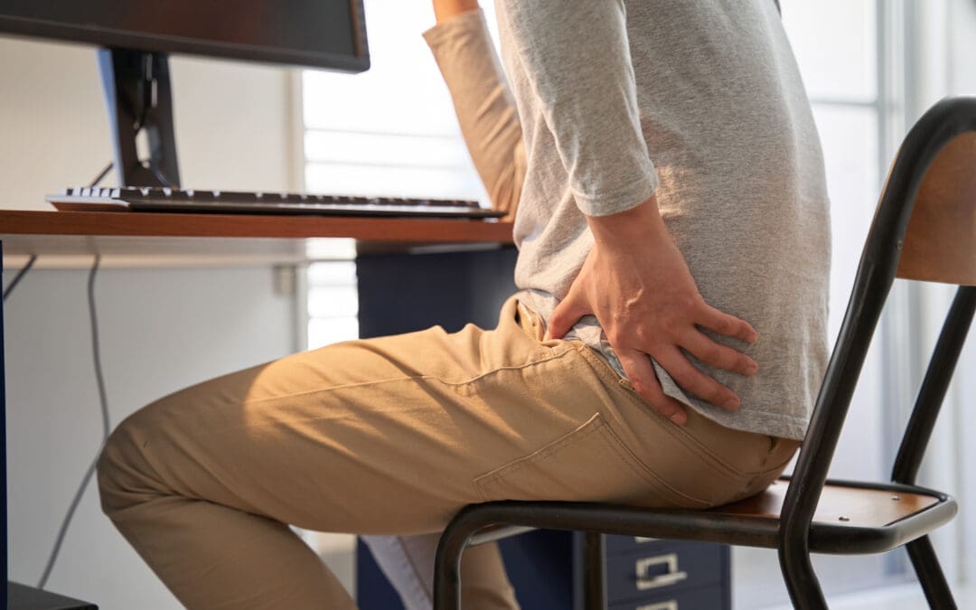
Back pain is a widespread issue that affects millions of people, whether they’re sitting at a desk for hours, unwinding at home, or managing daily stress. Research indicates that approximately 80% of adults will experience low back pain at some point, making it a leading cause of missed workdays and medical visits (Will et al., 2018). Fortunately, practical solutions like ergonomic chairs and chiropractic care can significantly alleviate discomfort and improve spinal health. This comprehensive guide explores the causes of back pain, the impact of poor seating, the benefits of ergonomic chairs in home and office settings, the risks of prolonged sitting, and how nonsurgical treatments, including chiropractic adjustments, targeted exercises, and integrative therapies, can promote natural healing and prevent long-term issues.
Drawing on evidence-based insights and expertise from a chiropractor and family nurse practitioner in El Paso, Texas, this article emphasizes holistic, patient-centered care that supports the body’s natural recovery processes. By the end, you’ll have actionable strategies to enhance posture, reduce pain, and maintain a healthy spine.
Understanding Back Pain: Causes and Contributors
Back pain can range from a mild ache to debilitating discomfort, impacting daily activities like work, sleep, and exercise. The lower back, or lumbar region, is particularly susceptible due to its role in supporting body weight and facilitating movement. Back pain is classified as specific (caused by identifiable conditions, such as fractures or infections) or non-specific (lacking a clear cause, often linked to lifestyle factors), with non-specific pain accounting for approximately 90% of cases (Chenot et al., 2017).
Key Factors Contributing to Back Pain
Several factors contribute to back pain, many of which are influenced by daily habits:
Poor Posture: Slouching or leaning forward strains spinal muscles and ligaments, weakening the spine’s natural support over time.
Sedentary Lifestyle: Prolonged sitting reduces blood flow to the spine and increases pressure on intervertebral discs, leading to stiffness and pain. Studies show that office workers who sit for extended periods have a higher incidence of low back pain (Bontrup et al., 2019).
Muscle Imbalances: Weak core muscles or tight hamstrings can misalign the spine, causing discomfort.
Injuries: Sudden strains from improper lifting, sports, or motor vehicle accidents (MVAs) can trigger acute pain.
Aging and Degeneration: As we age, spinal discs may degenerate, leading to conditions like herniated discs or osteoarthritis.
Stress and Emotional Factors: Psychological stress can cause muscles to tighten, exacerbating pain and contributing to chronic issues.
Occupational Risks: Jobs involving heavy lifting, vibrations, or irregular schedules, such as night shifts, increase the risk of back pain by 31%, particularly in healthcare workers (Chen et al., 2023).
Mechanical low back pain, stemming from issues with the spine or surrounding tissues, is the most common type (Will et al., 2018). Lumbar instability, where weakened ligaments allow excessive vertebral movement, can also lead to persistent pain (Hauser et al., 2022). Understanding these factors is crucial for effective prevention and treatment.
Impact of Motor Vehicle Accidents
Motor vehicle accidents (MVAs) are a significant cause of back pain, often resulting in injuries like whiplash-associated disorders (WAD), sprains, or disc issues. These injuries can disrupt spinal alignment and lead to chronic pain if not addressed properly. Chiropractic care and integrative therapies play a vital role in MVA recovery by restoring function and reducing inflammation.
How Poor Seating Affects Your Spine
Using a broken or poorly designed chair can directly harm your spinal health. The spine consists of 33 vertebrae, cushioned by discs that absorb shock. A faulty chair disrupts this delicate system in several ways:
Uneven Weight Distribution: A sagging or uneven seat forces the body into awkward positions, compressing specific vertebrae and discs, which can cause inflammation or pain.
Lack of Lumbar Support: Without a backrest that supports the spine’s natural “S” curve, the lower back flattens, stressing lumbar vertebrae and increasing the risk of subluxations—slight misalignments that irritate nerves.
Improper Height: A chair that’s too high or low disrupts leg and pelvis positioning, pulling on hip muscles connected to the spine and misaligning vertebrae.
Instability: A wobbly chair forces constant adjustments, fatigues back muscles, and risks minor vertebral shifts.
Reduced Circulation: Poorly designed chairs can prevent feet from resting flat, thereby limiting blood flow to the legs and spine, which can slow healing and exacerbate pain.
Prolonged use of such chairs can accelerate spinal degeneration, causing vertebrae to rub abnormally and potentially leading to conditions like osteoarthritis or facet joint issues (Jimenez, 2023b). Research confirms that static sitting, common with poor chairs, increases disc pressure and low back pain risk (Bontrup et al., 2019).
Lower Back Pain Relief After Gym Injury- Video
Advantages of Ergonomic Chairs for Home and Office
Ergonomic chairs are designed to support the body’s natural alignment, reducing strain and enhancing comfort. With adjustable features like seat height, lumbar support, and tilt mechanisms, they’re ideal for preventing back pain in various settings. Here’s how they benefit users at work and home.
Benefits in the Office
For those spending long hours at a desk, ergonomic chairs offer:
Customizable Seat Height: Adjust the chair so feet are flat and knees form a 90-degree angle, reducing thigh and lower back pressure while maintaining vertebral alignment.
Lumbar Support: A contoured backrest supports the spine’s natural curve, preventing slouching and reducing stress on lumbar discs.
Adjustable Armrests and Swivel Base: Armrests reduce shoulder tension, which can pull on the back, while a swivel base allows movement without twisting the spine.
Enhanced Productivity: Comfort reduces fatigue, improving focus and reducing errors. Studies show ergonomic seating decreases low back pain and boosts efficiency (Bontrup et al., 2019).
Injury Prevention: Tilt and recline features promote dynamic sitting, keeping muscles active and reducing stiffness.
Longevity: Durable materials ensure long-term use, making them a cost-effective investment.
Benefits at Home
Ergonomic chairs are equally valuable at home for remote work, relaxation, or hobbies:
Versatility: Adjustable features accommodate tasks like reading, gaming, or working, minimizing strain.
Family-Friendly: Easy adjustments allow multiple users to find comfortable positions, reducing back pain risk for all.
Health Maintenance: Supports proper posture during leisure, preventing pain buildup from long days.
Cost Efficiency: Reduces the need for medical interventions by preventing chronic pain.
Stylish Design: Modern options seamlessly blend with home decor, combining function with aesthetic appeal.
Support for Recovery: For those with existing back issues, ergonomic chairs aid healing by maintaining spinal alignment.
Adjustable seat depth and tilt mechanisms ensure users of all sizes can find a comfortable position, reducing spinal strain (Jimenez, 2023a).
Key Features to Look For
Casters: Five-point base with wheels for stability and mobility.
Seat Pan: At least one inch wider than hips, with dense foam or spring coils for lasting comfort.
Backrest: Adjustable lumbar support fitting the lower back and pelvis.
Tilt/Recline: Allows weight redistribution to ease disc pressure.
Breathable Fabric: Prevents heat buildup for prolonged comfort.
Risks of Prolonged Sitting
Even with an ergonomic chair, prolonged sitting poses health risks. Regular movement is essential to counteract these effects:
Spinal Pressure: Sitting increases disc pressure by 40-90% compared to standing, risking bulges or herniations (Will et al., 2018).
Muscle Weakness: Inactive back and core muscles weaken, compromising posture and increasing pain risk.
Poor Circulation: Restricted blood flow causes leg swelling and limits nutrient delivery to the spine, slowing recovery.
Weight Gain: Reduced calorie burn contributes to obesity, which in turn adds spinal stress.
Mental Health: Chronic pain from sitting can lead to stress or depression, worsening physical symptoms.
Chronic Diseases: Prolonged sitting is linked to heart disease, diabetes, and cancer.
Shift workers, especially those on night shifts, face a 31% higher risk of low back pain (Chen et al., 2023). Standing or stretching every 30 minutes can mitigate these risks.
Chiropractic Care: A Cornerstone of Back Pain Relief
Chiropractic care is a nonsurgical, drug-free approach focusing on spinal alignment and nervous system function. It’s effective for managing back pain, particularly mechanical and non-specific types.
How It Helps
Chiropractors assess the spine for subluxations—misalignments that irritate nerves and cause pain. Gentle adjustments restore alignment, offering:
Spinal Support: Evenly distributes weight across vertebrae, preventing wear.
Posture Improvement: Trains the body for better positioning, reducing strain.
Pain Relief: Effective for acute low back pain, often outperforming medications (Kinkade, 2007).
Enhanced Function: Improves nervous system performance, boosting overall health.
Chiropractic care prioritizes precision and patient communication, not force, ensuring tailored treatments (Jimenez, 2023a). For non-specific pain, it promotes natural healing without drugs (Chenot et al., 2017).
Chiropractic for MVA Injuries
Post-MVA, chiropractic care addresses injuries like whiplash or disc issues by restoring alignment and reducing inflammation. It’s a key component of recovery plans, often paired with other therapies for optimal results.
Synergy of Ergonomic Chairs and Chiropractic Care
Combining ergonomic chairs with chiropractic care creates a powerful approach to managing back pain. The clinical rationale includes:
Dual Support System: Ergonomic chairs provide daily spinal support, while chiropractic adjustments correct underlying misalignments, ensuring long-term alignment and reduced disc pressure (Hauser et al., 2022).
Pain Reduction: Adjustments offer immediate relief, and chairs prevent pain recurrence, supported by studies on mechanical low back pain (Will et al., 2018).
Natural Healing: Both methods support the body’s repair processes, avoiding surgery and preventing chronic issues like lumbar instability.
For example, a chair-related misalignment can be corrected with adjustments, while an ergonomic chair prevents further strain, creating a cycle of healing and prevention.
Complementary Nonsurgical Treatments
Other nonsurgical therapies enhance chiropractic and ergonomic interventions:
Targeted Exercises: Core exercises like planks or the McKenzie method strengthen spinal support and reduce pain recurrence (Will et al., 2018).
Acupuncture stimulates natural pain relief and is effective for managing chronic pain (Graf et al., 2023).
Integrative Medicine: Addresses nutrition, stress, and lifestyle to reduce inflammation and support overall health.
These therapies emphasize prevention and patient education, ensuring long-term results through tailored plans, not just physical strength.
Expert Insights from El Paso
A chiropractor and family nurse practitioner in El Paso, Texas, with dual expertise in chiropractic and functional medicine, advocates for integrative care. His approach combines:
Holistic Assessments: Using tools like the Living Matrix to identify pain triggers.
Personalized Plans: Tailoring treatments to address physical, nutritional, and emotional factors.
Patient Education: Empowering patients with knowledge about posture and lifestyle.
Learn more at dralexjimenez.com or linkedin.com/in/dralexjimenez. He emphasizes, “Support your spine daily with proper tools and professional care to unlock your body’s healing potential.”
Prevention Strategies for a Healthy Back
Prevent back pain with these practical tips:
Optimize Your Workspace: Adjust chair height, lumbar support, and monitor position for neutral posture.
Take Breaks: Stand or stretch every 30 minutes to reduce disc pressure.
Exercise Daily: Engage in 30 minutes of low-impact activities like yoga or swimming.
Maintain a Healthy Weight: Eat anti-inflammatory foods to reduce spinal stress.
Sleep Smart: Use a medium-firm mattress to support spinal alignment.
Seek Early Care: Consult professionals for persistent pain to prevent chronic issues.
Conclusion
Back pain doesn’t have to limit your life. Ergonomic chairs and chiropractic care offer a powerful combination to support your spine, improve posture, and reduce discomfort. Paired with exercises, massage, acupuncture, and integrative medicine, these nonsurgical approaches promote natural healing and prevent long-term problems. Start with small changes—adjust your chair, move regularly, and consider professional care—to enjoy a healthier, pain-free back.
References
Bontrup, C., Taylor, W. R., Fliesser, M., Visscher, R., Green, T., Wippert, P. M., & Zemp, R. (2019). Low Back Pain and Its Relationship with Sitting Behavior among Sedentary Office Workers. Applied Ergonomics, 81, 102894. https://pubmed.ncbi.nlm.nih.gov/31422243/
Chen, H.-M., Liu, C.-H., Yang, C.-H., Chen, Y.-J., & Wang, C.-L. (2023). Association of low back pain with shift work: A meta-analysis. International Journal of Environmental Research and Public Health, 20(2), 918. https://pubmed.ncbi.nlm.nih.gov/36673675/
Chenot, J.-F., Greitemann, B., Kladny, B., Petzke, F., Pfingsten, M., & Schorr, S. G. (2017). Non-specific low back pain. Deutsches Ärzteblatt International, 114(51-52), 883–890. https://pubmed.ncbi.nlm.nih.gov/29321099/
Graf, F., Nater, U. M., & Biedermann, L. (2023). [Lower back pain: Specific or non-specific?] Therapeutische Umschau, 80(4), 167–173. https://pubmed.ncbi.nlm.nih.gov/37122186/
Hauser, R. A., Matias, L. I., Woznica, D., Rawlings, B., & Woldin, B. A. (2022). Lumbar instability as an etiology of low back pain and its treatment by prolotherapy: A review. Journal of Back and Musculoskeletal Rehabilitation, 35(4), 701–712. https://pubmed.ncbi.nlm.nih.gov/34957989/
Emotional Driving & How El Paso Back Clinic Heals the Mind–Body Divide
Introduction
Driving is more than a mechanical task. When strong emotions—anger, grief, stress, excitement—take over, driving becomes risky. Emotional driving occurs when your attention is diverted from the road toward internal feelings. This impairs focus, slows reaction time, and can lead to serious crashes (Lawyer Don, n.d.; Car Accident Help, n.d.).
At El Paso Back Clinic, under the care of Dr. Alexander Jimenez, DC, APRN, FNP-BC, we see many patients who, after an auto accident or work injury, also report being emotionally shaken before or during the event. The clinic’s integrative model doesn’t just treat symptoms—it aims to heal both the physical damage and the emotional stress behind it. By doing so, El Paso Back Clinic helps people drive (and live) more safely and fully.
What Exactly Is Emotional Driving?
Emotional driving refers to operating a vehicle while strong emotions distract from safe driving. These emotions may be:
Anger or road rage
Sadness or grief
Stress or anxiety
Over-excitement
Even positive feelings, if overwhelming, can reduce awareness of surroundings. The brain only has so much capacity for processing, so when so much is going on emotionally, it can’t give full attention to driving (Pintas & Mullins, n.d.; Car Accident Help, n.d.).
Why It’s Dangerous
Here’s how emotional driving leads to danger:
Delayed reactions: A driver under stress or emotional overload may take longer to brake or swerve.
Impaired judgment: Anger or anxiety can lead to risky choices, such as speeding, tailgating, and ignoring traffic signals.
Tunneled attention: Emotions narrow attention, making it easier to miss hazards (Lawyer Don, n.d.).
Physical symptoms: Stress causes muscular tension (especially in neck/shoulders), elevated heart rate, and poor posture—all of which reduce control and focus while driving (Genesis Medical, n.d.; Spine Clinic Salem, n.d.).
How Emotional States Also Affect Injuries
When injuries happen (auto accidents, sports injuries, work accidents), emotional states often worsen the physical situation:
Tense muscles around injured areas slow healing.
Anxiety or stress can cause inflammation to persist.
Poor sleep and unresolved emotional stress weaken immune response and recovery (Denver Chiropractic, n.d.; HelloNote, n.d.).
In El Paso Back Clinic, many patients with motor vehicle injuries arrive not only with physical symptoms like whiplash or back pain but also emotional distress—panic, fear, or anger. These get documented, addressed, and integrated into treatment.
El Paso Back Clinic’s Dual-Scope & Integrative Approach
Dr. Jimenez has a special position: he’s both a Doctor of Chiropractic and a Board-Certified Nurse Practitioner (FNP-BC). This allows him to diagnose using medical tools and treat with chiropractic, integrative medicine, and other modalities. (a4m.com)
Key Components of Care at El Paso Back Clinic
Advanced imaging & diagnostics: X-rays, MRI, ultrasound, where needed, to see not just the injury but also how the nervous and musculoskeletal systems are impacted. (El Paso, TX Doctor Of Chiropractic)
Chiropractic adjustments: To restore spinal alignment, relieve nerve compression, and reduce tension.
Integrative medicine: Nutrition, functional medicine assessments, and lifestyle changes to address systemic stress. (El Paso, TX Doctor Of Chiropractic)
Acupuncture, massage, and soft tissue work: These help relax the body, reduce inflammation, and promote emotional calm.
Rehabilitation and exercise therapy: Customized programs to restore strength, flexibility, and better body awareness.
Legal & Injury Case Documentation
For patients injured in auto accidents or work incidents, the clinic provides:
Coordination of care with legal and insurance entities
Follow-up care plans that cover both physical injury and emotional well-being
How This Care Helps With Emotional Driving
When someone uses El Paso Back Clinic’s services, here’s how it helps reduce the risk of emotional driving:
Reduced pain and tension → Less physical distraction or discomfort when driving.
Improved emotional regulation (through integrative methods) → Less reactive driving, more stable mood.
Better sleep and recovery → More alertness, sharper reflexes.
Holistic awareness of how stressors (work, injury, emotional trauma) are influencing both mind and body.
Patient Scenario (Hypothetical but Based on Clinic Observations)
Maria, a patient in El Paso, is in a car accident. She has neck pain (whiplash) and lower back strain. She also reports feeling extremely anxious when thinking of driving again. At El Paso Back Clinic:
She receives imaging confirming soft tissue damage and alignment shifts.
Chiropractic adjustments relieve spinal tension and reduce nerve irritation.
Dr. Jimenez assesses her nutrition and stress levels and recommends changes to reduce inflammation.
Massage and acupuncture help her relax and sleep better.
She is coached in breathing, mindfulness, and coping techniques, so when she must get back in the car, she’s calmer and more aware.
Over weeks, Maria notices less pain, less anxiety when driving, better posture, and fewer physical flares when emotional stress arises.
Practical Tips to Use Before Driving
Even outside of clinic visits, patients are encouraged to:
Take a few minutes of deep breathing before starting the car
Stretch neck, shoulders, and back to release tension
Recognize emotional state: avoid driving when feeling overwhelmed, if possible
Use mindfulness or brief meditation when stressed
Make sure you rest well: fatigue makes emotional driving worse
Conclusion
Emotional driving is a hidden risk on El Paso roads and everywhere. Strong emotions steal attention, slow responses, and make driving dangerous. But at El Paso Back Clinic, the combined chiropractic, medical, integrative, and rehabilitative care led by Dr. Alex Jimenez offers a powerful solution. By addressing both body and mind, reducing pain and stress, improving sleep, and providing tools for emotional self-regulation, patients gain safety, health, and resilience.
For folks in El Paso, committing to this kind of holistic treatment isn’t just about recovery—it’s about preventing future accidents and driving with confidence, clarity, and control.
Live Pain-Free: Chiropractic and Integrative Care for Injury Recovery at El Paso Back Clinic
In the vibrant heart of El Paso, Texas, where desert trails beckon and hardworking days define our community, injuries can derail your active lifestyle. From car accidents to workplace strains or sports mishaps, overexertion and trauma often lead to pain, stiffness, or chronic issues that linger without proper care. These setbacks can limit your ability to work, play, or enjoy El Paso’s unique spirit. At El Paso Back Clinic, led by Dr. Alexander Jimenez, DC, APRN, FNP-BC, chiropractic and integrative care offer a path to recovery. Through spinal adjustments, soft tissue therapy, and neuromuscular re-education, the clinic accelerates healing, restores flexibility, enhances balance, and boosts heart and lung function. With holistic nutrition and stress management plans, Dr. Jimenez’s team crafts personalized strategies to prevent future injuries, empowering El Pasoans to live pain-free and thrive.
This article explores how injuries arise, the benefits of integrative care, and how El Paso Back Clinic delivers top-tier recovery solutions.
The Impact of Overuse and Accidents: Why Pain Persists
El Paso’s dynamic lifestyle—hiking the Franklin Mountains, working long shifts, or driving busy roads—can strain the body. Overexertion from repetitive tasks like lifting or intense workouts causes sprains, strains, or joint issues. Motor vehicle accidents (MVAs) bring sudden trauma, with 60% of cases leading to lingering pain if untreated (Jimenez, n.d.). Even minor falls at home can spark chronic discomfort.
Dr. Alexander Jimenez, a chiropractor and nurse practitioner with over 30 years of experience, sees these patterns daily. “Our dual-scope diagnostics, combining chiropractic and nursing insights, uncover how trauma or overuse triggers pain cycles,” he shares on his clinic’s site (Jimenez, n.d.). Using advanced neuromusculoskeletal imaging, his team pinpoints root causes, from workplace injuries to MVA trauma. Ignoring early signs, such as stiffness or fatigue, can lead to reduced mobility, increased stress, and sleep disturbances. El Paso Back Clinic’s integrative approach breaks this cycle, restoring health naturally.
Everyday Injuries: From Crashes to Chronic Strains
Injuries vary but share a common impact: they disrupt your life. MVAs cause neck and back pain, limiting movement. Work-related strains, like those from lifting or repetitive tasks, create nagging discomfort. Sports injuries, such as twisted ankles or knees, sideline active El Pasoans. Personal falls at home can lead to shoulder or hip pain, while untreated stress may cause chronic conditions like joint stiffness.
Dr. Jimenez’s clinic tackles these with precision. “We connect injury origins—crashes, work tasks, or sports—to customized treatments,” he explains. MVAs receive urgent care with legal documentation for claims. Work injuries get rehab to restore function, and sports or personal injuries benefit from targeted plans to prevent recurrence. Without care, these issues worsen, lowering the quality of life. El Paso Back Clinic’s chiropractic and integrative methods pave the way to recovery.
Realigning for Relief: The Power of Spinal Adjustments
Spinal adjustments are the foundation of chiropractic care at El Paso Back Clinic. These precise, hands-on techniques realign vertebrae, easing nerve pressure and restoring balance to the body. Injuries from accidents or overuse misalign the spine, causing pain and impaired movement. Adjustments can boost blood flow, reduce inflammation, and alleviate pain by up to 25% in as little as a few weeks (Trident Health Chiropractic, n.d.).
For MVA patients, adjustments relieve neck stiffness, restoring mobility. Work injury patients regain strength for daily tasks. Dr. Jimenez’s approach is unique: “We use imaging to guide adjustments, targeting issues from trauma or strains,” he says. Legal reports ensure MVA patients have clear records for claims. From athletes to office workers, adjustments help El Pasoans move freely and heal quickly.
Healing Muscles: Soft Tissue Therapy for Recovery
Injuries tighten muscles, creating knots that misalign joints and prolong pain. Soft tissue therapy, like massage or myofascial release, targets these areas, breaking up scar tissue and boosting circulation. This delivers nutrients to damaged tissues, speeding recovery. A single session can significantly reduce healing time, getting you back to work or play faster (Yoder Chiropractic Center, n.d.).
Picture a construction worker with shoulder pain from heavy lifting. Therapy loosens tightness, improving arm range. MVA patients find relief from neck strain. Dr. Jimenez’s team pairs therapy with imaging for precision. “We treat trauma from accidents or sports non-surgically,” he notes. Legal documentation tracks progress for claims, prioritizing natural healing. Patients feel relaxed, move more easily, and recover more quickly.
Injuries disrupt nerve-muscle communication, resulting in shaky balance or impaired movements. Neuromuscular re-education uses exercises like balance drills or resistance training to retrain these pathways, reducing fall risks and boosting confidence. A soccer player with a sprained ankle, for example, regains stability, thereby lowering the odds of re-injury (Integrative Chiropractic, n.d.).
Dr. Jimenez’s clinic excels here. “We link nerve issues to injury histories, guiding re-education for MVA, work, or sports recovery,” he says. A retail worker with back pain learns core-strengthening moves; an MVA patient rebuilds neck control. Legal reports detail progress for claims, ensuring comprehensive care. This sharpens coordination, making daily tasks and active pursuits feel natural again.
Faster Healing, Better Mobility: Recovery and Flexibility Gains
Chiropractic care at El Paso Back Clinic speeds healing by optimizing body systems. Adjustments and therapy reduce swelling, allowing tissues to mend faster—often in weeks, not months (Abundant Life Chiropractor, n.d.). Flexibility improves as tight muscles and joints stretch safely. A warehouse worker lifts without strain; an accident victim moves freely again.
Dr. Jimenez’s holistic plans amplify results. “Targeted exercises and adjustments build lasting mobility, preventing chronic issues,” he says. Nutrition tips, like anti-inflammatory foods, fuel healing. MVA and work cases get legal-grade documentation, aligning care with claims. El Pasoans recover quickly, staying active in our vibrant community.
Balance and coordination are key to preventing injuries and enhancing daily function. Re-education drills steady wobbly steps, helping MVA victims or athletes avoid falls. A delivery driver navigates uneven terrain easily post-care. Chiropractic also boosts stamina by freeing the spine for deeper breaths, improving oxygen flow and endurance (ASR Sports Medicine, n.d.).
Jimenez’s integrative approach shines: “Acupuncture and massage enhance flow, boosting stamina for work or sports.” Virtual coaching reinforces gains, and legal support ensures MVA patients have clear records. Patients work longer, play harder, and live stronger.
Whole-Person Healing: Nutrition, Stress, and Custom Plans
El Paso Back Clinic’s functional medicine approach goes beyond physical fixes. Nutrition advice—like omega-3s or antioxidant-rich fruits—fights inflammation and boosts energy. Stress management, such as mindfulness or breathing exercises, eases tension, aiding sleep and recovery. Personalized plans fit your injury, lifestyle, and goals.
Dr. Jimenez leads the way. “We uncover root causes—poor diet, stress—and craft plans with acupuncture or massage,” he says. MVA or work injuries get detailed reports for legal cases, prioritizing natural healing. Patients receive plans tailored to their El Paso lives, ensuring lasting wellness.
El Paso Back Clinic: Your Trusted Recovery Partner
At El Paso Back Clinic, Dr. Alexander Jimenez combines chiropractic and nursing expertise for exceptional care. Awarded from 2015 to 2024, his team treats MVAs, work strains, sports injuries, and personal falls with precision. “Our imaging and dual expertise catch hidden issues,” he says. A crash victim drives pain-free in weeks. A nurse lifts patients again. Legal documentation supports MVA and work cases, while virtual coaching and nutrition webinars empower long-term health.
Patients praise the results: “Dr. Jimenez restored my mobility and energy,” one shares. From veterans to families, his care transforms lives, helping El Pasoans thrive.
Preventing Future Pain: A Strategy for Lifelong Wellness
Prevention keeps you active. Regular chiropractic checkups spot misalignments early, cutting injury risks by 20% (Erie Chiro, n.d.). Holistic habits—such as balanced diets, stress relief, and smart exercise—build resilience. Dr. Jimenez’s team creates plans for workers, athletes, or retirees. “We flag risks like posture or stress early, ensuring lasting health,” he notes.
With care, education, and documentation, El Pasoans live pain-free, embracing our city’s vibrant spirit.
Discover the connection between garlic and its anti-inflammatory properties for alleviating musculoskeletal pain in your body.
Contents
Chiropractic Care and Anti-Inflammatory Diets: A Holistic Approach to Musculoskeletal Pain Relief
Musculoskeletal pain, including back pain, neck pain, and joint discomfort, affects millions worldwide, often disrupting daily activities, work, and overall quality of life. While medications and surgery are common treatments, nonsurgical approaches like chiropractic care combined with an anti-inflammatory diet provide a holistic, effective way to manage and reduce pain. This comprehensive guide explores the clinical rationale for integrating chiropractic care with an anti-inflammatory diet, emphasizing the role of foods like garlic in reducing inflammation and supporting immune function. We’ll also examine the factors contributing to musculoskeletal pain, the nutritional benefits of garlic, and how integrative, nonsurgical treatments promote the body’s natural healing processes. Drawing on evidence-based insights, this article offers actionable strategies for pain relief and improved health.
Understanding Musculoskeletal Pain
Musculoskeletal pain refers to discomfort in muscles, bones, joints, ligaments, tendons, or nerves. It can be acute (short-term) or chronic (lasting over three months), ranging from mild aches to severe, debilitating pain. Low back pain, for instance, affects approximately 80% of adults at some point, making it a leading cause of disability globally (World Health Organization, 2023). Chronic musculoskeletal pain can lead to emotional distress, reduced mobility, and diminished quality of life, underscoring the need for effective, sustainable management strategies.
Factors Contributing to Musculoskeletal Pain
Several factors contribute to musculoskeletal pain, and addressing these is key to effective treatment. These include:
Injuries and Trauma: Acute injuries from motor vehicle accidents (MVAs), sports, or falls, such as sprains, strains, or whiplash, can damage muscles, ligaments, or joints, causing pain. For example, a herniated disc from an MVA can lead to persistent back pain (El Paso Back Clinic, 2025a).
Poor Posture and Ergonomics: Prolonged sitting, slouching, or improper lifting techniques strain the musculoskeletal system. Office workers who sit for extended periods without proper lumbar support are prone to lower back pain due to spinal misalignment.
Chronic Inflammation: Inflammation, triggered by stress, a poor diet, or conditions such as arthritis, exacerbates musculoskeletal pain by increasing pressure on nerves and tissues (Sala-Climent et al., 2023).
Sedentary Lifestyle: A lack of physical activity weakens muscles, reduces joint flexibility, and increases the risk of conditions such as sciatica or muscle stiffness. Regular movement is essential for musculoskeletal health.
Obesity: Excess body weight stresses weight-bearing joints like the spine, hips, and knees. Obesity also promotes systemic inflammation, worsening pain (Imaizumi et al., 2023).
Stress and Psychological Factors: Chronic stress or anxiety causes muscle tension, particularly in the neck and shoulders, contributing to pain. Psychological factors can also amplify pain perception (Sala-Climent et al., 2023).
Degenerative Conditions: Osteoarthritis, degenerative disc disease, and scoliosis cause chronic pain due to wear and tear on joints, discs, or connective tissues.
Nutritional Deficiencies: Diets lacking anti-inflammatory nutrients, such as omega-3 fatty acids, vitamin D, or antioxidants, impair tissue repair and inflammation control, prolonging pain (Rawson et al., 2018).
By addressing these factors through chiropractic care, dietary changes, and lifestyle modifications, individuals can significantly reduce musculoskeletal pain and enhance their quality of life.
Clinical Rationale for Chiropractic Care and Anti-Inflammatory Diets
Chiropractic care and anti-inflammatory diets work synergistically to address the root causes of musculoskeletal pain, such as inflammation, spinal misalignment, and poor tissue health. This integrative approach is grounded in evidence-based practices and aligns with functional medicine principles, focusing on holistic care rather than symptom suppression.
Chiropractic Care: A Nonsurgical Solution
Chiropractic care involves manual adjustments, spinal manipulation, and other nonsurgical techniques to treat musculoskeletal disorders, particularly those affecting the spine. The goal is to restore proper alignment, improve joint mobility, and reduce nerve irritation, alleviating pain and supporting natural healing (El Paso Back Clinic, 2025b).
Benefits of Chiropractic Care
Correcting Spinal Misalignments: Vertebral subluxations can compress nerves, causing pain and dysfunction. Chiropractic adjustments realign the spine, reducing nerve pressure and improving function.
Reducing Inflammation: Spinal manipulations lower pro-inflammatory cytokines, proteins that contribute to inflammation and pain, helping to alleviate discomfort (Dragan et al., 2020).
Enhancing Mobility: Pain and stiffness often restrict joint movement. Chiropractic techniques restore joint function, improving movement and reducing pain.
Promoting Natural Healing: By enhancing blood flow, reducing muscle tension, and optimizing nervous system function, chiropractic care supports the body’s innate healing processes without relying on medications or surgery (El Paso Back Clinic, 2025b).
Chiropractic care is particularly effective for conditions like whiplash-associated disorders (WAD), sciatica, and degenerative arthritis, which are common after MVAs or due to chronic conditions. Personalized treatment plans ensure patients receive care tailored to their specific needs (El Paso Back Clinic, 2025a).
The Role of Anti-Inflammatory Diets
An anti-inflammatory diet, rich in foods like garlic, fruits, vegetables, nuts, and omega-3 fatty acids, complements chiropractic care by targeting systemic inflammation, a key driver of musculoskeletal pain. Chronic inflammation occurs when the immune system remains activated, releasing chemicals that damage tissues and intensify pain (Sala-Climent et al., 2023).
How Anti-Inflammatory Diets Work
Lowering Inflammatory Markers: Foods like garlic, turmeric, and berries reduce C-reactive protein (CRP) and other inflammatory markers, alleviating pain and protecting tissues (Dragan et al., 2020).
Supporting Tissue Repair: Nutrients such as vitamin C, zinc, and omega-3 fatty acids promote collagen production and tissue repair, essential for healing muscles, tendons, and ligaments (Rawson et al., 2018).
Boosting Immune Function: Anti-inflammatory foods strengthen the immune system, helping it regulate inflammation effectively, particularly in conditions like rheumatoid arthritis (Ahmed et al., 2021).
Improving Overall Health: A diet low in pro-inflammatory foods (e.g., processed sugars, red meat) and high in whole foods supports cardiovascular health, blood sugar regulation, and weight management, reducing musculoskeletal stress (Imaizumi et al., 2023).
A pilot study by Sala-Climent et al. (2023) found that an anti-inflammatory Mediterranean diet (AnMeD-S), excluding red meat, gluten, and cow’s milk, significantly reduced pain, stress, and sleep disturbances in patients with chronic pain due to rheumatic diseases. This evidence supports the integration of dietary interventions with chiropractic care to enhance pain relief.
Synergy of Chiropractic Care and Diet
Combining chiropractic care with an anti-inflammatory diet creates a powerful synergy for pain management. Chiropractic adjustments address structural and neurological issues, while an anti-inflammatory diet reduces systemic inflammation, creating an optimal environment for healing. Patients adopting both approaches often report faster recovery, reduced pain, and improved energy levels (El Paso Back Clinic, 2025b). This integrative model prioritizes long-term health over temporary symptom relief.
Fighting Inflammation Naturally- Video
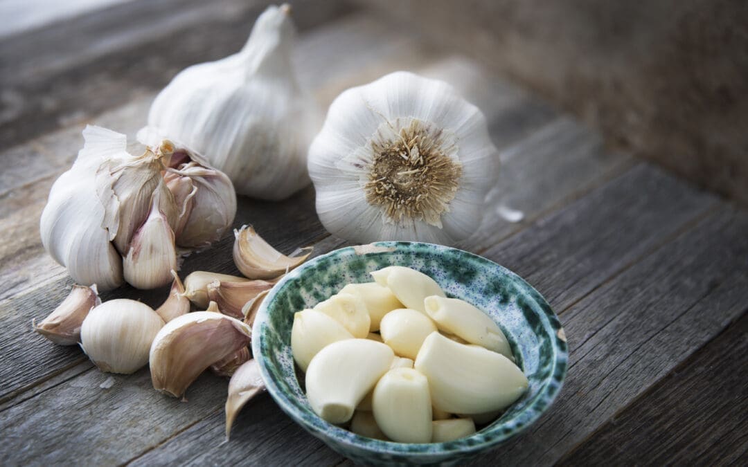
Garlic: A Key Anti-Inflammatory Food
Garlic (Allium sativum) is a nutrient-dense food with a long history in traditional medicine and cuisine. Its anti-inflammatory and immune-boosting properties make it an ideal addition to an anti-inflammatory diet for managing musculoskeletal pain.
Nutritional Facts About Garlic
According to the U.S. Department of Agriculture (USDA), one clove (3 grams) of raw garlic contains:
Calories: 4.5 kcal
Protein: 0.2 grams
Carbohydrates: 1 gram
Fiber: 0.1 grams
Vitamin C: 0.9 mg (1% of the Daily Value)
Manganese: 0.05 mg (2% of the Daily Value)
Sulfur Compounds: Allicin, diallyl disulfide, and S-allyl cysteine, responsible for garlic’s health benefits
Garlic’s organosulfur compounds, particularly allicin, contribute to its anti-inflammatory, antioxidant, and antimicrobial properties (Quesada et al., 2020).
Health Benefits of Garlic
Garlic’s therapeutic effects are well-documented, making it a valuable tool for reducing musculoskeletal pain and supporting overall health. Key benefits include:
Anti-Inflammatory Effects: Allicin and other organosulfur compounds inhibit pro-inflammatory cytokines, reducing inflammation in muscles and joints, which alleviates pain in conditions like arthritis or back pain (Quesada et al., 2020).
Immune System Support: Garlic enhances immune function by stimulating white blood cell activity and increasing antioxidant levels, helping regulate inflammation and prevent pain exacerbation (Ahmed et al., 2021).
Antioxidant Properties: Garlic’s polyphenols and sulfur compounds neutralize free radicals, reducing oxidative stress that damages tissues and contributes to inflammation (Imaizumi et al., 2023).
Cardiovascular Benefits: Garlic lowers blood pressure, cholesterol, and triglycerides, which are linked to systemic inflammation and musculoskeletal stress. Improved blood flow supports muscle and joint recovery (Imaizumi et al., 2023).
Antidiabetic Effects: Garlic improves insulin sensitivity and blood sugar control, reducing inflammation in patients with diabetes-related musculoskeletal pain (Liu et al., 2007).
Affordability and Accessibility: Garlic is inexpensive (often less than $1 per bulb) and widely available, making it an accessible option for daily consumption (El Paso Back Clinic, n.d.).
Garlic’s Role in Musculoskeletal Pain Relief
Garlic’s anti-inflammatory and immune-boosting properties directly address the mechanisms of musculoskeletal pain. Chronic inflammation increases pressure on nerves and tissues, worsening conditions like lower back pain or fibromyalgia. By reducing inflammatory markers, garlic alleviates this pressure, improving pain levels and mobility (Quesada et al., 2020). Its immune-enhancing effects also support tissue repair, aiding recovery from injuries or chronic conditions (Ahmed et al., 2021).
To maximize garlic’s benefits, allow chopped or crushed garlic to sit for 5–10 minutes before cooking to activate allicin. Incorporate it into meals like soups, stir-fries, or a simple spaghetti dish with oil and garlic, combining flavor with health benefits (El Paso Back Clinic, n.d.).
Integrative Nonsurgical Treatments for Musculoskeletal Pain
In addition to chiropractic care and dietary interventions, other nonsurgical treatments enhance pain relief and promote healing, aligning with integrative medicine’s focus on addressing the root causes of pain.
Targeted Exercises
Exercise is a cornerstone of musculoskeletal pain management. Tailored exercises include:
Core Strengthening: Planks or bridges strengthen core muscles, supporting the spine and reducing back pain.
Stretching: Yoga or Pilates stretches improve flexibility and relieve muscle tension.
Low-Impact Aerobics: Swimming or walking enhances blood flow and promotes healing without stressing joints (El Paso Back Clinic, 2025a).
These exercises are customized to the patient’s condition and fitness level for safety and effectiveness.
Massage Therapy
Massage therapy complements chiropractic care by relaxing tight muscles, improving circulation, and reducing stress. Techniques such as deep tissue massage or myofascial release target specific pain areas, thereby enhancing the effects of spinal adjustments (El Paso Back Clinic, 2025b).
Acupuncture
Acupuncture involves inserting thin needles into specific points to relieve pain and reduce inflammation. It modulates pain signals and improves immune function, making it a valuable adjunct to chiropractic care (Dragan et al., 2020).
Integrative Medicine Approach
Integrative medicine combines chiropractic care, diet, exercise, and therapies like massage and acupuncture into personalized treatment plans. This holistic approach addresses biological, psychological, and social factors, ensuring comprehensive care (El Paso Back Clinic, 2025a).
Preventing Long-Term Complications
Nonsurgical treatments correct underlying issues, such as misalignments or inflammation, preventing long-term complications like chronic pain, reduced mobility, or the need for surgery. Regular chiropractic care and adherence to an anti-inflammatory diet reduce the risk of recurrent injuries (El Paso Back Clinic, 2025b).
Practical Tips for Incorporating Garlic and Anti-Inflammatory Foods
To maximize the benefits of an anti-inflammatory diet, consider these tips:
Incorporate Garlic Daily: Add raw or lightly cooked garlic to soups, stir-fries, or salads. Try a spaghetti with oil and garlic recipe for a delicious, anti-inflammatory meal (El Paso Back Clinic, n.d.).
Focus on Whole Foods: Include anti-inflammatory foods like berries, leafy greens, nuts, seeds, and fatty fish. Avoid pro-inflammatory foods like processed sugars, fried foods, and red meat (Sala-Climent et al., 2023).
Be Consistent: Consistent dietary changes are key to reducing inflammation. Consult a healthcare provider or nutritionist for a sustainable plan.
Combine with Lifestyle Changes: Pair dietary changes with regular chiropractic visits, exercise, and stress management techniques like meditation for optimal health.
The Importance of Patient Communication
Clear communication between healthcare providers and patients is essential for successful outcomes. Educating patients about their condition, treatment options, and lifestyle changes empowers them to take an active role in their recovery, improving adherence to treatment plans (El Paso Back Clinic, 2025b).
Conclusion
Musculoskeletal pain can significantly impact daily life, but integrative approaches like chiropractic care and anti-inflammatory diets offer a natural, effective solution. Chiropractic adjustments address structural and neurological issues, while anti-inflammatory foods like garlic reduce systemic inflammation and support immune function, creating a synergistic effect that promotes healing. Additional nonsurgical treatments, such as targeted exercises, massage therapy, and acupuncture, further enhance pain relief and prevent long-term complications. Garlic, with its potent anti-inflammatory and immune-boosting properties, is an accessible and affordable addition to any pain management diet.
This holistic model, combining evidence-based chiropractic care with dietary and lifestyle interventions, empowers individuals to manage pain and improve overall health. Whether dealing with chronic back pain, arthritis, or post-MVA injuries, exploring chiropractic care and an anti-inflammatory diet can unlock the body’s natural healing potential for lasting relief.
References
Ahmed, T., Wang, R., & Chen, W. (2021). Black garlic and its bioactive compounds on human health diseases: A review. Molecules, 26(16), 5028. https://doi.org/10.3390/molecules26165028
Dragan, S., Șerban, M. C., Damian, G., Buleu, F., Valcovici, M., & Christodorescu, R. (2020). Dietary patterns and interventions to alleviate chronic pain. Nutrients, 12(9), 2510. https://doi.org/10.3390/nu12092510
Imaizumi, V. M., Queiroz, N. P., & Berretta, A. A. (2023). Garlic: A systematic review of the effects on cardiovascular diseases. Critical Reviews in Food Science and Nutrition, 63(24), 6797–6819. https://doi.org/10.1080/10408398.2022.2043821
Liu, C. T., Hse, H., Lii, C. K., Chen, P. S., & Sheen, L. Y. (2007). Does garlic have a role as an antidiabetic agent? Molecular Nutrition & Food Research, 51(11), 1353–1364. https://doi.org/10.1002/mnfr.200700082
Quesada, I., de Paola, M., & Álvarez, C. (2020). Effect of garlic’s active constituents in inflammation, obesity, and cardiovascular disease. Current Hypertension Reports, 22(1), 6. https://doi.org/10.1007/s11906-019-1009-9
Rawson, E. S., Miles, M. P., & Larson-Meyer, D. E. (2018). Dietary supplements for health, adaptation, and recovery in athletes. International Journal of Sport Nutrition and Exercise Metabolism, 28(2), 188–199. https://doi.org/10.1123/ijsnem.2017-0340
Sala-Climent, M., López-García, E., & Alemany, M. (2023). The effect of an anti-inflammatory diet on chronic pain: A pilot study. Frontiers in Nutrition, 10, 1205526. https://doi.org/10.3389/fnut.2023.120552
Adaptive, Low-Impact Exercises for Seniors & Mobility-Limited Patients at El Paso Back Clinic
Introduction
Many people in El Paso struggle with mobility issues, chronic back pain, or injuries—from work, auto accidents, or sports—that limit their ability to move freely. For seniors and others with limited abilities, movement may be painful, risky, or overwhelming.
El Paso Back Clinic, under Dr. Alex Jimenez (DC, APRN, FNP-BC, CFMP, IFMCP), specializes in helping these patients. By combining chiropractic care, functional medicine, advanced diagnostics, and adaptive exercise plans, the clinic helps improve strength, flexibility, balance, and spinal health—while managing pain and reducing risks.
This article explains:
Why adaptive, low-impact exercise is especially helpful
What kinds of exercises are safest and most useful at El Paso Back Clinic
How those exercises are integrated into treatment plans there
Tips for starting safely and getting results
Understanding El Paso Back Clinic & Dr. Alex Jimenez
To appreciate how exercise fits into care, here are some key features of El Paso Back Clinic:
Dual licensure & integrative approach: Dr. Jimenez is both a Chiropractor and a Nurse Practitioner. This gives him the ability to assess not just the spine and joints, but also medical issues, systemic health, inflammation, etc. (El Paso Back Clinic® • 915-850-0900)
Functional medicine & wellness focus: The clinic offers services including nutrition, lab testing, diagnostics, wellness coaching, injury care, and chronic pain management programs. (El Paso Back Clinic® • 915-850-0900)
Injury recovery & rehabilitation: For work, auto, sports, or accident injuries, El Paso Back Clinic has specialized rehab, soft tissue work, spinal care, and exercise therapy. (El Paso Back Clinic® • 915-850-0900)
Local presence & commitment: Located in El Paso, the clinic serves older adults, people recovering from trauma, and those who want to maintain or regain mobility. The facility, staff, and equipment are set up for people at all levels of ability. (El Paso Back Clinic® • 915-850-0900)
Because of these factors, adaptive exercises at El Paso Back Clinic are not generic—they’re tailored to the specific needs of local patients, the most common injuries, and the safe movement patterns available in the facilities.
Why Adaptive, Low-Impact Exercise Matters
Here are some reasons why these kinds of exercises are especially important in the El Paso Back Clinic setting:
Preventing worsening injury or chronic pain
Many patients come with existing spinal issues, nerve compression, or soft tissue damage. Without safe movement, muscles weaken and joints stiffen, which can worsen conditions like herniated discs or sciatica.
Supporting spinal adjustments and chiropractic work
Adjustments realign vertebrae or relieve pressure on nerves, but supporting muscles and improving flexibility help keep those corrections effective. El Paso Back Clinic uses exercises to reinforce what adjustments accomplish.
Improving mobility & independence
Seniors especially need to retain mobility—walking, standing, turning, sitting without pain. Adaptive movement helps preserve or restore these abilities.
Reducing risk of falls
Balance, coordination, and joint stabilization reduce fall risk—a major concern for older adults.
Managing inflammation and systemic issues
Because Dr. Jimenez’s clinic uses functional medicine, exercises are chosen not just to move muscles but to improve circulation, reduce inflammation, and support overall health—which helps with chronic pain, arthritis, nerve health.
Best Adaptive, Low-Impact Exercises Used at El Paso Back Clinic
Here are specific exercises often prescribed for seniors or mobility-limited individuals in El Paso, tailored by Dr. Jimenez and his team. Also included: how to adapt them locally.
Exercise
How It’s Done
Adaptations/Local Considerations
Benefits & Precautions
Seated Chair Movements
Seated marches, seated leg extensions, arm raises while seated, torso twists in chair
Use a sturdy chair in the clinic or home. In the clinic, staff can assist. As strength improves, shift into a partial standing position.
Builds strength without stressing joints. Precaution: make sure the chair is stable, avoid sharp pain or sudden movements.
Cat-Cow Stretch (modified)
Either on hands & knees, or sitting in a chair and arching/rounding back gently
In the clinic, maybe use a padded table or mat. For patients with knee or wrist issues, do it while seated.
Improves spinal flexibility, reduces stiffness. Avoid if you have an acute spinal injury or flare-up, unless approved.
Heel-to-Toe Walking / Balance Walks
Walk, placing the heel of the front foot to the toe of the back foot; use support (wall or parallel bars)
El Paso Back Clinic has a rehab facility space. Use rails or staff guidance initially. Progress gradually.
Enhances balance and spatial awareness. Precaution: do in a safe area; supervision for initial sessions.
Water Therapy / Pool Work
Walking in water, gentle kicks, supported leg lifts
If patients have access to a local pool. The clinic may partner or refer. Warm water is best; avoid cold water that could stiffen joints.
Very low impact; good for arthritis, joint pain. Precautions: entering/exiting pool safely; water cleanliness.
Gentle Strengthening
Chair squats, resistance bands, light-weight hand holds, bridges
In-clinic, a trainer/therapist supervises. At home, use bands with manageable resistance. With imaging, pick exercises that avoid aggravating injured structures.
Supports joints and builds muscle mass, which declines with age. Precaution: avoid overloading; monitor form.
Core Stabilization
Bridges, pelvic tilts, modified planks (knees or seated leaning on countertop)
Use a clinic mat, assistive props (bolsters), and do shorter holds, then increase time.
Protects the spine; supports posture. Precaution: Avoid core strain if herniation or post-surgery without clearance.
Exercise choice depends on the patient’s injury, pain level, mobility, and goals.
For example, a patient with a herniated disc receives core stabilization, gentle stretching, and aquatic work before progressing to more intense strength training.
Spinal adjustments or decompression relieve structural stress. Exercises help maintain alignment.
Soft tissue work, acupuncture, or massage help relax muscles so movement is easier.
Wellness & Functional Medicine Support
Diet, nutrition, and supplements to reduce inflammation.
Lifestyle coaching: sleep, stress, ergonomics.
Progression & Monitoring
As stiffness/pain improves, increase the duration, intensity, and complexity of exercises.
Use outcome measures: pain score, mobility improvements, and imaging when appropriate.
Supporting Local & Legal Needs
Many patients at El Paso Back Clinic are there after auto or work injuries. Documentation of progress, exercise prescription, treatments, and diagnostics is key for insurance or legal cases.
Case Snapshot (Local, Hypothetical Based on Typical Patients)
Here’s a local-style case to illustrate:
Patient: 68-year-old El Paso resident, history of office work, now with lower back pain, some sciatic pain in the right leg; imaging shows a mild lumbar disc bulge; also has mild knee arthritis limiting walking.
Plan at El Paso Back Clinic:
Begin with pain control: spinal decompression, chiropractic adjustments of the lumbar spine.
Gentle seated and aquatic work: chair-based core work, light pool walking if possible.
Stretching for hamstrings and hip flexors; soft tissue work on glutes and lower back.
Balance drills near parallel bars or a wall, heel-to-toe walking.
Progressive strengthening using resistance bands, partial squats, and core stabilization.
Goals: Reduce sciatic pain, increase walking distance, improve ability to climb stairs, reduce fall risk, and stay off pain medications more often.
Starting Safely: Tips for Patients in El Paso
If you’re considering adaptive exercise under care at El Paso Back Clinic, here are important things to keep in mind:
Talk to Dr. Jimenez or the team first, especially if you have imaging, recent surgery, or severe pain.
Start slow, maybe just 5-10 minutes a day. Utilize clinic facilities or participate in supervised sessions if available.
Use supports: chairs, walls, rails. Safety first.
Listen to your body: discomfort is different from sharp pain or numbness; stop or modify if symptoms worsen.
Stay consistent. Small gains add up over time.
Combine exercise with other treatments. The clinic offers a range of tools, including chiropractic care, functional medicine, and massage, which collectively enhance results.
Local Benefits & Why El Paso Back Clinic Is Especially Well-Suited
Being in El Paso, the clinic has advantages in delivering this kind of care:
Community familiarity: understanding typical local injuries (auto accidents, work injuries, and older adults with joint issues) makes treatment more tailored.
Holistic care under one roof: medical + chiropractic + diagnostics + exercise + wellness minimizes the need to travel to various specialists.
Legal/documentation support: Many patients have cases involving personal injury or insurance. The clinic maintains excellent documentation of treatments and exercise plans.
Summary
If you’re a senior, recovering from an injury, or have limited mobility in El Paso, low-impact, adaptive exercise is a powerful tool—especially when combined with the integrative, dual-licensed care available at El Paso Back Clinic under the guidance of Dr. Alex Jimenez.
Through personalized diagnostic work, carefully selected exercises, support from wellness and functional medicine, and steady progression, many patients see improvements in pain, mobility, flexibility, balance, and overall quality of life.
Discover how chiropractic care for chronic inflammation plays a vital role in alleviating chronic pain and improving quality of life.
Contents
Managing Chronic Inflammation with Chiropractic and Integrative Care at El Paso Back Clinic
Inflammation is a natural process that helps the body heal from injuries and infections. However, when inflammation becomes chronic, it can silently contribute to serious health issues like arthritis, heart disease, and fibromyalgia. Unlike acute inflammation, which is a short-term response to harm, chronic inflammation persists and can damage tissues over time. At El Paso Back Clinic in El Paso, Texas, Dr. Alexander Jimenez, DC, APRN, FNP-BC, combines chiropractic care, integrative medicine, and lifestyle interventions to address chronic inflammation and promote long-term health. This article examines the distinctions between acute and chronic inflammation, their respective roles in the body, environmental factors that trigger chronic inflammation, and how non-surgical treatments provided at El Paso Back Clinic can help reduce inflammation, alleviate pain, and improve overall well-being.
Acute vs. Chronic Inflammation: Understanding the Difference
Inflammation is the body’s defense mechanism against injury, infection, or harmful substances, involving immune cells, blood vessels, and chemical signals. Acute and chronic inflammation serve different purposes and have distinct effects on health.
Acute Inflammation: The Body’s Quick Response
Acute inflammation occurs when the body responds to a specific event, like a cut, sprain, or infection. For example, when you twist your ankle, the area becomes red, swollen, and painful as immune cells like neutrophils rush to fight infection and begin healing (Germolec et al., 2018). Key features include:
Short-Term: Lasts hours to days, resolving once the threat is neutralized.
Visible Symptoms: Redness, swelling, heat, and pain signal increased blood flow to the area.
Protective Role: Helps eliminate pathogens, repair tissue, and restore function (Arulselvan et al., 2016).
For instance, a scraped knee triggers acute inflammation to prevent infection and promote healing.
Chronic Inflammation: A Hidden Health Risk
Chronic inflammation is a prolonged, low-grade inflammatory state that can persist for months or years, often without obvious symptoms initially. It can damage tissues and contribute to diseases like diabetes, osteoarthritis, and fibromyalgia (Suzuki, 2019). Characteristics include:
Long-Lasting: Persists due to ongoing stressors or immune dysfunction.
Silent Damage: Affects organs and tissues, leading to conditions like heart disease or cancer.
Pain and Dysfunction: Increased pain sensitivity, as seen in fibromyalgia (García-Domínguez, 2025).
For example, chronic inflammation in joints can lead to arthritis, causing persistent pain and reduced mobility.
The Role of Inflammation in the Body
Inflammation is essential for survival, but it can become harmful when it becomes chronic. Understanding its roles helps explain why managing chronic inflammation is critical.
Acute Inflammation’s Benefits
Acute inflammation protects the body by:
Fighting Infections: Immune cells attack bacteria or viruses to prevent illness (Arulselvan et al., 2016).
Repairing Tissues: Delivers nutrients and oxygen to injured areas for healing.
Clearing Debris: Removes dead cells and pathogens, cleaning the injury site.
For example, a sore throat during a cold is acute inflammation fighting the virus, aiding recovery.
Chronic Inflammation’s Harmful Effects
Chronic inflammation can disrupt normal bodily functions by:
Damaging Tissues: Prolonged inflammation breaks down healthy tissues, contributing to conditions like fatty liver or atherosclerosis (Suzuki, 2019).
Altering Organ Function: Disrupts normal processes, leading to diseases like diabetes or depression (El Paso Back Clinic, n.d.).
Several lifestyle and environmental factors contribute to chronic inflammation. Addressing these can help reduce its impact.
Unhealthy Diet
Diet significantly influences inflammation. Consuming too many refined sugars, flours, and processed oils—common in American diets—spikes blood sugar and fat levels, promoting inflammation (El Paso Back Clinic, n.d.). Key dietary triggers include:
Excess Calories: Overeating, especially processed foods, leads to obesity, a known inflammatory state (Suzuki, 2019).
Low Nutrient Intake: Diets lacking fiber, antioxidants, or healthy fats are ineffective in combating inflammation (Strasser et al., 2021).
Inflammatory Foods: Aspartame and monosodium glutamate may increase inflammation in some people (Kurapatti et al., 2023).
For example, frequent fast food consumption can elevate inflammatory markers like C-reactive protein (CRP).
Sedentary Lifestyle
Lack of physical activity promotes inflammation by contributing to obesity and poor circulation. Regular movement helps clear inflammatory mediators and supports immune balance (Metsios et al., 2020).
Chronic Stress
Ongoing stress releases cortisol, disrupting immune function and increasing inflammation. For instance, work-related stress can elevate pro-inflammatory cytokines like IL-6 (Suzuki, 2019).
Poor Sleep
Sleep deprivation impairs tissue repair and increases inflammatory markers, such as IL-6 and TNF-α, contributing to the development of chronic conditions (El Paso Back Clinic, n.d.).
Environmental Toxins
Exposure to pollutants like air pollution or cigarette smoke activates the immune system, causing low-grade inflammation (Arulselvan et al., 2016).
Non-Surgical Treatments at El Paso Back Clinic
El Paso Back Clinic, led by Dr. Alexander Jimenez, offers a comprehensive approach to managing chronic inflammation through chiropractic care, integrative medicine, and lifestyle interventions. These non-surgical treatments support the body’s natural detoxification processes, reduce pain, and promote long-term health.
Chiropractic Care
Chiropractic adjustments correct spinal misalignments (subluxations) that contribute to inflammation and pain. Dr. Jimenez uses hands-on techniques to improve joint mobility and nerve function, offering benefits like:
Enhanced Circulation: Adjustments improve blood flow, delivering oxygen and nutrients while removing inflammatory waste.
Pain Relief: Reducing nerve irritation alleviates pain caused by inflammation.
Holistic Recovery: Dr. Jimenez combines adjustments with advanced diagnostics, such as neuromusculoskeletal imaging, to tailor treatments (Jimenez, n.d.).
For example, a patient with chronic low back pain may receive adjustments to restore spinal alignment, reducing inflammation and improving mobility.
Integrative Medicine
Dr. Jimenez integrates complementary therapies to address inflammation holistically:
Acupuncture stimulates specific points to reduce inflammation and pain by lowering cytokines, such as IL-6 (Wickenheisser et al., 2019).
Massage Therapy: Improves circulation and lymphatic drainage, helping clear inflammatory mediators (Metsios et al., 2020).
Nutrition Counseling: Emphasizes anti-inflammatory diets, such as Mediterranean or vegan, rich in fiber, antioxidants, and omega-3s (Kurapatti et al., 2023).
These therapies enhance the body’s natural detoxification systems, like the lymphatic system, to eliminate waste efficiently.
Physical Activity and Sports
Exercise is a cornerstone of inflammation management. Dr. Jimenez designs personalized plans to boost circulation and reduce inflammation, including:
Low-Impact Aerobics: Activities such as walking, swimming, or cycling increase blood flow and lower CRP levels (Suzuki, 2019).
Strength Training: Builds muscle to regulate blood sugar and reduce inflammation (Strasser et al., 2021).
Yoga and stretching improve flexibility, reduce muscle tension, and lower stress hormones (Metsios et al., 2020).
For instance, a patient recovering from a motor vehicle accident (MVA) might follow a swimming routine to enhance circulation and reduce joint inflammation.
Anti-Inflammatory Nutrition
Dr. Jimenez advises patients to adopt diets that combat inflammation:
Avoid Refined Foods: Reduce sugar, flour, and processed oils (El Paso Back Clinic, n.d.).
Emphasize Plant-Based Foods: Vegetables, fruits, and whole grains provide antioxidants and fiber (Kurapatti et al., 2023).
Incorporate Omega-3s: Foods like salmon or chia seeds reduce inflammatory cytokines (Strasser et al., 2021).
A Mediterranean diet, for example, can help lower inflammatory markers and alleviate symptoms in conditions such as arthritis.
Low-Level Laser Therapy (LLLT)
LLLT uses light to reduce inflammation and promote tissue repair by stimulating ATP production and reducing reactive oxygen species (ROS) (Wickenheisser et al., 2019; Rayegani et al., 2017). Dr. Jimenez may use LLLT for patients with musculoskeletal pain, such as those with fibromyalgia, to complement chiropractic care.
Exploring Integrative Medicine- Video
Dr. Alexander Jimenez’s Expertise at El Paso Back Clinic
Dr. Jimenez, a dual-licensed chiropractor and nurse practitioner, brings a unique approach to managing inflammation and injuries at El Paso Back Clinic. His expertise includes:
Dual-Scope Diagnosis
Using advanced neuromusculoskeletal imaging (e.g., X-rays, MRIs) and medical assessments (e.g., blood tests for CRP or hemoglobin A1c), Dr. Jimenez identifies inflammation and injury causes. This dual-scope approach ensures the development of precise and personalized treatment plans (Germolec et al., 2018; Jimenez, n.d.).
Treating Diverse Injuries
The clinic addresses injuries from MVAs, work, sports, and personal accidents:
MVAs: Whiplash, herniated discs, and sciatica are treated with adjustments, LLLT, and rehabilitation exercises to reduce inflammation and restore function (El Paso Back Clinic, n.d.).
Work Injuries: Repetitive strain injuries are managed with chiropractic care, acupuncture, and ergonomic advice to prevent chronic inflammation.
Sports Injuries: Sprains or tendonitis are treated with targeted exercises and massage to reduce inflammation and promote healing.
Personal Injuries: Falls or minor traumas are addressed with integrative therapies to prevent long-term issues.
Medical and Legal Support
For injury cases, Dr. Jimenez provides detailed medical care and legal documentation, critical for insurance claims or legal proceedings in MVA or workplace injury cases. His clinic ensures accurate records of diagnoses, treatments, and progress (Jimenez, n.d.).
Synergistic Healing
Dr. Jimenez’s treatments work together to enhance the body’s natural healing processes:
Circulation and Detoxification: Exercise and massage boost blood and lymph flow, clearing inflammatory waste (Metsios et al., 2020).
Inflammation Reduction: Acupuncture and LLLT target inflammatory pathways, while nutrition neutralizes free radicals (Wickenheisser et al., 2019; Kurapatti et al., 2023).
Pain Management: Adjustments and stretching reduce nerve irritation and muscle tension (El Paso Back Clinic, n.d.).
Prevention: Addressing root causes like poor diet or stress prevents chronic conditions (García-Domínguez, 2025).
For example, a patient with fibromyalgia might receive adjustments to improve spinal alignment, acupuncture to reduce pain, and a tailored exercise plan to boost circulation, all supported by a plant-based diet.
Addressing Fibromyalgia and Chronic Pain
Fibromyalgia, often linked to chronic inflammation, requires careful management. Dr. Jimenez uses blood tests to monitor markers like IL-6 and TNF-α, which are elevated in fibromyalgia (García-Domínguez, 2025). His integrative approach, which includes acupuncture, exercise, and nutrition, reduces these markers, thereby alleviating pain and fatigue.
Conclusion
Chronic inflammation can lead to serious health issues, but El Paso Back Clinic, led by Dr. Alexander Jimenez, offers effective non-surgical solutions. By combining chiropractic adjustments, integrative therapies like acupuncture and massage, targeted exercise, and anti-inflammatory nutrition, the clinic supports the body’s natural detoxification and healing processes. Whether recovering from an MVA, managing fibromyalgia, or seeking overall wellness, patients benefit from personalized care that reduces inflammation, relieves pain, and promotes long-term health. Contact El Paso Back Clinic at 915-850-0900 to start your journey to better health.
References
Arulselvan, P., Fard, M. T., Tan, W. S., Gothai, S., Fakurazi, S., Norhaizan, M. E., & Kumar, S. S. (2016). Role of antioxidants and natural products in inflammation. Oxidative Medicine and Cellular Longevity, 2016, 5276130. https://doi.org/10.1155/2016/5276130
Germolec, D. R., Shipkowski, K. A., Frawley, R. P., & Evans, E. (2018). Markers of inflammation. Methods in Molecular Biology, 1803, 57–79. https://doi.org/10.1007/978-1-4939-8549-4_5
Kurapatti, M., Ratheesh, M., & Jose, R. (2023). Diet composition’s effect on chronic musculoskeletal pain: A narrative review. Pain Physician, 26(7), 527–534. https://pubmed.ncbi.nlm.nih.gov/37976478/
Metsios, G. S., Moe, R. H., & Kitas, G. D. (2020). Exercise and inflammation. Best Practice & Research Clinical Rheumatology, 34(2), 101504. https://doi.org/10.1016/j.berh.2020.101504
Paroli, M., Gioffrè, F. M., & Caccamo, V. (2024). Inflammation, autoimmunity, and infection in fibromyalgia: A narrative review. International Journal of Molecular Sciences, 25(11), 5922. https://doi.org/10.3390/ijms25115922
Rayegani, S. M., Raeissadat, S. A., Heidari, S., & Moradi-Joo, M. (2017). Safety and Effectiveness of Low-Level Laser Therapy in Patients With Knee Osteoarthritis: A Systematic Review and Meta-analysis. Journal of lasers in medical sciences, 8(Suppl 1), S12–S19. https://pubmed.ncbi.nlm.nih.gov/29071029/
Strasser, B., Wolters, M., Weyh, C., Krüger, K., & Ticinesi, A. (2021). The effects of lifestyle and diet on gut microbiota composition, inflammation, and muscle performance in our aging society. Nutrients, 13(6), 2045. https://doi.org/10.3390/nu13062045
Suzuki, K. (2019). Chronic inflammation as an immunological abnormality and effectiveness of exercise. Biomolecules, 9(6), 223. https://doi.org/10.3390/biom9060223
Wickenheisser, V. A., Zywot, E. M., Rabjohns, E. M., Lee, H. H., Lawrence, D. S., & Tarrant, T. K. (2019). Laser light therapy in inflammatory, musculoskeletal, and autoimmune disease. Current Allergy and Asthma Reports, 19(8), 37. https://doi.org/10.1007/s11882-019-0869-z
Why El Paso Back Clinic Cares About Your Fast Food and Packaged-Food Intake
At El Paso Back Clinic, your health is approached in a whole-person way. That means when you come in with back pain, sports injuries, auto accident trauma, or chronic conditions, the team led by Dr. Alexander Jimenez doesn’t just look at the injury. They also ask questions about your diet—especially about fast food, convenience meals, and packaged foods. Those questions help Dr. Jimenez and his staff understand not only what is hurting, but why your body isn’t healing as fast as it could.
Here’s what you should know:
How El Paso Back Clinic Works
Dr. Jimenez holds dual credentials: Doctor of Chiropractic (DC) and Board-Certified Nurse Practitioner (FNP-BC), plus Functional Medicine training (CFMP, IFMCP). (A4M)
The clinic treats injuries from car accidents, work strains, sports, whiplash, and chronic spinal problems. (El Paso Back Clinic® • 915-850-0900)
They use advanced diagnostics: imaging (X-ray, MRI), functional medicine intake, blood/lab assessments, and motion evaluations. (El Paso Back Clinic® • 915-850-0900)
Treatment is integrative, incorporating chiropractic adjustments, targeted exercise, massage/acupuncture, as well as nutrition counseling and legal & medical documentation when needed (for personal injury or auto accidents), to provide patients with comprehensive support. (El Paso Back Clinic® • 915-850-0900)
Why Diet Matters in Injury Recovery & Chronic Pain
1. Inflammation
Foods high in processed sugars, unhealthy fats, and additives—common in fast food and many packaged items—can lead to chronic inflammation. Inflammation slows healing of soft tissue (muscles, tendons), nerves, and joints. At El Paso Back Clinic, reducing inflammation is a key part of helping patients recover faster. (El Paso Back Clinic® • 915-850-0900)
2. Nutrient Deficiencies & Poor Absorption
Your body needs certain nutrients (protein, vitamins, minerals, antioxidants) to repair damage. Packaged/fast foods often lack those key nutrients. If your diet doesn’t supply enough, or your body can’t absorb them well due to inflammation or gut issues, healing stalls. Dr. Jimenez’s functional medicine components aim to assess and correct such deficiencies. (El Paso Back Clinic® • 915-850-0900)
3. Weight & Mechanical Stress
Extra body weight from high-calorie but low-nutrient diets increases stress on the spine, joints, and discs. When you’re recovering from an injury, the added mechanical load makes adjustments, rehabilitation exercises, and mobility work harder. The clinic works to factor those stressors into treatment plans. (El Paso Back Clinic® • 915-850-0900)
4. Diet Interacting with Treatments and Medical Issues
Since Dr. Jimenez also provides medical/nurse practitioner scope, many patients may have comorbid conditions (e.g., metabolic issues, inflammatory diseases, compromised immune function). Poor diet can interfere with medications, exacerbate diseases (like diabetes), affect healing rates, or even affect legal documentation for injury claims. (El Paso Back Clinic® • 915-850-0900)
How El Paso Back Clinic Uses Diet Information in Your Care
Here’s how and when the clinic will ask about your diet and how they will use that info:
Intake & Diagnostics: When you first come in, there are functional medicine questionnaires, blood/lab testing, imaging, your past medical history, and lifestyle habits. Diet is part of that. (El Paso Back Clinic® • 915-850-0900)
Treatment Planning: Your plan may include chiropractic adjustments, as well as nutritional counseling (e.g., an anti-inflammatory diet, avoiding certain processed foods), supplements or dietary changes, and physical therapies. (El Paso Back Clinic® • 915-850-0900)
Monitoring Progress: Over time, the clinic tracks improvements in pain, mobility, lab markers, and also how you feel overall (energy, inflammation). They adjust diet guidance if needed. (El Paso Back Clinic® • 915-850-0900)
Legal / Injury Documentation: For auto accidents or work-related injuries, careful medical records are needed. Demonstrating how non-dietary and dietary factors, when combined, add credibility to recovery timelines. (El Paso Back Clinic® • 915-850-0900)
Practical Tips Based on What the Clinic Recommends
Here are actionable changes El Paso Back Clinic might suggest to sync with their holistic, diet-aware care:
Replace at least one fast-food meal per week with freshly prepared whole foods (vegetables, lean protein, healthy fats).
Include anti-inflammatory foods: leafy greens, berries, fatty fish (like salmon), nuts/seeds.
Choose complex carbs over refined sugars; avoid sodas and sugary beverages.
Prioritize protein intake to support soft-tissue repair after injury.
Stay hydrated; good hydration supports nutrient delivery and spinal disc health.
Reduce processed foods high in preservatives or trans fats.
Why Honesty About Your Diet Matters
If you underreport or downplay your use of fast food or convenience foods, the treatment plan might fail to hit an important cause of pain or slow recovery. Dr. Jimenez’s clinic needs accurate info to:
Set realistic recovery expectations
Tailor nutrition and functional medicine interventions properly
Document recovery in injury/legal cases
Prevent recurring injuries
Conclusion
El Paso Back Clinic cares about your fast-food, packaged-food, and convenience-food habits because food deeply influences inflammation, nutrient supply, weight stress, and your ability to heal. With Dr. Alex Jimenez’s dual roles (chiropractic, nurse practitioner, and functional medicine) and the clinic’s integrative model, your diet becomes an integral part of the treatment, not an afterthought.
Being honest and detailed with your diet history helps the clinic design a care plan that doesn’t just reduce symptoms but addresses root causes—so you can heal stronger, recover faster, and reduce the chance of long-term problems.
Chiropractic care and spinal manipulation offer a natural solution for back pain. Explore its effectiveness and benefits today.
Contents
Chiropractic Care for Back Pain Relief: A Holistic Approach to Healing
Back pain is a pervasive issue that affects millions globally, disrupting daily routines, work, and overall quality of life. From a nagging ache in the lower back to sharp, radiating pain down the leg, back pain can range from mildly bothersome to severely debilitating. Fortunately, nonsurgical treatments such as chiropractic care, spinal manipulation, targeted exercises, and integrative therapies like massage and acupuncture provide effective solutions for managing and alleviating back pain. This comprehensive guide explores the clinical rationale for chiropractic care and spinal manipulation, the factors contributing to back pain, and how a patient-centered, integrative approach can promote natural healing and prevent long-term complications. Drawing on scientific research and clinical expertise, this article offers actionable insights to help you understand and address back pain effectively.
The Global Impact of Back Pain
Back pain is one of the leading causes of disability worldwide, affecting approximately 11% of the global population (Gevers-Montoro et al., 2021). It ranks as the fourth leading cause of years lost to disability, posing significant personal, social, and economic challenges (Gevers-Montoro et al., 2021). For many individuals, back pain is not a one-time occurrence but a recurrent condition marked by periods of relief and flare-ups (Von Korff et al., 1996). Studies show that 66% to 75% of patients experience at least mild pain one month after seeking treatment, with about one in three reporting moderate to severe pain after a year (Von Korff et al., 1996).
Most back pain cases are classified as non-specific, meaning no single structural or pathological cause can be pinpointed (Gevers-Montoro et al., 2021). This complexity makes diagnosis and treatment challenging, as the pain often stems from a combination of musculoskeletal, lifestyle, and psychological factors. Chronic low back pain, defined as pain lasting beyond three months, can significantly impair mobility and daily activities, underscoring the need for effective, non-invasive interventions (Petrozzi et al., 2020).
Why Addressing Back Pain Matters
Back pain affects people across all age groups, though its prevalence increases with age, with fewer individuals over 65 experiencing it compared to younger adults (Borenstein, 2001). It can result from acute injuries, such as improper lifting, or chronic issues like poor posture or sedentary habits. Beyond physical discomfort, back pain can lead to emotional distress, reduced productivity, and increased healthcare costs. Understanding its causes and effective treatments is essential for managing symptoms and preventing long-term disability.
Factors Contributing to Back Pain
Back pain arises from a complex interplay of physical, lifestyle, psychological, and environmental factors. Below are the primary contributors to back pain, supported by research and clinical insights.
1. Musculoskeletal Factors
Muscle Imbalances and Weakness: Weak or imbalanced core and paraspinal muscles can compromise spinal stability, increasing the risk of pain and injury (Alrwaily et al., 2019). For instance, weak core muscles may fail to support the spine during movement, placing excessive stress on vertebrae and discs.
Herniated Discs: A herniated disc occurs when the soft inner material of an intervertebral disc protrudes, potentially compressing nerves and causing localized or radiating pain (sciatica) (Borenstein, 2001).
Spinal Misalignments: Misalignments or subluxations in the spine can disrupt normal biomechanics, leading to pain and restricted movement (Personal Injury Doctor Group, 2017).
Degenerative Conditions: Conditions such as spinal stenosis or osteoarthritis can narrow the spinal canal or degrade joint cartilage, resulting in pain and stiffness (Borenstein, 2001).
2. Lifestyle Factors
Sedentary behavior, particularly prolonged sitting with poor posture, weakens back muscles and increases spinal pressure, contributing to pain (Lis et al., 2015).
Physical Inactivity: A lack of regular exercise reduces muscle strength and flexibility, thereby heightening the risk of back pain (Alrwaily et al., 2019).
Obesity: Excess weight places additional strain on the spine, particularly the lower back, exacerbating pain (Borenstein, 2001).
Improper Lifting Techniques: Lifting heavy objects incorrectly can strain back muscles or cause acute injuries like sprains or disc herniations (Von Korff et al., 1996).
3. Psychological and Social Factors
Stress and Anxiety: Psychological stress can cause muscle tension, particularly in the back and neck, worsening pain (Pinheiro et al., 2016).
Fear-Avoidance Beliefs: Fear of pain or reinjury can lead to reduced activity, which may exacerbate symptoms and contribute to chronicity (Alrwaily et al., 2019).
Work-Related Factors: Jobs involving repetitive motions, heavy lifting, or prolonged sitting increase the risk of back pain. Lower work ability is also a predictor of worse outcomes in chronic low back pain (Petrozzi et al., 2020).
4. Medical and Genetic Factors
Previous Injuries: A history of back injuries can predispose individuals to recurrent pain or chronic conditions (Von Korff et al., 1996).
Genetic Predispositions: Genetic variations related to disc degeneration may increase susceptibility to back pain (Borenstein, 2001).
Comorbid Conditions: Conditions like depression or fibromyalgia can amplify pain perception and complicate recovery (Pinheiro et al., 2016).
5. Environmental and Occupational Factors
Poor Ergonomics: Inadequate workstation setups, such as non-ergonomic chairs or desks, can contribute to back strain (Lis et al., 2015).
High Physical Demands: Occupations involving heavy lifting, bending, or twisting elevate the risk of back pain and injury (Petrozzi et al., 2020).
Understanding these factors is crucial for developing personalized treatment plans that address the underlying causes of back pain. Chiropractic care, combined with spinal manipulation and integrative therapies, targets many of these contributors to promote healing and prevent recurrence.
Lower Back Pain Relief- Video
The Clinical Rationale for Chiropractic Care and Spinal Manipulation
Chiropractic care focuses on diagnosing and treating musculoskeletal disorders, particularly those affecting the spine, through manual techniques like spinal manipulation. Spinal manipulative therapy (SMT) involves applying controlled force to specific joints to restore mobility, reduce pain, and improve function. Below, we explore the clinical rationale for why chiropractic care and spinal manipulation are effective for back pain, supported by scientific evidence and clinical expertise.
How Spinal Manipulation Works
Spinal manipulation, often referred to as a chiropractic adjustment, involves high-velocity, low-amplitude thrusts or gentler mobilization techniques to realign the spine, reduce joint restrictions, and alleviate pain. According to the Personal Injury Doctor Group (2017), spinal manipulation restores proper alignment and motion to the spine, which can:
Reduce Nerve Irritation: Misaligned vertebrae or subluxations can compress or irritate spinal nerves, causing pain or radiating symptoms like sciatica. Manipulation relieves this pressure, reducing pain signals (Personal Injury Doctor Group, 2017).
Improve Joint Mobility: Restricted spinal joints can limit movement and cause stiffness. SMT restores range of motion, enhancing flexibility and function (Gevers-Montoro et al., 2021).
Decrease Muscle Tension: Manipulation can relax tight muscles and reduce spasms, which are common in back pain patients (Personal Injury Doctor Group, 2017).
Enhance Blood Flow: Improved circulation to the affected area supports tissue healing and reduces inflammation (Gevers-Montoro et al., 2021).
Scientific Evidence Supporting Chiropractic Care
Research supports the effectiveness of chiropractic care and spinal manipulation for managing back pain, particularly non-specific and chronic low back pain:
Effectiveness Compared to Other Treatments: A 2021 review found that spinal manipulative therapy is as effective as other recommended therapies, such as physical therapy or standard medical care, for managing non-specific and chronic primary spine pain (Gevers-Montoro et al., 2021). This positions chiropractic care as a viable first-line treatment for low back pain.
Clinical Practice Guidelines: Most clinical guidelines recommend SMT in combination with exercise for neck pain and as a frontline intervention for low back pain (Gevers-Montoro et al., 2021), reflecting its acceptance in evidence-based practice.
Short-Term Benefits: Patients with acute low back pain often experience significant improvement within the first four weeks of treatment, with chiropractic care contributing to reduced pain and improved function (Von Korff et al., 1996).
Prognostic Factors: Patients with lower work ability or recent consultations with medical specialists may have worse outcomes, indicating the need for tailored chiropractic interventions to address these risk factors (Petrozzi et al., 2020).
Clinical Insights for Holistic Care
Chiropractic care goes beyond symptom relief to address the underlying causes of back pain. A holistic approach includes:
Personalized Treatment Plans: Each patient’s condition is unique, requiring individualized assessments to identify specific musculoskeletal imbalances or lifestyle factors contributing to pain.
Integrative Care: Combining spinal manipulation with targeted exercises, nutritional guidance, and stress management enhances outcomes and promotes long-term wellness.
Patient Education: Clear communication empowers patients to understand their condition and actively participate in their recovery, reducing fear-avoidant behaviors and promoting adherence to treatment.
This approach aligns with research indicating that multimodal treatments, including SMT, exercise, and education, are effective for managing chronic low back pain (Petrozzi et al., 2020; Gevers-Montoro et al., 2021).
Mechanisms of Pain Relief
The clinical effectiveness of spinal manipulation can be attributed to several physiological mechanisms:
Neurophysiological Effects: SMT may modulate pain perception by influencing the central nervous system, reducing pain sensitivity, and altering pain processing pathways (Gevers-Montoro et al., 2021).
Biomechanical Corrections: By restoring proper spinal alignment, SMT reduces stress on surrounding muscles, ligaments, and discs, alleviating pain and improving function (Personal Injury Doctor Group, 2017).
Inflammation Reduction: Manipulation may decrease inflammatory markers in the affected area, promoting tissue healing (Gevers-Montoro et al., 2021).
Muscle Activation: SMT can enhance neuromuscular function, improving muscle coordination and strength, which supports spinal stability (Alrwaily et al., 2019).
These mechanisms collectively address the musculoskeletal and neurological components of back pain, making chiropractic care a comprehensive treatment option.
Nonsurgical Treatments for Back Pain
In addition to chiropractic care and spinal manipulation, other nonsurgical treatments can complement back pain management, promoting the body’s natural healing processes and preventing long-term issues.
1. Targeted Exercises
Stabilization Exercises: Exercises targeting the core and paraspinal muscles, such as abdominal, side support, and quadruped exercises, strengthen the spine’s supporting structures, reducing pain and preventing recurrence (Alrwaily et al., 2019).
Aerobic Exercise: Regular aerobic activities like walking or swimming are as effective as more complex exercise programs for chronic low back pain, improving fitness and reducing pain (Borenstein, 2001).
Stretching and Flexibility: Stretching exercises improve flexibility, reduce muscle tension, and enhance range of motion, complementing chiropractic adjustments.
2. Massage Therapy
Massage therapy, often integrated into chiropractic care, reduces muscle tension, improves circulation, and promotes relaxation. It is particularly effective when combined with SMT as part of a multimodal approach (Petrozzi et al., 2020).
Clinical evidence suggests that massage can alleviate pain and improve function in patients with chronic low back pain, especially when paired with other therapies.
3. Acupuncture
Acupuncture involves inserting thin needles into specific points to stimulate the body’s natural healing processes and reduce pain. It is thought to modulate pain signals and promote endorphin release (Borenstein, 2001).
While evidence for acupuncture’s efficacy is mixed, it can be a valuable complementary therapy for some patients, particularly those with chronic pain (Borenstein, 2001).
4. Integrative Medicine
Integrative medicine combines conventional and complementary approaches to address the whole person. Nutritional guidance, such as anti-inflammatory diets rich in omega-3 fatty acids and antioxidants, can reduce inflammation, while mindfulness practices can mitigate stress-related muscle tension (Pinheiro et al., 2016).
This holistic approach supports recovery by addressing lifestyle factors that contribute to back pain.
5. Patient Education and Communication
Educating patients about their condition, treatment options, and self-care strategies is critical for long-term success. Clear communication helps patients adhere to exercise regimens and lifestyle changes.
Addressing fear-avoidance beliefs through education can reduce the risk of chronicity and improve outcomes (Alrwaily et al., 2019).
Specialized Care for Motor Vehicle Accident (MVA) Injuries
Motor vehicle accidents (MVAs) can cause a range of musculoskeletal injuries, from whiplash-associated disorders (WAD) to spinal misalignments and soft tissue damage. Chiropractic care plays a crucial role in MVA recovery by:
Addressing Whiplash: Whiplash, a common MVA injury, involves rapid neck movement that can strain muscles and ligaments. Chiropractic adjustments and soft tissue therapies help restore alignment and reduce pain.
Rehabilitating Spinal Injuries: SMT corrects misalignments caused by the force of a collision, while targeted exercises strengthen supporting muscles.
Integrating Massage Therapy: Massage therapy complements chiropractic care by reducing muscle tension and promoting relaxation in MVA patients.
A comprehensive rehabilitation plan, including durable medical equipment like braces or supports, can enhance recovery and prevent long-term complications.
Nutrition for Recovery
Nutrition plays a crucial role in the healing of musculoskeletal injuries. A diet focused on reducing inflammation and supporting tissue repair can enhance chiropractic outcomes. Key recommendations include:
Anti-Inflammatory Foods: Foods rich in omega-3 fatty acids (such as salmon and walnuts), antioxidants (like berries and leafy greens), and anti-inflammatory spices (like turmeric) can help reduce inflammation and support healing.
Adequate Protein: Protein is essential for muscle repair and recovery, particularly after injuries like those sustained in MVAs.
Hydration: Proper hydration supports tissue health and reduces muscle stiffness.
Preventing Long-Term Problems
Preventing chronic back pain and long-term disability requires a proactive, multifaceted approach. Chiropractic care and nonsurgical treatments contribute by:
Promoting Natural Healing: Techniques like SMT and targeted exercises enhance the body’s ability to heal without invasive procedures or medications (Gevers-Montoro et al., 2021).
Addressing Root Causes: Correcting musculoskeletal imbalances, improving posture, and addressing lifestyle factors reduce the likelihood of recurrence (Personal Injury Doctor Group, 2017).
Empowering Patients: Education and clear communication enable patients to take control of their health, reducing their reliance on passive treatments and promoting self-management.
Tailoring Treatment to Risk Factors: Identifying patients with risk factors like low work ability or recent specialist consultations allows for customized interventions to improve prognosis (Petrozzi et al., 2020).
Challenges and Future Directions
While chiropractic care and spinal manipulation are effective for many patients, challenges remain:
Limited Evidence on Efficacy: The efficacy of SMT compared to placebo or no treatment is uncertain due to low-quality evidence, highlighting the need for further research (Gevers-Montoro et al., 2021).
Individual Variability: Not all patients respond equally to chiropractic care, and factors like psychological distress or comorbidities can influence outcomes (Pinheiro et al., 2016).
Access to Care: Ensuring access to qualified chiropractors and integrative care options is essential for widespread adoption of these treatments.
Future research should focus on identifying predictors of treatment success, optimizing multimodal approaches, and clarifying the specific effects of SMT to validate its role in back pain management (Gevers-Montoro et al., 2021).
Conclusion
Back pain is a complex condition with physical, lifestyle, and psychological contributors; however, nonsurgical treatments such as chiropractic care, spinal manipulation, targeted exercises, and integrative therapies offer effective solutions. Chiropractic care addresses the musculoskeletal and neurological components of back pain through spinal manipulation, which reduces nerve irritation, improves joint mobility, and promotes natural healing. Complementary approaches, such as massage, acupuncture, and nutrition, enhance outcomes, while patient education fosters long-term wellness. By addressing the root causes of back pain and tailoring treatments to individual needs, chiropractic care provides a holistic, patient-centered path to recovery.
References
Alrwaily, M., Timko, M., Schneider, M., Stevans, J., Bise, C., Hariharan, K., & Delitto, A. (2019). Stabilization exercises combined with neuromuscular electrical stimulation for patients with chronic low back pain: A randomized controlled trial. Brazilian Journal of Physical Therapy, 23(6), 506–515. https://doi.org/10.1016/j.bjpt.2018.10.003
Borenstein, D. G. (2001). Epidemiology, etiology, diagnostic evaluation, and treatment of low back pain. Current Opinion in Rheumatology, 13(2), 128–134. https://doi.org/10.1097/00002281-200103000-00006
Gevers-Montoro, C., Provencher, B., Descarreaux, M., Ortega de Mues, A., & Piché, M. (2021). Clinical effectiveness and efficacy of chiropractic spinal manipulation for spine pain. Frontiers in Pain Research, 2, 765921. https://doi.org/10.3389/fpain.2021.765921
Lis, A. M., Black, K. M., Korn, H., & Nordin, M. (2015). Association between sitting and occupational LBP. European Spine Journal, 26(2), 49–54. https://pubmed.ncbi.nlm.nih.gov/16736200/
Petrozzi, M. J., Rubinstein, S. M., Ferreira, P. H., Leaver, A., & Mackey, M. G. (2020). Predictors of low back disability in chiropractic and physical therapy settings. Chiropractic & Manual Therapies, 28(1), 41. https://doi.org/10.1186/s12998-020-00328-3
Pinheiro, M. B., Ferreira, M. L., Refshauge, K., Maher, C. G., Ordoñana, J. R., Andrade, T. B., … Ferreira, P. H. (2016). Symptoms of depression as a prognostic factor for low back pain: A systematic review. The Spine Journal, 16(1), 105–116. https://pubmed.ncbi.nlm.nih.gov/26523965/
IFM's Find A Practitioner tool is the largest referral network in Functional Medicine, created to help patients locate Functional Medicine practitioners anywhere in the world. IFM Certified Practitioners are listed first in the search results, given their extensive education in Functional Medicine