Causation Explained Using Chiropractic Care for Headaches
Learn how chiropractic care, combined with causation, can impact headaches and support your journey to wellness.
Abstract
Headaches are among the most common neurological complaints affecting people worldwide. Although many people think of a headache as pain limited to the forehead, temples, or back of the skull, headache disorders often involve a more complex relationship among the brain, cervical spine, nervous system, neck muscles, shoulders, and upper extremities. Depending on the underlying cause, headache symptoms may occur alongside neck stiffness, shoulder discomfort, muscle tenderness, dizziness, fatigue, sleep disturbances, reduced concentration, and pain or abnormal sensations extending into the arms and hands. Understanding these connections becomes especially important following motor vehicle collisions, workplace injuries, sports-related trauma, falls, and other personal injury events. Sudden acceleration and deceleration forces can affect the cervical joints, supporting ligaments, muscles, neural pathways, and, in some circumstances, the brain. These injuries may trigger new headaches, aggravate preexisting migraine conditions, or contribute to persistent post-traumatic symptoms.
However, not every headache originates in the neck, and not every headache following an accident is necessarily caused by that accident. Establishing an accurate diagnosis and understanding medical causation requires a detailed clinical history, appropriate neurological and musculoskeletal examinations, consideration of preexisting conditions, and diagnostic testing when indicated. This educational article explores the physiological mechanisms responsible for headache disorders, including migraine, tension-type headache, cervicogenic headache, occipital neuralgia, post-traumatic headache, and headaches associated with whiplash. It also examines the relationship between headaches and symptoms involving the shoulders, arms, and hands.
We will review how modern clinical research supports an individualized, multidisciplinary approach involving chiropractic treatment, therapeutic exercise, functional rehabilitation, appropriate medical management, lifestyle interventions, and coordinated specialty care. Special attention is given to the integrative clinical framework used by Dr. Alexander Jimenez, a chiropractor and board-certified family nurse practitioner, working alongside Dr. Maria Guadalupe Cardenas, MD, a board-certified internal medicine physician with more than four decades of medical experience who serves as Medical Director and Collaborative Physician at Injury Medical Clinic PA in El Paso, Texas. The objective is to help patients understand not only what headaches are, but also why they develop, how they can affect the entire upper body, how injury-related causation is evaluated, and how evidence-informed nonsurgical care may improve pain, movement, function, and overall quality of life.
Understanding Headaches and Why They Affect More Than the Head
A headache is a symptom characterized by discomfort or pain involving structures of the head, face, scalp, or upper neck. The sensation may feel throbbing, like pressure, tightness, burning, stabbing, aching, or pulsating. Some headaches remain localized to one area, while others spread across multiple anatomical regions. Patients may experience pain behind the eyes, along the forehead, at the temples, around the ears, or beneath the base of the skull. Physiologically, brain tissue itself is largely insensitive to pain. Instead, headache pain involves sensory information generated by pain-sensitive structures, including the meninges, certain blood vessels, cranial nerves, cervical tissues, and muscles.
The nervous system receives these sensory signals, processes their intensity and meaning, and integrates them with other information concerning movement, emotion, sleep, inflammation, and the body’s stress response. Consequently, headache disorders cannot always be understood as isolated mechanical problems. A person with migraine may experience neck pain because migraine-related neural pathways influence sensory processing throughout the head and cervical region. Another individual may develop a headache because an injured upper cervical joint sends pain signals that the brain interprets as head pain.
Both patients may complain of similar symptoms, but their underlying conditions and treatment needs may differ considerably. The International Classification of Headache Disorders, Third Edition (ICHD-3), divides headache disorders into major diagnostic categories, including primary headaches, secondary headaches, and painful cranial neuropathies (Headache Classification Committee of the International Headache Society, 2018).
Primary Headaches
Primary headaches are disorders in their own right, not symptoms caused by another identifiable disease or injury.
Common examples include:
- Migraine, a neurological disorder frequently accompanied by sensitivity to light, sound, movement, or nausea.
- Tension-type headache, usually characterized by bilateral pressure or tightening discomfort.
- Cluster headache, which produces severe attacks commonly involving one eye or the surrounding region.
- Other primary headache disorders associated with particular triggers or characteristic patterns.
Primary headache disorders may coexist with musculoskeletal conditions. For example, a patient can have migraine and cervical muscle dysfunction simultaneously. The presence of neck tenderness does not automatically establish that cervical dysfunction caused the migraine.
Secondary Headaches
Secondary headaches occur because another condition contributes to or causes the headache.
Examples include:
- Cervicogenic headache associated with a cervical disorder
- Headache attributed to whiplash
- Post-traumatic headache following head injury
- Medication-overuse headache
- Headache associated with certain infections
- Headache related to intracranial or vascular conditions
- Headache resulting from selected systemic or metabolic disturbances
Accurate classification matters because treating the wrong mechanism may delay recovery. For instance, stretching may improve some forms of cervical muscle discomfort but will not appropriately treat a dangerous intracranial hemorrhage. Likewise, migraine-specific medication may control neurological headache symptoms without resolving a separate cervical radiculopathy responsible for arm weakness. The first responsibility of headache care is to identify the headache pattern, exclude dangerous conditions, and determine which contributing factors can be safely treated.
The Anatomy of the Head, Neck, and Shoulders in Headache Disorders
The head, neck, and shoulders operate as a connected biomechanical and neurological system. The cervical spine supports the skull and consists of seven vertebrae, associated joints, intervertebral discs, ligaments, muscles, and nerve pathways. The neck must provide stability while permitting substantial movement.
It allows individuals to rotate their heads, look upward or downward, maintain visual orientation, respond to their surroundings, and position the head during walking, lifting, driving, and working. These tasks require coordination among the upper cervical joints, deep cervical stabilizers, superficial neck muscles, shoulder girdle, vestibular system, and visual system. When one component becomes painful or poorly coordinated, other regions may increase their muscular activity to compensate. This compensation may temporarily preserve function, but persistent excessive muscle activation can contribute to fatigue, tenderness, movement restriction, and additional discomfort.
The Upper Cervical Spine
The upper cervical spine includes the atlas (C1), the axis (C2), and the adjacent C2-C3 region. These segments play an important role in head positioning and rotation. The C1-C2 joint accounts for much of cervical rotation. The adjacent upper cervical joints and surrounding tissues contribute sensory information that helps the brain determine head position. Pain-sensitive structures in these regions may refer discomfort toward the back of the skull, temples, forehead, or orbital region. This referred pain helps explain why a cervical disorder can sometimes produce pain that feels as though it originates inside the head.
The Lower Cervical Spine
The lower cervical spine contributes to neck flexion, extension, rotation, and movement coordination. It also contains structures closely associated with the nerves that travel into the arms. The nerve roots emerging from the lower cervical spine contribute to the brachial plexus, the major neural network supplying the shoulders, arms, forearms, and hands. When lower cervical nerve roots become irritated or compressed, individuals may develop arm pain, tingling, numbness, weakness, or altered reflexes. This condition is known as cervical radiculopathy. Cervical radiculopathy does not ordinarily explain a primary migraine, but it can coexist with headache disorders, particularly after neck trauma.
The Shoulder Girdle
The shoulder girdle includes the clavicles, scapulae, surrounding muscles, and functional relationships with the upper thoracic spine. Muscles such as the upper trapezius, levator scapulae, and cervical extensors connect the neck and shoulder region. The scapulae provide a stable platform for arm movement, while the neck helps maintain head position during upper-extremity activity. When shoulder mechanics are inefficient, certain cervical muscles may work harder than necessary.
For someone already experiencing headache-related neck sensitivity, this added muscular demand may worsen discomfort. However, posture and shoulder positioning should be considered modifiable contributors rather than universal explanations for headache disorders. Not everyone with forward head posture develops headaches, and correcting posture alone does not reliably eliminate migraine.
The Trigeminocervical Complex and Why Neck Pain Can Travel Into the Head
One of the most important concepts in understanding headache physiology is the trigeminocervical complex. This term describes interconnected sensory processing regions involving the trigeminal system and upper cervical spinal cord. The trigeminal nerve carries sensory information from large portions of the face, head, and pain-sensitive intracranial structures. The upper cervical nerves carry sensory information from the upper neck and adjacent tissues. These pathways converge within interconnected regions of the brainstem and upper cervical spinal cord. Because the nervous system receives overlapping sensory information from these anatomical regions, pain originating in the upper neck may sometimes be perceived in the head.
This phenomenon is called referred pain. Consider a person who has irritation involving an upper cervical joint. Sensory nerve fibers transmit information toward the central nervous system. Because some of the neural processing regions also receive input associated with the head, the individual may experience pain behind the eye, near the temple, or along the occipital region. The source of the pain and where the pain is perceived are not necessarily the same.
Why the Trigeminocervical Connection Matters
The trigeminocervical relationship offers a physiological explanation for several clinical observations.
- Upper cervical disorders can generate certain headache patterns.
- Neck movement may aggravate some headaches.
- Headache symptoms may coexist with cervical tenderness.
- Some headache treatments directed toward cervical structures can reduce symptoms in appropriately selected patients.
- Migraine can involve neck pain even when the neck is not the primary cause.
The final point is especially important. Neck discomfort is frequently reported during migraine attacks, and it may occur before, during, or after the headache phase. This means that neck pain does not automatically establish a diagnosis of cervicogenic headache. Clinical reasoning must determine whether cervical dysfunction is the principal pain generator, a secondary contributor, or part of the neurological headache process.
Research by Pensri and colleagues (2025) identified several cervical musculoskeletal differences among individuals with migraine and tension-type headache. However, the authors emphasized that the evidence was very uncertain and that these findings do not establish a simple causal relationship. Clinical significance: Cervical assessment can be useful in headache care, but clinicians must avoid assuming that every painful or restricted neck is the original cause of the headache.
The Muscles That Connect Headaches to the Neck and Shoulders
The muscular system is another important component of headache-related discomfort. Muscles provide movement, stabilize joints, protect sensitive structures, and help the nervous system maintain orientation. Muscular tenderness may occur because of local tissue loading, injury, pain-related guarding, altered movement strategies, or heightened nervous-system sensitivity.
Suboccipital Muscles
The suboccipital muscles are small muscles located beneath the base of the skull. They help fine-tune head position and cervical movement. These muscles contain sensory receptors that communicate information about head and neck position. Following certain cervical injuries, patients may experience suboccipital tenderness, movement sensitivity, and discomfort extending toward the occipital region. In some individuals, gentle manual therapy and graded movement exercises may reduce symptoms by decreasing local sensitivity and improving movement tolerance. However, tenderness in this region does not independently prove that the suboccipital muscles are causing the headache.
Upper Trapezius
The upper trapezius connects portions of the skull, cervical spine, clavicle, and scapula. It contributes to shoulder elevation, scapular rotation, and stabilization. Patients with headaches may report persistent tightness across the tops of their shoulders.
This can occur during prolonged computer work, repetitive reaching, emotional stress, or recovery from neck injury. Increased tenderness can reflect muscular loading, altered pain processing, or both. Treatment may include movement variation, strengthening, exercise, soft-tissue techniques, and attention to relevant workplace demands.
Levator Scapulae
The levator scapulae connects the upper cervical region with the shoulder blade. It helps elevate and position the scapula during cervical movement. When this muscle becomes painful, patients may experience discomfort near the upper inner border of the shoulder blade and along the neck.
Certain head movements can reproduce symptoms. This pattern may accompany cervical pain and occasionally headache, but it is not a specific diagnostic sign of a particular headache disorder.
Sternocleidomastoid
The sternocleidomastoid is a prominent muscle running along the front and side of the neck. It contributes to head rotation, flexion, and stabilization. Tenderness involving this muscle may coexist with head, facial, or neck discomfort. The muscle is also anatomically near important vascular and neurological structures. For that reason, clinicians must use appropriate examination techniques and avoid aggressive pressure over sensitive vascular regions.
Temporalis and Masseter Muscles
The temporalis and masseter participate in chewing and jaw stabilization. These muscles may become tender in people with temporomandibular disorders, jaw clenching, or bruxism. Patients may describe temple headaches, jaw soreness upon waking, or discomfort with chewing. Treatment requires distinguishing muscular jaw pain from migraine, dental conditions, and other causes of facial pain. A key clinical principle is that muscles can contribute to headache symptoms without necessarily being the only or primary cause.
How the Nervous System Amplifies Headache Pain
Pain is not simply a direct measurement of tissue damage. The nervous system actively processes sensory information and adjusts its responses according to previous experience, inflammatory signals, sleep, attention, emotional state, and many other biological influences. Two concepts are particularly relevant to persistent headaches.
Peripheral Sensitization
Peripheral sensitization describes increased responsiveness of pain-sensitive nerve endings. After an injury, chemical mediators released in affected tissues may increase nociceptor sensitivity. These specialized sensory receptors respond to potentially harmful mechanical, thermal, or chemical stimuli. Inflammatory mediators may include prostaglandins, cytokines, and other signaling substances. As a result, movements or pressures that were previously comfortable may begin to cause pain. For example, an injured cervical muscle may become more sensitive to stretching early in recovery. This heightened sensitivity can serve a protective purpose by discouraging excessive loading of injured tissue. However, persistent sensitivity may become a barrier to normal movement.
Central Sensitization
Central sensitization involves increased responsiveness within the central nervous system’s pain-processing networks. When neural pathways become more responsive, sensory input may produce more intense or prolonged pain. Patients may notice that relatively mild pressure causes significant discomfort or that painful sensations spread beyond the original injury region. In chronic headache disorders, central sensitization may contribute to persistent tenderness and pain sensitivity. Research by Bendtsen (2003) described how peripheral nociceptive input and central sensory amplification may contribute to tension-type headache. More recent research continues to investigate how peripheral and central mechanisms interact across different headache disorders.
Why This Matters for Treatment
A patient with long-standing headache symptoms may benefit from several complementary approaches:
- Reducing excessive mechanical irritation where clinically relevant
- Restoring comfortable movement
- Improving sleep quality
- Addressing medication overuse
- Using appropriate headache-specific preventive treatment
- Managing significant stress and emotional distress
- Gradually increasing physical activity
- Improving confidence in movement
The purpose is not to suggest that pain is imaginary. Instead, it recognizes that persistent pain reflects a real biological experience influenced by multiple interacting systems.
How Headaches Affect the Shoulders, Arms, and Hands
A common question is whether headaches can cause symptoms extending into the upper extremities. The answer requires an important distinction. Headaches can coexist with upper-extremity symptoms, but you should not automatically attribute true arm numbness, weakness, or radiating nerve pain to the headache itself. Several mechanisms may explain this combination of symptoms.
Muscular Referral Into the Shoulders
Patients with cervical tenderness or tension-type headaches may experience discomfort across the upper trapezius and shoulder girdle. The discomfort may feel like aching, fatigue, heaviness, or muscular tightness. This type of pain generally does not follow a precise neurological distribution. It may worsen with prolonged static positions or repetitive activities and improve with movement, rest, or appropriate treatment.
Cervical Nerve Root Irritation
When an injury or degenerative process affects a cervical nerve root, pain can travel from the neck into the shoulder, arm, or hand. Depending on the affected nerve root, patients may experience altered sensation in certain fingers, muscular weakness, or changes in reflexes. For example, C6-related symptoms may involve the lateral forearm and thumb, although real clinical presentations often overlap and do not always follow textbook patterns. C7 involvement can affect the middle-finger region and certain elbow or wrist functions. C8 involvement may affect the medial hand and selected finger movements. These patterns help guide examination but should never replace a complete neurological assessment.
Peripheral Nerve Entrapment
Symptoms in the upper extremities may arise independently of the cervical spine. Carpal tunnel syndrome affects the median nerve at the wrist. Cubital tunnel syndrome affects the ulnar nerve near the elbow. These conditions may cause tingling, numbness, weakness, or hand discomfort. An individual can have migraine, cervical discomfort, and peripheral nerve entrapment at the same time. Multiple conditions require separate diagnostic consideration.
Migraine Aura
Some individuals experience sensory aura before or during migraine. Sensory aura may involve temporary tingling or numbness that develops gradually and spreads across part of the face or an upper extremity. Typical aura symptoms generally evolve over several minutes and are reversible. However, sudden one-sided weakness, speech difficulty, or other possible stroke symptoms require immediate emergency evaluation rather than an assumption of migraine.
Thoracic Outlet and Shoulder Conditions
Certain shoulder girdle or thoracic outlet disorders can produce upper-extremity symptoms. These conditions require careful differentiation from cervical radiculopathy, peripheral neuropathy, and vascular disorders. Clinical takeaway: A headache associated with shoulder tightness may involve musculoskeletal contributors. A headache associated with arm weakness or significant neurological abnormalities requires a more comprehensive medical and neurological evaluation.
Migraine Headaches and Their Effects on the Entire Body
Migraine is a complex neurological disorder characterized by recurring attacks involving headache and frequently other sensory, autonomic, gastrointestinal, and cognitive symptoms. Not every migraine attack includes severe throbbing head pain. Some individuals experience prominent dizziness, visual disturbance, nausea, light sensitivity, or difficulty concentrating. Migraine frequently includes several overlapping phases.
The Prodrome Phase
The prodrome occurs before the main headache phase in some patients.
Symptoms can include:
- Fatigue
- Increased yawning
- Changes in appetite
- Difficulty concentrating
- Mood changes
- Sensitivity to light
- Neck discomfort or stiffness
These symptoms suggest that migraine involves changes across neurological networks rather than beginning only when head pain becomes noticeable.
The Aura Phase
Some patients experience neurological symptoms called aura. Visual aura may include shimmering lights, zigzag patterns, or expanding areas of altered vision. Sensory aura may involve tingling or numbness that gradually spreads. Language disturbances may also occur. Aura requires appropriate clinical assessment, particularly when symptoms are new, atypical, prolonged, or associated with weakness.
The Headache Phase
Migraine headache may be unilateral or bilateral. It often worsens with ordinary physical activity and may involve nausea, vomiting, and sensitivity to light or sound. Patients may find it difficult to tolerate routine activities such as reading, working, driving, or exercising. Neck and shoulder discomfort may become more noticeable during this phase.
The Postdrome Phase
After the most intense symptoms improve, patients may continue experiencing fatigue, reduced concentration, sensitivity, or a feeling of exhaustion. This period is sometimes described as a migraine hangover.
The Physiology of Migraine
Research by Ashina (2020) describes the central importance of the trigeminovascular system. This system includes trigeminal sensory pathways associated with pain-sensitive intracranial structures. A key signaling molecule is calcitonin gene-related peptide, or CGRP. CGRP participates in migraine-related sensory signaling and has become an important target for modern migraine medications.
Migraine also involves altered processing within brainstem, hypothalamic, thalamic, and cortical networks. In migraine with aura, cortical spreading depolarization, a slowly propagating wave of altered cortical electrical activity, is a leading physiological explanation for many aura symptoms. These mechanisms demonstrate why migraine should not be reduced to muscular tightness or spinal misalignment.
Where Chiropractic Care Fits
Chiropractic care may be relevant when a person with migraine also has a treatable neck or shoulder condition. Gentle mobilization, appropriate exercise, and rehabilitation may improve associated musculoskeletal symptoms and movement tolerance. However, the evidence does not establish chiropractic manipulation as a reliable stand-alone treatment for the neurological mechanisms of migraine. Migraine-specific medications, behavioral treatment, and individualized preventive strategies remain important. The objective of integrative care is to address musculoskeletal contributors without neglecting migraine as a neurological disorder.
Tension-Type Headaches and the Relationship to Muscle Sensitivity
Tension-type headache is commonly characterized by a sensation of pressure or tightening around the head. Patients sometimes describe the discomfort as a tight band across the forehead, temples, or back of the skull. Symptoms are often bilateral and mild to moderate in intensity. Unlike typical migraine, ordinary physical activities generally do not substantially aggravate tension-type headache. Significant nausea or vomiting is not characteristic. The ICHD-3 provides specific criteria for different forms of tension-type headache, including infrequent episodic, frequent episodic, and chronic presentations (Headache Classification Committee of the International Headache Society, 2018).
Physiological Mechanisms
The precise mechanisms remain incompletely understood. Researchers have identified increased tenderness involving pericranial muscles in many individuals with tension-type headache. These include muscles around the temples, jaw, forehead, and neck. Peripheral nociceptive input may contribute to episodic symptoms, while central sensitization appears relevant in some patients with chronic tension-type headache. This does not mean that all tension-type headaches are produced by muscle contraction. The older explanation that these headaches result from muscles becoming too tight is incomplete. A more accurate model considers sensory processing, muscular tenderness, individual susceptibility, sleep, and other contributing factors.
Why Neck and Shoulder Symptoms Occur
Individuals with tension-type headache may develop tenderness in the upper trapezius, cervical muscles, and pericranial tissues. Prolonged desk work, reduced movement variability, disrupted sleep, or persistent stress may influence symptom severity. These factors can increase discomfort without necessarily being the original biological cause. The condition may also coexist with migraine, making diagnosis more difficult.
Treatment Considerations
Management may include:
- Education about the headache pattern
- Appropriate acute medication when indicated
- Avoidance of frequent analgesic overuse
- Therapeutic exercise
- Sleep improvement
- Stress management
- Physical therapy
- Selected manual therapies
- Preventive medication for frequent or chronic symptoms
Qualified medical professionals may consider amitriptyline and other prescribed preventive options when clinically appropriate. In chiropractic practice, soft-tissue care, gentle mobilization, exercise, and ergonomic guidance may help patients with associated neck discomfort. However, manipulation should not be represented as a guaranteed cure. The best approach depends on headache frequency, disability, associated conditions, patient preferences, and response to care.
Cervicogenic Headaches and the Connection to Cervical Spine Dysfunction
Cervicogenic headache is a secondary headache attributed to a disorder affecting the cervical spine or associated neck structures. The headache may begin in the neck or occipital region and extend toward the forehead, temple, or eye. Some patients report that particular neck movements reproduce their familiar headache. Others experience reduced cervical range of motion or pain associated with sustained head positions.
What Makes a Headache Cervicogenic
Cervicogenic headache requires more than neck pain. According to ICHD-3, the diagnosis depends on evidence of a cervical disorder capable of causing headache and clinical evidence supporting causation.
That evidence may include:
- Headache beginning in temporal association with the cervical disorder
- Headache improving as the cervical disorder improves
- Reduced cervical motion with reproduction of familiar headache symptoms
- Headache relief following an appropriate diagnostic block
The clinician must also determine whether another headache diagnosis better explains the symptoms.
Potential Pain Generators
Potential cervical pain generators include selected joints, discs, and other pain-sensitive structures. The upper cervical region is particularly important because of its neural connections with cranial sensory processing. For example, certain C2-C3 joint disorders can refer pain toward the occipital region. The third occipital nerve innervates the C2-C3 facet joint and may be relevant in selected cases. However, degenerative findings visible on imaging are common among people without headaches. An abnormal image does not automatically identify the source of pain.
Why Manual Care May Help
When a cervical structure contributes meaningfully to headache symptoms, appropriate treatment may reduce mechanical sensitivity, improve movement, and decrease nociceptive input. Manual therapy can include joint mobilization, selected manipulation techniques, and soft-tissue treatment. Therapeutic exercise supports strength, endurance, and movement control. A 2026 systematic review by Martins and colleagues found that manual therapy may provide short-term relief for cervicogenic headache, although evidence certainty was low and long-term benefit remained uncertain.
A separate 2025 network meta-analysis by Xu and Ling reported potentially favorable short-term outcomes for selected cervical manual therapy techniques. These findings are encouraging but should be interpreted alongside study limitations, patient selection, and treatment risks. For appropriately diagnosed cervicogenic headache, cervical-focused treatment can be reasonable, particularly when combined with active rehabilitation and careful reassessment.
Post-Traumatic Headaches After Motor Vehicle Accidents and Other Injuries
Post-traumatic headache develops in temporal association with an injury affecting the head or neck. It may follow a motor vehicle collision, fall, sports injury, workplace incident, or other traumatic event. The headache may resemble migraine, tension-type headache, or a combination of patterns. This creates an important diagnostic challenge.
A patient may experience a throbbing headache, nausea, light sensitivity, and neck pain after a collision. Although the symptoms resemble migraine, the timing may support classification as a secondary headache attributed to trauma. The ICHD-3 generally requires headache onset within seven days of the injury, or within seven days of regaining consciousness or the ability to report symptoms, for its standard post-traumatic headache diagnoses. Persistent post-traumatic headache refers to symptoms continuing beyond three months. The classification also recognizes circumstances in which an existing headache condition becomes substantially worse after injury.
Biological Mechanisms
Post-traumatic headache may involve several mechanisms simultaneously.
- Neurometabolic changes can occur following traumatic brain injury, affecting cellular energy demands and neural function.
- Neuroinflammatory signaling may contribute to altered sensory processing.
- Trigeminovascular activation may produce symptoms resembling migraine.
- Cervical tissue injury may add neck pain signals.
- Sleep disturbances, emotional stress, dizziness, and visual dysfunction may further increase disability.
Importantly, these possible mechanisms differ among patients. A mild traumatic brain injury cannot be diagnosed merely because a patient develops headache after an accident. Similarly, a normal conventional CT or MRI does not necessarily exclude concussion.
Acute Versus Persistent Post-Traumatic Headache
During the acute period, priorities include identifying emergencies, assessing neurological status, treating symptoms appropriately, and providing a safe recovery plan. When symptoms persist, assessment should become broader. Clinicians may need to investigate migraine features, cervical dysfunction, vestibular symptoms, sleep disturbances, medication overuse, psychological factors, and functional limitations. Research synthesized by Patterson Gentile and colleagues (2024), specifically addressing post-traumatic headache in youth, supports individualized treatment while acknowledging substantial gaps in the evidence.
Careful phenotyping and multidisciplinary evaluation also matter in adults, although pediatric evidence should not automatically generalize to every adult patient. Effective post-traumatic headache treatment starts by identifying the mechanisms actually present rather than assuming that all symptoms have one cause.
Whiplash-Associated Headaches and Their Effects on the Cervical Spine
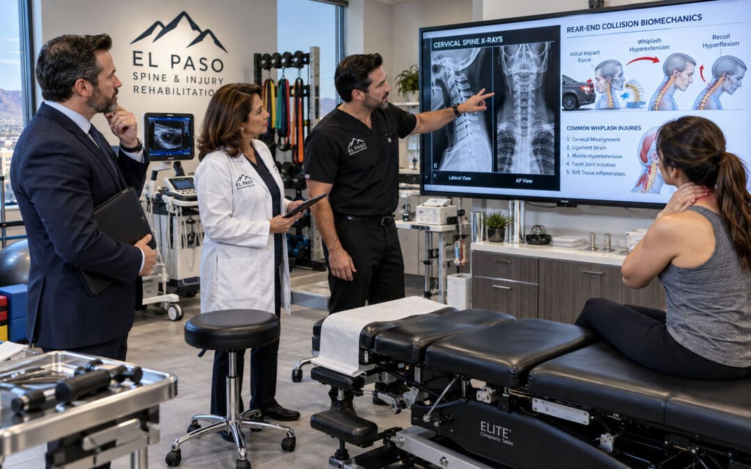
Whiplash describes a mechanism involving rapid acceleration and deceleration of the head and neck. Although commonly associated with rear-end motor vehicle collisions, whiplash-type mechanisms may occur during side impacts, sports injuries, falls, or other sudden movements. The head and neck move in response to applied forces, which can place mechanical demands on muscles, ligaments, joints, and other tissues. The severity and consequences of the injury depend on numerous factors, including the collision circumstances, the individual’s position, preexisting health conditions, and the actual biomechanical forces involved.
What Happens During Whiplash
During rapid acceleration and deceleration, cervical segments may experience complex combinations of motion and loading. Muscles may respond reflexively to stabilize the head. Certain tissues may become painful or irritated.
Patients may subsequently develop:
- Neck pain or stiffness
- Headache
- Shoulder discomfort
- Reduced neck movement
- Muscular tenderness
- Dizziness
- Difficulty sleeping
- Reduced tolerance for normal activities
Some individuals also experience neurological symptoms requiring further evaluation.
Why Headaches May Develop
Whiplash-associated headache may involve cervical nociceptive pathways and altered pain processing. Symptoms may also resemble migraine or tension-type headache. The ICHD-3 recognizes both acute and persistent headache attributed to whiplash. In its standard classification, headache must begin within seven days of the whiplash event and be associated with the qualifying injury history. Persistent headache continues for longer than three months.
Why Early Evaluation Is Important
Following a collision, clinicians must first determine whether there is a potentially serious injury. Fracture, neurological compromise, vascular injury, or traumatic brain injury may require emergency assessment. Once clinicians have excluded serious conditions, treatment generally emphasizes reassurance, appropriate activity, gradual restoration of movement, and individualized rehabilitation. Prolonged unnecessary immobilization is usually discouraged for uncomplicated whiplash-associated disorders. Patients should not be instructed to force painful neck movements or receive aggressive cervical treatment before adequate evaluation.
Role of Chiropractic Care
For selected patients without contraindications, chiropractic care may provide part of a nonsurgical rehabilitation program. The treatment approach may include gentle joint mobilization, soft-tissue care, therapeutic exercise, and movement education. High-velocity cervical manipulation is not automatically necessary. The clinician should consider injury acuity, irritability, neurological findings, vascular concerns, patient preferences, and the availability of safer alternatives. The objective is to restore function safely, not simply to produce joint movement or a temporary reduction in pain.
Concussion, Post-Concussion Symptoms, and Headache Rehabilitation
A concussion is a form of traumatic brain injury resulting from biomechanical forces affecting the brain. It may occur after a direct head impact or through forces transmitted to the head during a collision or other injury. Loss of consciousness is not required for concussion. Symptoms may include headache, dizziness, nausea, visual sensitivity, difficulty concentrating, balance disturbances, fatigue, and sleep changes. Because these symptoms overlap with cervical and vestibular disorders, careful assessment is essential.
Why Concussion Can Produce Headaches
Following concussion, brain cells may experience temporary disturbances in ionic regulation and energy metabolism. Changes in neuronal activity can increase metabolic demand while normal physiological regulation is recovering. These processes may influence headache susceptibility, sensory sensitivity, and cognitive symptoms. The clinical course varies substantially. Some patients improve relatively quickly, while others develop persistent symptoms requiring specialized rehabilitation.
The Cervical Contribution
A person who sustains a concussion during a collision may also experience cervical injury. The cervical region can contribute to headache, dizziness, and movement sensitivity. This creates the possibility of overlapping neurological and musculoskeletal symptoms. Clinicians must avoid treating cervical tenderness as proof that all post-concussion symptoms originate in the neck. Instead, evaluate the cervical contribution alongside neurological, vestibular, visual, and other findings.
Cervicovestibular Rehabilitation
The Amsterdam international consensus statement on concussion in sport supports cervicovestibular rehabilitation for appropriate patients experiencing persistent neck pain, headache, dizziness, or balance problems (Patricios et al., 2023). This approach may combine cervical rehabilitation with exercises targeting balance, gaze stabilization, and vestibular function. Treatment must be individualized. A patient with significant dizziness may need carefully progressed exercises that do not provoke excessive symptom exacerbation. A patient with substantial neck pain may require initial cervical assessment and movement rehabilitation. A person with persistent cognitive or visual symptoms may need additional specialist input.
Returning to Activity
Modern concussion management generally avoids prolonged complete inactivity. Following the initial period of relative rest, patients may benefit from a structured, symptom-limited return to physical and cognitive activity. The exact plan depends on symptoms, clinical findings, occupation, and other risks.
Athletes require appropriate return-to-sport protocols. Workers may need temporary adjustments to screen exposure, physical demands, driving, or concentration-intensive tasks. Concussion rehabilitation is most effective when it addresses the patient’s specific impairments rather than following one identical protocol for everyone.
Occipital Neuralgia and Sharp Pain Behind the Skull
Occipital neuralgia is a pain disorder involving the nerves that supply the posterior scalp. It commonly affects the greater, lesser, or third occipital nerves. Patients may describe sharp, electric, shooting, or stabbing pain extending from the upper neck toward the back of the head. The scalp may feel unusually sensitive. Certain patients experience discomfort when brushing their hair or resting the back of the head against a surface. Occipital neuralgia can overlap symptomatically with cervicogenic headache and migraine. Therefore, do not diagnose it solely because pain occurs in the occipital region.
Potential Mechanisms
Occipital nerve irritation may involve trauma, compression, inflammation, or other conditions. However, the exact cause is not always identifiable. The involved nerves travel through anatomical regions containing muscles, connective tissues, and other structures. Painful sensitivity may develop along their distribution.
Diagnostic Assessment
Evaluation may include a detailed pain history, neurological examination, assessment of scalp sensitivity, and investigation of potential cervical or neurological causes. In selected cases, a diagnostic occipital nerve block can help support the diagnosis. Interpret a temporary response to a block in context rather than as absolute proof of a particular mechanism.
Treatment Options
Conservative treatment may involve medication, modification of aggravating activities, and appropriate management of associated cervical symptoms. Selected patients may benefit from image-guided or anatomically guided nerve blocks performed by appropriately trained clinicians. More persistent cases may require specialist pain or neurological evaluation. Chiropractic care can address coexisting cervical mobility or muscular problems when clinically appropriate. However, spinal manipulation is not established as a definitive treatment for occipital nerve pathology.
Cluster Headaches and Other Severe Primary Headache Disorders
Cluster headache is a distinct neurological condition characterized by attacks of extremely severe pain, often around one eye or the temple.
Attacks may include tearing, nasal congestion, eyelid changes, facial sweating, or restlessness on the affected side.
Some patients experience repeated attacks during characteristic periods.
Because cluster headache can be intensely disabling, early recognition and appropriate medical treatment are essential.
Why Cluster Headache Is Different
Cluster headache involves trigeminal autonomic pathways and biological processes that regulate attack timing.
Neck muscle tension does not adequately explain the disorder.
Appropriate treatment may include high-flow oxygen and certain migraine-related medications, such as subcutaneous sumatriptan, under medical supervision.
Preventive options often require specialist oversight and careful monitoring.
Role of Integrative Care
Integrative care can help address sleep disruption, general conditioning, stress, and associated musculoskeletal complaints.
However, chiropractic manipulation is not a proven primary treatment for cluster headache.
Patients with suspected cluster headache should receive appropriate medical evaluation.
Other severe unilateral headache disorders may require different treatments, reinforcing the importance of accurate diagnosis.
Medication-Overuse Headaches and the Cycle of Repeated Pain Relief
Patients with frequent headaches may begin using acute pain medication increasingly often.
This is understandable because medication may provide temporary relief.
However, repeated use of certain acute headache medications can contribute to medication-overuse headache.
The ICHD-3 generally defines medication-overuse headache as headache occurring on at least 15 days per month in a person with a preexisting headache disorder, associated with regular overuse of acute medication for more than three months.
The relevant frequency threshold depends on the medication.
For example, triptans and combination analgesics generally have a lower overuse threshold than simple analgesics.
Why Medication Overuse Can Become a Problem
Repeated exposure to acute medication may interact with an already susceptible headache system.
Over time, headache frequency may increase.
Patients may respond by taking additional medication, inadvertently reinforcing the pattern.
The exact biological mechanisms vary and are not fully understood.
Medication-overuse headache frequently coexists with migraine or tension-type headache.
How Clinicians Identify It
A headache diary is particularly useful.
Patients can record:
- Number of headache days
- Type of medication used
- Number of medication-use days
- Headache intensity
- Associated symptoms
- Functional limitations
The number of days medication is used is often more informative than the number of individual tablets taken.
Treatment Principles
Management generally includes education, evaluation of the underlying headache disorder, development of an appropriate medication plan, and consideration of preventive treatment.
Withdrawal strategies differ depending on the medication.
Certain medications, including opioids and barbiturate-containing products, may require medically supervised withdrawal.
Patients should not abruptly discontinue potentially dependence-producing medication without professional guidance.
The updated 2026 AAN/AHS migraine-prevention guideline supports offering appropriate preventive treatment to people with migraine and medication overuse.
Integrative care must include medication review. Treating neck stiffness while ignoring frequent analgesic use may leave an important contributor unaddressed.
Headaches Associated With Jaw Dysfunction, Sleep Disorders, and Systemic Illness
Not every headache arises primarily from neurological or cervical musculoskeletal mechanisms.
Some headaches coexist with temporomandibular disorders, sleep-related breathing disorders, anemia, endocrine abnormalities, medication effects, or other medical conditions.
These possibilities become especially relevant when symptoms do not fit a typical headache pattern.
Temporomandibular Disorders
The temporomandibular joints connect the lower jaw to the skull.
Dysfunction involving these joints or associated muscles may produce facial pain, temporal discomfort, difficulty chewing, and jaw tenderness.
Some patients also report neck and shoulder discomfort.
Treatment may involve dental evaluation, education, jaw exercises, behavioral strategies, and selected physical therapy.
Nighttime tooth grinding may require further evaluation.
Sleep Disorders
Poor sleep can increase headache susceptibility and reduce pain tolerance.
Patients with suspected obstructive sleep apnea may report snoring, daytime sleepiness, unrefreshing sleep, and morning headaches.
A sleep evaluation may be appropriate when the history supports this possibility.
Treating the underlying sleep disorder can improve overall health and may reduce associated headache symptoms.
Metabolic and Medical Conditions
Selected medical conditions can contribute to headache symptoms.
Examples include anemia, thyroid dysfunction, dehydration, medication reactions, and certain inflammatory or infectious disorders.
Laboratory testing is most useful when guided by the history and physical examination.
There is no evidence-based reason to order extensive laboratory panels for every person with an uncomplicated primary headache.
Why Medical Oversight Matters
A multidisciplinary clinic can help distinguish a cervical musculoskeletal complaint from a broader medical condition.
This is particularly valuable among patients with complex medication regimens, cardiovascular disease, metabolic disorders, or persistent unexplained symptoms.
A complete headache evaluation considers the whole patient without assuming that every biological system is abnormal or requires testing.
Headache Red Flags That Require Immediate Medical Evaluation
Although most headaches are not caused by dangerous disease, some presentations require urgent investigation.
Identifying these situations is a fundamental responsibility of every healthcare professional managing headache patients.
The SNNOOP10 framework summarizes important warning features that may suggest secondary headache disorders (Do et al., 2019).
Seek Emergency Care for These Symptoms
- A sudden, extremely severe headache reaching maximum intensity rapidly
- New headache associated with weakness, facial drooping, speech difficulty, or confusion
- Headache with loss of consciousness or seizure
- New severe headache after significant head or neck trauma
- Severe headache with fever, neck stiffness, or altered mental status
- New severe headache with visual loss or major neurological changes
- Headache accompanied by concerning symptoms of cervical artery dissection
- New concerning headache during pregnancy or shortly after childbirth
Other warning signs that warrant prompt medical assessment include progressive worsening, a major change from a previous headache pattern, new headache beginning after age 50, a history of cancer or immunosuppression, and symptoms suggestive of elevated intracranial pressure.
Cervical Artery Dissection
One particularly important consideration in head and neck care is cervical artery dissection.
A dissection involves injury to the wall of a cervical artery.
It may present with new or unusual headache, neck pain, and sometimes neurological symptoms.
Certain patients experience pain before other neurological findings become apparent.
The American Heart Association has published scientific statements addressing cervical artery dissection and its relationship to cervical manipulative therapy (Biller et al., 2014; Yaghi et al., 2024).
The relationship between manipulation and dissection is complex, and temporal association alone does not establish causality.
Nevertheless, the potential seriousness of the condition makes risk screening and informed consent essential.
A Safety-First Chiropractic Approach
Before considering cervical manual treatment, clinicians should evaluate the clinical history, new or unusual symptoms, neurological status, trauma history, and relevant contraindications.
When a vascular or other serious condition is suspected, cervical manipulation should not proceed.
The patient should receive the appropriate medical or emergency evaluation.
No single physical screening maneuver can reliably rule out every vascular concern.
Patient safety takes priority over completing a planned adjustment or rehabilitation session.
Personal Injury and Headache Causation: Understanding the Medical Connection
In personal injury care, the word causation refers to the relationship between an event and a resulting medical condition or change in health status.
A patient may develop headaches after a motor vehicle collision.
However, the fact that symptoms appeared after the collision does not by itself prove that the collision caused them.
Medical causation requires a structured evaluation of the available evidence.
This assessment should examine the injury mechanism, symptom chronology, clinical findings, preexisting history, alternative explanations, and the recovery course.
Temporal Relationship
The timing of symptoms is important.
A headache beginning shortly after a collision may support a possible injury-related relationship.
The clinician should document the onset as precisely as possible.
Questions include:
- Did the headache begin immediately or later?
- Was neck pain present at the time of injury?
- Did the patient strike their head?
- Were dizziness, nausea, confusion, or neurological symptoms present?
- Did the patient have a history of headache before the event?
- Did the headache pattern change after the incident?
The ICHD-3 uses specific temporal criteria for trauma-related headache.
These criteria are valuable for diagnosis but do not match legal standards of causation.
Biological Plausibility
The proposed relationship should be compatible with established anatomy and physiology.
For example, sudden neck loading during a collision may plausibly contribute to certain cervical soft-tissue injuries and headache patterns.
A head impact may produce symptoms consistent with concussion.
However, evaluate the event using the actual history and findings rather than assuming a collision always produces a particular injury.
Clinical Consistency
A consistent pattern of symptoms and findings may strengthen the clinical explanation.
For example, a patient reporting new headache and neck pain after trauma may demonstrate cervical movement restriction, reproducible pain with selected movements, and functional limitations.
These findings support the presence of symptoms and impairment.
They do not necessarily prove which tissue was injured or establish legal responsibility.
Alternative Explanations
A careful clinician considers other plausible causes.
These may include preexisting migraine, medication overuse, infection, sleep disorders, or unrelated neurological conditions.
Failure to consider alternative explanations can weaken diagnostic accuracy.
Changes Over Time
The patient’s clinical course provides additional information.
Improvement in cervical function alongside improvement in headache may support a cervical contribution.
Nevertheless, improvement after treatment is not, by itself, definitive proof of the original injury mechanism.
Medical causation is a reasoned clinical opinion based on converging evidence, not simply the statement that symptoms started after an accident.
Preexisting Headaches and How an Injury May Aggravate Them
Many personal injury patients have a history of headache symptoms before the event in question.
Preexisting migraine or tension-type headache does not exclude the possibility that a later injury worsened the condition.
The important issue is whether the post-injury presentation differs meaningfully from the previous baseline.
A patient may have experienced occasional migraine before a collision.
After the event, headache frequency, severity, or disability may increase.
Such changes require documentation and investigation.
Establishing the Pre-Injury Baseline
Clinicians should ask about the patient’s previous headache history.
Relevant details include frequency, duration, severity, associated symptoms, medication use, and functional effects.
If a patient previously experienced two headache days per month and now experiences substantially more frequent symptoms, that difference may be clinically meaningful.
However, the reason for the change still requires evaluation.
Distinguishing Aggravation From New Disease
An injury may plausibly contribute to an increase in an existing headache disorder without creating a completely different condition.
The ICHD-3 allows appropriate recognition of preexisting primary headache disorders that become substantially worse after trauma.
This approach can more accurately describe patients with both longstanding headache susceptibility and a new injury-related change.
Why Accurate Documentation Matters
Medical documentation should distinguish:
- Symptoms clearly present before the event
- Newly reported symptoms
- Changes in headache frequency or intensity
- Changes in medication requirements
- New cervical or neurological findings
- Functional limitations developing after injury
- Other possible contributors
A balanced report should acknowledge uncertainty when the evidence does not support a definitive conclusion.
The goal is accurate clinical characterization, not assuming every symptom is new or entirely injury-related.
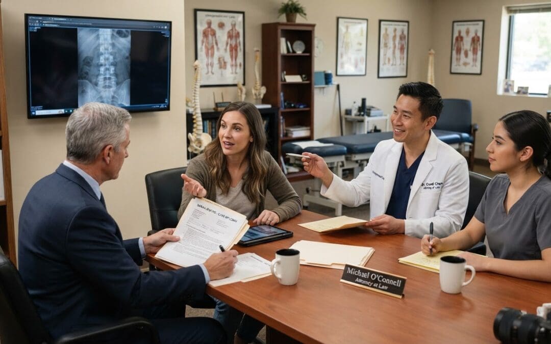
Medical Documentation in Personal Injury Headache Cases
Good documentation supports continuity of care, clinical reasoning, and communication among healthcare professionals.
It can also provide a clear record of the patient’s reported symptoms and objectively observed findings.
In personal injury cases, documentation should be sufficiently detailed to distinguish the patient’s history from examination findings and clinician interpretations.
Injury History
The record should describe the circumstances the patient reports.
For motor vehicle collisions, this may include vehicle position, direction of impact, use of restraints, head contact, immediate symptoms, and emergency treatment.
Information about vehicle damage may be relevant background, but it cannot establish the extent of a person’s injury on its own.
Symptom History
The clinician should document headache location, intensity, frequency, duration, and associated symptoms.
Specific questions should address neck pain, shoulder pain, dizziness, visual symptoms, sleep problems, and upper-extremity complaints.
The chronology should remain consistent with the information the patient actually provides.
Examination Findings
The medical record should distinguish subjective symptoms from objective observations.
For example, a patient may report hand numbness.
The clinician may then document sensory testing, reflex findings, strength examination, and other relevant observations.
These should not be represented as interchangeable.
Diagnostic Reasoning
A useful clinical note explains why the available information supports a working diagnosis.
It should also document significant alternative diagnoses under consideration.
When diagnostic certainty is limited, acknowledge that limitation.
Treatment Response
Follow-up notes should describe changes in pain, movement, neurological function, medication use, and daily activities.
Statements such as “patient improving” are less informative than specific observations.
For example, the clinician might document increased comfortable cervical rotation or fewer headache days recorded in a diary.
Ethical Considerations
Clinicians should never exaggerate clinical findings to support a legal claim.
Documentation should not automatically attribute every symptom to an accident or imply that imaging abnormalities prove traumatic causation when they may be degenerative or incidental.
A patient’s treatment plan should be guided by medical need.
Accurate documentation protects the integrity of clinical care and creates a reliable record of the recovery process.
Diagnostic Assessment of Headaches in an Integrative Clinical Setting
The diagnostic process begins with a comprehensive history.
The goal is to determine whether the presentation fits a known primary headache disorder, a secondary headache, or a potentially urgent medical condition.
The clinician should then evaluate which additional assessments are necessary.
Chiropractic Care for Neck Pain Relief- Video
Headache History
Important questions include:
- When did the headaches begin?
- Where is the pain located?
- How does the pain feel?
- How long does each episode last?
- How frequently do symptoms occur?
- Are nausea, light sensitivity, or visual disturbances present?
- Does movement aggravate symptoms?
- Is the headache associated with neck movement?
- Are there new neurological symptoms?
- What medications are being used?
- Has there been a recent injury?
The patient’s answers help distinguish migraine, tension-type headache, cervicogenic headache, and other presentations.
General Medical Assessment
The clinician may assess vital signs, including blood pressure, and review relevant medical conditions.
Medication history should include prescription drugs, over-the-counter medications, supplements, and recent medication changes.
Medical assessment may also include evaluation for fever, systemic illness, pregnancy-related concerns, or other factors depending on the presentation.
Neurological Examination
A neurological examination evaluates the function of relevant nervous-system pathways.
It may include assessment of cranial nerves, strength, sensation, reflexes, coordination, balance, and gait.
New or progressive abnormalities can indicate the need for further investigation.
Cervical Musculoskeletal Examination
The neck examination may include active movement, tenderness assessment, movement control, and selected orthopedic testing.
Clinicians may assess whether specific neck movements reproduce familiar headache symptoms.
However, provoking pain does not automatically identify a specific tissue or prove causation.
Functional Assessment
The examination should also evaluate how symptoms affect the patient’s life.
Questions about work, driving, lifting, sleep, and household activities help identify meaningful treatment goals.
The most useful examination is not the one with the most tests, but the one that answers the most important diagnostic questions safely and accurately.
Cervical Range of Motion and Joint Mobility Assessment
Cervical range-of-motion testing helps identify movement limitations and symptom responses.
The patient may be asked to turn the head, look upward or downward, or move into side bending.
The clinician observes movement quality, available range, and symptom reproduction.
The clinician may obtain measurements with appropriate instruments.
Why Range of Motion Matters
Reduced cervical motion can affect driving, reaching, visual orientation, and daily activities.
Movement restriction may occur because of pain, muscular guarding, stiffness, fear of movement, or other factors.
In selected cervicogenic headache presentations, restricted motion and reproduction of the familiar headache can support the diagnosis.
The Cervical Flexion-Rotation Test
The cervical flexion-rotation test is commonly discussed in cervicogenic headache assessment.
It is intended to evaluate upper cervical rotation, particularly involving the C1-C2 region.
The clinician positions the cervical spine in flexion and assesses available rotation.
Restricted motion or reproduction of familiar symptoms may support a cervical contribution.
However, no isolated physical test establishes cervicogenic headache with complete certainty.
Interpret results alongside the history and other findings.
Why Movement Testing Guides Rehabilitation
Range-of-motion assessment can help establish a baseline.
After treatment begins, repeated measurements may indicate whether movement is improving.
The results also help guide exercise selection.
A patient with highly irritable symptoms may begin with small, comfortable movements.
Another patient with low irritability and reduced endurance may benefit from a more active strengthening program.
The goal is to improve functional movement rather than force the cervical spine toward an arbitrary range.
Evaluating Arm Pain, Numbness, and Weakness During Headache Assessment
When a patient reports upper-extremity symptoms, the clinician must investigate whether the problem arises from the cervical spine, peripheral nerves, shoulder structures, or another medical condition.
This is particularly important after personal injury events.
Sensory Testing
The clinician may examine light touch or other sensory responses in relevant regions of the arms and hands.
The purpose is to identify patterns that may correspond to nerve root or peripheral nerve involvement.
Interpret sensory findings cautiously because symptoms can overlap across anatomical distributions.
Muscle Strength Testing
Strength assessment helps identify possible motor impairment.
Relevant muscle groups may include shoulder abductors, elbow flexors and extensors, wrist muscles, and intrinsic hand muscles.
Significant or progressive weakness requires prompt attention.
Reflex Testing
Deep tendon reflexes provide additional information about neurological function.
Commonly examined upper-extremity reflexes include the biceps, brachioradialis, and triceps reflexes.
Asymmetry may be clinically relevant when combined with other findings.
Spurling’s Test
Spurling’s test is used in selected patients when cervical radiculopathy is suspected.
It attempts to reproduce characteristic radiating symptoms under controlled positioning.
A positive result may increase suspicion of cervical radiculopathy, but the test is not definitive.
A 2026 systematic review found substantial variation in diagnostic accuracy across studies and emphasized the low or very low certainty of evidence for several examination tests.
Upper-Limb Neurodynamic Testing
Neurodynamic tests assess symptom responses during movements that place mechanical demands on peripheral neural structures.
They may help determine whether neural mechanosensitivity contributes to symptoms.
Interpret these tests in conjunction with the neurological examination.
When Further Testing Is Needed
MRI may be appropriate when significant neurological deficits, concerning findings, or persistent symptoms suggest a structural cervical disorder requiring further investigation.
Electromyography and nerve conduction studies may help distinguish cervical radiculopathy from peripheral nerve entrapment in selected cases.
Arm weakness, altered reflexes, and persistent neurological symptoms deserve specific assessment rather than being grouped under a general diagnosis of headache or muscle tension.
Diagnostic Imaging for Headaches and Neck Injuries
Imaging can be valuable when clinical findings suggest a condition that may change management.
However, routine imaging is not necessary for every patient with headache.
The American College of Radiology’s headache appropriateness criteria distinguish patients with typical headache presentations from those with concerning features (American College of Radiology, 2022).
CT Imaging
Computed tomography, or CT, is frequently used in emergency settings when clinicians need to investigate certain acute intracranial conditions.
It may be particularly important following head trauma or sudden severe headache.
CT can detect many acute hemorrhages and certain structural abnormalities.
However, a normal CT does not exclude every neurological disorder or concussion.
MRI
Magnetic resonance imaging, or MRI, provides detailed images of soft tissues and neural structures.
Consider brain MRI when the headache history or neurological examination suggests a concerning intracranial process.
Cervical MRI may help evaluate selected disc, spinal cord, or nerve root conditions.
Imaging decisions depend on the specific clinical question.
Cervical Radiographs
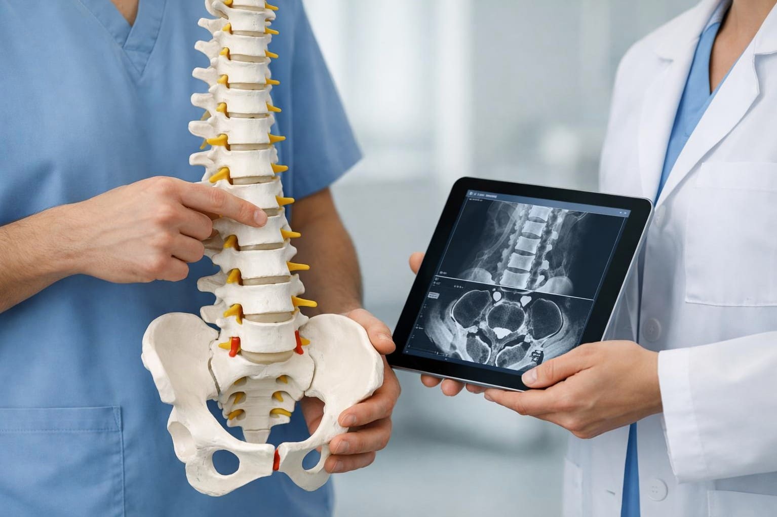
Radiographs may identify certain bony abnormalities, alignment features, or degenerative changes.
However, many degenerative findings occur in people who have no symptoms.
Reduced disc space or cervical spondylosis does not prove that the finding caused a headache.
Vascular Imaging
When cervical artery dissection or another vascular disorder is suspected, clinicians may use appropriate vascular imaging.
This may include CT angiography or magnetic resonance angiography depending on the clinical situation.
Such decisions require urgent medical evaluation when warning features are present.
The Limitations of Imaging
Imaging reveals anatomical structures, not the entire biological experience of pain.
Many painful conditions do not produce clearly visible abnormalities.
Conversely, many visible abnormalities are not painful.
The most accurate diagnosis integrates imaging with the history and examination.
Imaging should answer a meaningful clinical question rather than serve as a routine requirement before every headache treatment.
Functional Outcome Measures for Monitoring Headache Recovery
Effective treatment requires more than asking whether a patient feels better.
Clinicians should evaluate whether headache frequency, disability, movement, sleep, and daily functioning are improving.
Validated questionnaires and symptom diaries can help measure these changes.
Headache Diaries
A headache diary can record headache days, duration, severity, medication use, and associated symptoms.
It helps clinicians identify patterns and evaluate response to treatment.
For migraine, monthly migraine days and overall headache days are commonly used outcomes.
Headache Impact Test
The Headache Impact Test, commonly known as HIT-6, measures the effect of headaches on daily life.
It assesses pain severity, social functioning, role limitations, and cognitive effects.
Changes over time may help demonstrate whether treatment is improving the patient’s experience.
Migraine Disability Assessment
The Migraine Disability Assessment, or MIDAS, evaluates lost or reduced productivity associated with migraine.
It considers effects on work, school, household activities, and social participation.
Neck Disability Index
The Neck Disability Index, or NDI, assesses limitations associated with neck pain.
It may be helpful when a patient has both headache and cervical dysfunction.
Patient-Specific Functional Scale
A patient-specific measure helps individuals identify the activities that matter most to them.
Examples include turning the head while driving, completing a work shift, carrying groceries, or sleeping comfortably.
The clinician can track progress toward these personalized goals.
Why Measurement Matters
Outcome measurement helps distinguish temporary symptom relief from meaningful recovery.
A patient may experience less neck pain after treatment while headache frequency remains unchanged.
That result suggests the cervical treatment may be helping one problem without sufficiently addressing the headache disorder.
This information should guide reassessment and modification of the treatment plan.
Evidence-informed care requires ongoing measurement, not assumptions about effectiveness.
Chiropractic Care as Part of Nonsurgical Headache Treatment
Chiropractic care primarily focuses on evaluating and treating selected musculoskeletal conditions, particularly involving the spine and associated joints and muscles.
In headache management, chiropractic care is most relevant when cervical or related musculoskeletal impairments contribute to pain or functional limitations.
Treatment should be based on a working diagnosis, not the assumption that spinal adjustments are appropriate for all headache disorders.
Potential Treatment Goals
A chiropractic rehabilitation plan may seek to:
- Reduce associated neck pain
- Improve comfortable cervical movement
- Address selected joint mobility restrictions
- Reduce muscular tenderness
- Improve neck and shoulder endurance
- Restore movement confidence
- Support return to work and daily activities
- Reduce the impact of musculoskeletal contributors to headaches
Individualize and measure these goals.
Why a Multimodal Approach Is Important
Manual treatment may provide temporary pain relief or improvement in movement.
However, long-term function often depends on the patient’s ability to maintain activity, develop physical capacity, and manage relevant headache triggers.
This is why an integrative chiropractic program may combine hands-on treatment with exercise, education, and coordination with medical professionals.
What the Research Shows
The 2026 guideline developed by Trager and colleagues provides recommendations for chiropractic management of cervicogenic and tension-type headache.
It supports appropriately selected nonpharmacological interventions while emphasizing diagnostic assessment, informed consent, referral, and individualized care.
A separate systematic review published in 2025 found that evidence for chiropractic spinal manipulation across headache disorders remained uncertain and generally of very low certainty.
The results are not necessarily contradictory.
Different reviews include different populations, intervention definitions, comparison groups, and outcome measures.
A treatment may benefit selected cervicogenic headache presentations without being established as effective for all headaches.
Chiropractic care is an evidence-informed musculoskeletal treatment within a broader diagnostic and therapeutic plan.
Spinal Manipulation and Its Potential Role in Cervicogenic Headache
Spinal manipulation involves a controlled manual technique applied to a joint.
Certain techniques involve a rapid, low-amplitude movement.
The intended purpose is to influence movement, pain sensitivity, or neuromuscular responses.
The precise mechanisms responsible for treatment effects remain under investigation.
Potential effects may involve changes in sensory input, short-term pain modulation, and movement tolerance.
Why It May Be Considered
In selected patients with cervicogenic headache, cervical joint dysfunction may contribute to pain.
A clinician may consider manipulation when the patient’s symptoms, examination, preferences, and risk profile support its use.
Some clinical trials and reviews suggest improvement in headache pain or disability.
However, the magnitude and durability of benefits vary.
What Manipulation Does Not Necessarily Do
Manipulation should not be described as permanently repositioning displaced vertebrae in patients with ordinary headaches.
It does not necessarily correct a structural abnormality.
It also does not establish the cause of headache merely because symptoms improve afterward.
Temporary pain relief can occur through multiple physiological pathways.
Safety Considerations
Potential adverse effects include temporary soreness, headache exacerbation, or discomfort.
Serious complications are uncommon, but cervical manipulation requires careful consideration because of the potential severity of vascular or neurological events.
The possibility of cervical artery dissection must be included in appropriate risk discussions.
Patients with concerning vascular symptoms, suspected instability, significant acute trauma, or other contraindications should not undergo cervical manipulation.
Informed Consent
Patients should understand the expected benefits, uncertainties, potential harms, alternatives, and option to decline treatment.
Alternatives may include mobilization, exercise, soft-tissue treatment, or other nonmanipulative approaches.
High-velocity manipulation is one possible technique, not an essential requirement for effective cervical rehabilitation.
Cervical Joint Mobilization and Gentle Manual Therapy
Joint mobilization generally uses controlled, lower-velocity movements within an appropriate range.
Clinicians may select it when they want to improve movement tolerance without using a high-velocity technique.
Mobilization can be particularly relevant when symptoms are irritable, or the patient prefers a gentler treatment.
Potential Mechanisms
Mobilization may influence mechanoreceptor activity, pain processing, and movement confidence.
It may also gradually expose the patient to previously uncomfortable movement.
This process can be combined with active exercise.
Sustained Natural Apophyseal Glides
Researchers have investigated certain mobilization approaches, including sustained natural apophyseal glides, for cervicogenic headache.
These techniques involve guided movement while a clinician applies a controlled mobilizing force.
Research has reported potential benefits for pain and function in selected patients.
However, results depend on study quality and clinical context.
Why Gentle Care May Be Appropriate After Injury
After whiplash, patients may show significant pain sensitivity.
A forceful technique may be poorly tolerated.
Gentle mobilization can offer a more conservative way to assess treatment response and gradually restore motion.
The treatment should remain symptom-guided.
Combining Mobilization With Exercise
Manual treatment may make movement temporarily easier.
Exercise can then help patients practice controlled movement and build capacity.
For example, a patient may receive gentle cervical mobilization followed by active rotation exercises and low-load strengthening.
The objective is to improve function beyond the treatment session.
Manual therapy is most useful when it supports a larger rehabilitation strategy rather than replacing active recovery.
Soft-Tissue Therapy and Myofascial Treatment for Headache-Related Muscle Pain
Soft-tissue treatment refers to a range of techniques applied to muscles and associated connective tissues.
These may include massage, gentle myofascial techniques, and other clinically appropriate manual approaches.
Soft-tissue treatment can help when patients report muscular tenderness, stiffness, or discomfort with movement.
Why Muscles Become Sensitive
After injury, patients may reduce movement because of pain.
Muscles can become guarded or tender.
Prolonged static positions, poor sleep, and increased pain sensitivity may further contribute.
These changes can affect the neck, upper back, shoulders, and jaw.
Potential Benefits
Soft-tissue treatment may provide temporary pain relief and improve comfort.
Some patients experience a short-term reduction in muscular tenderness.
This may create an opportunity to participate more comfortably in exercise.
However, short-term relief does not necessarily mean the underlying headache disorder has resolved.
Trigger Point Treatment
Some clinicians identify sensitive regions within muscles commonly referred to as myofascial trigger points.
Pressure applied to these areas may reproduce local or referred discomfort.
Clinicians may use trigger point treatment to address associated muscle symptoms.
However, the precise definition and diagnostic reliability of trigger points remain subjects of scientific discussion.
Treatment should not assume that every tender point represents a distinct pathological lesion.
Avoiding Excessive Treatment Intensity
Aggressive pressure can worsen symptoms in highly sensitive patients.
Adjust treatment intensity to the individual’s tolerance.
A patient with persistent post-traumatic headache may benefit more from gentle techniques than forceful deep-tissue treatment.
Long-Term Management
Soft-tissue care should usually be paired with exercise, movement variation, ergonomic changes, or other measures that address the patient’s functional needs.
The goal is not to eliminate all muscular tension, which is neither realistic nor desirable, but to improve comfort and healthy movement.
Deep Neck Flexor Rehabilitation for Headache and Cervical Stability
The deep neck flexor muscles help control and support cervical movement.
They include muscles such as the longus colli and longus capitis.
These muscles help coordinate subtle neck movements and maintain head position.
Some patients with cervical pain or headache demonstrate reduced endurance or altered activation patterns.
However, these findings are not universal and should not be assumed without assessment.
Why Deep Neck Flexor Exercises Are Used
Low-load cervical exercises can help patients practice controlled movement.
They may improve muscular endurance, coordination, and tolerance for daily activities.
For example, gentle cranio-cervical flexion exercises may help develop controlled neck movement without excessive activation of superficial muscles.
Beginning Rehabilitation
Match early exercises to symptom irritability.
A patient with acute pain may begin with comfortable movements in supported positions.
The clinician may monitor breathing, muscle substitution, and symptom response.
Progressing the Exercises
As tolerance improves, the patient may progress to longer holds, controlled head movement, or functional positions.
Later stages may involve maintaining cervical control during arm movement, lifting, or work-related tasks.
Why Strength Alone Is Not Enough
The neck must respond to changing demands.
A patient may have adequate isolated muscle strength but struggle to maintain a comfortable head position during prolonged activity.
Therefore, rehabilitation should include endurance, coordination, and functional integration.
Avoiding Excessive Exercise
More exercise is not always better.
High-volume strengthening during a highly irritable headache episode may worsen symptoms.
Treatment progression should follow the patient’s clinical response.
The purpose of deep cervical rehabilitation is to restore useful movement capacity, not to impose a rigid or supposedly ideal neck posture.
Scapular Stabilization and Upper-Extremity Rehabilitation
The shoulder blades play a central role in upper-extremity movement.
They move in coordination with the arms and provide attachment sites for numerous muscles.
When patients experience neck pain and shoulder discomfort, evaluating scapular function may help identify relevant rehabilitation needs.
Why Shoulder Function Matters
During lifting, reaching, and carrying, the cervical and shoulder muscles work together.
If the shoulder girdle lacks endurance or movement capacity, the patient may experience increased fatigue during daily activities.
This can contribute to discomfort in already sensitive neck tissues.
Muscles Commonly Addressed
Rehabilitation may target the middle and lower trapezius, serratus anterior, rotator cuff, and other shoulder girdle muscles.
The exercise program should address the patient’s specific impairments.
Not every patient needs the same muscle exercises.
Exercise Progression
Early rehabilitation may begin with low-load scapular movements and supported exercises.
As symptoms improve, the program may include resistance training, reaching tasks, carrying activities, and occupational movements.
For example, an office worker may need endurance for prolonged computer tasks.
A warehouse employee may need progressive lifting and carrying capacity.
Relation to Headache Symptoms
Improving shoulder function may reduce associated neck discomfort.
In patients whose cervical symptoms contribute to headaches, this improvement may indirectly reduce headache burden.
However, evidence does not establish scapular strengthening as a direct cure for migraine or other primary neurological headache disorders.
Upper-Extremity Neurological Symptoms
When numbness or weakness is present, rehabilitation must follow neurological findings.
Patients with cervical radiculopathy may require modified exercises, symptom monitoring, and medical reassessment.
Do not manage progressive weakness solely by adding resistance exercises.
A functional rehabilitation program considers how the head, neck, shoulder girdle, and arms work together during real activities.
Vestibular Rehabilitation for Headaches With Dizziness and Balance Problems
Some patients experience headache together with dizziness, motion sensitivity, nausea, or balance problems.
These symptoms may occur with vestibular migraine, concussion, certain inner-ear disorders, or cervical conditions.
Treatment depends on the underlying cause.
Vestibular Migraine
Vestibular migraine is associated with episodes of vestibular symptoms in individuals with a relevant migraine history and clinical pattern.
Patients may report dizziness, vertigo, motion sensitivity, or difficulty tolerating visually complex environments.
Management may require migraine-specific medical treatment and selected vestibular rehabilitation.
Post-Concussion Dizziness
After concussion, dizziness may reflect vestibular, visual, autonomic, cervical, or other factors.
A comprehensive examination helps determine which systems require treatment.
Gaze Stabilization
Gaze stabilization exercises may be prescribed when indicated to improve the coordination between head movement and visual stability.
The exercises are progressed gradually.
Balance Training
Balance exercises challenge the body’s ability to integrate visual, vestibular, and proprioceptive information.
Training may include stance changes, walking tasks, or controlled head movements.
Cervical Proprioception
The cervical muscles and joints provide sensory information about head position.
Selected exercises may address movement accuracy and coordination.
However, no single cervical proprioception test can diagnose every cause of dizziness.
Why Collaboration Matters
Dizziness can also indicate conditions requiring urgent medical assessment.
Start treatment only after appropriate screening.
Patients with complex presentations may benefit from collaboration among medical providers, vestibular therapists, neurologists, and rehabilitation professionals.
Successful dizziness rehabilitation depends on identifying the affected system rather than assuming that the neck is responsible for every balance complaint.
Therapeutic Exercise and Gradual Return to Activity After Headache-Related Injury
Exercise is an important component of many rehabilitation programs because it helps restore physical capacity and functional independence.
However, exercise prescriptions should consider the headache type, associated injury, symptom severity, and medical history.
Aerobic Exercise
Regular aerobic activity may help some individuals with migraine or tension-type headache.
Activities may include walking, cycling, swimming, or other forms of comfortable cardiovascular exercise.
Potential benefits include improved physical conditioning, sleep, mood, stress regulation, and general health.
The VA/DoD headache guideline supports consideration of aerobic exercise and progressive strengthening for selected headache conditions.
Starting at an Appropriate Level
Patients recovering from injury may have reduced exercise tolerance.
An initial program should begin at a manageable intensity.
The clinician may recommend short activity periods followed by gradual progression.
Resistance Training
Resistance exercise can improve muscular strength and endurance.
It may be especially helpful for patients whose work or daily activities require lifting, carrying, or prolonged postural demands.
The program should incorporate the upper back, shoulders, neck, trunk, and other relevant regions.
Movement Confidence
Persistent pain can lead to fear of movement.
Patients may worry that normal activity will cause additional damage.
Education and gradual exposure can help them regain confidence.
This does not mean patients should ignore significant warning symptoms.
Instead, progress movement based on clinical findings and symptom tolerance.
Individualization
An exercise plan for a patient with episodic migraine may emphasize general fitness and consistency.
A patient with whiplash-related cervical pain may require more specific movement restoration.
A person with post-concussion symptoms may need symptom-limited aerobic conditioning and specialized rehabilitation.
Exercise is a therapeutic tool that must match the diagnosis, not a one-size-fits-all prescription.
Medical Treatment for Acute Migraine and Severe Headache Episodes
Many headache patients benefit from appropriate medical treatment alongside rehabilitation.
Acute headache medication aims to reduce symptoms during an attack and help restore normal function.
Medication selection depends on diagnosis, severity, contraindications, prior response, and associated medical conditions.
Simple Analgesics
Acetaminophen and certain nonsteroidal anti-inflammatory medications may help with selected headaches.
However, they are not appropriate for every patient.
NSAIDs can pose risks involving gastrointestinal bleeding, kidney function, cardiovascular health, and medication interactions.
The clinician must consider the patient’s medical history.
Triptans
Triptans are migraine-specific medications that act on selected serotonin receptors.
They can reduce migraine-related headache symptoms in appropriately selected patients.
Because of their pharmacological effects, they have important contraindications in certain cardiovascular or vascular conditions.
Gepants
Gepants target CGRP-related signaling pathways.
They provide additional options for selected patients with migraine.
Depending on the medication, gepants may be used acutely, preventively, or both.
Antiemetic Medication
Patients experiencing substantial nausea or vomiting may require appropriate treatment.
This can improve comfort and make other therapies easier to tolerate.
Emergency Department Treatment
The American Headache Society’s 2025 emergency department guideline evaluated parenteral migraine treatments.
It identified strong evidence for intravenous prochlorperazine and greater occipital nerve block in eligible patients requiring emergency treatment.
The guideline also recommended against intravenous hydromorphone for migraine pain.
These recommendations apply to the specific emergency-care context and should not be generalized to every outpatient headache situation.
Coordinating With Chiropractic Care
Medical management may reduce neurological headache symptoms while chiropractic rehabilitation addresses a separate cervical impairment.
For example, a patient may receive migraine-specific treatment from an appropriate medical clinician while participating in supervised exercise for neck discomfort.
Integrative care works best when medical and musculoskeletal treatments address their respective indications without unnecessarily competing with or replacing one another.
Migraine Prevention and the Latest 2026 Clinical Guidelines
Preventive migraine treatment aims to reduce headache frequency, severity, disability, and the need for acute medication.
It may be appropriate for patients with frequent attacks or substantial functional impairment.
In August 2026, the American Academy of Neurology and American Headache Society published an updated joint guideline on pharmacological migraine prevention.
The guideline incorporates a systematic review of 217 studies and evaluates evidence confidence for different medications (Potrebic et al., 2026; Pringsheim et al., 2026).
When Prevention Should Be Considered
The guideline recommends offering preventive treatment to patients experiencing at least four migraine days per month or at least four moderate to severe headache days per month.
It also supports preventive treatment for patients with substantial migraine-related disability.
The decision should involve patient preferences, treatment goals, side effects, medical history, and medication access.
CGRP-Targeted Prevention
CGRP-targeted therapies have expanded the available options.
These include certain monoclonal antibodies and oral medications.
Some demonstrated high or moderate confidence of benefit in the 2026 evidence review.
The choice depends on whether the patient has episodic or chronic migraine, prior treatment response, contraindications, and other factors.
Traditional Preventive Medications
Certain established medications remain important options.
These include selected beta blockers, topiramate, and other evidence-supported medications.
Medication choice must account for individual risks.
For example, beta blockers may be inappropriate for some patients with certain cardiovascular or respiratory conditions.
Topiramate may cause cognitive or sensory adverse effects and carries important reproductive safety considerations.
Valproate carries particularly serious pregnancy-related risks and other adverse effects requiring careful assessment.
OnabotulinumtoxinA
OnabotulinumtoxinA is an established preventive treatment for selected patients with chronic migraine.
Trained medical professionals administer it according to an appropriate clinical protocol.
Its use is not interchangeable with cosmetic injection procedures.
Treatment Monitoring
Patients should track headache frequency, disability, medication use, and adverse effects.
A preventive medication requires an appropriate trial and reassessment.
Treatment should not continue indefinitely without evaluating its benefit.
Why This Matters in Integrative Clinics
The 2026 guideline reinforces the importance of combining current neurological evidence with individualized care.
A patient with migraine and cervical pain may benefit from both preventive medication and targeted rehabilitation.
The two approaches address potentially different contributors.
Modern headache care should neither dismiss effective medications nor overlook appropriate nonpharmacological options.
Diagnostic Nerve Blocks and Other Interventional Headache Treatments
Some headache conditions require additional diagnostic or therapeutic interventions.
Clinicians generally consider these procedures when symptoms are persistent, severe, or insufficiently responsive to initial treatment.
Select them based on an established clinical indication.
Greater Occipital Nerve Block
A greater occipital nerve block involves injection near the greater occipital nerve.
Depending on the situation, it may be used for selected headache conditions.
The procedure can provide temporary symptom relief in some patients.
The 2025 American Headache Society emergency department guideline supports greater occipital nerve block in eligible adults presenting with migraine attacks requiring parenteral treatment.
Interpret this recommendation in its clinical context.
Diagnostic Cervical Blocks
For certain suspected cervical pain generators, a specialist may use diagnostic blocks to help determine whether a specific structure contributes to pain.
Examples include selected medial branch or third occipital nerve blocks.
These procedures are not routine requirements for every headache patient.
Radiofrequency Procedures
In carefully selected cases, pain specialists may consider radiofrequency procedures targeting specific cervical pain pathways.
Such interventions require appropriate diagnostic confirmation and risk-benefit assessment.
Evidence and outcomes vary by diagnosis and technique.
Botulinum Toxin Treatment
OnabotulinumtoxinA has an established role in chronic migraine prevention.
It does not have the same evidence base for every form of cervicogenic or post-traumatic headache.
Why Conservative Care Usually Comes First
When there are no emergencies or progressive neurological deficits, many musculoskeletal headache presentations can be managed conservatively at first.
Rehabilitation may improve symptoms without invasive intervention.
However, prolonged ineffective treatment should not delay appropriate referral.
Interventional treatment is most useful when it addresses a defined clinical indication within a coordinated care plan.
Functional Medicine and the Search for Modifiable Headache Contributors
Functional medicine emphasizes individualized evaluation of health patterns, lifestyle factors, nutrition, and relevant physiological systems.
Within headache care, this approach can be useful when it incorporates validated medical assessment and established treatments.
However, functional medicine should not replace conventional diagnostic standards or encourage unvalidated testing.
Nutrition and Meal Consistency
Irregular meals may contribute to headache susceptibility in some individuals.
Consistent nutrition can help, particularly when patients identify a link between missed meals and symptoms.
A balanced dietary pattern supports general health.
No single diet has been proven to eliminate all headache disorders.
Hydration
Dehydration can contribute to headache in susceptible individuals.
Patients may benefit from maintaining appropriate fluid intake.
However, drinking excessive water does not provide additional protection and can be harmful.
Hydration recommendations should consider kidney function, cardiovascular conditions, medications, and activity levels.
Caffeine
Caffeine may improve certain acute headaches, but excessive or inconsistent intake can contribute to symptoms.
Abrupt withdrawal may also produce headaches.
Patients benefit from reviewing their caffeine pattern rather than assuming that all caffeine is harmful.
Dietary Triggers
Some people identify specific foods or beverages associated with their headache episodes.
However, perceived triggers do not always establish causation.
Prospective symptom diaries can help identify consistent patterns.
Unnecessarily restrictive diets may create nutritional problems.
Appropriate Laboratory Testing
Testing may be useful when symptoms suggest a specific deficiency or medical condition.
For example, evaluation for anemia or thyroid dysfunction may be relevant when supported by the history and examination.
Broad testing without a clinical indication can produce confusing results and unnecessary expense.
Avoiding Unsupported Testing
Commercial food IgG sensitivity panels are not established tools for diagnosing food allergy or headache causes.
The American Academy of Allergy, Asthma & Immunology advises against using these tests to guide food avoidance.
A Balanced Integrative Philosophy
Functional medicine is most valuable when it promotes healthy behaviors, identifies clinically meaningful medical conditions, and supports evidence-based treatment.
The objective is to investigate relevant contributors, not to assume that every headache results from inflammation, toxicity, hormones, or hidden nutritional imbalance.
Nutritional Supplements and Headache Prevention
Some nutritional supplements have been studied for migraine prevention.
They may be considered in selected patients, particularly when patients prefer additional nonprescription options.
However, supplements are not automatically safer than medications.
They can cause adverse effects, interact with prescription drugs, and vary in manufacturing quality.
Magnesium
Magnesium participates in numerous physiological processes, including neuronal signaling and muscle function.
Research supports a possible role for certain magnesium preparations in migraine prevention.
The most appropriate formulation and use depend on individual circumstances.
Patients with kidney disease require particular caution.
Gastrointestinal adverse effects can occur.
Riboflavin
Riboflavin, also known as vitamin B2, contributes to cellular energy metabolism.
It has been studied as a migraine preventive supplement.
Evidence supports consideration in selected adults.
The potential benefit generally relates to prevention rather than immediate relief of an acute attack.
Coenzyme Q10
Coenzyme Q10 participates in mitochondrial energy processes.
Some studies suggest possible benefit in migraine prevention, although the evidence is less certain than for established preventive medications.
Melatonin
Melatonin influences sleep-wake regulation.
Evidence for migraine prevention is limited and inconsistent.
Supplement quality can vary.
Butterbur: Researchers have investigated butterbur for migraine prevention.
However, concerns regarding liver toxicity and potentially harmful plant compounds limit its suitability.
Clinicians should not casually recommend it as a natural headache treatment.
Why Individual Assessment Is Necessary
A patient’s medication list, kidney and liver function, pregnancy status, and medical history can influence supplement safety.
Clinicians should review supplements as carefully as other treatments.
Nutritional support may complement established headache management, but it should not displace treatments with stronger evidence when those treatments are indicated.
Sleep, Stress, Mental Health, and Headache Comorbidities
Headache disorders often coexist with sleep disturbances, anxiety, depression, fatigue, and other chronic pain conditions.
These relationships are frequently bidirectional.
Poor sleep may increase headache susceptibility.
Frequent headaches may make restful sleep more difficult.
Persistent pain can increase stress, while stress may influence pain processing and coping.
Sleep Regulation
Consistent sleep and wake times can help stabilize daily physiological rhythms.
Patients may benefit from reducing disruptive evening habits, reviewing caffeine timing, and addressing untreated sleep disorders.
When chronic insomnia is present, cognitive behavioral therapy for insomnia may be appropriate.
Stress and Pain Processing
Stress influences autonomic and neuroendocrine activity.
It can also affect muscle activity, attention, sleep, and central pain processing.
However, stress should not be the default explanation for unexplained headache symptoms.
Medical causes must still be considered.
Cognitive Behavioral Therapy
Cognitive behavioral approaches can help patients build skills to manage chronic pain and disability.
These approaches may address sleep, coping, activity pacing, and unhelpful fear patterns.
The objective is not to suggest that the pain is psychological.
Instead, behavioral treatment can influence real physiological and functional outcomes.
Biofeedback
Biofeedback techniques provide information about physiological processes such as muscle activity or other bodily responses.
Selected patients may learn strategies to improve relaxation and self-regulation.
Evidence supports a role for behavioral interventions in certain headache conditions, although effectiveness varies.
Addressing Anxiety and Depression
Persistent headaches can affect emotional health.
Patients may experience frustration, reduced activity, social withdrawal, or fear about their symptoms.
Screening for clinically significant anxiety or depression can be appropriate.
Offer a mental health referral when indicated.
Why Comorbidity Management Matters
A patient may experience improvement in neck pain but continue to have poor sleep and frequent migraine.
Unless these other factors are addressed, disability may remain substantial.
Comprehensive care treats the headache disorder while also recognizing the medical, functional, and psychological conditions that may influence recovery.
How Chiropractic Rehabilitation May Reduce Headache-Related Comorbidities
Chiropractic rehabilitation may help improve certain conditions that accompany headache disorders.
These benefits should be described accurately.
A reduction in neck pain does not necessarily mean that a neurological headache disorder has been cured.
Similarly, improved shoulder function does not prove the shoulder caused the original headache.
Neck Pain and Restricted Movement
When cervical musculoskeletal dysfunction is present, appropriate treatment may improve movement tolerance and reduce discomfort.
This can help patients turn their heads, drive, work, and perform daily activities.
Shoulder Pain and Muscular Fatigue
Shoulder-focused strengthening and movement education may reduce associated discomfort.
This may help individuals tolerate repetitive activity or prolonged tasks.
Activity-Related Disability
Reduced pain and improved physical capacity can make normal activities easier.
Patients may become more comfortable exercising, working, and participating in recreational activities.
Sleep Disturbance
When musculoskeletal pain interferes with sleep, successful pain management may indirectly improve sleep comfort.
However, chiropractic treatment is not a substitute for evaluating insomnia, sleep apnea, or other sleep disorders.
Fear of Movement
Education and progressive activity may reduce unnecessary fear related to normal movement.
This can support physical recovery.
Reduced Reliance on Symptom-Relief Strategies
Some patients may need less symptomatic treatment when associated musculoskeletal pain improves.
However, patients should discuss medication changes with the prescribing clinician.
Limitations
Evidence does not establish that chiropractic care directly cures depression, migraine-related neurobiology, metabolic disorders, or all systemic comorbidities.
The most defensible benefits involve selected musculoskeletal symptoms and functional outcomes.
Integrative treatment is successful when each component addresses a condition for which it has a reasonable clinical role.
A Multidisciplinary Treatment Model for Headaches and Personal Injury in El Paso
At Injury Medical Clinic PA, also known as Mission Plaza Injury Medical Clinic, the integrative model described in the practice’s published materials combines chiropractic evaluation, medical oversight, functional medicine, personal injury care, and rehabilitation services.
This structure reflects the reality that post-injury headache symptoms may involve several medical systems.
A patient may simultaneously experience cervical pain, migraine-like symptoms, disrupted sleep, shoulder dysfunction, and neurological complaints.
A multidisciplinary framework allows professionals to evaluate these issues from different perspectives.
Dr. Alexander Jimenez and Chiropractic Clinical Care
Dr. Alexander Jimenez, DC, APRN, FNP-BC, CFMP, IFMCP, ATN, CCST, combines chiropractic training with advanced nursing and family nurse practitioner credentials.
His clinical philosophy emphasizes the interaction between musculoskeletal mechanics, neurological symptoms, functional rehabilitation, and broader health considerations.
His published educational materials discuss headache disorders, cervical dysfunction, whiplash, physical rehabilitation, and nonsurgical treatment approaches.
Within the appropriate professional scope, his role may include musculoskeletal evaluation, chiropractic treatment, functional assessment, rehabilitation planning, medical assessment, and care coordination.
Dr. Maria Guadalupe Cardenas and Medical Oversight. The clinic’s materials describe
Dr. Maria Guadalupe Cardenas, MD, as board-certified in internal medicine, with over 40 years of experience.
She serves as the practice’s Medical Director and Collaborative Physician.
Her Texas medical license is listed as J2933, and publicly indexed NPPES records identify her NPI as 1164426748.
Her medical role complements musculoskeletal treatment.
Internal medicine expertise can support evaluation of systemic conditions, chronic disease considerations, medication-related risks, and complex medical presentations.
How Collaboration Benefits Patients
Consider a patient who presents with headache, neck pain, and arm tingling after a collision.
A coordinated approach may include cervical and neurological examination, medical risk assessment, appropriate imaging decisions, symptom-directed medication management, and rehabilitation.
If the patient also has hypertension, diabetes, sleep difficulties, or other medical conditions, those factors can influence treatment selection.
Medical oversight helps ensure rehabilitation decisions align with the patient’s broader medical needs.
Referral Beyond the Clinic
Multidisciplinary care does not mean that every service must occur in one facility.
Patients may require referral to neurologists, emergency physicians, physical therapists, vestibular specialists, pain medicine physicians, or other professionals.
Effective coordination is the most important feature.
Multidisciplinary care matches each patient’s problem with the appropriate expertise while maintaining a unified treatment plan.
The Role of Dr. Maria Guadalupe Cardenas in Coordinated Headache Care
Dr. Cardenas’s internal medicine background is especially relevant because headaches can reflect more than cervical musculoskeletal dysfunction.
A patient may present with a familiar migraine pattern but also have elevated blood pressure, medication interactions, kidney disease, or another condition requiring medical attention.
Another patient may develop a headache after an injury while taking anticoagulant medication.
These factors can substantially affect diagnostic and treatment decisions.
Medical Risk Assessment
An internal medicine evaluation helps identify medical conditions that may affect the safety of rehabilitation or medication use.
For example, certain medications may increase bleeding risk or influence the appropriateness of specific interventions.
Chronic Disease Management
Conditions such as diabetes, hypertension, thyroid disorders, and renal disease can affect overall health and treatment decisions.
These conditions do not automatically cause headache, but they may require consideration during care.
Diagnostic Coordination
Medical oversight can help determine whether additional testing or specialist referral is needed.
This may be particularly important when the headache pattern is atypical or neurological symptoms are present.
Medication Review
Medication selection requires consideration of adverse effects, contraindications, interactions, and patient preferences.
A coordinated medical approach helps avoid unnecessary duplication or conflicts between treatments.
Support for Rehabilitation Planning
Medical findings may influence exercise intensity, procedural decisions, and recovery expectations.
For example, patients with cardiovascular limitations may require modified conditioning programs.
Integrative Collaboration
Dr. Jimenez’s chiropractic and rehabilitation perspective can address relevant movement and musculoskeletal impairments.
Dr. Cardenas’s medical oversight can support the evaluation of systemic risks and broader medical needs.
Together, the model allows clinicians to consider multiple potential contributors while maintaining appropriate professional responsibilities.
The strongest multidisciplinary model is not one in which every provider performs the same treatment, but one in which each professional contributes complementary expertise.
Clinical Observations From Dr. Jimenez’s Published Headache and Injury Care Resources
Dr. Jimenez’s educational materials emphasize several recurring clinical themes.
These include the relationship between headaches and cervical pain, the importance of evaluating whiplash-associated symptoms, the potential relevance of soft-tissue dysfunction, and the role of functional rehabilitation.
These observations should be understood as clinical perspectives and practice descriptions, not as substitutes for controlled research.
Headaches After Automobile Collisions
The clinic’s published headache resources discuss the occurrence of headache and cervical pain following motor vehicle accidents.
This clinical emphasis is consistent with the recognized category of headache attributed to whiplash.
However, accurate diagnosis requires attention to symptom timing, mechanism of injury, clinical findings, and alternative explanations.
Neck Pain Radiating Toward the Head
The practice frequently discusses the relationship between cervical dysfunction and pain perceived in the head.
This concept is compatible with established trigeminocervical physiology and the diagnostic framework for cervicogenic headache.
Nevertheless, similar symptoms can occur with migraine and other headache conditions.
The Importance of Rehabilitation
Dr. Jimenez’s published philosophy emphasizes treatment strategies intended to restore mobility, improve muscular function, and support activity.
From an evidence-based perspective, these goals align with contemporary rehabilitation principles.
The Whole-Patient Perspective
The clinic’s integrative approach also considers nutrition, sleep, medical conditions, and functional health.
This perspective can improve care when a specific clinical indication supports each assessment and intervention.
Interpreting Clinical Experience
Clinical experience helps identify symptom patterns and monitor treatment response.
However, observations from routine practice cannot establish treatment efficacy in the same way as appropriately designed randomized clinical trials.
They may be affected by natural recovery, placebo effects, co-interventions, patient selection, and other factors.
The most responsible use of clinical experience is to combine it with high-quality research and careful patient-specific evaluation.
A Step-by-Step Integrative Care Pathway for Headaches Following Personal Injury
Patients recovering from injury often benefit from a structured pathway that guides evaluation and treatment.
The following framework describes how a coordinated clinical process may be organized.
Stage One: Safety Screening
The first step is to determine whether emergency evaluation is required.
The clinician reviews the injury history, current symptoms, neurological status, and warning signs.
Potential fracture, vascular injury, intracranial injury, or progressive neurological deficit must be addressed appropriately.
Conservative treatment should not delay necessary emergency care.
Stage Two: Headache Classification
After considering urgent conditions, the clinician evaluates the headache phenotype.
The symptoms may be most consistent with migraine, tension-type headache, cervicogenic headache, post-traumatic headache, or another disorder.
Some patients meet criteria for multiple headache diagnoses.
Stage Three: Musculoskeletal and Functional Assessment
The clinician evaluates cervical movement, muscle tenderness, shoulder function, and relevant neurological findings.
The clinician assesses upper-extremity symptoms separately when present.
Functional limitations are documented.
Stage Four: Shared Treatment Planning
The clinician explains the working diagnosis and proposed treatment options.
The patient participates in decisions regarding conservative care, medication, referral, and rehabilitation goals.
The clinician discusses the potential benefits and risks.
Stage Five: Early Symptom Management
Treatment may include education, appropriate activity modification, gentle manual therapy, medication management when indicated, and basic therapeutic movement.
The objective is to reduce disability while avoiding unnecessary immobilization.
Stage Six: Progressive Rehabilitation
As symptoms permit, treatment progresses toward strength, endurance, coordination, and functional tasks.
Work-specific and recreational activities may be incorporated.
Stage Seven: Reassessment
Monitor headache frequency, pain intensity, cervical movement, neurological symptoms, and functional limitations.
If meaningful improvement does not occur, the diagnosis and treatment plan are reconsidered.
Stage Eight: Return to Function
Later rehabilitation emphasizes normal activity, work capacity, physical conditioning, and independent symptom management.
The patient receives a long-term maintenance strategy based on actual needs rather than an automatic requirement for indefinite treatment.
This pathway encourages safety, diagnostic accuracy, measurable progress, and appropriate escalation of care.
Example Clinical Scenario Following a Rear-End Collision
Consider a hypothetical adult patient who presents several days after a rear-end collision.
The patient reports new headaches beginning near the base of the skull, neck stiffness, upper trapezius discomfort, and difficulty turning the head while driving.
The patient reports no loss of consciousness, but describes fatigue and mild dizziness.
This scenario is presented for education and does not represent a documented patient from Dr. Jimenez’s practice.
Initial Assessment
The clinician begins by reviewing the collision history and screening for concerning neurological or vascular symptoms.
The patient is asked about head contact, confusion, memory gaps, vomiting, worsening headaches, and other relevant symptoms.
The clinician performs a neurological examination and an appropriate cervical assessment.
If the findings suggest concussion, further evaluation is indicated.
If serious injury cannot be reasonably excluded, additional medical assessment or imaging may be necessary.
Developing the Differential Diagnosis
Potential diagnoses might include headache attributed to whiplash, cervical musculoskeletal pain, concussion-related headache, or exacerbation of an existing primary headache disorder.
The clinician should not assign all these diagnoses automatically.
Each requires support from the history and examination.
Early Treatment
If serious conditions have been excluded and conservative care is appropriate, treatment may include education, gentle cervical movement, and activity guidance.
Manual treatment may be considered according to the patient’s clinical findings and preferences.
Aggressive manipulation is not necessary.
Progressive Rehabilitation
As symptoms improve, rehabilitation may incorporate cervical endurance exercises, shoulder strengthening, and functional movements.
The patient gradually resumes driving, household activities, and work tasks.
Monitoring Recovery
A headache diary and disability measure provide information about progress.
If dizziness persists, vestibular assessment may be needed.
If neurological symptoms develop, additional medical evaluation becomes necessary.
Clinical Reasoning
Treatment targets identified impairments.
Improvement in neck function may help headache symptoms if the cervical region is a meaningful contributor.
However, persistent migraine-like symptoms may require additional headache-specific treatment.
This example illustrates why a coordinated diagnosis is more useful than automatically labeling all post-collision headaches as muscular or spinal.
Example Clinical Scenario Involving Migraine and Cervical Radiculopathy
Consider another hypothetical patient with longstanding migraine who develops new neck pain and arm tingling after lifting a heavy object at work.
The patient reports that the familiar migraine attacks continue, but the new arm symptoms worsen during certain neck positions.
Why Two Diagnoses May Be Necessary
The patient may have an established migraine disorder and a separate cervical nerve root problem.
Treating the migraine alone may not resolve the arm symptoms.
Similarly, treating the cervical spine may not eliminate the underlying migraine susceptibility.
Neurological Evaluation
The clinician evaluates arm strength, sensation, reflexes, and symptom distribution.
Selected tests may support suspicion of cervical radiculopathy.
If neurological findings are significant or progressive, appropriate imaging and referral may be needed.
Migraine Management
The medical team reviews headache frequency, medication use, and disability.
If preventive treatment is indicated, the team considers medication options according to current guidelines.
Cervical Rehabilitation
Once serious pathology has been excluded, conservative rehabilitation may address cervical movement, shoulder function, and activity tolerance.
Exercises should not repeatedly aggravate neurological symptoms.
Coordinated Outcome Measurement
The patient tracks migraine days separately from neck pain and arm symptoms.
This distinction helps clinicians determine which treatment is addressing which problem.
For example, shoulder and arm symptoms may improve while migraine frequency remains unchanged.
That result would not mean the rehabilitation failed.
It would mean that one component of the patient’s condition responded while another requires continued management.
Accurate treatment depends on recognizing that multiple conditions can coexist without sharing one identical cause.
Example Clinical Scenario Involving Chronic Headache, Poor Sleep, and Shoulder Pain
Consider a hypothetical office worker experiencing frequent bilateral headaches, upper shoulder tenderness, and poor sleep.
The patient spends long periods at a computer and reports using over-the-counter pain medication on many days.
Initial Evaluation
The clinician reviews headache characteristics and medication frequency.
The presentation may suggest tension-type headache, but migraine and medication-overuse headache must also be considered.
Sleep history is evaluated.
Cervical and shoulder examinations identify relevant functional limitations.
Why a Single Treatment May Be Insufficient
Soft-tissue therapy might temporarily reduce shoulder tenderness.
However, frequent analgesic use may be contributing to ongoing headaches.
Sleep disturbance may also increase pain sensitivity.
Without addressing these factors, symptoms may continue.
Treatment Planning
A coordinated plan might include medication review, headache education, appropriate medical management, sleep interventions, and graded exercise.
If clinically indicated, add cervical mobilization or other conservative care.
Functional Rehabilitation
The patient begins a tolerable shoulder and neck endurance program.
Review workstation setup and movement opportunities.
The goal is to reduce excessive static loading without encouraging rigid posture or constant self-monitoring.
Monitoring
The clinician tracks headache days, medication-use days, sleep quality, and functional tolerance over time.
The clinician modifies treatment based on the response.
This scenario demonstrates how headache care becomes more effective when clinicians address interacting contributors rather than relying on one procedure.
Ergonomic Strategies for Preventing Headache-Related Neck and Shoulder Strain
Ergonomics refers to adapting tasks and environments to support comfortable and efficient activity.
For patients with neck pain and headache-related disability, ergonomic strategies may reduce unnecessary physical demands.
However, ergonomics should not be presented as a universal cure for headache disorders.
Computer Workstations
A workstation should allow comfortable viewing and movement.
Adjust the monitor, keyboard, chair, and work surface to reduce unnecessary strain.
No single perfect posture exists that every patient must maintain throughout the day.
Movement variability is often more realistic and helpful than attempting to remain rigidly upright.
Frequent Movement
Changing position periodically may reduce fatigue associated with prolonged static tasks.
Patients can incorporate short movement opportunities into the workday.
Phone Use
Holding a phone between the shoulder and ear may create uncomfortable sustained neck loading.
Using a headset or speakerphone can reduce that demand.
Driving
Patients recovering from cervical injury may experience difficulty turning the head.
Assess driving safety based on neck movement, pain, dizziness, medications, and cognitive symptoms.
Lifting and Carrying
Workers who lift or carry objects may benefit from improved conditioning and task-specific movement training.
The goal is to develop capacity for real-world demands.
Individualized Recommendations
Ergonomic changes should address activities that reproduce the patient’s symptoms.
Generic recommendations may be less useful than adjustments based on actual work tasks.
Good ergonomics supports movement comfort and function while recognizing that the human body is designed to tolerate a variety of positions.
Preventing Headache Recurrence Through Long-Term Rehabilitation
After symptoms improve, many patients want to prevent future headaches.
Prevention depends on the headache diagnosis.
Strategies that help cervicogenic headache may differ from those used to prevent migraine.
Consistent Physical Activity
Regular exercise supports cardiovascular fitness, muscular endurance, sleep, and general health.
Patients should select activities they can perform consistently.
Maintaining Cervical and Shoulder Capacity
For people with recurring mechanical neck symptoms, maintaining strength and movement tolerance may reduce disability.
Exercises should be sustainable, not excessively demanding.
Managing Migraine Triggers
Patients with migraine may benefit from regular sleep, consistent meals, appropriate hydration, and individualized preventive treatment.
Not every perceived trigger requires complete avoidance.
Monitoring Medication Use
Patients with frequent headaches should review acute medication use to reduce the risk of medication-overuse headache.
Managing Workload
Gradual activity progression can help patients tolerate occupational demands.
Temporary modifications may be appropriate during recovery from injury.
Recognizing Recurrence Early
Patients should understand their typical headache pattern and recognize meaningful changes.
New neurological findings or unusual severe symptoms require medical evaluation.
Avoiding Dependence on Passive Treatment
Manual therapy may be useful during certain phases of recovery.
However, long-term care should emphasize independence, physical capacity, and appropriate self-management.
Some patients may choose periodic follow-up care for recurring musculoskeletal symptoms, but indefinite treatment is not necessarily needed.
The most sustainable recovery plan helps patients understand their condition and participate actively in maintaining their health.
Frequently Asked Questions About Headaches, Neck Pain, and Chiropractic Care
Can neck problems cause headaches?
Yes. Certain cervical spine disorders can cause cervicogenic headache through pain-sensitive structures and neural pathways connecting the upper neck with the head. However, neck pain is also common in migraine and other headache disorders. A cervical examination helps determine whether neck dysfunction is a likely cause or contributor.
Can headaches cause shoulder pain?
Headache disorders frequently coexist with shoulder discomfort. The association may involve muscular tenderness, altered activity, cervical dysfunction, or shared pain-processing mechanisms. Evaluate shoulder pain separately when symptoms suggest a different disorder.
Why do my headaches begin at the base of my skull?
Pain beginning near the base of the skull can occur with several conditions, including cervicogenic headache, occipital neuralgia, tension-type headache, and migraine. The location alone does not establish a diagnosis. The clinician should evaluate pain quality, duration, associated symptoms, neurological findings, and responses to cervical movement.
Can migraines cause arm numbness?
Some migraine auras involve temporary sensory symptoms affecting the arm or hand. However, arm numbness may also result from cervical radiculopathy, peripheral nerve disorders, or other neurological conditions. Sudden weakness or possible stroke symptoms require emergency evaluation.
Can a car accident cause long-term headaches?
Yes. Headaches may persist after certain traumatic head or neck injuries. Potential contributors include post-traumatic headache, cervical dysfunction, migraine-like mechanisms, sleep disturbances, and other factors. The diagnosis requires an appropriate injury history and clinical assessment.
What is the difference between cervicogenic headache and migraine?
Cervicogenic headache is attributed to a cervical disorder. Migraine is a primary neurological disorder involving characteristic sensory and headache pathways. Both may include neck pain, but migraine more commonly involves features such as nausea, light sensitivity, and activity-related aggravation. The conditions can coexist.
Can chiropractic care help headaches?
Chiropractic care may help selected patients, particularly when cervical musculoskeletal dysfunction contributes to cervicogenic headache or associated neck pain. The evidence varies by headache diagnosis and treatment technique. Chiropractic manipulation is not a proven cure for all headaches.
Is spinal manipulation necessary to treat a headache?
No. Many conservative treatment options do not require high-velocity manipulation. Alternatives include mobilization, exercise, soft-tissue treatment, education, and medical management. Treatment selection should reflect the diagnosis, patient preferences, and safety considerations.
How are headaches diagnosed after whiplash?
Diagnosis involves reviewing the injury history, headache timing, symptom pattern, and clinical findings. Clinicians also evaluate neurological status, cervical function, and other possible causes. Imaging is used when indicated, not automatically for every patient.
Can physical therapy and chiropractic care be combined?
Yes. Appropriate manual therapy and rehabilitation can be integrated when the treatments serve complementary purposes. The plan should avoid unnecessary duplication and emphasize measurable functional outcomes.
When should someone with headaches see a neurologist?
Neurological referral may be appropriate when headaches are difficult to diagnose, unusually severe, progressively worsening, associated with neurological abnormalities, or insufficiently responsive to appropriate treatment. Certain presentations require emergency evaluation rather than routine referral.
Can stress make headaches worse?
Stress can affect headache susceptibility, sleep, muscle tension, and pain processing. However, stress should not automatically be assumed to be the primary cause of a headache. Medical evaluation remains important when symptoms are new or concerning.
Can diet affect migraine?
Some individuals identify dietary patterns associated with migraine attacks. Consistent meals, proper hydration, and individualized trigger assessment may help. There is no single diet that reliably cures migraine.
ADoes inflammation always cause headaches
No. Headache disorders involve diverse mechanisms. Some conditions include inflammatory signaling, while others involve neurological, muscular, vascular, medication-related, or systemic processes. Do not use “inflammation” as a universal explanation.
Can headaches improve without surgery?
Yes. Most primary headaches and many cervical musculoskeletal headache conditions are managed without surgery. Treatment may include medications, rehabilitation, exercise, behavioral interventions, and selected medical procedures. Surgery is reserved for specific structural conditions when clinically indicated.
How long does headache rehabilitation take?
Recovery depends on the diagnosis, symptom severity, associated conditions, and individual response. Some patients improve relatively quickly. Others require longer-term management, particularly when chronic migraine, persistent post-traumatic headache, or significant neurological impairment is present. A fixed recovery timeline cannot be guaranteed.
Why are my headaches worse after working at a computer?
Prolonged static work may increase associated neck or shoulder discomfort. Screen brightness, visual demands, sleep, stress, and migraine susceptibility may also influence symptoms. A comprehensive evaluation can help distinguish these contributors.
Should I avoid exercise when I have headaches?
Not necessarily. Regular activity can benefit many headache patients. However, certain acute symptoms or medical conditions require temporary activity modification. Progress exercise based on the diagnosis and clinical response.
Can headache treatment help my sleep?
Improving painful musculoskeletal symptoms may indirectly improve sleep comfort. Headache-specific treatment can also reduce nighttime disability. However, persistent insomnia or suspected sleep apnea requires appropriate evaluation.
Why is medical collaboration important in chiropractic headache care?
Medical collaboration helps ensure clinicians recognize neurological, systemic, medication-related, and vascular concerns. It also supports safe treatment selection and appropriate specialist referral. A coordinated approach allows musculoskeletal rehabilitation and medical treatment to complement each other.
Understanding What Successful Headache Treatment Really Means
The objective of headache treatment is not always complete and permanent elimination of every symptom. For many patients, successful care means experiencing fewer headache days, reduced pain severity, improved movement, less disability, and greater independence.
A patient with migraine may achieve meaningful improvement through preventive medication and lifestyle management. Another patient with cervicogenic headache may benefit from manual therapy and targeted cervical rehabilitation. A person recovering from concussion may need coordinated neurological, vestibular, cervical, and functional treatment. These approaches are not interchangeable. Clinicians select them based on the patient’s diagnosis and needs.
The Importance of Reassessment: Reevaluate treatment when symptoms change, or progress does not occur. Persistent or worsening neurological findings require medical attention. A treatment that provides temporary comfort but no meaningful functional improvement should not continue indefinitely without reconsideration.
Why Patient Participation Matters
Patients play an active role in recovery. Understanding the diagnosis, tracking symptoms, participating in rehabilitation, and communicating changes can improve care quality. Clinicians must provide accurate information, appropriate treatment, and a clear plan.
Integrating Research With Clinical Experience
Clinical research provides evidence about treatment effectiveness and safety. Clinical examination helps determine whether that evidence applies to an individual patient. Patient preferences and circumstances influence treatment choices. Together, these elements form the foundation of evidence-based practice.
Conclusion: A Comprehensive Approach to Headaches, Neck Pain, and Personal Injury Recovery
Headaches are complex conditions that may involve the brain, nervous system, cervical spine, muscles, and other physiological systems. Their effects frequently extend beyond head pain. Patients may experience neck stiffness, shoulder discomfort, dizziness, fatigue, sleep disturbance, and reduced functional ability. In certain situations, upper-extremity symptoms may accompany headache because of associated cervical radiculopathy, peripheral nerve disorders, migraine aura, or other conditions requiring separate assessment. Following personal injury, accurate medical causation requires careful consideration of injury mechanics, symptom timing, preexisting conditions, clinical findings, and alternative explanations.
A headache appearing after trauma deserves evaluation, not an automatic assumption about its cause. Chiropractic care can serve an important role in nonsurgical treatment when appropriate musculoskeletal problems are present. Manual therapy, therapeutic exercise, movement education, and functional rehabilitation may help selected patients reduce pain and restore function. However, the effectiveness of these approaches varies by headache type, and treatment must remain consistent with contemporary scientific evidence and safety guidelines. At Injury Medical Clinic PA in El Paso, Texas, the collaborative model described by Dr. Alexander Jimenez integrates chiropractic and rehabilitation perspectives with medical oversight provided by Dr. Maria Guadalupe Cardenas, MD, a board-certified internist serving as Medical Director and Collaborative Physician. This approach considers headache symptoms through multiple clinical perspectives while coordinating appropriate treatment. For patients, the central message is straightforward. A successful headache treatment plan begins with the correct diagnosis, addresses the real contributors to pain and disability, emphasizes safe nonsurgical options when appropriate, and measures progress by meaningful improvements in everyday life.
References
- American Academy of Allergy, Asthma & Immunology. (n.d.). The myth of IgG food panel testing. AAAAI.
- American Academy of Physical Medicine and Rehabilitation. (n.d.). Cervical radiculopathy. AAPM&R.
- American College of Radiology. (2022). ACR Appropriateness Criteria: Headache.
- Ashina, M. (2020). Migraine. The New England Journal of Medicine, 383, 1866-1876.
- Bendtsen, L. (2003). Central and peripheral sensitization in tension-type headache. Current Pain and Headache Reports, 7(6), 460-465.
- Biller, J., Sacco, R. L., Albuquerque, F. C., Demaerschalk, B. M., Fayad, P., Long, P. H., Noorollah, L. D., Panagos, P. D., Schievink, W. I., Schwartz, N. E., Shuaib, A., Thaler, D. E., & Tirschwell, D. L. (2014). Cervical arterial dissections and association with cervical manipulative therapy: A statement for healthcare professionals from the American Heart Association/American Stroke Association. Stroke, 45(10), 3155-3174.
- Centers for Disease Control and Prevention. (2025). Mild traumatic brain injury management guideline.
- Do, T. P., Remmers, A., Schytz, H. W., Schankin, C., Nelson, S. E., Obermann, M., Hansen, J. M., Sinclair, A. J., Gantenbein, A. R., & Schoonman, G. G. (2019). Red and orange flags for secondary headaches in clinical practice: SNNOOP10 list. Neurology, 92(3), 134-144.
- Headache Classification Committee of the International Headache Society. (2018). The International Classification of Headache Disorders, 3rd edition. Cephalalgia, 38(1), 1-211. https://doi.org/10.1177/0333102417738202
- Headache Classification Committee of the International Headache Society. (2018). Cervicogenic headache. International Classification of Headache Disorders, Third Edition.
- Headache Classification Committee of the International Headache Society. (2018). Acute headache attributed to whiplash. International Classification of Headache Disorders, Third Edition.
- Headache Classification Committee of the International Headache Society. (2018). Persistent headache attributed to whiplash. International Classification of Headache Disorders, Third Edition.
- Headache Classification Committee of the International Headache Society. (2018). Medication-overuse headache. International Classification of Headache Disorders, Third Edition.
- Jimenez, A. (n.d.). Headaches and chiropractic treatment. El Paso Back Clinic.
- Jimenez, A. (n.d.). Dr. Alex Jimenez: Professional background and clinical philosophy. El Paso Back Clinic.
- Jimenez, A. (n.d.). Dr. Maria Cardenas, MD: Board-certified internal medicine specialist. Dr. Alex Jimenez.
- Martins, L., Collet, P., Lafrance, S., & Demont, A. (2026). Efficacy of nonsurgical interventions for the management of adults with cervicogenic headache: A systematic review and meta-analyses. Annals of Physical and Rehabilitation Medicine, 69(4), 102070.
- Patterson Gentile, C., Rosenthal, S., Blume, H., Rastogi, R. G., McVige, J., Bicknese, A., Ladak, A., Zaveri, H., Greene, K., Barlow, K., et al. (2024). American Headache Society white paper on treatment of post-traumatic headache from concussion in youth. Headache, 64(9), 1148-1162.
- Pensri, C., Liang, Z., Treleaven, J., Jull, G., & Thomas, L. (2025). Cervical musculoskeletal impairments in migraine and tension-type headache and relationship to pain-related factors: An updated systematic review and meta-analysis. Musculoskeletal Science and Practice, 76, 103251.
- Potrebic, S., Tanveer, S., Becker, W. J., Burch, R., Cooke, L. J., Fenton, T., Fletcher, J. J., Gordon Perue, G. L., Hershey, A. D., Jackson, J. L., et al. (2026). Pharmacologic treatment for migraine prevention in adults: Practice guideline recommendations. Neurology, 107(7), e214881.
- Pringsheim, T., Smith, D. B., Tanveer, S., Becker, W. J., Burch, R., Cooke, L. J., Fenton, T., Fletcher, J. J., Gordon Perue, G. L., Hershey, A. D., et al. (2026). Systematic review of pharmacologic treatment for migraine prevention in adults. Neurology.
- Trager, R. J., Daniels, C. J., Hawk, C., Taylor, D. N., Walters, S. A., Price, M. R., Anderson, K. R., Crivelli, L. S., Mooring, S. A., Staab, C. A., & Oakley, C. B. (2026). Chiropractic management of adults with cervicogenic or tension-type headaches: Development of a clinical practice guideline. Journal of Integrative and Complementary Medicine, 32(4), 325-344.
- U.S. Department of Veterans Affairs & U.S. Department of Defense. (2023). VA/DoD clinical practice guideline for management of headache.
- Xu, X., & Ling, Y. (2025). Comparative safety and efficacy of manual therapy interventions for cervicogenic headache: A systematic review and network meta-analysis. Frontiers in Neurology, 16, 1566764.
- Yaghi, S., Engelter, S., Del Brutto, V. J., Field, T. S., Jadhav, A. P., Kicielinski, K., Madsen, T. E., Mistry, E. A., Salehi Omran, S., Pandey, A., & Raz, E. (2024). Treatment and outcomes of cervical artery dissection in adults: A scientific statement from the American Heart Association. Stroke, 55, e91-e106.
SEO Tags: headaches, headache treatment, chronic headaches, migraine headaches, tension-type headaches, cervicogenic headaches, post-traumatic headaches, whiplash headaches, headache causation, personal injury headaches, personal injury care, motor vehicle accident injuries, concussion headaches, neck pain, shoulder pain, upper-extremity pain, arm numbness, cervical radiculopathy, occipital neuralgia, trigeminocervical complex, cervical spine, cervical rehabilitation, headache diagnosis, neurological assessment, chiropractic care, chiropractic headache treatment, nonsurgical headache treatment, evidence-based chiropractic, spinal manipulation, cervical mobilization, therapeutic exercise, physical rehabilitation, functional medicine, integrative medicine, multidisciplinary medicine, headache prevention, migraine prevention, Dr. Alexander Jimenez, Dr. Alex Jimenez, Dr. Maria Guadalupe Cardenas, Injury Medical Clinic PA, Mission Plaza Injury Medical Clinic, El Paso Back Clinic, El Paso chiropractor, El Paso personal injury doctor, El Paso Texas
Medical Disclaimer
This article provides general educational information and does not replace individualized medical assessment, diagnosis, or treatment. Headaches can occasionally indicate serious neurological or vascular conditions. A new severe headache, sudden neurological symptoms, significant head injury, or other warning signs require appropriate urgent medical evaluation. Select chiropractic and other conservative interventions only after considering the patient’s diagnosis, medical history, contraindications, available evidence, and informed preferences.