For individuals dealing with plantar fasciitis, every step can be painful. Can taking an integrative approach and utilizing acupuncture help treat this condition and accelerate symptom relief?
Acupuncture Plantar Fasciitis
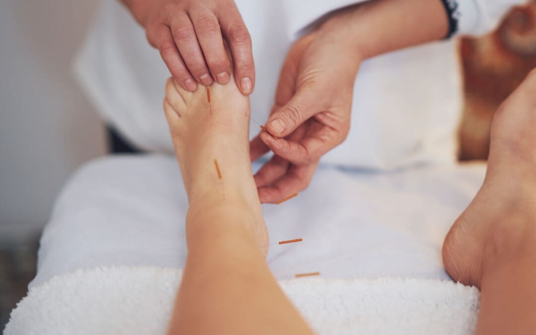
Plantar fasciitis is when the supportive tissue running under the foot, from the heel to the base of the toes, becomes irritated and painful. The disorder can be difficult to manage, but there are alternative treatment options. Acupuncture plantar fasciitis therapy is one potential method of relief, alleviating pain, and returning the individual to regular activities. Acupuncture involves inserting extremely thin needles into points in the body to restore and balance the normal flow of energy and improve overall health. (Johns Hopkins University. 2024) In traditional Chinese medicine or TCM, the body comprises a series of meridians/channels that supply energy flow or qi/chi.
Facts
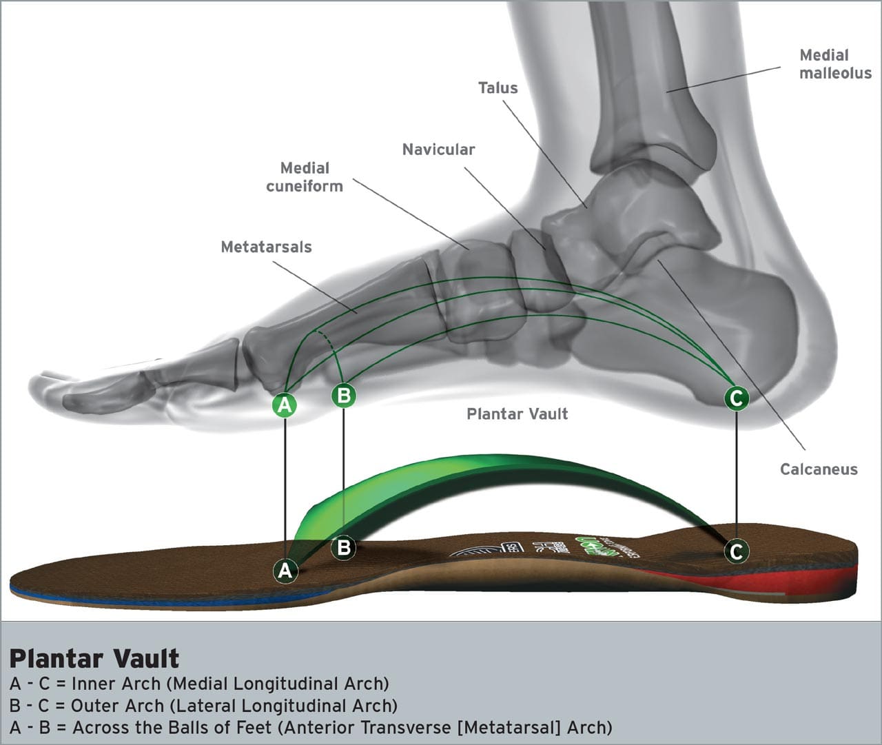
Plantar fasciitis is a common disorder affecting the foot. The condition occurs when the plantar fascia, designed to absorb the forces that travel through the arch of the foot, becomes overloaded. When the bottom of the foot is constantly subjected to high amounts of strain, it leads to ligament degeneration, pain, and inflammation. The most common symptom is heel pain, the first thing an individual experiences in the morning or after a long day of work and activities. Anyone can get plantar fasciitis, but those who are more prone to the condition include individuals with:
Acupuncture and its effectiveness are still being studied, but there is evidence suggesting that it is beneficial in plantar fasciitis treatment.
One review found significant pain improvements in individuals who had acupuncture for the condition compared to individuals who received standard treatments like stretching, orthotics, and strengthening. (Anandan Gerard Thiagarajah 2017) The same review also found benefits when comparing acupuncture to a placebo version of the treatment, further reinforcing the findings.
Another medical review found that acupuncture helped alleviate heel pain and improve daily function when combined with nonsteroidal anti-inflammatory medications/NSAIDs such as ibuprofen or naproxen. (Richard James Clark, Maria Tighe 2012)
Side Effects
While acupuncture plantar fasciitis therapy is beneficial, it is important to remember there can be potential side effects that can include:
Pain in the area where the needles were placed.
Bleeding in the area where the needles were placed.
Bruising or skin discoloration.
Allergic reaction or contact dermatitis/itchy rash.
The chances of a serious adverse side effect are relatively low when undergoing acupuncture on the foot.
Acupuncture Points and Sensations
The ways acupuncture works are not yet fully understood, but like other neuromusculoskeletal therapies, the process activates the body’s healing properties.
Inserting a needle into the body’s points stimulates the central nervous system.
This leads to the release of chemicals in the brain, spinal cord, and muscles that promote healing.
These same chemicals and reactions also reduce the body’s sensation of pain. (Teng Chen et al., 2020)
Number of Sessions
The amount of sessions that acupuncture takes to provide pain relief varies from person to person and case to case.
One review found that treating plantar fasciitis weekly with acupuncture produced significant pain relief after four to eight weeks. (Anandan Gerard Thiagarajah 2017)
This corresponds to another medical review that included a study showing significantly improved pain levels in individuals undergoing weekly acupuncture sessions for four weeks. (Richard James Clark, Maria Tighe 2012)
Individuals are recommended to consult a healthcare provider about personalized treatment plans and if they have a bleeding disorder, are on blood thinner medications, or are pregnant.
American Academy of Orthopaedic Surgeons. (2022). Plantar fasciitis and bone spurs. (Diseases and Conditions, Issue. https://orthoinfo.aaos.org/en/diseases–conditions/plantar-fasciitis-and-bone-spurs
Thiagarajah A. G. (2017). How effective is acupuncture for reducing pain due to plantar fasciitis?. Singapore medical journal, 58(2), 92–97. https://doi.org/10.11622/smedj.2016143
Clark, R. J., & Tighe, M. (2012). The effectiveness of acupuncture for plantar heel pain: a systematic review. Acupuncture in medicine : journal of the British Medical Acupuncture Society, 30(4), 298–306. https://doi.org/10.1136/acupmed-2012-010183
Chan, M. W. C., Wu, X. Y., Wu, J. C. Y., Wong, S. Y. S., & Chung, V. C. H. (2017). Safety of Acupuncture: Overview of Systematic Reviews. Scientific reports, 7(1), 3369. https://doi.org/10.1038/s41598-017-03272-0
Chen, T., Zhang, W. W., Chu, Y. X., & Wang, Y. Q. (2020). Acupuncture for Pain Management: Molecular Mechanisms of Action. The American journal of Chinese medicine, 48(4), 793–811. https://doi.org/10.1142/S0192415X20500408
Progress can be challenging for individuals in post total ankle replacement surgery. How can physical therapy help in recovery and restoring leg function?
Total Ankle Replacement Post Surgery Physical Therapy
Total ankle replacement surgery is a major procedure that takes time to recover. A total ankle replacement surgery or arthroplasty can benefit individuals with chronic ankle pain or disability. This procedure can significantly improve an individual’s overall pain and function with time. Physical therapy is essential to regaining movement in the ankle and restoring full mobility. A physical therapist will work with the individual to control pain and swelling, restore the ankle’s range of motion, train on walking gait and balance, and rebuild strength in the leg. This will help maximize the chances of a successful outcome after surgery.
Total Ankle Replacement
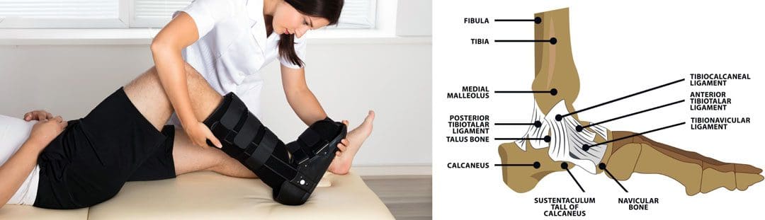
The ankle joint is the section of the lower leg where the shinbone/tibia meets the talus bone on the top of the foot. What can happen is the slippery surface/articular cartilage that coats the ends of these bones begins to thin or deteriorate. As the deterioration progresses, it can lead to significant pain, disability, and difficulty walking. (Cleveland Clinic. 2021) This is where a specialist may recommend total ankle replacement for the best results. Various conditions can be helped by this procedure, including:
During an ankle replacement procedure, an orthopedic surgeon removes the damaged ends of the tibia and talus bones and replaces them with an artificial covering. A polyethylene component is also secured between the two structures to support the smooth movement of the new joint endings. (Massachusetts General Hospital. N.D.) Following the procedure, individuals are typically placed in a protective boot or splint. The healthcare provider will recommend staying off the leg for 4 to 8 weeks to allow healing.
Physical Therapy
Outpatient physical therapy is usually initiated several weeks after the ankle operation. (UW Health Orthopedics and Rehabilitation. 2018) Physical therapy can last for five months or more, depending on the severity of the condition and injury. The physical therapist will focus on different areas to get the best results. (Cort D. Lawton et al., 2017)
Pain and Swelling Control
Post-operative pain and swelling are normal after a total ankle replacement. It is not unusual for an ankle to be swollen for even six to 12 months after the operation. (UW Health Orthopedics and Rehabilitation. 2018) The surgeon will normally prescribe medication to help manage discomfort early on, and physical therapy also plays an important role in addressing the symptoms. Treatments used can include:
Electrical stimulation – mild electrical pulses applied to the muscles.
Ice
Vasopneumatic compression, where an inflatable sleeve is used to create pressure around the area, is commonly utilized at the beginning of physical therapy to reduce pain or swelling.
Other modalities, such as stretching and targeted exercises, are combined with other treatments.
Range of Motion
Early after the procedure, the ankle will be very stiff and tight. This is due to several factors, including the inflammation and swelling after surgery and the time spent immobilized in a boot.
The physical therapist will employ various techniques to improve the ankle joint’s range of motion to rotate and flex.
The physical therapist may employ passive stretching induced by an outside force such as the therapist or a resistance band) to help improve mobility.
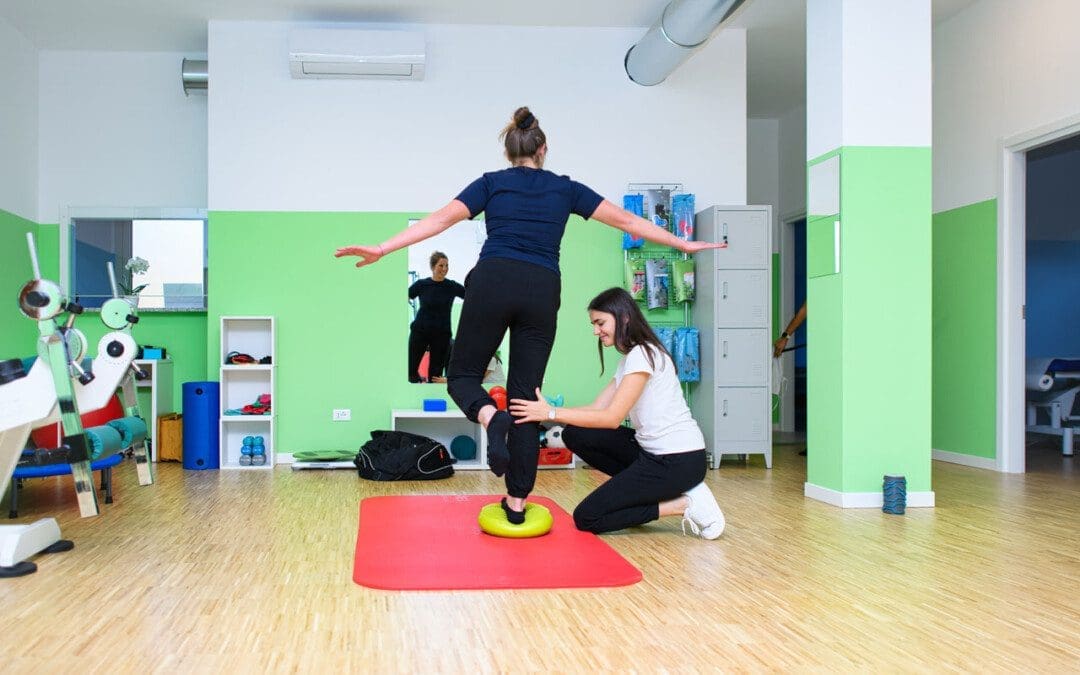
After multiple weeks of reduced movement and lack of bearing any weight on the ankle, the muscles that surround the ankle have often atrophied/weakened, which can impact balance.
When the individual can begin placing weight on the leg, the therapist will apply proprioceptive/sense of body position training to improve overall stability. (UW Health Orthopedics and Rehabilitation. 2018)
Balance exercises will be added to the home program and will progress from week to week.
Strength
The muscles in the leg, ankle, and foot become weak from the surgery and the time spent in a splint or boot. These structures have a significant role in balance, the ability to stand, walk, and go up or down the stairs.
Regaining the strength and power of these muscles is a critical goal of rehabilitation.
In the first weeks, the physical therapist will focus on gentle strengthening exercises.
Isometrics lightly activate the muscles but avoid irritating the surgical site.
As time passes and weight-bearing is allowed, these gentle moves are replaced with more challenging ones, like resistance bands and standing exercises, to accelerate strength gains.
Lawton, C. D., Butler, B. A., Dekker, R. G., 2nd, Prescott, A., & Kadakia, A. R. (2017). Total ankle arthroplasty versus ankle arthrodesis-a comparison of outcomes over the last decade. Journal of orthopaedic surgery and research, 12(1), 76. https://doi.org/10.1186/s13018-017-0576-1
Individuals in post-surgery recovery or dealing with illness or an injury can experience weakened muscles and endurance that can cause temporary loss of sleeping mobility and not being able to move around normally because of weakness, decreased range of motion, or pain. Can they benefit from physical therapy to help get back to normal functional mobility?
Sleeping Mobility
For individuals who are hospitalized or homebound from injury, illness, or surgical recovery, a physical therapist will assess various areas of functional mobility. These include transfers – from sitting to standing positions, walking, and sleeping mobility. Sleeping mobility is the ability to perform specific motions while in bed. A therapist can assess sleeping or bed mobility and recommend strategies and exercises to improve movements. (O’Sullivan, S. B., Schmitz, T. J. 2016) A therapist may have the individual use specific devices, like an over-the-bed trapeze or a sliding board, to help move around.
All of these movements require strength in different muscle groups. By checking out individual motions in sleeping mobility, a therapist can work out specific muscle groups that may be weak and require targeted exercises and stretches to restore mobility to normal. (O’Sullivan, S. B., Schmitz, T. J. 2016) Individuals visiting a therapist in an outpatient clinic or rehabilitation area may have the individual work on sleeping mobility on a treatment table. The same motions on the treatment table can be done in the bed.
Importance
The body is meant to move.
For individuals who cannot move comfortably on their bed, the body may suffer disuse atrophy or the wasting away of muscular strength, which can lead to increased difficulties. Not being able to move can also lead to pressure ulcers, especially for individuals who are severely deconditioned and/or remain in one position for a long period. Skin health may start to break down, leading to painful wounds that require specialized care. Being able to move around in bed can help prevent pressure ulcers. (Surajit Bhattacharya, R. K. Mishra. 2015)
Improvement
A physical therapist can prescribe specific exercises to strengthen muscle groups and improve sleeping mobility. The muscles include:
Shoulder and rotator cuff muscles.
Triceps and biceps in the arms.
Gluteus muscles of the hips.
Hamstrings
Quadriceps
Calf muscles
The shoulders, arms, hips, and legs work together when moving the body around the bed.
Various Exercises
To improve bed movement, physical therapy exercises can include:
Physical therapists are trained to assess these motions and functions and prescribe treatments to improve body movement. (O’Sullivan, S. B., Schmitz, T. J. 2016) Maintaining appropriate physical fitness can help the body stay active and mobile. Performing mobility exercises prescribed by a physical therapist can keep the right muscle groups working properly, and working with a physical therapist can ensure the exercises are correct for the condition and are performed properly.
Bhattacharya, S., & Mishra, R. K. (2015). Pressure ulcers: Current understanding and newer modalities of treatment. Indian journal of plastic surgery : official publication of the Association of Plastic Surgeons of India, 48(1), 4–16. https://doi.org/10.4103/0970-0358.155260
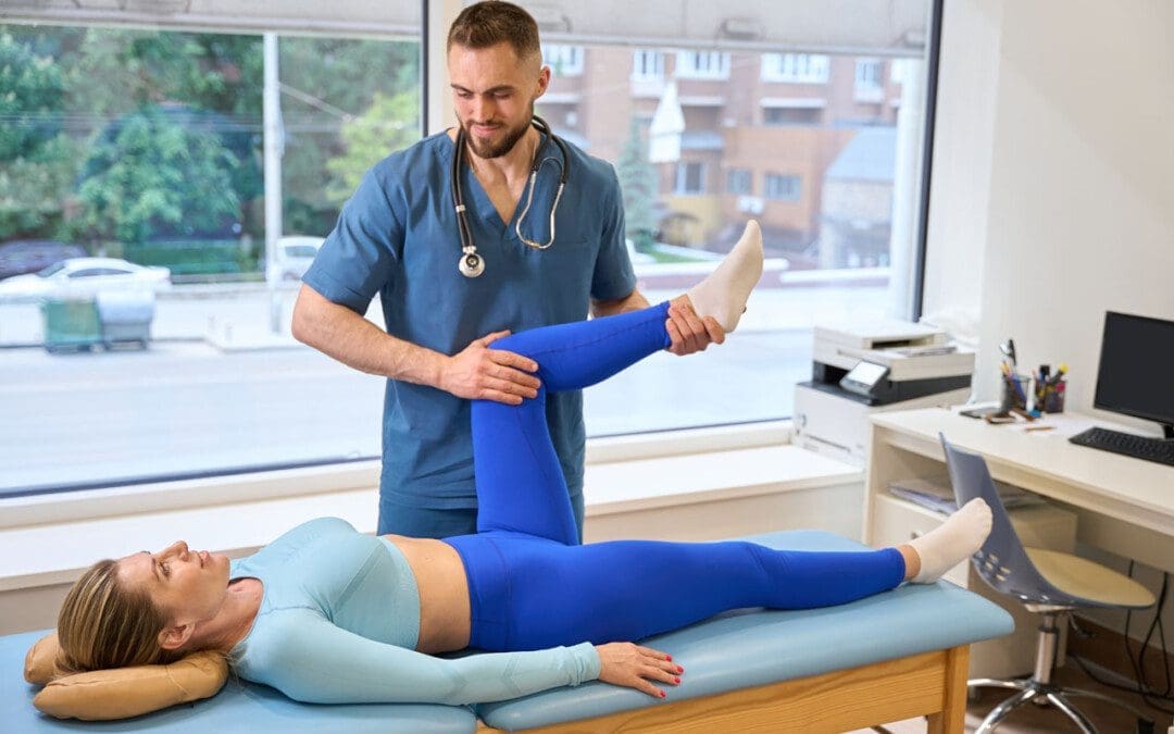
For individuals experiencing pelvis pain symptoms and associated problems, can integrating pelvic floor physical therapy exercises help with treatment and prevention?
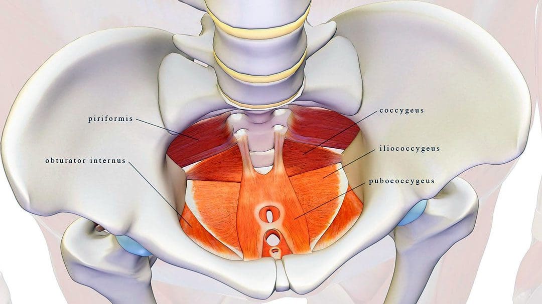
Pelvic Floor Physical Therapy
The pelvic floor muscles are located at the base of the pelvis and protect the pelvic organs like the vagina, cervix, uterus, bladder, urethra, and rectum. (U.S. Food and Drug Administration. 2019)
When the muscles fail to function correctly, individuals can experience symptoms like:
Painful intercourse
Prolapse – when an organ or tissue drops or shifts out of place.
Urinary incontinence
Constipation problems
These conditions are common in pregnant individuals or older women.
These symptoms can be treated with pelvic floor physical therapy to alleviate discomfort. Pelvic floor physical therapy can help women and individuals with vaginas:
Alleviate issues like painful sex, urinary leakage, and prolapse.
In physical therapy, individuals work on breathing, relaxation, and lengthening and strengthening techniques to train their muscles to function optimally.
Causes of Pelvic Floor Issues
Pelvic floor dysfunction tends to happen with age, during pregnancy, or in combination with events like the postpartum period and menopause, which can lower hormone levels.
Individuals who are pregnant are especially prone to pelvic floor issues but might not know they have a problem.
The pregnancy weight of a uterus can pressure and strain the muscles.
If left untreated, these symptoms can worsen over time.
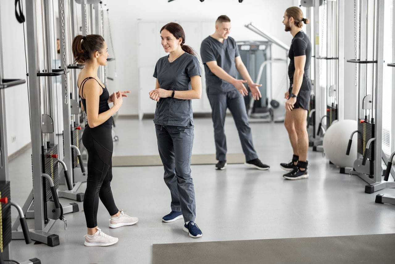
Pelvic Floor Physical Therapy
An individual will meet with a specialist to discuss symptoms and undergo a physical examination that includes:
Pelvic floor exam.
Evaluation of posture, mobility, and core strength.
Once the initial exams and evaluation are complete, the practitioner will go over pelvic floor exercises and provide a treatment plan.
Recommended exercises vary based on symptoms but focus on relaxing, stretching, and/or strengthening muscles.
Muscle Relaxation
To relax the muscles, a therapist may recommend breathing exercises.
For pregnant individuals, this means timing breaths with contractions.
For individuals experiencing constipation, breathing exercises can help the body relax and reduce strain.
Stretching Muscles
Stretching can help relieve muscle tightness and stiffness.
A therapist may help stretch the pelvic floor through various therapy modalities.
This type of physical therapy can help loosen tight muscles or help gently reset dislocated organs back into place.
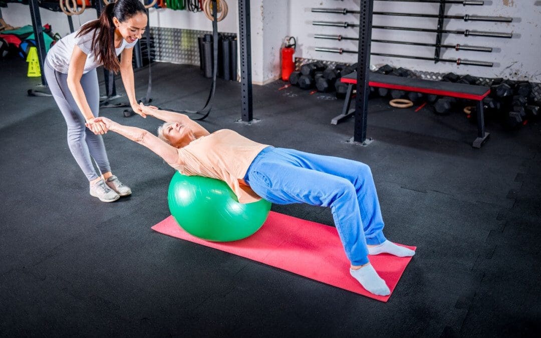
Strengthening Muscles
After the pelvic floor is loose and relaxed, the focus typically switches to strengthening the muscles.
Strength work may target abdominal muscles or the pelvic floor muscles themselves.
With time, commitment, and targeted treatment, individuals can use pelvic floor physical therapy to loosen tissues, strengthen muscles, and restore function.
Sartori, D. V. B., Kawano, P. R., Yamamoto, H. A., Guerra, R., Pajolli, P. R., & Amaro, J. L. (2021). Pelvic floor muscle strength is correlated with sexual function. Investigative and clinical urology, 62(1), 79–84. https://doi.org/10.4111/icu.20190248
Raizada, V., & Mittal, R. K. (2008). Pelvic floor anatomy and applied physiology. Gastroenterology clinics of North America, 37(3), 493–vii. https://doi.org/10.1016/j.gtc.2008.06.003
Soave, I., Scarani, S., Mallozzi, M., Nobili, F., Marci, R., & Caserta, D. (2019). Pelvic floor muscle training for prevention and treatment of urinary incontinence during pregnancy and after childbirth and its effect on urinary system and supportive structures assessed by objective measurement techniques. Archives of gynecology and obstetrics, 299(3), 609–623. https://doi.org/10.1007/s00404-018-5036-6
Individuals who have gone through recent low back surgery, like a lumbar laminectomy and discectomy, could they benefit from physical therapy for full recovery? (Johns Hopkins Medicine. 2008)
Rehabilitation Exercise Program
A lumbar laminectomy and discectomy is a surgical procedure performed by an orthopedic or neurologic surgeon to help decrease pain, relieve associated symptoms and sensations, and improve flexibility and mobility. The procedure involves cutting away disc and bone material that presses against, irritates, and damages the spinal nerves. (Johns Hopkins Medicine. 2023)
Post-Surgery
The therapist will work with the individual to develop a rehabilitation exercise program. The objective of a rehabilitation exercise program is to help the individual:
Relax their muscles to prevent muscle tensing and becoming over-cautious
Regain full range of motion
Strengthen their spine
Prevent injuries
A guide on what to expect in physical therapy.
Postural Retraining
After back surgery, individuals have to work to maintain proper posture when sitting and standing. (Johns Hopkins Medicine. 2008)
Postural control is important to learn as it maintains the lower back in the optimal position to protect and expedite the healing of lumbar discs and muscles.
A physical therapist will teach the individual how to sit with proper posture and use lumbar support.
Attaining and maintaining proper posture is one of the most important things to help protect the back and prevent future back problems.
Walking helps to improve cardiovascular health and blood circulation throughout the body.
This helps to provide added oxygen and nutrients to the spinal muscles and tissues as they heal.
It is an upright exercise that puts the spine in a natural position, which helps to protect the discs.
The therapist will help set up a program tailored to the individual’s condition.
Prone Press Up
One of the exercises to protect the back and lumbar discs is prone press-ups. (Johns Hopkins Medicine. 2008) This exercise helps keep the spinal discs situated in the proper position. It also helps to improve the ability to bend back into lumbar extension.
To perform the exercise:
Lie facing down on a yoga/exercise mat and place both hands flat on the floor under the shoulders.
Keep the back and hips relaxed.
Use the arms to press the upper part of the body up while allowing the lower back to remain against the floor.
There should be a slight pressure in the lower back while pressing up.
Hold the press-up position for 2 seconds.
Slowly lower back down to the starting position.
Repeat for 10 to 15 repetitions.
Sciatic Nerve Gliding
Individuals who had leg pain coming from the back prior to surgery may have been diagnosed with sciatica or an irritation of the sciatic nerve. Post-surgery, individuals may notice their leg feels tight whenever straightening it out all the way. This could be a sign of an adhered/trapped sciatic nerve root, a common problem with sciatica.
After lumbar laminectomy and discectomy surgery, a physical therapist will prescribe targeted exercises called sciatic nerve glides to stretch and improve how the nerve moves. (Richard F. Ellis, Wayne A. Hing, Peter J. McNair. 2012)
Nerve glides can help free the stuck nerve root and allow for normal motion.
To perform the exercise:
Lie on the back and bend one knee up.
Grab underneath the knee with the hands.
Straighten the knee while supporting it with the hands.
Once the knee is fully straightened, flex and extend the ankle about 5 times.
Return to the starting position.
Repeat the sciatic nerve glide 10 times.
The exercise can be performed several times to help improve how the nerve moves and glides in the lower back and leg.
Supine Lumbar Flexion
After surgery, gentle back flexion exercises can help safely stretch the low-back muscles and gently stretch the scar tissue from the surgical incision. Supine lumbar flexion is one of the simplest exercises to improve lumbar flexion range of motion.
To perform the exercise:
Lie on the back with the knees bent.
Slowly lift the bent knees towards the chest and grasp the knees with both hands.
Gently pull the knees toward the chest.
Hold the position for 1 or 2 seconds.
Slowly lower the knees back to the starting position.
Perform for 10 repetitions.
Stop the exercise if experiencing an increase in pain in the lower back, buttocks, or legs.
Hip and Core Strengthening
Once cleared, individuals can progress to an abdominal and core strengthening program. This involves performing specific motions for the hips and legs while maintaining a pelvic neutral position. Advanced hip strengthening exercises help generate strength and stability in the muscles that surround the pelvic area and lower back. A physical therapist can help decide which exercises are recommended for the specific condition.
Return-to-Work and Physical Activities
Once individuals have gained an improved lumbar range of motion, hip, and core strength, their doctor and therapist may recommend working on specific activities to help them return to their previous level of work and recreation. Depending on job occupation, individuals may need to:
Work on proper lifting techniques.
Require an ergonomic evaluation if they spend time sitting at a desk or workstation.
Some surgeons may have restrictions on how much an individual can bend, lift, and twist from two to six weeks after surgery.
Low-back surgery can be difficult to rehab properly. Working with a healthcare provider and physical therapist, individuals can be sure to improve their range of motion, strength, and functional mobility to return to their previous level of function quickly and safely.
Ellis, R. F., Hing, W. A., & McNair, P. J. (2012). Comparison of longitudinal sciatic nerve movement with different mobilization exercises: an in vivo study utilizing ultrasound imaging. The Journal of orthopaedic and sports physical therapy, 42(8), 667–675. https://doi.org/10.2519/jospt.2012.3854
For individuals managing osteoarthritis, could massage therapy provide added treatment benefits?
Osteoarthritis Massage Therapy
Osteoarthritis happens when the cartilage between the joints wears away, causing stiffness and pain. Massage therapy is a treatment used to relieve various types of pain symptoms.
There are many types of massage therapy, that healthcare providers utilize to manipulate the muscles and other soft tissues to relieve symptoms, relax muscles, increase circulation, reduce inflammation, release trigger points, and restore mobility, flexibility, and function. (Ergonomic Trends. 2023)
Professional therapists can help relieve osteoarthritis joint pain by relaxing the surrounding muscles and other soft tissues to release stiffness. (Adam Perlman, et al., 2019)
Massage Objectives and Types
Massage therapists use their hands and fingers, forearms, elbows, and/or instruments to manipulate the body’s soft tissues. Soft tissues support and surround body structures and include muscle, fat, tendons, and ligaments.
The goal of osteoarthritis massage therapy is to relax muscles and soft tissues, increase blood and oxygen circulation, warm the affected area/s, relieve pain, and restore mobility and function.
Depending on the location of the muscles being massaged, individuals may be seated or lie down on a specialized table.
The amount of pressure and direction of movement depend on the body area.
Therapeutic oils and/or massage creams may be used to increase the therapy.
Types include:
Swedish
The therapist uses long strokes, kneading, and friction on the muscles.
Joints are moved to increase flexibility.
Deep Tissue
The therapist uses deep finger or instrument pressure, focusing on muscles that are tight or knotted.
Trigger Point
Trigger points represent a source of radiating pain symptoms.
The therapist focuses pressure on these myofascial tissue points using various strokes to release them.
Shiatsu
The therapist applies rhythmic pressure with their thumbs, fingers, and palms to redirect and increase energy or chi/qi.
A massage session lasts around 30–60 minutes depending on the severity of the condition and the number of sessions the patient has undergone. Chronic pain patients usually go through a series of specialized sessions that focus on specific areas and gradually build.
Risk Factors
Certain precautions must be taken before getting osteoarthritis massage therapy. Although there are a few serious risks, certain individuals are not suitable candidates and should not receive massage therapy. The conditions include: (Medical Massage Therapy Resource & Reference. 2023)
Damaged nerves.
Damaged blood vessels.
Infection and inflammation in the area to be massaged.
Open wounds.
Fever.
Taking a blood thinner.
Deep vein thrombosis – blood clots.
Bleeding disorders.
Osteoporosis – weak and brittle bones.
Recent fractures – broken bones.
Tumors.
Cancer.
Individuals who have recently undergone surgery.
Individuals with a skin condition that is contagious, like warts or herpes, or noncontagious, like psoriasis, could be aggravated by touch or pressure.
Individuals who have cancer, fragile skin, heart problems, or dermatomyositis are recommended to discuss osteoarthritis massage therapy with their healthcare provider.
Research on the effects of massage therapy on various health conditions is ongoing. Massage therapy promotes relaxation while reducing stress, which can help with chronic joint issues like osteoarthritis.
Perlman, A., Fogerite, S. G., Glass, O., Bechard, E., Ali, A., Njike, V. Y., Pieper, C., Dmitrieva, N. O., Luciano, A., Rosenberger, L., Keever, T., Milak, C., Finkelstein, E. A., Mahon, G., Campanile, G., Cotter, A., & Katz, D. L. (2019). Efficacy and Safety of Massage for Osteoarthritis of the Knee: a Randomized Clinical Trial. Journal of general internal medicine, 34(3), 379–386. https://doi.org/10.1007/s11606-018-4763-5
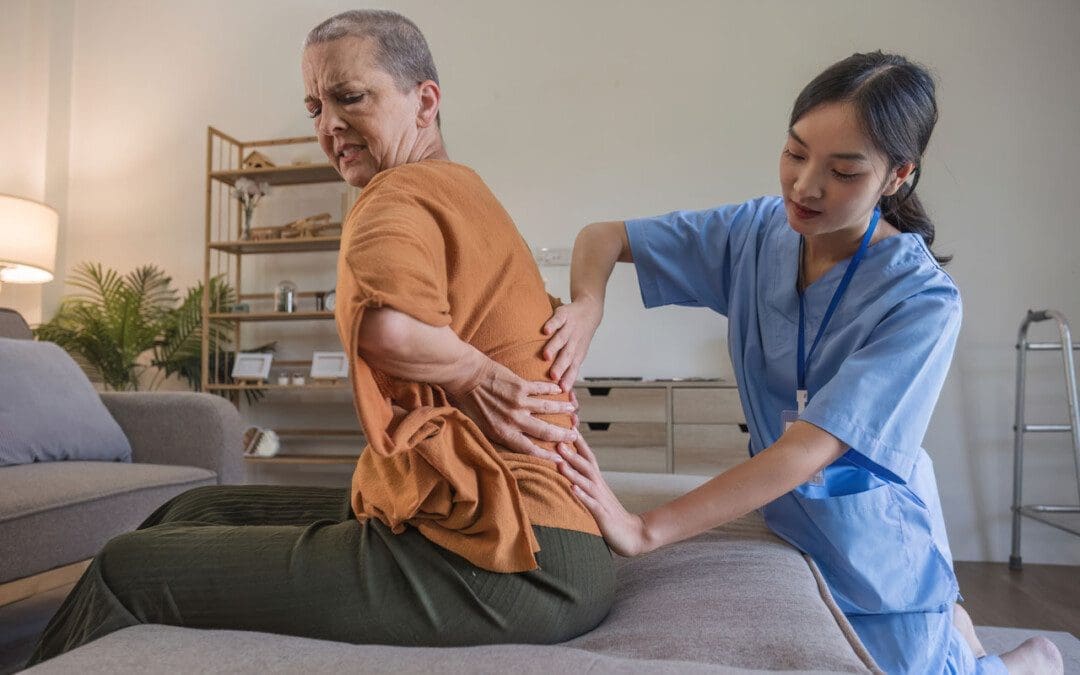
Individuals dealing with back pain problems could be suffering from a bulging disc. Could knowing the difference between slipped and herniated disc symptoms help with treatments and finding relief?
Bulging Disc Pain
Back pain can become debilitating if not treated properly. A bulging disc is a common cause of cervical, thoracic, and lower back pain symptoms. It happens when one of the fluid-filled cushions between the vertebrae begins to shift out of place. Instead of being aligned with the edges, the disc bulges over. This begins to generate pressure on the nerves causing pain and inflammation.
Bulging discs are often caused by age, but repetitive movements and/or lifting heavy objects can contribute to the condition.
Symptoms can resolve on their own, but individuals are recommended to consult with a physical therapist and/or chiropractor to make sure the disc healed properly, otherwise, it can lead to worsening and/or further injuries.
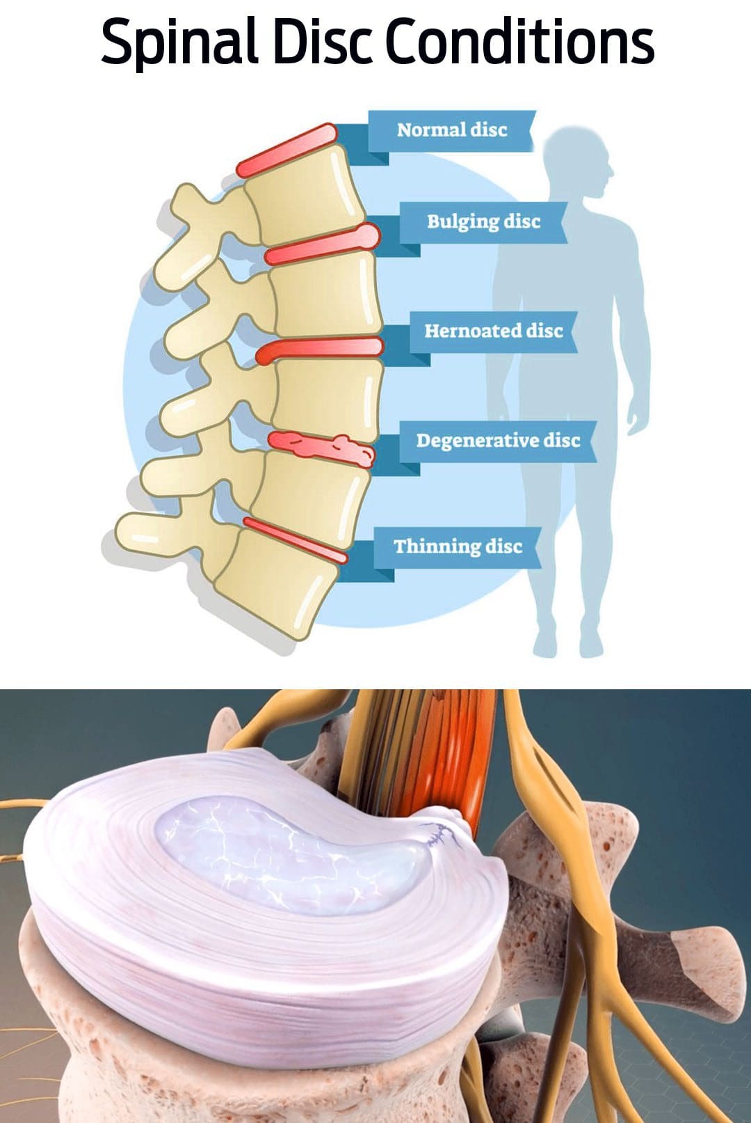
Bulging Disc vs. Herniated Disc
Bulging and herniated discs cause pain symptoms.
They both can be linked to injuries and degenerative disc disease but are not the same condition. (Penn Medicine. 2018)
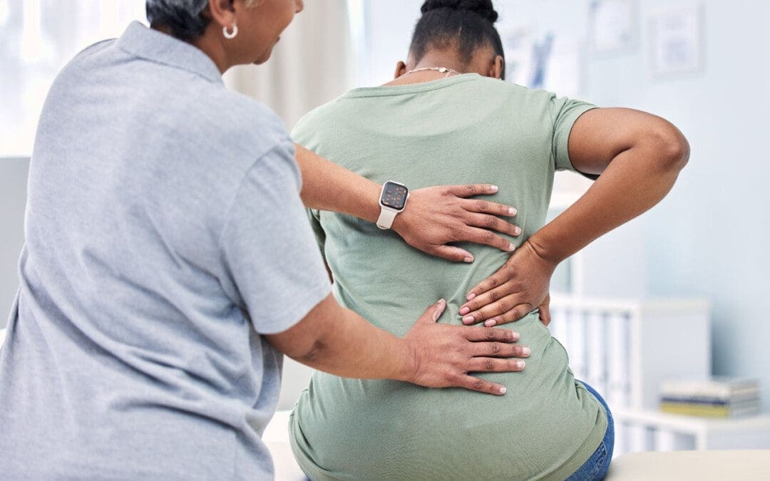
This is because the lower back is subject to all kinds of pressure and movement with daily activities, increasing the chances of pain and injuries.
The next most common place is the neck/cervical spine where there are constant movements making it prone to injury and pain symptoms.
Causes
Bulging discs are most often caused by body aging and normal wear and tear. As time goes on the intervertebral discs naturally degenerate, known as degenerative disc disease. This can cause the discs to pull downward, causing them to bulge from their placement. (Penn Medicine. 2018) Factors that can cause or worsen the condition include:
Practicing unhealthy postures.
Repetitive motions.
Lifting heavy objects
Spinal injuries.
Medical history of spinal or disc disease in the family.
Individuals with back pain that interferes with daily functions or has lasted longer than six weeks, should see a healthcare provider for a diagnosis. They will order a magnetic resonance imaging scan/MRI, which can show where a disc is protruding. (American Academy of Neurological Surgeons. 2023)
Rest
For bulging disc pain, resting the back is necessary. However,
IFM's Find A Practitioner tool is the largest referral network in Functional Medicine, created to help patients locate Functional Medicine practitioners anywhere in the world. IFM Certified Practitioners are listed first in the search results, given their extensive education in Functional Medicine