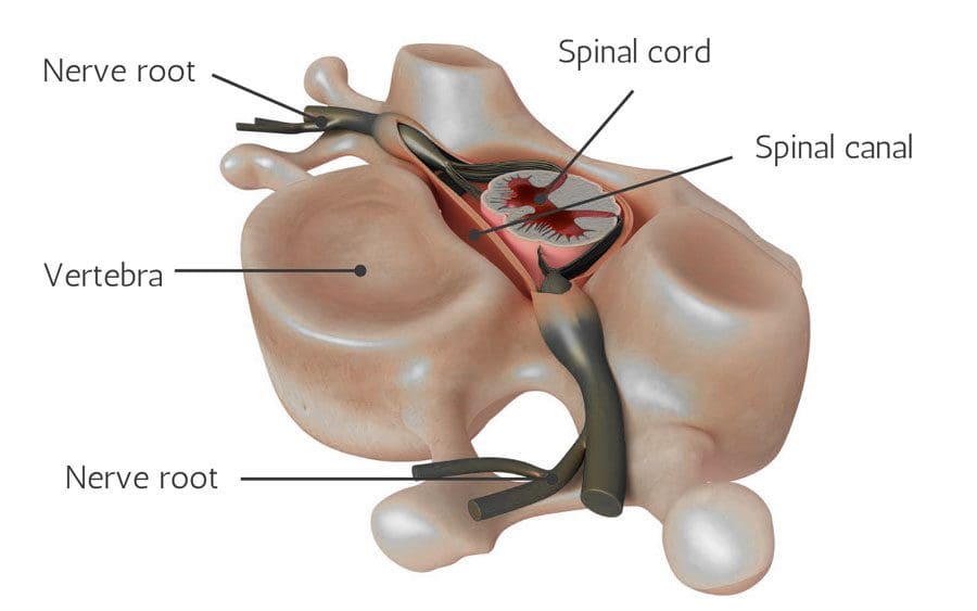
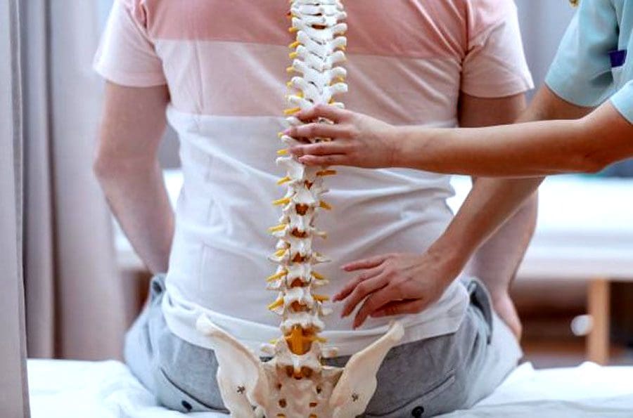
Spinal nerves send motor, sensory, and autonomic signals between the central nervous system and the body and are part of the peripheral nervous system. They are essential for carrying information that controls body movements and sensations to the brain. When a nerve gets injured, compressed, or damaged, it can cause discomfort, increased sensitivity, numbness, muscle weakness, and pain.
Damaged Nerve Roots
Nerve root pain is often caused by other underlying conditions that have caused compression or damage to the nerve root. Causes of nerve root pain can include:
Spinal nerves impacted by injuries or infection can lose their ability to control the body areas, lose their functional capacity, lose sensation, and die.
Spinal Imaging
Nerve damage can be diagnosed on a neurological exam and correlated with MRI and X-ray imaging. Conditions that MRI can identify include herniated discs, spinal cord compression or fracture, arthritic development, tumors, or cysts pressing on a nerve.
MRI images are obtained with a magnetic field and radio waves.
MRI shows spine images from the side/sagittal view and cross-sectional/axial views.
This allows the chiropractic doctor to see the vertebrae and discs and identify abnormalities.
The spinal cord is a gray area in the middle surrounded by the spinal fluid, which appears white.
Little white channels on either side of the spinal cord are where the nerve roots branch off.
X-rays can show the alignment of the bones along the spine and determine any narrowing or damage to the discs.
It is important to be evaluated and diagnosed for signs and symptoms of nerve injury as soon as possible, as nerve damage accelerates and worsens.
Function Restoration
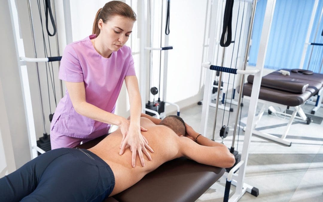
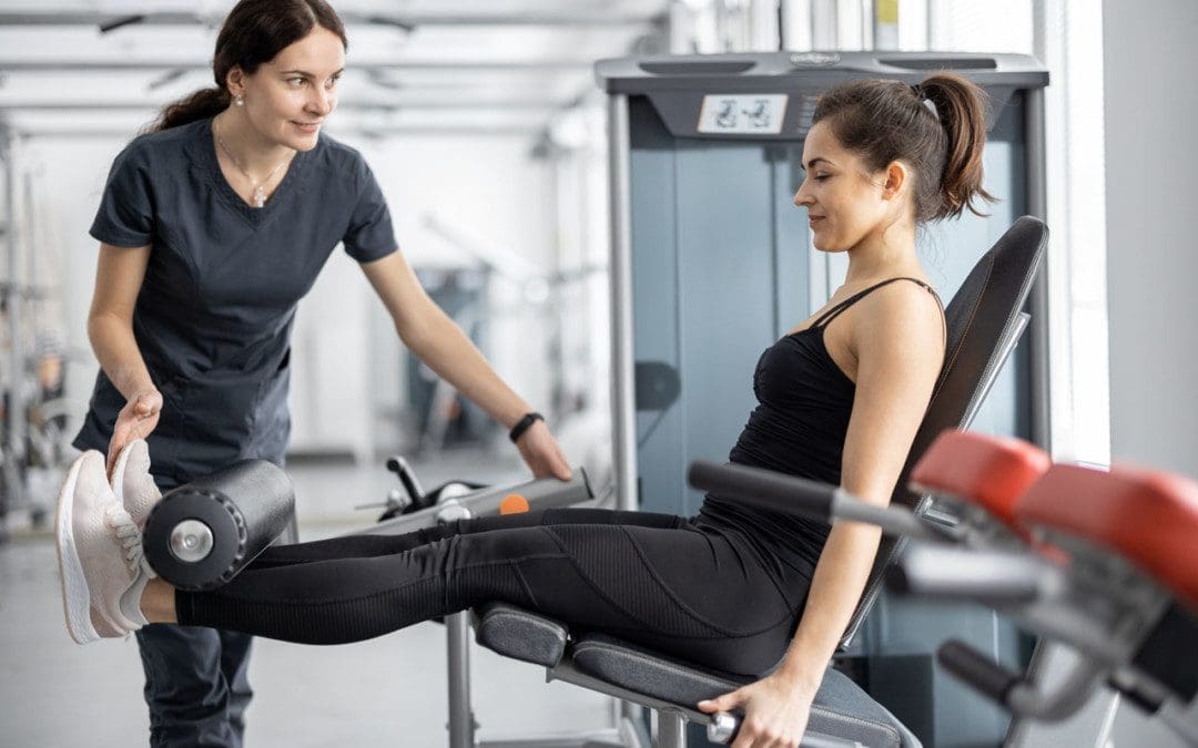
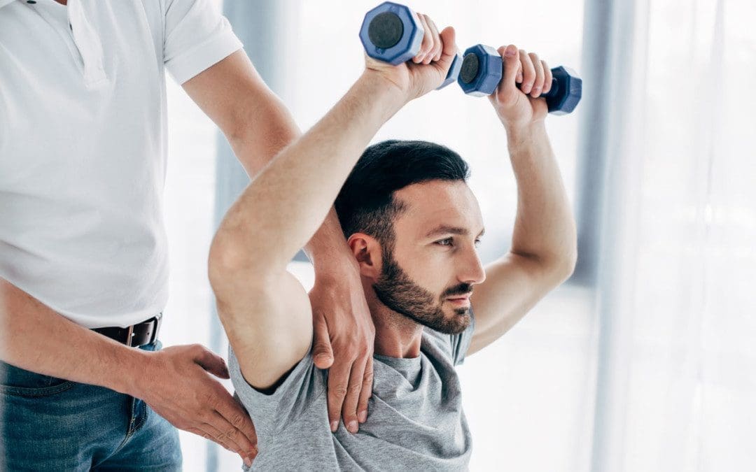
Sometimes, the symptoms improve by themselves and do not require treatment. Nonetheless, physicians begin with conservative, non-surgical approaches to treat nerve root pain. Chiropractic and physical massage therapy involves specific movements, stretches, and exercises to keep the affected muscles and joints active,prevent stiffness and help restore function and feeling. Treatment can include:
Therapeutic massage
Manual adjustment/resistance treatment
Trigger point therapy
Instrument-assisted soft tissue therapy
Decompression
Traction
Joint stretching
Electrical stimulation
Ultrasound
Specialized exercise
Activity modification
Anti-inflammatory diet
Nerve Chiropractor
References
Liu, Yan, and Huan Wang. “Peripheral nerve injury-induced changes in the spinal cord and strategies to counteract/enhance the changes to promote nerve regeneration.” Neural regeneration research vol. 15,2 (2020): 189-198. doi:10.4103/1673-5374.265540
Menorca, Ron M G, et al. “Nerve physiology: mechanisms of injury and recovery.” Hand clinics vol. 29,3 (2013): 317-30. doi:10.1016/j.hcl.2013.04.002
Shehab, Safa Al-Deen Saudi. “Fifth lumbar spinal nerve injury causes neurochemical changes in corresponding and adjacent spinal segments: a possible mechanism underlying neuropathic pain.” Journal of chemical neuroanatomy vol. 55 (2014): 38-50. doi:10.1016/j.jchemneu.2013.12.002
Stoll, G, and H W Müller. “Nerve injury, axonal degeneration, and neural regeneration: basic insights.” Brain pathology (Zurich, Switzerland) vol. 9,2 (1999): 313-25. doi:10.1111/j.1750-3639.1999.tb00229.x
Ye, Xuan, et al. “Nerve fascicle transfer using a part of the C-7 nerve for spinal accessory nerve injury.” Journal of neurosurgery. Spine vol. 28,5 (2018): 555-561. doi:10.3171/2017.8.SPINE17582
Individuals that have experienced a muscle strain, pull, spasm, etc., that has healed can begin to behave overly cautious, avoiding putting full weight on the area or using full motion out of fear of re-injuring it. This can and does strain other body areas because of the imbalance and awkward positioning. It also leads to anxiety, emotional distress, and decreased self-confidence in everyday movement. Adjustments, massage, and decompression therapy can maintain musculoskeletal health, and a chiropractor can help retrain individuals on healthy posture and confident movement.
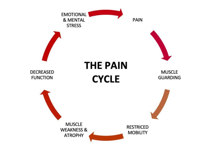
Muscle Guarding
Muscles can be held in a position of readiness to act, like the stress response of fight or flight. When this happens, the muscles are partially contracted in preparation for action and is a form of muscle-guarding. But once the fight or flight passes, the muscles relax into their normal position. With injury muscle guarding, the fears and stresses after recovering from an injury can cause the injured and non-injured muscles to stay in the guarded/semi-contracted position. The longer the muscle guarding continues, fatigue begins to set in, decreasing function, restricting mobility, and making the body more vulnerable to damage and injury.
The Brain
The discomfort, pain, or just the thought reinforces the need to guard the area. The brain will find a way to move without causing pain and create compensating but unhealthy movement patterns that strain the other areas of the body. The body adapts to not using the formerly injured muscles and now relies on the other muscles to perform the functions in a non-relaxed state that can become normal, causing stiffness, soreness, tenderness, tendon tension, and pain.
An example is a hip strain, pull or spasm that has been treated and has healed, but the individual is fearful of another injury or going through the painful experience again and begins walking by shifting all their weight to the other side and steps with a limp or some abnormal motion that strains and/or injures the rest of the body.
Chiropractic Treatment and Retraining
Individuals experiencing muscle guarding can find help through chiropractic to retrain their muscles to return to their normal position and regain confidence in their movements. The body will be rebalanced by releasing and relaxing the tight muscles. Then therapeutic repetitive movements, specialized exercises, stretches, and relaxation techniques will help the individual relearn to use the muscles without fear.
Protective Muscle Guarding
References
Hanlon, Shawn et al. “Examining Ankle-Joint Laxity Using 2 Knee Positions and With Simulated Muscle Guarding.” Journal of athletic training vol. 51,2 (2016): 111-7. doi:10.4085/1062-6050-51.3.06
Olugbade, Temitayo et al. “The relationship between guarding, pain, and emotion.” Pain reports vol. 4,4 e770. 22 Jul. 2019, doi:10.1097/PR9.0000000000000770
Prkachin, Kenneth M et al. “Pain behavior and the development of pain-related disability: the importance of guarding.” The Clinical journal of pain vol. 23,3 (2007): 270-7. doi:10.1097/AJP.0b013e3180308d28
Spinal disc deterioration from aging is normal, but health issues or injuries can advance the degenerative process. Disc protrusions are related to herniated discs but are the mildest form of the condition and are a common form of spinal disc deterioration that can cause neck and back issues. However, individuals may have a small protruding disc that can go undetected unless it irritates or compresses the surrounding nerves. Chiropractic care, decompression, and massage therapy can realign the disc back into position, relieving discomfort and pain.
Disc Protrusion
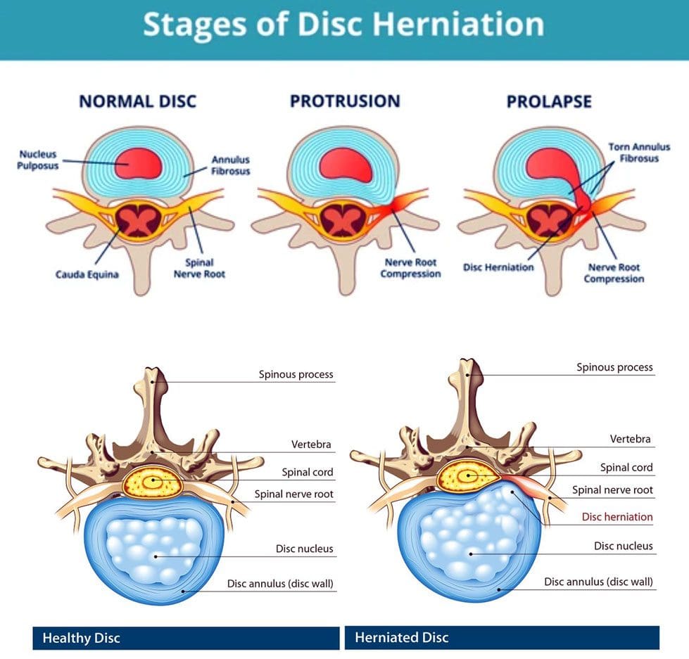
A disc is like a sturdy soft rubber shock absorber/cushion with added gel inside. The gel acts as a shock absorber. When the gel begins to protrude out slightly, this is a disc protrusion. Once a protruding disc begins to develop, it usually remains in that position. The disc can sometimes reabsorb on its own and realign back into position, but there is no way of knowing that will happen or how long it will take. With age and/or injuries, the body’s parts change. The spine’s discs dehydrate and lose elasticity weakening the discs and making them more vulnerable to herniation stages:
First Stage
Following natural weakening can be classified as a disc protrusion when the disc’s core begins pushing into the spinal column.
Disc protrusions can be tiny or push out an entire side of the disc.
Second Stage
Disc deterioration often consists of a bulging disc when the core pushes out farther around the circumference beyond the disc’s outer layer, called the annulus fibrosus, creating the telltale bulge.
A bulging disc involves more than 180 degrees of the disc’s circumference.
Third Stage
The third stage is a herniated disc, meaning the disc’s outer wall has torn, allowing the inner gel to leak out, usually irritating the surrounding nerves.
Fourth Stage
The fourth stage is sequestration, a herniated disc in which a piece of the nucleus breaks free of the vertebral disc fragments and falls into the spinal canal.
Types
A disc protrusion is one type of disc herniation that pushes out but remains connected. Different types compress and irritate the discs differently and produce various symptoms, including:
Paracentral
This is the most common, where the disc protrusion jams the space between the central canal and the foramen.
Central
This is where the disc protrusion impinges into the spinal canal, with or without spinal cord compression.
Foraminal
The disc intrudes into the foramen, the space through which nerve roots branch off the spinal cord and exit the vertebrae.
Symptoms, Diagnosis, and Chiropractic Care
Individuals with a disc protrusion can have symptoms similar to sciatica, which includes back, buttock, and leg discomfort, numbness, and pain sensations.
Treatment for disc protrusion will be based on the individual’s symptoms.
A chiropractor will take a detailed medical history and perform a physical examination.
A spinal MRI test could be ordered depending on the injury or condition.
A customized treatment plan will be developed to fit the individual’s medical needs.
Most disc protrusions improve after a few weeks of rest, avoiding strenuous activities, activity modification, an anti-inflammatory diet, and gentle exercises that the chiropractic team will provide.
True Spinal Decompression
References
Fardon, David F et al. “Lumbar disc nomenclature: version 2.0: Recommendations of the combined task forces of the North American Spine Society, the American Society of Spine Radiology and the American Society of Neuroradiology.” The spine journal: official journal of the North American Spine Society vol. 14,11 (2014): 2525-45. doi:10.1016/j.spinee.2014.04.022
Mysliwiec, Lawrence Walter, et al. “MSU classification for herniated lumbar discs on MRI: toward developing objective criteria for surgical selection.” The European spine journal: official publication of the European Spine Society, the European Spinal Deformity Society, and the European Section of the Cervical Spine Research Society vol. 19,7 (2010): 1087-93. doi:10.1007/s00586-009-1274-4
Sciatica pain can radiate to the knee. Individuals with sciatica do report unique/unusual knee pain that was never there and no past or recent physical injuries. Sciatica is the culprit, as the knee muscles are powered and controlled by nerves in the lower spine. Irritation or compression of these nerves can cause symptoms that can include: random back pain, hamstring tightness, weakness in the hips or quadriceps, the development of bunions, and knee pain and/or weakness. Chiropractic treatment can release the compression, heal the sciatic nerve, and alleviate knee problems.
Sciatica Pain Can Radiate To The Knee
Spine conditions that can cause sciatica include:
Disc herniation – Where the inside of the discs leak out and compress and/or irritate surrounding nerves.
Spinal stenosis – The spinal canal begins to narrow, not allowing enough space for the nerves to rest comfortably, resulting in compressed nerves.
Spondylolisthesis – A condition that occurs when a vertebrae slips forward onto the vertebrae below it.
Any can cause irritation, inflammation, or compression of the sciatic nerve leading to painful sensations that extends from the lower back down through the leg.
Symptoms
Common knee symptoms that may be experienced with sciatica include:
A dull ache, warm sensation, or sharp pain around the knee.
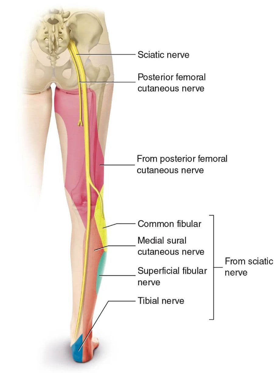
Bunions form from weakened stabilizing muscles that affect walking, running, and standing posture.
As sciatica pain can radiate to the knee, individuals will usually also experience pain in their buttocks, thigh, calf, and/or foot. The nerve sensations and other symptoms in the knee can be felt through a branch of the sciatic nerve known as the peroneal nerve.
Duration
The knee pain will last as long as sciatica does, depending on the type of sciatica, whether it is acute or chronic.
An acute sciatic episode usually resolves after a few weeks, with possible future flare-ups.
Chronic sciatica is a long-term condition that does not resolve independently and necessitates intervention by a specialist.
Chiropractic Treatment Plan
Depending on the diagnosis, a chiropractor will develop a personalized treatment plan to address the root cause and heal the injury. The treatment plan will include therapeutic massage, posture training, and at-home self-care to help heal and prevent future sciatica.
Massage Rehabilitation
Physical therapy and therapeutic massage will loosen and relax the muscles, nerves, tendons, and ligaments.
Heat and ice, exercises, and stretches will prepare the muscles and nerves for chiropractic decompression adjustments.
Posture Training
Training will be provided to maintain the back, hips, knees, and feet in proper alignment.
Training on removing pressure from the lower back and restabilizing the rest of the body.
Training on proper body mechanics, safe lifting techniques, and injury prevention.
Self-Pain Management
Training on self-care habits that include healthy weight, core strengthening exercises and stretches for the back muscles, and proper rest for a full recovery.
An anti-inflammatory diet to reduce/eliminate inflammation and achieve a healthy weight and a nutrition plan to maintain overall health.
Surgery
Surgery is the final option when conservative treatments are not working.
Treating Severe & Complex Sciatica Syndromes
References
Dydyk AM, Khan MZ, Singh P. Radicular Back Pain. [Updated 2021 Nov 2]. In: StatPearls [Internet]. Treasure Island (FL): StatPearls Publishing; 2022 Jan-. Available from: https://www.ncbi.nlm.nih.gov/books/NBK546593/
Hirabayashi, Hiroki, et al. “Characteristics of L3 nerve root radiculopathy.” Surgical neurology vol. 72,1 (2009): 36-40; discussion 40. doi:10.1016/j.surneu.2008.08.073
Jandre Reis, Felipe Jose, and Adriana Ribeiro Macedo. “Influence of Hamstring Tightness in Pelvic, Lumbar and Trunk Range of Motion in Low Back Pain and Asymptomatic Volunteers during forwarding Bending.” Asian spine journal vol. 9,4 (2015): 535-40. doi:10.4184/asj.2015.9.4.535
Jeong, Ui-Cheol, et al. “The effects of self-mobilization techniques for the sciatic nerves on physical functions and health of low back pain patients with lower limb radiating pain.” Journal of physical therapy science vol. 28,1 (2016): 46-50. doi:10.1589/jpts.28.46
Herniated, slipped, or ruptured discs affect 80% or more of the population. Most individuals don’t even realize they suffered a vertebral subluxation, as it shifted slightly but returned on its own and healed itself. Herniated disc/s symptoms can subside over time and can heal on their own. However, there are times when chiropractic is necessary to help the slipped or ruptured disc back into correct alignment and to help prevent re-injury or the development of new ones.
When Chiropractic Is Necessary
When an individual’s ability to move is limited is definitely when chiropractic is necessary. Individuals twist and turn their bodies, and the rotational force that comes from lifting and moving objects at home, work, school, sports, or lifting weights increases the risk of disc injury.
The lumbar spine or lower back is the most common location for a herniated disc injury.
The pain can spread to the glutes and legs, causing sciatica or sciatica-like symptoms.
When back pain spreads to the shoulder through the arm, it s caused by a herniated neck/cervical disc.
When the cushioning material from the disc/nucleus pulposus presses on surrounding nerves, it causes inflammation, pain, and numbness.
Individuals can suffer a herniated disc after changing a flat tire, stepping/slipping out of the bath/shower, or coughing and sneezing.
Healing
Herniated discs can be treated with ice packs and heat, over-the-counter medications, and anti-inflammatories. However, if these approaches are not producing results, chiropractic and physical therapy could be necessary to address the pain, reactivate the body’s healing system, and get the body’s circulation energy flowing. Exercises/movements are recommended depending on the injury to allow the musculoskeletal system to realign and circulate the nutrient-rich blood.
Evaluation
The chiropractic team must check if the individual is cleared for chiropractic care. Some individuals cannot undergo chiropractic adjustments because of the following:
The chiropractor will assess the injury and damage by evaluating the spine’s overall health, not just the painful areas.
They will inquire about medical history and conduct a physical examination.
Diagnostic tests could be necessary depending on the condition.
The team will evaluate the following criteria:
If reflexes are normal.
If there is muscle loss or decreased muscle strength.
If there is numbness or loss of sensation.
Loss of reflexes, muscle strength, and sensation could indicate the need for more aggressive treatment.
Depending on what is found, they may refer the individual to a spinal surgeon or specialist.
Techniques
Chiropractic focuses on restoring structural integrity to the body, reducing pressure on neurological tissue, and re-establishing a normal range of motion. With this treatment, pain and inflammation will be reduced or eliminated, and regular movement and reflexes will return. The body is realigned, stress is reduced, and the body’s natural energy can repair the damage. Adjustments involve:
HVLA is a high velocity, low amplitude short thrust to vertebrae that are out of position.
Mobilization involves low-velocity manipulation, stretching, and moving affected muscles and joints.
Joint cavitation expels oxygen, nitrogen, and carbon dioxide from the vertebrae and releases pressure on the affected area.
This technique uses a drop table while the chiropractor uses quick thrust and release manipulation.
Logan Basic Technique
This technique uses a light touch to level the sacrum.
Thompson Terminal Point Technique or Thompson Drop
This table technique adjusts with a weight mechanism to keep the patient in the correct position before the thrust is applied.
DOC Decompression Table
References
Danazumi, Musa S et al. “Two manual therapy techniques for management of lumbar radiculopathy: a randomized clinical trial.” Journal of osteopathic medicine vol. 121,4 391-400. 26 Feb. 2021, doi:10.1515/jom-2020-0261
Kerr, Dana, et al. “What Are Long-term Predictors of Outcomes for Lumbar Disc Herniation? A Randomized and Observational Study.” Clinical orthopedics and related research vol. 473,6 (2015): 1920-30. doi:10.1007/s11999-014-3803-7
Lurie, Jon D et al. “Surgical versus nonoperative treatment for lumbar disc herniation: eight-year results for the spine patient outcomes research trial.” Spine vol. 39,1 (2014): 3-16. doi:10.1097/BRS.0000000000000088
Wang, Jeffrey C et al. “Epidural injections for the treatment of symptomatic lumbar herniated discs.” Journal of spinal disorders & techniques vol. 15,4 (2002): 269-72. doi:10.1097/00024720-200208000-00001
Yussen, P S, and J D Swartz. “The acute lumbar disc herniation: imaging diagnosis.” Seminars in ultrasound, CT, and MR vol. 14,6 (1993): 389-98. doi:10.1016/s0887-2171(05)80032-0
Functional fitness conditioning exercises train the muscles for everyday activities safely and efficiently. It refers to exercises that simulate daily movements like standing, bending, reaching, jumping, twisting, pushing, pulling, squatting, lunging, turning, walking, and running. These exercises improve functional body strength to train the muscles to work together and prepare for daily tasks and chores that individuals do at home, at work, or in sports activities. Strengthening the muscles the same way they are needed for everyday tasks reduces the risk of injury, allowing individuals to go through the day without worrying about straining or pulling something.
Functional Fitness Conditioning
Functional fitness conditioning exercises can be done at home, park, or gym and is an excellent way to combat restlessness and keep the body moving. Training the muscles to work the way they do daily prepares the body to move optimally and efficiently in various situations. Functional fitness exercises simultaneously utilize upper and lower body muscles to increase core stability and train the whole body. Examples include:
Combining strength training with exercises that mirror the movements of daily life increase overall strength and improves:
Balance
Endurance
Flexibility
Mobility
Various gyms or fitness centers may offer functional fitness conditioning classes or incorporate functional fitness into their exercise courses. Exercise equipment can include:
Primarily using body weight makes this strength training simple and safe for almost anybody. Functional fitness conditioning is laidback, requiring less equipment and intensity. The objective is to develop and strengthen the entire body to handle daily life while minimizing the risk of injury, making it suitable for individuals of all ages and fitness levels.
Benefits
These include benefits for overall health and physical performance.
Everyday life
Quality of life improves because of the stress relief factor.
This type of training has been reported to be more enjoyable because the training is for everyday life.
Increased muscle memory
Builds muscle and core strength and exercises the brain to increase memory.
Increased mobility
Improves balance
Coordination
Flexibility
Muscle strength
Agility
Improves balance and posture
Training and strengthening the muscles to manage/balance weight correctly reduces stress on the body.
Faster injury recovery
A strengthened body operates at optimal levels that improve repairing and healing capabilities.
It is recommended to check with your doctor before starting any new exercise program, and pregnant women should check with their doctors. A personal trainer, functional medicine, or sports chiropractor can discuss available conditioning options specific to the individual.
Functional Fitness
References
Fahlman, Mariane M et al. “Effects of resistance training on functional ability in elderly individuals.” American Journal of health promotion: AJHP vol. 25,4 (2011): 237-43. doi:10.4278/ajhp.081125-QUAN-292
Gerards, Marissa H G, et al. “Perturbation-based balance training to improve balance control and reduce falls in older adults – study protocol for a randomized controlled trial.” BMC geriatrics vol. 21,1 9. 6 Jan. 2021, doi:10.1186/s12877-020-01944-7
Pacheco, Matheus Maia, et al. “Functional vs. Strength training in adults: specific needs define the best intervention.” International Journal of sports physical therapy vol. 8,1 (2013): 34-43.
Pullyblank, Kristin, et al. “Effects of the Strong Hearts, Healthy Communities Intervention on Functional Fitness of Rural Women.” The Journal of rural health: official Journal of the American Rural Health Association and the National Rural Health Care Association vol. 36,1 (2020): 104-110. doi:10.1111/jrh.12361
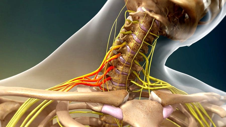
The body’s nerves are the communication system that carries messages between the brain and the rest of the body. Some nerves transmit messages from the brain to muscles to make the body move, while others relay pain, pressure, or temperature signals. Tiny fibers bundled inside each nerve carry the messages with an outer layer/sheathing that insulates and protects the nerves. The brachial plexus is a network of nerves that send signals from the spinal cord to the shoulders, arms, and hands. A brachial plexus nerve injury occurs when the nerves are over-stretched, compressed, torn, cut, or ripped from the spinal cord.
Brachial Plexus Nerve Injury
The injury involves the head or neck hitting or getting hit and shifting to one side while the shoulder is stretched/pulled in the opposite direction.
Minor brachial plexus injuries are commonly known as stingers or burners and are common in sports like football, wrestling, hockey, soccer, and basketball.
Severe brachial plexus injuries can cause arm paralysis and usually result from vehicle or motorcycle accidents.
Other conditions like inflammation or tumors can affect the brachial plexus.
Sometimes babies can sustain brachial plexus injuries during birth.
Pressure and stretching injuries do not physically sever the nerve but can disrupt communication.
Cutting injuries vary depending on the severity of the cut and because the nerves are in a protective canal that can also be fractured or broken. If the canal remains intact, the nerve fibers could grow back with time.
However, surgery is necessary to repair the damage if the canal is broken.
Signs and symptoms of a brachial plexus nerve injury can vary, depending on the severity and location of the injury. Usually, only one arm is affected.
Minor Injuries
Minor damage comes from over-stretching or mild compression.
An electric or burning sensation shoots down the arm.
Numbness and weakness in the arm.
Neck pain.
These symptoms usually last for a few seconds or minutes but can linger for days or longer.
Severe Injuries
More-severe symptoms result from injuries that impact, tear, or rupture the nerves.
The most severe injury occurs when the nerve root is torn from the spinal cord.
Symptoms include:
Intense pain.
Writhing neck pain.
Weakness or inability to use specific shoulder, arm, and/or hand muscles.
Complete lack of movement and feeling in the shoulder, arm, and/or hand.
Symptoms in both arms.
Complications
With time, most brachial plexus injuries in children and adults heal with minimal long-term damage. But some injuries can cause long-lasting problems that include:
Joint Stiffness
The joints can stiffen, making movement difficult.
Healthcare providers often recommend ongoing chiropractic and physical rehabilitation during recovery.
Atrophy
Nerves regrow slowly and can take some time to completely heal after the injury.
During that time, lack of use can cause the muscles to break down.
Chronic Pain
Nerve damage can cause pain signals to be constantly firing.
Numbness
It can occur in the arm or hand, increasing the risk of worsening the injury or causing new injuries.
Disability
Recovery from a severe brachial plexus injury depends on age, damage, location, and severity.
Even with surgery, individuals can experience long-term muscle weakness or paralysis.
Chiropractic Treatment and Rehabilitation
Treatment depends on the severity of the damage. Chiropractic can help realign, rehabilitate, stretch, and strengthen the muscles, nerves, tendons, joints, and ligaments to expedite recovery. For less severe injuries:
Muscle strengthening and posture exercises help maintain motion.
Therapeutic massage will stimulate circulation and keep the muscles loose.
For severe injuries:
Surgery
Continued chiropractic and physical rehabilitation to maintain thorough circulation, range of motion, and relaxed muscles.
The Brachial Plexus
References
Brucker, J et al. “Brachial plexus birth injury.” The Journal of neuroscience nursing: Journal of the American Association of Neuroscience Nurses vol. 23,6 (1991): 374-80. doi:10.1097/01376517-199112000-00006
Gutkowska, Olga, et al. “Brachial plexus injury after shoulder dislocation: a literature review.” Neurosurgical review vol. 43,2 (2020): 407-423. doi:10.1007/s10143-018-1001-x
Joyner, Benny, et al. “Brachial plexus injury.” Pediatrics in review vol. 27,6 (2006): 238-9. doi:10.1542/pir.27-6-238
Noland, Shelley S et al. “Adult Traumatic Brachial Plexus Injuries.” The Journal of the American Academy of Orthopaedic Surgeons vol. 27,19 (2019): 705-716. doi:10.5435/JAAOS-D-18-00433
IFM's Find A Practitioner tool is the largest referral network in Functional Medicine, created to help patients locate Functional Medicine practitioners anywhere in the world. IFM Certified Practitioners are listed first in the search results, given their extensive education in Functional Medicine