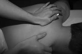
Chiropractic adjustment has various applications. From the treatment of chronic pain and pre-existing conditions to early or sudden onset pain. Perhaps most frequently cited, sufferers of back and neck pain, sciatica, migraines and more have found relief from chiropractic medicine.
In fact, many medical doctors recommend that their patients seek chiropractic care for a variety of conditions before seeking more invasive measures such as surgery. This sentiment was (echoed by the American Medical Association) as recently as 2013.
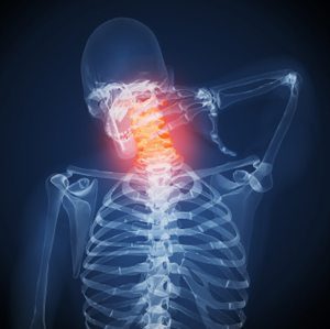
Relieving Back & Neck Pain
Roughly 80% of adults have experienced back pain at some point. (In a study by Consumer Reports), �14,000 sufferers were surveyed. None of these individuals had undergone back surgery of any type. By the end of the study, Chiropractic adjustment was rated as the #1 treatment option.
At the conclusion of the twelve-week study, patients who underwent regular chiropractic adjustments were twice as likely to be pain free as those who were treated with medication. (Further research demonstrates) the validity of chiropractic adjustments as easily seen via magnetic resonance imaging (MRI).
Patients with lower back pain often experience limited mobility in the lumbar spine region that produces degeneration as well as adhesions within the joints of the vertebrae. When patients received an MRI scan following a chiropractic adjustment, the imaging showed an increase in spinal gapping. This breaks up the adhesions, allows the joints to move freely, and lead to a reduction in pain.
Headaches and Migraines
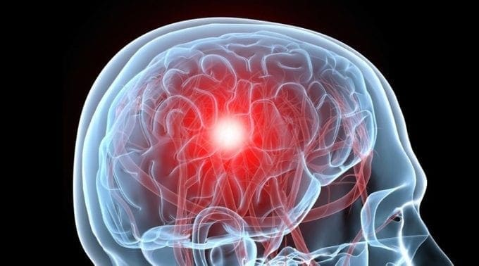
Chronic head pain, including migraines is another common condition that chiropractic adjustment can provide relief for. Through manipulation, chiropractic medicine is able to relieve pressure off of the nerves and provide relief. Headache and migraine sufferers may find long-term relief with continued adjustments, resulting in lessening the severity of symptoms or the frequency of onset.
There is also published research showing the validity of these treatment options. In an (Australian study that tracked 127 migraine sufferers), those who received regular chiropractic adjustments reported a decrease in migraine attacks as well as the need to take less medication.
Considering the rising costs of healthcare, specifically prescription medication, mitigating pharmaceutical costs can provide extra benefits.
Foundation for Chiropractic Progress
In Support of Chiropractic Care
Chiropractic care is becoming increasingly more widespread. Support from medical research and organizations such as The American Medical Association, as well as high profile supporters such as Tiger Woods, Jerry Rice, Aaron Rodgers have helped bring the treatments into the mainstream.
Today, Chiropractic medicine is practiced all over the world, and is an increasingly popular as well as effective form of treatment for a variety of conditions, including but not limited to the ones mentioned above.
(Some more statistics)
Utilizing chiropractic adjustments for treatment of back pain may help save Medicare costs by more than $80 million per year.
Back pain sufferers whose first point of treatment included a medical doctor or surgeon, went on to receive surgery 42.7% of the time. For sufferers who sought chiropractic adjustment first, surgery only occurred 1.5% of the time.
Chronic pain sufferers who sought treatment through chiropractic adjustment experienced a 20% reduction in overall care costs.
Is Chiropractic Adjustment Right for You?
The effectiveness of chiropractic adjustment in treating chronic and recent onset conditions is supported through research, scientific study, as well as patients just like you. If you are suffering from pain or discomfort in your neck, back, joints, or elsewhere,�contact a well qualified Chiropractor near you.
About the Author:
Dr. Alec�with Proactive Chiropractic and Rehab Centre extensively studied human anatomy, physiology, radiology, kinesiology and post graduate seminars in topics such as herniated disc, whiplash, functional movement, car accident rehabilitation. With over 10 years of experience, Dr. Alec helps care for patients with back pain, neck pain, headaches, knee pain, shoulder pain, foot pain, whiplash, etc. Treatment services include: spinal decompression therapy, functional rehabilitation, active release technique, gua sha, electro-stimulation, intersegmental traction, hands-on chiropractic adjustment, instrument assisted chiropractic adjustment and rehabilitation. Dr. Alec takes pride in providing individualized treatment for each patient, with lasting results.
Some People don’t believe in Chiropractic Treatment. However, chiropractic care is becoming increasingly widespread through�medical research. And�top supporters, such as Tiger Woods, Jerry Rice, and Aaron Rodgers have helped to bring�chiropractic�treatment into the mainstream.
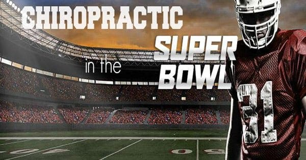
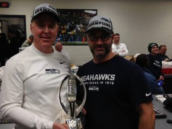
This Sunday over 111 million people will tune into watch the SuperBowl. Behind the scenes both teams and coaches have been fine tuning their game plans as they prepare for the biggest game of their lives. As part of the players preparation there have been a few lucky individuals with direct access to care for these athletes. �Referred to as the �magic workers� by some players these men are the team Chiropractors who serve both the Seattle Seahawks and New England Patriots.
For the last 12 years, Dr. Gerry Ramogida and Dr. Jim Kurtz have had front-row seats to the spectacle of NFL games as chiropractic consultants for the Seattle Seahawks. Attending every game, �Dr. Ramogida was supposed to stay with the team for just a couple of years so he could show the trainers how to use some of his soft-tissue techniques. But as players got used to getting adjusted by him on�game days, a temporary job turned into a permanent one.
�There�s so much behind the scenes that you don�t get a sense for when you�re watching a game. It�s a massive effort. I am just happy to be a part of the preparation as these guys get ready to go out and do what they do best, compete.�
In a interview with Global News�Dr. Ramogida stated, �Over the period of that first season and into the next year, things very quickly became integrated. It�s been a great experience,� he says.
In recent news, The Professional Football Chiropractic Society (PFCS) has taken pride in announcing that all 32 teams in the National Football League offer their players and personnel chiropractic physician services as part of the triage in managing and preventing injuries. According to the Foundation for Chiropractic Progress, this distinction is a benchmark for the profession and documents the important role that chiropractic care plays in optimizing athletic performance.
�The robust need for chiropractic care in the NFL has been deeply driven by the players� desire for peak physical conditioning and not simply for injuries,� states Spencer Baron, D.C., �From the earliest years of full contact football, their bodies are subject to structural stress that doctor of Chiroprctic (DCs) are specially trained to care for. �
Attending this weekend to the New England Patriots is Dr. Mike Miller. �Over twenty five years ago he became the official chiropractor of the New England Patriots. Since that time, he�s treated hundreds of players, watched ownership of the team change hands three times, and seen the Patriots go from one of the worst franchises in the National Football League to a team readying themselves to play in the Superbowl this weekend.
In a interview with Dynamic Chiropractic Dr. Miller states that his position with the Patriots involves him being present during mini-camps, training camp, preseason games, regular-season games (both home and away), and postseason games.
During the games, I see an average of at least 40 players, coaches, and other personnel who are affiliated with the team. I would say just about 90 percent get chiropractic services, because the present coaching staff of the Patriots has almost mandated chiropractic care with the players. The coaches speak about it at team meetings, as the new players and rookies come into the team in the preseason. During mini-camps, they explain the significance of chiropractic, and that we have a chiropractor who has had phenomenal results in dealing with injuries and preventing them from occurring, and that they would like the players to proactively be treated [by] me and begin a chiropractic course of care.
During the season, if there are any injuries, they (the medical staff) will generally send the player to my office to be evaluated. By game time, just about everyone on the roster is adjusted, and you start to learn the idiosyncrasies of each player, because each one wants certain things checked on them. Some are very firm with extremity adjusting; others enjoy use of a specialized technique that we use, called Graston Technique. Basically, it takes me about four hours before the game to go through the entire roster.
The NFL has just released a statement advocating for�all NFL teams as now employing a Chiropractor as part of their medical staff.
If you are interested in learning more about how to be an NFL Chiropractor you can connect with the Professional Football Chiropractic Association�on their website or on their Facebook page.
No matter what the outcome of this weekends game may be, rest assured that both teams will have been well adjusted and their nervous systems tuned on and ready to perform on the biggest stage of all.
Behind the bright lights of the stadium, there is another team that keeps these players and their performance at their optimal level. The healers�if you will, that audiences don’t see, but should know about. These are the Chiropractors!
Super Bowl LI kicks-off with a team of doctors of chiropractic (DC)�to help these professional athletes prevent, manage and care for injuries, as well as, achieve peak performance. The Foundation for Chiropractic Progress� (F4CP), the leading voice of the chiropractic profession, points to the role of DCs with all four play-off teams,and cites data from the Professional Football Chiropractic Society (PFCS) showing on average, a professional football DC performs between 30 to 50 weekly treatments during the regular season � not including training camp or the playoffs.
F4CP� celebrates Super Bowl LI with NFL doctors of chiropractic
New England Patriots Team Chiropractor of 35+ years and proud to attend his ninth Super Bowl, Dr. Michael Miller, states, �During the NFL season, I regularly visit the stadium on my afternoon off from my office, as well as on game days both at home and away. Chiropractic care is emphasized by the head coach, trainers and medical staff as a proactive regimen to prevent injuries, with most of the players receiving adjustments roughly 1-2 times per week.�
He adds that chiropractic care has proven itself over the years in the sports injury arena to be well-accepted as the choice of champions and an integral part of any injury prevention program. �We�ve also earned the respect of other team physicians as a necessary protocol, and are all committed to one goal: keeping the athletes performing at their maximum potential and preventing and managing injuries as they occur.�
He says that chiropractic care provides the athletes with the confidence they need to play without the distraction of pain. �The players are educated about the principles of chiropractic and that it is designed to eliminate the cause of their problem rather than just masking their symptoms.�
Joseph Krzemien, DC, team chiropractor, Atlanta Falcons, says that there is growing evidence and a correlation between football players who receive consistent chiropractic care and a decline in injuries during practice or play:
�During the six seasons I�ve spent with the Atlanta Falcons, my goal has always been to prepare each player�s body to better resist trauma and to speed its natural recovery time,� he says. �This season, the Falcons have been successful for a lot of reasons, but I firmly believe that regular chiropractic care has played an important role in helping us stay healthy and get to Super Bowl LI � I am proud of the role I�ve played in their success.�
For Michael Zoelle, DC, team chiropractor, Green Bay Packers, the trauma experienced by the athletes� bodies during a game is very similar to that of a car accident.
�Ensuring that the joints are functioning properly is critical in the healing process, as well as for injury prevention and optimal performance,� says Dr. Zoelle. �The players recognize that chiropractic care helps them to perform better and heal faster, ultimately leading to better team success.�
DCs receive a minimum of seven years of higher level education, and are qualified to diagnose, treat and manage a broad spectrum of health conditions. They are the primary care professionals for spinal health and well-being. For athletes, chiropractic care helps to reduce the risk of injuries and improve health and performance through enhancements in range of motion, flexibility, balance, muscle strength and other key factors.
On every sideline all over the NFL and NCAA game you will now see chiropractors fixing and maintaining their local teams. In our very own back yard, you will see chiropractors treating El Paso’s finest at most high school games. �UTEP now has Dr. Paul Munoz standing tall ready to help our kids perform better. �The story is getting better each and every year how the public understands joint health as to how it applies to peak performance. �This last Olympics we saw Usain Bolt getting tuned just before his record breaking run. �The day of ushering in alternative care for high performance has for sure landed. �The spine was created to propel the creature faster and to transmit power. �No surprise that most sports have embraced the science and art. �Proud to be a part of the movement…..
Super Bowl 50 showcased the leading players in the NFL along with Joshua Kollmann, DC and Brad Wiest, DC � chiropractors for the Carolina Panthers � and Shawn Caldwell, DC for the Denver Broncos. These doctors of chiropractic helped the players achieve optimal performance.
The Foundation for Chiropractic Progress (F4CP), a not-for-profit organization dedicated to raising awareness about the value of chiropractic care, points out that all 32 NFL teams include the professional services of a doctor of chiropractic (DC) as part of their integrated health care team approach.
Marking their fifth year with the Carolina Panthers, Drs. Kollmann (pic. left) and Wiest (pic. right) highlight the integrative role of chiropractic care: �We are in the stadium training room twice a week � and more often during play-offs � addressing specific sports injuries or providing preventive, maintenance care that the athletes want in order to achieve peak performance. Every player is individually assessed and the treatment plan is communicated and discussed with the training staff. Since the physical nature of the sport really impacts body structure, many players look forward to their pre- and post-game spinal and extremity adjustments as well as other advanced approaches.�
Shawn Caldwell, DC, who has served the Denver Broncos since 2004 and is now preparing for his second Super Bowl, says, �I work hand-in-hand with the athletic trainers and focus on performing chiropractic spine and extremity adjustments that restore joint function. The goal is to enable players to perform optimally or heal from injuries. I am at the facility two-three times weekly or more if necessary. Some players get an adjustment every time I am in the training room, while others when they are symptomatic so they can return to the playing field.�
According to Kyle Prusso, DC, team chiropractor for the Oakland Raiders since 2005 and president of Pro Football Chiropractic Society, an organization of chiropractors who provide the highest quality chiropractic health care to the elite athletes of professional Football: �It�s great to see chiropractic care integrated in all facets of health care, especially in professional and amateur sports. One of the reasons is that athletes are asking for us, with increased player requests driving utilization of chiropractic across all sports. Athletes are very in tune with their bodies and recognize that chiropractic care can boost optimal performance levels.�
All doctors are passionate about their roles as team chiropractors to an NFL Super Bowl contender, as Dr. Kollmann says, �I am humbled by this position and want the world to know that this is a great time to be a chiropractor. It means the world to me to ignite and advance our profession � especially for those who are pursuing a chiropractic education as well as veteran professionals. We are igniting the profession and honor those DCs who pioneered the opportunities that have helped us to become a part of the NFL teams.�
Dr. Caldwell, who is also the chiropractor for the Colorado Rockies Major League Baseball team, sums it up, �Chiropractors are playing an important role in the health care and performance of professional athletes. This is a fantastic experience for us and for the players.�
Doctors of chiropractic receive a minimum of seven years of higher level education, and are qualified to diagnose, treat and manage a broad spectrum of health conditions. They are the primary care professionals for spinal health and well-being. For athletes, chiropractic care helps to reduce the risk of injuries, and improve health and performance through enhancements in range of motion, flexibility, balance, muscle strength and other key factors.
If you are interested in learning more about how to be an NFL Chiropractor you can connect with the Professional Football Chiropractic Association on their website or on their Facebook page.
No matter what the outcome of this weekends game may be, rest assured that both teams will have been well adjusted and their nervous systems tuned on and ready to perform on the biggest stage of all.
Chiropractors swarm the field! Ready for some adjustments to help these players perform their best. The Foundation for Chiropractic Progress (F4CP), a non-profit organization dedicated to raising awareness about the value of chiropractic care share the fact that all 32 NFL teams utilize doctors of chiropractic (DC), as part of their health care programs.
A new, unique test has been designed where it has the ability to search for more than 100 markers which could indicate the presence of a concussion, according to the authors of the research. In previous years, researchers looked for a single marker in the blood to indicate whether an individual had suffered a concussion or not.
“We were pleasantly surprised when we looked at the pattern of metabolites (or markers) and we could identify people who were injured with no other information at a greater than 90 percent certainty,” stated lead researcher Dr. Douglas Fraser, a consultant in pediatric critical care medicine at the Children’s Health Research Institute in London, Ontario.
During the research study, Dr. Fraser and his colleagues examined 29 teen hockey players for markers of concussion. Of these individuals, some had experienced head injuries while others had not. Regardless, everyone involved in the study was convinced of the test’s abilities.
“It might have potential for diagnosis of concussion but these are preliminary results with only 29 patients,” stated Dr. John Kuluz, director of traumatic brain injury and neuro-rehabilitation at Nicklaus Children’s Hospital in Miami. According to Dr. Kuluz, the test must first be validated in a lot more patients before its effectiveness can be determined.
Additionally, he stated how this type of testing isn’t necessary to utilize often. “There are only a small number of patients where the diagnosis is in doubt,” declared Dr. Kuluz. “However, in those cases, such a test could be helpful,” he noted.
Because properly diagnosing the presence of a concussion heavily relies on the observation of specific symptoms,such as dizziness, headaches, blurred vision, nausea and other overt complications, it can often be challenging to distinguish an individual’s cause of injury. In the same manner, it is similarly difficult to accurately determine when an individual has fully recovered from a concussion and if they can return to their regular activities.
“People have been searching for one or two proteins floating around in the blood which are released from the brains after experiencing an injury,” stated Dr. Fraser. “But that approach hasn’t yielded great results, probably because every patient is different and every injury is different. Therefore, it’s probably a little naive to believe one or two proteins are going to give us the answer we need,” he concluded.
The team of researchers narrowed down 174 markers to approximately between 20 to 40 specific ones which could diagnose a concussion with an accuracy of more than 90 percent.
Foremost, to accurately diagnose a concussion through this process, the blood must be tested within 72 hours after the individual has experienced a head injury. The report was published in the October 2016 issue of the journal Metabolomics.
The test was developed in hopes that it could be a widely available and inexpensive procedure to be utilized in emergency rooms. Furthermore, the test could be used to evaluate the individual’s healing process. According to Dr. Fraser, he quoted, “It looks like these patterns remain abnormal for up to three months at a time. There is a potential that following the profile for a period of time can reveal accurate information pertaining to the healing process.”
The researchers have tested the accuracy of the test in other groups, such as the military, to determine whether it functions equally in adults as it does in teens. They have also developed a machine which can run the test quickly using a single drop of blood.
The research study was funded by the Children�s Health Foundation in Canada. The authors have filed a patent application for their test.
A concussion can occur as a result of a traumatic sports injury or due to any other blow to the head. While several symptoms could indicate the presence of a concussion, symptoms can manifest differently for each individual, making it difficult to accurately determine the presence of a head injury. With the development of a new test, a concussion could be diagnosed using a single drop of blood.
For more information, please feel free to ask Dr. Jimenez or contact us at 915-850-0900 .
The piriformis muscle is commonly known among athletes and healthcare professionals as a significant muscle in the posterior hip. This muscle functions to control hip joint rotation and abduction and it is also a distinguishable muscle due to its inversion of action in rotation. The piriformis muscle also raises awareness as the various causes of piriformis syndrome, a condition suspected to be a potential source of pain and dysfunction, not only in athletes, but in the general population as well.
Anatomy of the Piriformis Muscle
The piriformis muscle originates on the anterior surface of the sacrum and it is securely held to it by three tissue attachments found between the first, second, third and fourth anterior sacral foramina. Occasionally, its origin may be so broad that it joins the capsule of the sacroiliac joint with the sacrotuberous and/or sacrospinous ligament. The piriformis muscle is a thick and strong muscle that travels out of the pelvis through the greater sciatic foramen, dividing the foramen into the suprapiriform and infra-piriform foramina. As it courses through the greater sciatic foramen, the muscle decreases to a point where it forms a tendon that attaches to the superior-medial surface of the greater trochanter, frequently integrating with the tendon of the obturator internus and gemelli muscles.
The nerves and blood vessels found within the suprapiriform foramen are known as the superior gluteal nerves and vessels, and those found in the infra-piriforma fossa are known as the inferior gluteal nerves and vessels, including the sciatic nerve. Because of its broad size in the greater sciatic foramen, there�s a risk the numerous vessels and nerves that exit the pelvis may become compressed.
The piriformis muscle is closely associated with other short hip rotators as well, such as the superior gemellus, obturator internus, inferior gemellus and obturator externus. The primary difference between this muscle and other short rotators is its connection to the sciatic nerve. The piriformis muscle passes behind the nerve while the other rotators pass before it.
Anatomical Variants
Several anatomical variations have been previously diagnosed among the piriformis muscle. First, there may be additional medial attachments to the first and fifth sacral vertebrae and to the coccyx. Second, the tendon may merge with the gluteus medius or minimus or with the gemellus. Also, in approximately less than 20 percent of cases, the piriformis muscle may be divided into two different segments, through which part or all of the sciatic nerve may travel. Then, the muscle may blend with the posterior hip joint capsule as a conjoined tendon with the obturator internus. Additionally, the distal attachment of the piriformis muscle has been demonstrated to vary in proportion and position on the supero-medial surface of the greater trochanter. It can stretch across 25 to 64 percent of the anterior-posterior length along the greater trochanter, with 57 percent of it attaching more anteriorly and 43 percent more posteriorly. Last but not least, researchers studied its insertion point broadly and discovered that four types of insertions existed and these were characterized based on the relationship to the obturator internus. The variation of placement and width of the distal attachment of the piriformis muscle may influence the effectiveness of the concept known as the inversion of action.
Furthermore, the connection between the piriformis muscle and the sciatic nerve has been a highly debated complication. It�s been previously concluded that there are several anatomical variations among the piriformis muscle and its connection to the sciatic nerve. The sub-types of this variation include: type 1-A, where the muscle is pear shaped with the nerve running anteriorly and inferiorly to this, found in 70 to 85 percent of cases; type 2-B, where the piriformis muscle is divided into two sections with the common peroneal nerve running between the two parts and the tibial nerve travels anteriorly and below, found in 10 to 20 percent of cases; type 3-C, where the peroneal portion loops over the top of the muscle and the tibial portion is found below, found in 2 to 3 percent of cases; and type 4-D, where the undivided nerve passes through the piriformis muscle, found in approximately 2 percent of cases.
Moreover, it is also speculated that two other, very rare variations may occur, demonstrated by letters E and F in the diagram. Type 1-A is the most common variation, displaying the sciatic nerve as it passes below the piriformis muscle.
Function of the Piriformis Muscle
The fundamental functions of the piriformis muscle are to provide hip external rotation and allow abduction at 90 degrees of hip flexion. During weight-bearing, the piriformis muscle restricts femoral internal rotation in the stance phase of walking and running. Also, it assists the short hip rotators in compressing the hip joint and stabilizing it. Because it can exert an oblique force on the sacrum, it may produce a strong rotary shearing force on the sacroiliac joint. Otherwise, this would dislocate the ipsilateral base of the sacrum forward and the apex of the sacrum backwards.
Since the piriformis muscle is the furthest behind of the hip external rotators because of its attachment on the anterior surface of the sacrum, it has the greatest influence to apply a rotation effect on the hip joint. Occasionally, healthcare specialists have found issues with the piriformis muscle where it appears to be tight and hypertonic, while the other short hip rotators which are found closer to the axis of rotation become inhibited and hypotonic.
Inversion of action
The most argumentative complication relating to the function of the piriformis muscle is its reversal-of-function role, best referred to as the inversion of action role. Researchers have suggested that as the hip approaches angles of 60 to 90 degrees and greater, the tendon of the piriformis muscle shifts on the greater trochanter. As a result, its line of pull becomes ineffective as a hip external rotator, however, it does contribute to internal hip rotation. Consequently, it reverses its rotation function at high hip flexion angles.
Nonetheless, more recent studies conducted through anatomical dissection have demonstrated that the attachment of the piriformis muscle onto the greater trochanter can change and, in some instances, it may insert in a position by which it may be unable to reverse its function, for example, in a more posteriorly placed attachment. Thus, stretching the piriformis muscle into external rotation when the hip is flexed beyond 90 degrees, based on the inversion of action role, would be ineffective as a treatment or misleading as an examination technique.
The role of the piriformis muscle at several joint angles is an essential consideration for healthcare professionals who evaluate and treat the causes of piriformis syndrome. Frequently, it�s recommended to stretch the hip into flexion, adduction and external rotation to stretch the piriformis muscle over the glutes by utilizing the reversal of function concept.
MSK Dysfunction and Causes of Piriformis Syndrome
Many decades ago, it was suggested that in some cases, sciatica symptoms may originate outside the spine as a result of the piriformis muscles. This hypothesis was supported soon after when specialists successfully improved an individual�s symptoms of sciatica by surgically dividing the piriformis muscle. Based on cadaver anatomical dissections, the researchers believed that the spasm of the piriformis muscle could be responsible for the irritation of the sciatic nerve.
The medical term piriformis syndrome then became associated to sciatica symptoms, believed to be caused by a usually traumatic abnormality in the piriformis muscle with a focus on ruling out more common causes of sciatica, such as nerve root impingement caused by a disc herniation. It soon became an accepted interpretation but with no consensus about the exact clinical signs and diagnostic tests to differentiate it from other sources of sciatica.
Understanding the Causes of Piriformis Syndrome
Piriformis syndrome can be defined as the interaction between the piriformis muscle and the sciatic nerve, where these may irritate the nerves and develop posterior hip pain with distal referral down the posterior thigh, resembling symptoms of true sciatica. Differentiating�the damage to this region typically follows exceptions of the more well-known causes of sciatica and buttock pain.
More specifically, reports of buttock pain with distal referral of symptoms are not unique to the causes of piriformis syndrome. Similar symptoms are prevalent with the more medically evident lower back pain syndromes and pelvic dysfunctions. Therefore, a complete evaluation of these areas must be performed to rule out any underlying pathology. It has been suggested that the causes of piriformis syndrome can be held responsible for approximately 5 to 6 percent of sciatica cases. In the majority of instances, it develops in middle-aged individuals, an average or 38 years and it�s more common among women.
Pathogenesis of Piriformis Syndrome
The causes of Piriformis syndrome can be associated to three primary causing factors: First, the referred pain may be the result of myofascial trigger points. Second, the entrapment of the nerve against the greater sciatic foramen as it passes through the infrapiriform fossa or within a variating piriformis muscle. And third, sacroiliac joint dysfunction causing piriformis muscle spasms.
Other researchers presented an additional number of factors behind the causes of piriformis syndrome as follows: gluteal trauma in the sacroiliac or gluteal regions, anatomical variations, myofascial trigger points, hypertrophy of the piriformis muscle or spasms of the piriformis muscle, secondary to spinal surgery such as laminectomy, space occupying lesions such as neoplasm, bursitis, abscess and myositis, intragluteal injections and femoral nailing.
Symptoms
The general symptoms described with the causes of piriformis syndrome include: a tight or cramping sensation in the buttock and/or hamstring, gluteal pain in up to 98 percent of cases, �calf pain in up to 59 percent of cases, aggravation through sitting and squatting if the trunk is inclined forward or the leg is crossed over the unaffected leg and possible peripheral nerve signs such as pain and paresthesia in the back, groin, buttocks, perineum and back of the thigh in up to 82 percent of cases.
Physical findings and examinations
It�s important to keep in mind that hip flexion with active external rotation or passive internal rotation may aggravate the symptoms of dysfunction. Additional findings for the evaluated causes of piriformis syndrome have demonstrated a positive SLR that is less than 15 degrees on the normal side. Other tests used to evaluate the causes of piriformis syndrome include, positive Freiberg�s sign, used in 32 to 63 percent of cases, involves the reproduction of pain on a passively forced internal rotation of the hip in the supine position, believed to result from passive stretching of the piriformis muscle and pressure of the sciatic nerve at the sacrospinous ligament. Pacers sign, used in 30 to 74 percent of cases, involves reproducing pain and weakness on resisted abduction and external rotation of the thigh in a sitting position. Pain in a FAIR position used to evaluate dysfunction, involves the reproduction of pain when the leg is held in flexion, adduction and internal rotation. Furthermore, an accentuated lumbar lordosis and hip flexor tightness predisposes an individual to increased compression of the sciatic nerve against the sciatic notch by a shortened piriformis. Electro-diagnostic tests may also prove useful to diagnose piriformis muscle complications.
When palpable spasm within the surrounding piriformis muscle occur and there is obturator internus pain and external tenderness over the greater sciatic notch, found in approximately 59 to 92 percent of cases, the individual must perform the Sims position to follow up an evaluation. The piriformis line should overlie the superior border of the piriformis muscle and extend immediately from above the greater trochanter to the cephalic border of the greater sciatic foramen at the sacrum. The examination will continue where the line is divided into equal thirds. The fully rendered thumb presses on the point of maximum trigger-point tenderness, which is usually found just lateral to the junction of the middle and last thirds of the line.
Investigations
Conventional imaging, such as X-ray, CT scan and MRI, tend to be ineffective in diagnosing the presence and causes of piriformis syndrome. However, some value may exist in electro-diagnostic testing. The purpose of these tests is to find conduction faults in the sciatic nerve. Findings such as long-latency potentials, for instance the H reflex of the tibial nerve and/or peroneal nerve, may be normal at rest but become delayed in positions where the hip external rotators are tightened.
It�s been confirmed that the tibial division of the sciatic nerve is usually spared, the inferior gluteal nerve that supplies the gluteus maximus may be affected and the muscle can become atrophied. However, testing of the peroneal nerve may provide more conclusive results as they�re more likely to be the impinged portion of the sciatic nerve. The H-wave may become inactive during the painful position of forced adduction-internal rotation of the affected leg.
Piriformis Syndrome Myths
Researchers discussed that piriformis syndrome is a commonly over-used term used to describe any non-specific gluteal tenderness with radiating leg pain. It was argued that only in rare cases is the piriformis muscle involved in nerve compression of the sciatic nerve which may then accurately qualify as one of the causes of piriformis syndrome. It was cited that there is only limited evidence and cases where the diagnosis of the causes of piriformis syndrome can be made, foremostly, where there is compressive damage to the sciatic nerve by the piriformis muscle. In several isolated studies, the sciatic nerve was seen to be compressed by the piriformis muscle in instances such as hypertrophy of the muscle, general anatomical abnormalities such as a bifid piriformis muscle and due to compression by fibrous bands.
Also, trauma and scarring to the piriformis muscle can involve the sciatic nerve. It is possible that rare cases of true piriformis syndrome have been caused by direct heavy trauma to the piriformis muscle due to a blunt trauma to the muscle. This is termed as post- traumatic piriformis syndrome.
Researchers supported this argument by stating that it is more likely that, given the anatomical relationship of the piriformis muscle to the various nerves in the deep gluteal region, the buttock pain�may be caused by an entrapment of the gluteal nerves and the hamstring pain may be due to an entrapment of the posterior cutaneous nerve of the thigh, rather than an entrapment of the sciatic nerve alone. This demonstrates the medically analyzed circumstance in the absence of distal sciatic neurological signs. Whether the piriformis muscle is the cause of the compression has not been clearly established. It is possible that the obturator internus/gemelli complex is an alternative cause of neural compression. The researchers have suggested utilizing the term deep gluteal syndrome rather than piriformis syndrome.
Treatment
When one of the several causes of piriformis syndrome is discovered and a healthcare specialist feels that an appropriate diagnosis has been made, the treatment will generally depend on the cause behind the dysfunction. If the piriformis muscle is tight and it spasms, then initially conservative treatment will focus on stretching and massaging the tight muscle to clear the piriformis muscle from being the source of the pain. If this fails, then the following have been suggested and may be attempted: local anesthetic block, typically performed by an anesthesiologist who has expertise in pain management and in performing nerve blocks; steroid injections into the piriformis muscle; botulinum toxin injections in the piriformis muscle; and surgical neurolysis.
Therapist-directed interventions, such as stretching of the piriformis muscle and direct trigger point massage, can also be used as treatment. It�s been encouraged that piriformis muscle stretches are done in positions of hip flexion greater than 90 degrees, adduction and external rotation to utilize the inversion of action effect of the piriformis muscle to isolate the stretch to this muscle independent of the other hip external rotators.
However, recent evidence utilizing ultrasound investigation determined that there was no connection between hip flexion angle and the thickness of the piriformis muscle tendon in both internal and lateral hip rotation stretching, which implies that the piriformis muscle does not invert its action. Furthermore, researchers who performed cadaveric studies concluded that the piriformis muscle insertion is different and a lot more complex than it was first believed to be. It is possible that the piriformis muscle may invert its action only in some individuals but not in others.
Accordingly�due to the disagreements and confusions over the concept of inversion of action, it is suggested that healthcare professionals should perform two variations of a piriformis muscle stretch: stretches in flexion, adduction and external rotation and stretches in flexion, adduction and internal rotation.
Pigeon Stretch for left piriformis muscle: hip flexion, neutral adduction and maximal hip external rotation.
Stretch for left piriformis muscle: hip is in flexion, neutral adduction and maximal external rotation.
Short leg posterior chain stretch for right piriformis muscle: hip is in 90 degree flexion, adduction and neutral rotation.
Trigger Points and Massage
The most appropriate suggestion to palpate the piriformis muscle trigger points is in the following recommended position. In this posture, the healthcare professional can feel for the deep piriformis muscle trigger points and apply a constant pressure to relieve the trigger points as well as apply a flush massage to the muscle in this position. In this position, the large gluteus maximus is relaxed and it is easier to feel the deeper piriformis muscle.
The piriformis muscle is a deep posterior hip muscle that is anatomically similar to both the sacroiliac joint and the sciatic nerve. It is a muscle that functions as a dominant hip rotator and stabilizer, with a propensity to shorten and become hypertonic. For that reason, stretching and massage techniques are best utilized and often recommended to reduce the tone through the muscle. In conclusion, it has also been implied in compression and irritation of the sciatic nerve, most frequently referred to as piriformis syndrome.
For more information, please feel free to ask Dr. Jimenez or contact us at 915-850-0900 .
IFM's Find A Practitioner tool is the largest referral network in Functional Medicine, created to help patients locate Functional Medicine practitioners anywhere in the world. IFM Certified Practitioners are listed first in the search results, given their extensive education in Functional Medicine


 he could show the trainers how to use some of his soft-tissue techniques. But as players got used to getting adjusted by him on�game days, a temporary job turned into a permanent one.
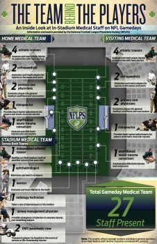
 In a interview with Dynamic Chiropractic Dr. Miller states that his position with the Patriots involves him being present during mini-camps, training camp, preseason games, regular-season games (both home and away), and postseason games.

 Joseph Krzemien, DC, team chiropractor, Atlanta Falcons, says that there is growing evidence and a correlation between football players who receive consistent chiropractic care and a decline in injuries during practice or play:
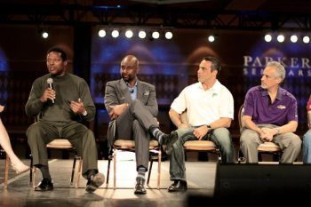

 Marking their fifth year with the Carolina Panthers, Drs. Kollmann (pic. left) and Wiest (pic. right) highlight the integrative role of chiropractic care: �We are in the stadium training room twice a week � and more often during play-offs � addressing specific sports injuries or providing preventive,
  maintenance care that the athletes want in order to achieve peak performance. Every player is individually assessed and the treatment plan is communicated and discussed with the training staff. Since the physical nature of the sport really impacts body structure, many players look forward to their pre- and post-game spinal and extremity adjustments as well as other advanced approaches.�
 Shawn Caldwell, DC, who has served the Denver Broncos since 2004 and is now preparing for his second Super Bowl, says, �I work hand-in-hand with the athletic trainers and focus on performing chiropractic spine and extremity adjustments that restore joint function. The goal is to enable players to perform optimally or heal from injuries. I am at the facility two-three times weekly or more if necessary. Some players get an adjustment every time I am in the training room, while others when they are symptomatic so they can return to the playing field.�