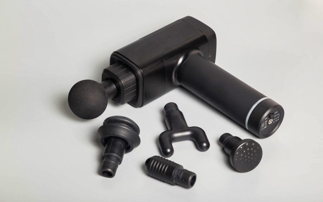
Massage guns can help relieve aching muscles and prevent soreness when used before and after physical activity, work, school, and exercise. They provide massage therapy benefits by targeting muscles with rapid burst pulses. Massage guns can be percussive or vibration-based. Percussive therapy helps increase blood flow to a targeted area, which reduces inflammation and muscle tension, and breaks up knots/trigger points that may have formed in the tissues from added stress or intense physical activity. One of the benefits is that they come with interchangeable massage gun head attachments that target different muscle groups and provide different types of massage. There are many types of interchangeable massage heads, we go over the most common to give a general idea of how they work. If experiencing joint pain, injury, acute muscle pain, or other musculoskeletal disorders, make sure to get clearance from a doctor before using a massage gun.
Massage Gun Head Attachments
The variations of attachments/heads are designed and shaped differently to effectively apply the right amount of pressure to rejuvenate the body’s pressure points, soothe tissues, and release tight and sore muscles. The different heads are designed with a distinctive purpose based on the muscle groups targeted. This maximizes effectiveness and ensures maximum comfort and safety.
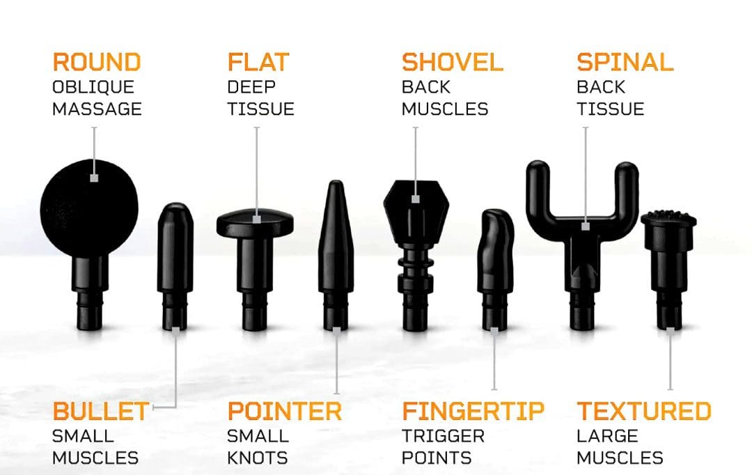
Ball Head
The ball attachment is for overall muscle recovery.
It provides a broad surface area and mimics the hands of a skilled massage therapist, delivering a soothing kneading sensation.
Made of durable material, the ball massage head can reach deep into the muscles.
Its round shape makes it more flexible to use anywhere, especially the large muscle groups like the quads and glutes.
U/Fork Shaped Head
A plastic, dual-pronged head also known as a fork head.
The attachment provides relief to areas like the shoulders, spine, neck, calves, and Achilles tendon.
Bullet Head
The plastic head is named as such because of its pointed shape.
This is recommended for tightness and discomfort in the joints, deep tissues, trigger points, and/or small muscle areas like the feet and wrist.
Flat Head
The multipurpose flat head is for full-body general massage.
It helps relieve stiffness and pain for total body muscle relaxation, including muscle groups closer to bone joints.
Shovel-Shaped Head
The shovel-shaped head is for the abdominal muscles and lower back.
The attachment provides stimulation to release stiff muscles.
Using The Right Head
Which head to use depends on the individual’s specific needs and preferences. Consider the following factors when selecting a massage gun head:
Targeted Areas
Identify the body areas requiring the most attention.
If muscle tightness or soreness is occurring in larger muscle groups, like the back or legs, the ball attachment is recommended.
For more precise areas like trigger points, the bullet head is recommended.
Heads can be used in conjunction – for example, a large surface area head is used to relax and loosen and relax a general area, then a more precise head is used to focus the massage on the actual tight spot or trigger point.
Massage Intensity
Massage intensity levels can be varied from a light massage to full force.
For a softer touch on sensitive muscles, flat head or forkhead attachments are recommended.
For deep muscle penetration and consistent pressure, the bullet head or shovel head attachments are recommended.
Specific Conditions
Consider any specific conditions or injuries previous and current.
For individuals recovering from an injury or with sensitive areas, it’s important to choose a massage gun head that provides the necessary relief without causing discomfort or worsening an injury.
Try Out Different Heads and Settings
Experiment with different massage head attachments and speeds to find the one that works best for the intended purpose.
Explore each to discover personal preferences.
Start with the lowest setting and gradually increase, based on comfort level.
Always consult with a qualified healthcare provider regarding any medical concerns before using a massage gun.
Choosing The Right Massage Head Attachment
References
Bergh, Anna, et al. “A Systematic Review of Complementary and Alternative Veterinary Medicine in Sport and Companion Animals: Soft Tissue Mobilization.” Animals: an open access journal from MDPI vol. 12,11 1440. 2 Jun. 2022, doi:10.3390/ani12111440
Imtiyaz, Shagufta, et al. “To Compare the Effect of Vibration Therapy and Massage in Prevention of Delayed Onset Muscle Soreness (DOMS).” Journal of Clinical and diagnostic research: JCDR vol. 8,1 (2014): 133-6. doi:10.7860/JCDR/2014/7294.3971
Konrad, Andreas, et al. “The Acute Effects of a Percussive Massage Treatment with a Hypervolt Device on Plantar Flexor Muscles’ Range of Motion and Performance.” Journal of sports science & medicine vol. 19,4 690-694. 19 Nov. 2020
Leabeater, Alana et al. “Under the Gun: The effect of percussive massage therapy on physical and perceptual recovery in active adults.” Journal of athletic training, 10.4085/1062-6050-0041.23. 26 May. 2023, doi:10.4085/1062-6050-0041.23
Lupowitz, Lewis. “Vibration Therapy – A Clinical Commentary.” International journal of sports physical therapy vol. 17,6 984-987. 1 Aug. 2022, doi:10.26603/001c.36964
Yin, Yikun, et al. “The effect of vibration training on delayed muscle soreness: A meta-analysis.” Medicine vol. 101,42 (2022): e31259. doi:10.1097/MD.0000000000031259
The lumbosacral plexus is located on the posterolateral wall of the lesser pelvis, next to the lumbar spine. A plexus is a network of intersecting nerves that share roots, branches, and functions. The sacral plexus is a network that emerges from the lower part of the spine. The plexus then embeds itself into the psoas major muscle and emerges in the pelvis. These nerves provide motor control to and receive sensory information from portions of the pelvis and leg. Sacral nerve discomfort symptoms, numbness, or other sensations and pain can be caused by an injury, especially if the nerve roots are compressed, tangled, rubbing, and irritated. This can cause symptoms like back pain, pain in the back and sides of the legs, sensory issues affecting the groin and buttocks, and bladder or bowel problems. Injury Medical Chiropractic and Functional Medicine Clinic can develop a personalized treatment plan to relieve symptoms, release the nerves, relax the muscles, and restore function.
Sacral Plexus
Anatomy
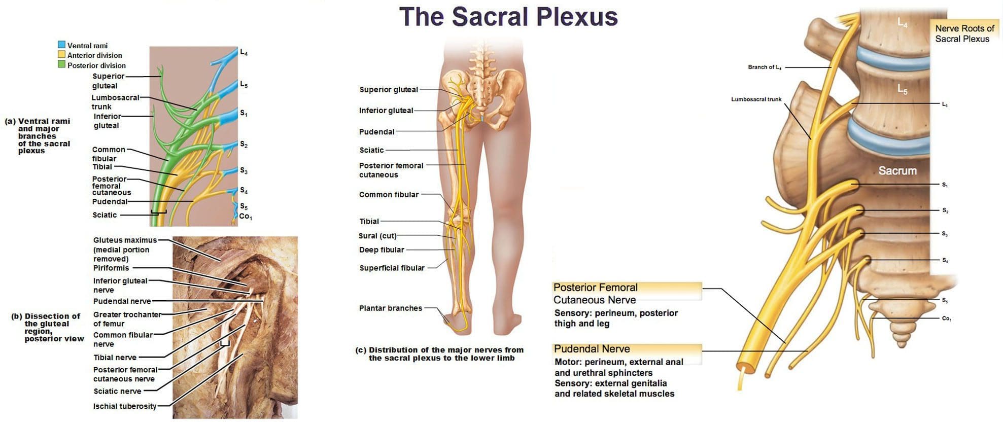
The sacral plexus is formed by the lumbar spinal nerves, L4 and L5, and sacral nerves S1 through S4.
Several combinations of these spinal nerves merge together and then divide into the branches of the sacral plexus.
Everybody has two sacral plexi – plural of plexus – one on the right side and left side that is symmetrical in structure and function.
Structure
There are several plexi throughout the body. The sacral plexus covers a large area of the body in terms of motor and sensory nerve function.
Spinal nerves L4 and L5 make up the lumbosacral trunk, and the anterior rami of sacral spinal nerves S1, S2, S3, and S4 join the lumbosacral trunk to form the sacral plexus.
Anterior rami are the branches of the nerve that are towards the front of the spinal cord/front of the body.
At each spinal level, an anterior motor root and a posterior sensory root join to form a spinal nerve.
Each spinal nerve then divides into an anterior – ventral – and a posterior – dorsal – rami portion.
Each can have motor and/or sensory functions.
The sacral plexus divides into several nerve branches, which include:
Superior gluteal nerve – L4, L5, and S1.
Inferior gluteal nerve – L5, S1, and S2.
The sciatic nerve – is the largest nerve of the sacral plexus and among the largest nerves in the body – L4, L5, S1, S2, and S3
The common fibular nerve – L4 through S2, and tibial nerves – L4 through S3 are branches of the sciatic nerve.
Posterior femoral cutaneous nerve – S1, S2, and S3.
Pudendal nerve – S2, S3, and S4.
The nerve to the quadratus femoris muscle is formed by L4, L5, and S1.
The obturator internus muscle nerve – L5, S1, and S2.
The piriformis muscle nerve – S1 and S2.
Function
The sacral plexus has substantial functions throughout the pelvis and legs. The branches provide nerve stimulation to several muscles. The sacral plexus nerve branches also receive sensory messages from the skin, joints, and structures of the pelvis and legs.
Motor
Motor nerves of the sacral plexus receive signals from the brain that travel down the column of the spine, out to the motor nerve branches of the sacral plexus to stimulate muscle contraction and movement. Motor nerves of the sacral plexus include:
Superior Gluteal Nerve
This nerve provides stimulation to the gluteus minimus, gluteus medius, and tensor fascia lata, which are muscles that help move the hip away from the center of the body.
Inferior Gluteal Nerve
This nerve provides stimulation to the gluteus maximus, the large muscle that moves the hip laterally.
Sciatic Nerve
The sciatic nerve has a tibial portion and a common fibular portion, which have motor and sensory functions.
The tibial portion stimulates the inner part of the thigh and activates muscles in the back of the leg and the sole of the foot.
The common fibular portion of the sciatic nerve stimulates and moves the thigh and knee.
The common fibular nerve stimulates muscles in the front and sides of the legs and extends the toes to straighten them out.
Pudendal Nerve
The pudendal nerve also has sensory functions that stimulate the muscles of the urethral sphincter to control urination and the muscles of the anal sphincter to control defecation.
The nerve to the quadratus femoris stimulates the muscle to move the thigh.
The nerve to the obturator internus muscle stimulates the muscle to rotate the hips and stabilize the body when walking.
The nerve to the piriformis muscle stimulates the muscle to move the thigh away from the body.
Conditions
The sacral plexus, or areas of the plexus, can be affected by disease, traumatic injury, or cancer. Because the nerve network has many branches and portions, symptoms can be confusing. Individuals may experience sensory loss or pain in regions in the pelvis and leg, with or without muscle weakness. Conditions that affect the sacral plexus include:
Injury
A traumatic injury of the pelvis can stretch, tear, or harm the sacral plexus nerves.
Bleeding can inflame and compress the nerves, causing malfunction.
Neuropathy
Nerve impairment can affect the sacral plexus or parts of it.
Neuropathy can come from:
Diabetes
Vitamin B12 deficiency
Certain medications – chemotherapeutic meds
Toxins like lead
Alcohol
Metabolic illnesses
Infection
An infection of the spine or the pelvic region can spread to the sacral plexus nerves or produce an abscess, causing symptoms of nerve impairment, pain, tenderness, and sensations around the infected region.
Cancer
Cancer developing in the pelvis or spreading to the pelvis from somewhere else can compress or infect the sacral plexus nerves.
Treatment of the Underlying Medical Condition
Rehabilitation begins with the treatment of the underlying medical condition causing the nerve problems.
Cancer treatment – surgery, chemotherapy, and/or radiation.
Antibiotic treatment for infections.
Neuropathy treatment can be complicated because the cause may be unclear, and an individual can experience several causes of neuropathy simultaneously.
Major pelvic trauma like a vehicle collision can take months, especially if there are multiple bone fractures.
Motor and Sensory Recovery
Sensory problems can interfere with walking, standing, and sitting.
Adapting to sensory deficits is an important part of treatment, rehabilitation, and recovery.
Chiropractic, decompression, massage, and physical therapy can relieve symptoms, restore strength, function, and motor control.
Sciatica Secrets Revealed
References
Dujardin, Franck et al. “Extended anterolateral transiliac approach to the sacral plexus.” Orthopaedics & traumatology, surgery & research: OTSR vol. 106,5 (2020): 841-844. doi:10.1016/j.otsr.2020.04.011
Eggleton JS, Cunha B. Anatomy, Abdomen and Pelvis, Pelvic Outlet. [Updated 2022 Aug 22]. In: StatPearls [Internet]. Treasure Island (FL): StatPearls Publishing; 2023 Jan-. Available from: https://www.ncbi.nlm.nih.gov/books/NBK557602/
Garozzo, Debora et al. “In lumbosacral plexus injuries can we identify indicators that predict spontaneous recovery or the need for surgical treatment? Results from a clinical study on 72 patients.” Journal of brachial plexus and peripheral nerve injury vol. 9,1 1. 11 Jan. 2014, doi:10.1186/1749-7221-9-1
Gasparotti R, Shah L. Brachial and Lumbosacral Plexus and Peripheral Nerves. 2020 Feb 15. In: Hodler J, Kubik-Huch RA, von Schulthess GK, editors. Diseases of the Brain, Head and Neck, Spine 2020–2023: Diagnostic Imaging [Internet]. Cham (CH): Springer; 2020. Chapter 20. Available from: https://www.ncbi.nlm.nih.gov/books/NBK554335/ doi: 10.1007/978-3-030-38490-6_20
Norderval, Stig, et al. “Sacral nerve stimulation.” Tidsskrift for den Norske laegeforening : tidsskrift for praktisk medicin, ny raekke vol. 131,12 (2011): 1190-3. doi:10.4045/tidsskr.10.1417
Neufeld, Ethan A et al. “MR Imaging of the Lumbosacral Plexus: A Review of Techniques and Pathologies.” Journal of Neuroimaging: official journal of the American Society of Neuroimaging vol. 25,5 (2015): 691-703. doi:10.1111/jon.12253
Staff, Nathan P, and Anthony J Windebank. “Peripheral neuropathy due to vitamin deficiency, toxins, and medications.” Continuum (Minneapolis, Minn.) vol. 20,5 Peripheral Nervous System Disorders (2014): 1293-306. doi:10.1212/01.CON.0000455880.06675.5a
Yin, Gang, et al. “Obturator Nerve Transfer to the Branch of the Tibial Nerve Innervating the Gastrocnemius Muscle for the Treatment of Sacral Plexus Nerve Injury.” Neurosurgery vol. 78,4 (2016): 546-51. doi:10.1227/NEU.0000000000001166
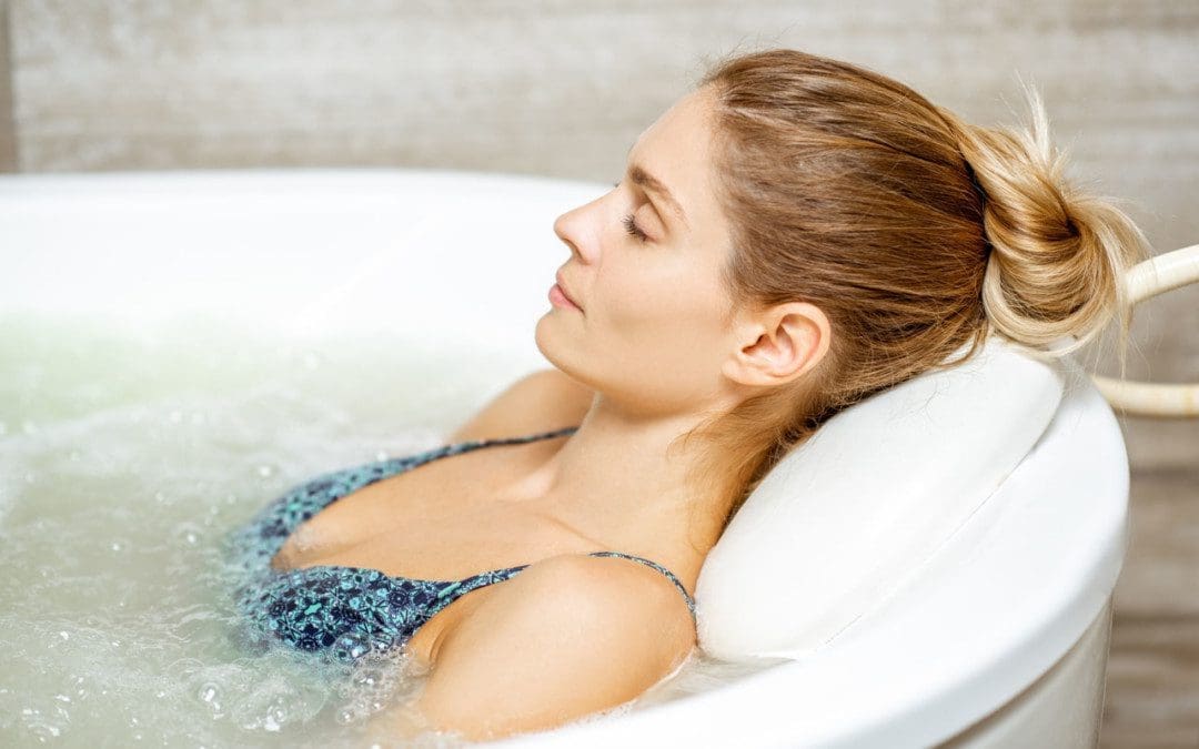
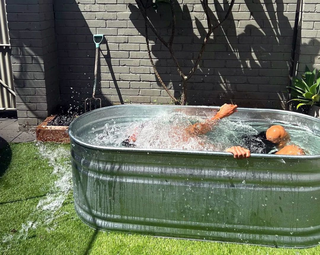
Athletes regularly take an ice-water bath after training or playing. It is known as cold water immersion/cryotherapy. It is used to relieve and reduce muscle soreness and pain after intense training or competition. From runners to professional tennis and football players, taking an ice bath is a common recovery practice. Many athletes utilize ice baths to help with faster recovery, prevent injury, and cool down the body. Here we provide some research on cold-water immersion therapy.
Ice Water Bath
Cold Immersion After Exercise or Physical Activity
Exercise causes microtrauma/tiny tears in the muscle fibers. The microscopic damage stimulates muscle cell activity to repair the damage and strengthen the muscles/hypertrophy. However, hypertrophy is linked with delayed onset muscle soreness and pain/DOMS, between 24 and 72 hours after physical activity. An ice water bath works by:
Constricting the blood vessels.
Flushes out waste products (lactic acid), out of the muscle tissues.
Reduces inflammation, swelling, and tissue breakdown.
Then, applying heat or warming up the water increases and speeds up blood circulation, improving the healing process.
There is no current ideal time and temperature for cold immersion, but most athletes and trainers who use the therapy recommend a water temperature between 54 to 59 degrees Fahrenheit and immersion of five to 10 minutes, and depending on the soreness, sometimes up to 20 minutes.
Pros and Cons
The effects of ice baths and cold water immersion on exercise recovery and muscle soreness.
Relieves Inflammation but Can Slow Down Muscle Growth
A study determined that cold water immersion can disrupt training adaptations.
Research suggests that icing muscles right after maximum exercise decreases inflammation, but can slow down muscle fiber growth, and delay muscle regeneration.
Athletes trying to increase muscle size and strength may need to adjust the therapy sessions.
Reduce Muscle Soreness
A review concluded there was some evidence that ice water immersion reduced delayed onset muscle soreness when compared to resting and rehabilitation or no medical treatment.
The most effects were seen in running athletes.
There was no substantial evidence to conclude whether it improved fatigue or recovery.
The studies did not have a standard for adverse effects or follow-up with the participants regularly.
There was no difference in muscle soreness between cold water immersion, active recovery, compression, or stretching.
Pain Relief
Cold water immersion after a physical activity offers temporary pain relief but can help with a faster recovery.
A study of jiu-jitsu athletes found that following a workout with cold water immersion could lead to decreased muscle aches and help reduce lactate levels.
Alternating cold water and warm water baths (contrast water therapy), may help athletes feel better and offer temporary pain relief.
Active Recovery Alternative
More research is needed before a firm conclusion can be reached on ice-water bath therapy. However, active recovery is a recommended alternative for athletes looking to recover faster.
A study suggested that ice baths were equally effective, but not more effective, as active recovery for reducing inflammation.
Cold water immersion is no greater than active recovery upon local and systemic inflammatory cellular stress.
Research determined that active recovery is still the most widely used, and currently the best way to recover after intense exercise or physical activity.
Low-impact workouts and stretches are still considered the most beneficial cool-down methods.
Cold Water Therapy
Ice Bath
Individuals can use their tub at home to perform cold water therapy.
Individuals may want to purchase a large bag of ice, but the cold water from the faucet will work.
Fill the tub with cold water, and if desired, pour in some ice.
Let the water and ice sit to get the cold temperature.
Measure the temperature if necessary before getting in.
Submerge the lower half of the body and adjust the temperature based on feel by adding more water, ice, or warm water if freezing.
It’s like icing with an ice pack, but the whole body swelling reduces and relaxes the muscles.
Don’t overdo it – one review found the best routine was 11 to 15 minutes of immersion at a temperature between 52 and 60 degrees Fahrenheit.
Cold Shower
A few minutes in a cold shower is another way to perform the therapy.
Individuals can get in a cold shower or start with warm water and slowly transition to cold.
This is the easiest and most time-efficient method of cold water therapy.
Safety
Consult with your doctor or a health care practitioner before practicing cold water therapy.
Exposure to cold water can affect blood pressure, circulation, and heart rate.
Cold water immersion can cause cardiac stress and can result in a heart attack.
Be mindful that exposure to cold temperatures can result in hypothermia.
Get out of the cold water if you experience numbness, tingling, discomfort, and/or pain.
Optimizing Wellness
References
Allan, R, and C Mawhinney. “Is the ice bath finally melting? Cold water immersion is no greater than active recovery upon local and systemic inflammatory cellular stress in humans.” The Journal of Physiology vol. 595,6 (2017): 1857-1858. doi:10.1113/JP273796
Altarriba-Bartes, Albert, et al. “The use of recovery strategies by Spanish first division soccer teams: a cross-sectional survey.” The Physician and sports medicine vol. 49,3 (2021): 297-307. doi:10.1080/00913847.2020.1819150
Bieuzen, François, et al. “Contrast water therapy and exercise-induced muscle damage: a systematic review and meta-analysis.” PloS one vol. 8,4 e62356. 23 Apr. 2013, doi:10.1371/journal.pone.0062356
Fonseca, Líllian Beatriz et al. “Use of Cold-Water Immersion to Reduce Muscle Damage and Delayed-Onset Muscle Soreness and Preserve Muscle Power in Jiu-Jitsu Athletes.” Journal of athletic training vol. 51,7 (2016): 540-9. doi:10.4085/1062-6050-51.9.01
Forcina, Laura, et al. “Mechanisms Regulating Muscle Regeneration: Insights into the Interrelated and Time-Dependent Phases of Tissue Healing.” Cells vol. 9,5 1297. 22 May. 2020, doi:10.3390/cells9051297
Shadgan, Babak, et al. “Contrast Baths, Intramuscular Hemodynamics, and Oxygenation as Monitored by Near-Infrared Spectroscopy.” Journal of athletic training vol. 53,8 (2018): 782-787. doi:10.4085/1062-6050-127-17
Sutkowy, Paweł, et al. “Postexercise impact of ice-cold water bath on the oxidant-antioxidant balance in healthy men.” BioMed research international vol. 2015 (2015): 706141. doi:10.1155/2015/706141
Individuals spend around one-third of their life sleeping or resting. Every person has their own preferred sleeping position. However, not all sleep positions are comfortable and supportive to the body, especially the spine. Individuals that sleep on their side or stomach that experience back pain might want to consider switching to sleeping on their back. Changing preferred sleeping positions can seem impossible, however, learning to sleep on your back is possible with a little training and adjustment period.
Learning To Sleep On Your Back
After side sleeping, back sleeping is the second most common position. Individuals that are stomach or side sleepers that suffer from:
Learning to sleep on your back is recommended because its health benefits can potentially solve all these problems and more.
Adapting this sleeping position can help maintain proper spinal alignment.
Relieves waking up with tension headaches.
Relieve sinus problems.
Individuals that are not natural back sleepers understand how difficult it is to force oneself to adapt to a new sleeping position. There are ways to condition the mind and body to fall and stay asleep on your back, resulting in healthy rest. These include:
A Pillow Under The Knees
It may help to place a supportive pillow under the knees.
The knees should be slightly bent and feel comfortable.
Check to make sure that the neck and spine feel comfortable and are in alignment.
Make adjustments as needed.
A Pillow Under The Low Back
In the beginning, switching to back sleeping can increase discomfort in the low back.
Placing a pillow under the lower back can help.
Using too large or thick a pillow could create added discomfort.
Try a few different pillows to find what works best and feels right.
Pillow Surround
Individuals that are active sleepers and tend to roll onto their side or stomach soon after falling asleep, can place pillows around the midsection and hips.
A small barrier of pillows around the body can assist in learning to sleep on your back.
The pillows help prevent the body from rolling.
It’s recommended to place pillows closely against either side of the body.
Using the pillows as an enclosure will force the body to remain in a neutral position throughout the night.
Sleeping On The Right Pillow
Individuals will want to make sure they are using the right sleeping pillow.
In addition to supporting the spine’s alignment, a quality pillow will also support the neck.
The recommended pillow for back sleeping should cradle the head and ensure it stays elevated.
A pillow that is too flat or too thick can cause the head to become unlevel with the body leading to:
Neck and upper body pain
Restricted airflow, which may cause you to snore or suffer from sleep apnea.
Digestive issues like acid reflux and heartburn.
Consider a pillow that’s made out of some type of memory foam to assist with learning to sleep on your back.
The thickness and hugging sensation can help to stay on the back and prevent inadvertently flipping over.
Sleeping On The Right Mattress
A positive back sleeping experience begins with the right mattress. There are so many mattress types to choose from. It is recommended to consider the materials, the firmness level, and the size. For comfortably sleeping on your back, the firmness level is essential.
Consider the positioning of your spine.
The objective is to keep the spine as straight as possible, which is achieved with the proper firmness.
A mattress that is too firm will create unwanted pressure and tension on the shoulders and the pelvic region.
A mattress that is too soft will cause the hips to sink, throwing spinal alignment off and causing back pain symptoms.
A medium-firm mattress is recommended.
Memory foam is a great option for learning to sleep on your back.
Memory foam cradles the natural curve of the body, and hugs the body during sleep, which helps avoid accidentally rolling onto your side or stomach.
Memory foam mattresses with integrated gel can provide cooling and ventilation to keep the body refreshed throughout the night.
A medium-firm memory foam mattress will make sure the body stays straight, with the proper cushioning around the pelvis and hips.
Training To Sleep On Your Back
References
Anderson, Ngaire H et al. “Association of Supine Going-to-Sleep Position in Late Pregnancy With Reduced Birth Weight: A Secondary Analysis of an Individual Participant Data Meta-analysis.” JAMA network open vol. 2,10 e1912614. 2 Oct. 2019, doi:10.1001/jamanetworkopen.2019.12614
Desouzart, Gustavo, et al. “Effects of sleeping position on back pain in physically active seniors: A controlled pilot study.” Work (Reading, Mass.) vol. 53,2 (2015): 235-40. doi:10.3233/WOR-152243
Khan, Bashir Ahmad, et al. “Effect of bed head elevation during sleep in symptomatic patients of nocturnal gastroesophageal reflux.” Journal of Gastroenterology and Hepatology vol. 27,6 (2012): 1078-82. doi:10.1111/j.1440-1746.2011.06968.x
Portale, G et al. “When are reflux episodes symptomatic?.” Diseases of the esophagus: official journal of the International Society for Diseases of the Esophagus vol. 20,1 (2007): 47-52. doi:10.1111/j.1442-2050.2007.00650.x
Skarpsno, Eivind Schjelderup, et al. “Sleep positions and nocturnal body movements based on free-living accelerometer recordings: association with demographics, lifestyle, and insomnia symptoms.” Nature and Science of Sleep vol. 9 267-275. 1 Nov. 2017, doi:10.2147/NSS.S145777
Surdea-Blaga, Teodora, et al. “Food and Gastroesophageal Reflux Disease.” Current medicinal chemistry vol. 26,19 (2019): 3497-3511. doi:10.2174/0929867324666170515123807
A nerve becomes pinched/compressed when added pressure is placed on it by surrounding structures that can include muscles, bones, ligaments, tendons, or a combination. This injures and damages the nerve causing function problems and symptoms and sensations in that area or other parts of the body that are supplied by that nerve. Medical practitioners refer to this as nerve compression or entrapment. Although compressed nerves are more commonly associated with the neck, arms, hands, elbows, and lower back, any nerve in the body can experience irritation, spasms, inflammation, and compression. The causes and treatment of a compressed nerve in the knee.
Compressed Nerve In The Knee
There’s only one nerve that goes through the knee that has an increased risk of getting compressed. It’s a branch of the sciatic nerve called the peroneal nerve. The nerve goes around the outside of the knee before traveling down the outside of the lower leg. At the bottom of the knee, it lies between the bone and skin, making it vulnerable to irritation or compression by anything that can put pressure on the outside of the knee.
Causes
Traumatic injuries over time can lead to pressure on the nerve from inside the knee. Common causes of a compressed nerve in the knee include:
Frequently Crossing Legs
Compression by the opposite knee, while the legs are crossed is the most common cause.
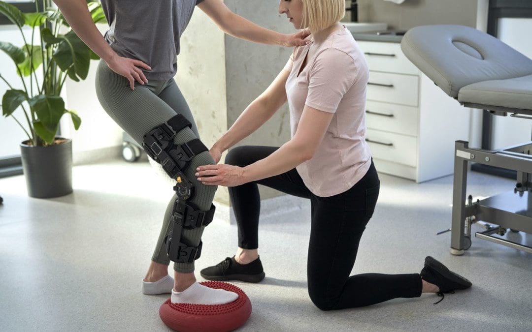
Knee Brace
A too-tight or strong brace can compress the leg and nerve.
Thigh-High Compression Stockings
Designed to maintain pressure on the legs, if too tight these stockings can compress the nerve.
Squatting Posture For Long Periods
This position places pressure on the side of the knee.
Fractures
A fracture of the large lower leg bone/tibia or sometimes the small bone/fibula near the knee can entrap the nerve.
Lower Leg Cast
The portion of the cast around the knee can be tight and compress the nerve.
Tell the doctor if a cast or brace feels tight or is causing numbness or pain in the leg.
Knee-High Boots
The top of a boot can land right below the knee and be too tight pinching the nerve.
Knee Ligament Injury
The nerve can become compressed due to bleeding or inflammation from an injured ligament.
Knee Surgery Complications
This is rare, but the nerve can inadvertently get pinched during knee replacement surgery or an arthroscopic procedure.
Prolonged Bed Rest
When lying down the legs tend to rotate outward and the knees flex.
In this position, the mattress can place pressure on the nerve.
Tumors or Cysts
Tumors or cysts can develop right on top or next to a nerve irritating and compressing the area.
Abdominal or Gynecologic Surgery
The equipment used to keep the legs rotated outward and the knees flexed for gynecologic and abdominal surgeries can compress the nerve.
Symptoms
The peroneal nerve supplies sensation and movement to the outside of the lower leg and the top of the foot. When compressed, it becomes inflamed, which causes the symptoms of a compressed nerve. Usually, only the lining/myelin sheath around the nerve is what gets injured. However, when the nerve gets damaged, the symptoms are similar but more severe. Common symptoms include:
Weakness that limits the ability to lift the foot toward the leg aka dorsiflexion.
This causes dragging the foot when walking.
The ability to turn the foot outward and extend the big toe is also affected.
Symptoms can be felt on the outside of the lower leg and on the top of the foot and include:
Tingling or pins and needles sensations.
Numbness.
Loss of sensation.
Pain.
Burning.
For individuals that have had a pinched nerve for two or more weeks, the muscles supplied by the nerve can begin to waste away or atrophy.
Symptoms can be intermittent or continuous depending on the cause.
The other common cause is a pinched nerve in the lumbar/lower spine.
When this is the cause, sensations, and pain will present in the lower back or the back and outside of the thigh.
Diagnosis
A doctor will look at medical history and perform an examination to make a diagnosis, determine the cause, and lay out a personalized treatment plan. The nerve in the knee can be felt as it travels around the top of the tibia, so a doctor may tap on it. If there is shooting pain down the leg, a pinched nerve may be present. Tests a doctor may order can include:
Knee X-ray
Shows any bone fractures or abnormal masses.
Knee MRI
Can confirm the diagnosis
Shows masses within the nerve.
Shows details of fractures or other problems in the bones.
Electromyogram – EMG
Tests electrical activity in the muscles.
Nerve Conduction Test
Tests the signal speed of the nerve.
Treatment
Treatment is aimed at reducing pain and improving mobility.
Over-the-Counter Pain Medication
OTC medication can reduce inflammation and improve symptoms short term.
Ice and Heat
Applying either heat or ice for 15 to 20 minutes at a time can provide relief from the symptoms.
An ice pack can make symptoms worse if it adds more pressure on the nerve.
Chiropractic and Physical Therapy
Chiropractic and physical therapy can release the compressed nerve, realign the structures, strengthen the muscles, and provide gait training.
Orthotic Boot
If walking gait is affected because the foot cannot bend, an orthotic boot can help.
This is a support that maintains the foot in a neutral position to walk normally.
Corticosteroid Injection
A corticosteroid injection can reduce inflammation and relieve pressure on the nerve.
Surgery
The nerve can suffer permanent damage if it has been pinched for a long time.
If that happens, surgery cannot repair the damage.
A doctor can perform surgery to correct a fracture, tumor, or other invasive problem causing a compressed nerve.
If conservative treatment doesn’t work, a peroneal nerve decompression procedure can be done to remove the pressure.
If surgery is needed, symptoms can disappear immediately, but it takes around four months to recover and rehabilitate.
Injury Rehabilitation
References
Krych, Aaron J et al. “Is peroneal nerve injury associated with worse function after knee dislocation?.” Clinical orthopedics and related research vol. 472,9 (2014): 2630-6. doi:10.1007/s11999-014-3542-9
Lezak B, Massel DH, Varacallo M. Peroneal Nerve Injury. [Updated 2022 Nov 14]. In: StatPearls [Internet]. Treasure Island (FL): StatPearls Publishing; 2023 Jan-. Available from: https://www.ncbi.nlm.nih.gov/books/NBK549859/
Soltani Mohammadi, Sussan, et al. “Comparing the squatting position and traditional sitting position for ease of spinal needle placement: a randomized clinical trial.” Anesthesiology and pain medicine vol. 4,2 e13969. 5 Apr. 2014, doi:10.5812/aapm.13969
Stanitski, C L. “Rehabilitation following knee injury.” Clinics in sports medicine vol. 4,3 (1985): 495-511.
Xu, Lin, et al. Zhongguo gu Shang = China Journal of Orthopedics and Traumatology vol. 33,11 (2020): 1071-5. doi:10.12200/j.issn.1003-0034.2020.11.017
Yacub, Jennifer N et al. “Nerve injury in patients after hip and knee arthroplasties and knee arthroscopy.” American Journal of physical medicine & Rehabilitation vol. 88,8 (2009): 635-41; quiz 642-4, 691. doi:10.1097/PHM.0b013e3181ae0c9d
Sitting at a desk or standing at a workstation in the same position for hours at a time every day or night can strain the body’s neuromusculoskeletal system. This causes hunching of the shoulders which leads to body tension, headaches, and neck, shoulder, back, leg, and foot pain. Taking regular breaks to move and stretch out provides various benefits that include pain symptom relief, increased circulation, improved posture, increased energy, muscle relaxation, and improve overall health. Here we look at stretches for sitting and standing jobs.
Stretches For Sitting and Standing Jobs
Sitting or standing for too long can affect an individual’s health. The body is made to move and not remain in one position for long periods. Standing and sitting for a prolonged period of time increases the risk for chronic conditions, sleep disorders, digestion issues, weight gain, obesity, heart disease, cancer, and diabetes. To help break the cycle of staying in one position, experts recommend taking movement breaks for one to three minutes every 30 to 45 minutes, or at least once an hour.
Individuals are encouraged to find ways to set up a job/work system where they are not limited to just sitting or standing but have a balance where they can move around, do some desk work, move some more, and so on where the body is regularly engaging all the muscles and not only using a few that often leads to overworked muscles and overcompensating postures that lead to injuries. This can be using a standing desk, taking quick short walks, or doing some stretches. Movement loosens up the body and helps maintain mental focus.
Flexibility Breaks
The following stretches for sitting and standing jobs are recommended to be done as often as possible to develop healthy habits.
Set an alarm for every 45 to 55 minutes and perform the stretches.
Hold each stretch for at least 15-30 seconds.
Avoid exercises or stretches that cause discomfort or pain.
Chest Stretch
Many individuals begin to hunch forward. Therefore, it is essential to stretch the pectoral/chest muscles and shoulders. Individuals with shoulder problems or injuries should avoid this stretch.
Stand with the feet shoulder-width apart.
The stretch can be performed seated in a chair.
Move the arms behind the body and, if possible, interlock the fingers together.
Straighten the arms and gently lift the hands up until you feel a stretch in the chest.
Hold for 10 to 30 seconds.
Repeat 5-10 times.
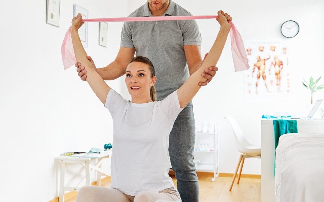
Individuals can also use a resistance band and hold it overhead.
Individuals can put their forearms on either side of a doorway, and gently press forward until the stretch is felt in the chest.
Upper Back Stretch
The upper back stretch will help get circulation moving in all the muscles between the shoulder blades as well as the traps and the shoulders.
Begin in a seated or standing position.
Stretch the arms straight out.
Place one hand on top of the other.
Reach away with your arms.
Relax and gently bend the head down.
Imagine the arms curving up and over an imaginary sphere.
Hold the stretch for 10 to 30 seconds.
Neck Stretch
Tension in the neck can lead to headaches and upper back pain. Forward head posture is common when working at a desk/workstation, which places extra weight and stress on the neck muscles. The head can weigh up to 11 pounds. Staying aware of posture and regular stretching can provide relief.
Start in a sitting position, with the back straight and the shoulders back.
Reach down and grab the side of the chair with your hand.
Gently pull on the chair, while tilting the head in the opposite direction, feeling the stretch down the side of the neck and shoulder.
Hold for 10 to 30 seconds and repeat on the other side.
Repeat five to 10 times on each side.
Inner Thigh Stretch
Stretching the inner thigh is important for the hips and groin. This stretch helps open the hips and gets rid of tightness and tension in the lower body.
In a seated position, widen the legs, toes outward, and lean forward with the elbows on the thighs.
Keep the back straight and the abs contracted.
Gently press forward while using the elbows to push the thighs out until a stretch is felt in the inner thighs.
Hold for 10 to 30 seconds.
Repeat as many times as necessary to get a thorough stretch.
Regular stretching will help to improve the range of motion and help the muscles move more efficiently. Stretching may also help individuals reach or maintain a healthy weight for improved posture.
Benefits of Stretching
References
Cooley D, Pedersen S. A pilot study of increasing non-purposeful movement breaks at work as a means of reducing prolonged sitting. J Environ Public Health. 2013;2013:128376. doi:10.1155/2013/128376
Daneshmandi H, Choobineh A, Ghaem H, Karimi M. Adverse effects of prolonged sitting behavior on the general health of office workers. J Lifestyle Med. 2017;7(2):69-75. doi:10.15280/jlm.2017.7.2.69
Fathollahnejad, Kiana, et al. “The effect of manual therapy and stabilizing exercises on the forward head and rounded shoulder postures: a six-week intervention with a one-month follow-up study.” BMC Musculoskeletal Disorders vol. 20,1 86. 18 Feb. 2019, doi:10.1186/s12891-019-2438-y
Feldman, Anatol G. “The Relationship Between Postural and Movement Stability.” Advances in experimental medicine and biology vol. 957 (2016): 105-120. doi:10.1007/978-3-319-47313-0_6
Korakakis, Vasileios, et al. “Physiotherapist perceptions of optimal sitting and standing posture.” Musculoskeletal Science & Practice vol. 39 (2019): 24-31. doi:10.1016/j.msksp.2018.11.004
Lurati AR. Health issues and injury risks associated with prolonged sitting and sedentary lifestyles. Workplace Health Saf. 2018;66(6):285-290. doi:10.1177/2165079917737558
Nakphet N, Chaikumarn M, Janwantanakul P. Effect of different types of rest-break interventions on neck and shoulder muscle activity, perceived discomfort and productivity in symptomatic VDU operators: A randomized controlled trial. Int J Occup Saf Ergon. 2014;20(2):339-53. doi:10.1080/10803548.2014.11077048
Sanders, Martha J, and Claudia Michalak Turcotte. “Posture makes perfect.” Today’s FDA: official monthly journal of the Florida Dental Association vol. 25,2 (2013): 62-5.
Shaghayegh Fard, B et al. “Evaluation of forward head posture in sitting and standing positions.” The European spine journal: official publication of the European Spine Society, the European Spinal Deformity Society, and the European Section of the Cervical Spine Research Society vol. 25,11 (2016): 3577-3582. doi:10.1007/s00586-015-4254-x
Overexertion and repetitive stress injuries make up a fourth of all work injuries. Repetitive pulling, lifting, punching in numbers, typing, pushing, holding, carrying, and scanning are the most common causes of job-related injuries. These types of injuries are the most common that cause missed days at work. Overexertion can lead to lasting physical chronic conditions, ranging from chronic back pain to chronic joint pain caused by the advanced wearing and tearing of the various musculoskeletal tissues. Chiropractic medicine takes a comprehensive and whole-body approach to treating neuromusculoskeletal injuries. Chiropractic relieves tight or damaged muscles, increases nerve energy flow, and properly aligns the joints through adjustments, spinal traction, decompression, and various forms of manual manipulation.
Overexertion and Repetitive Stress Injuries
Overexertion and repetitive stress injuries typically occur over time/years of engaging in the same strenuous activity regularly. However, an overexertion injury can occur with one sudden or extreme movement. A worker can injure muscles, tendons, joints, and ligaments. Overexertion can lead to musculoskeletal disorders with symptoms including:
Inflammation
Swelling
Numbness
Stiffness
Chronic pain
Limited or total loss of mobility in the muscles, tendons, ligaments, and joints.
Types
A few of the most common examples of overexertion injuries include:
Soft-Tissue
Injuries to muscles, ligaments, tendons, and joints.
Back
Pulled, strained back muscles.
Herniated discs.
Compressed nerve roots.
Fractured vertebrae.
Dehydration and Heat Stroke
Most common among workers doing outdoor manual labor.
Repetitive and Overuse
Injuries range from carpal tunnel syndrome to stress fractures.
Often the result of weeks, months, or years of repeated movements
In many cases, two or more injuries can happen simultaneously.
For example, a worker is more likely to sustain an injury if they are dehydrated or they are performing dual tasks.
Causes
Certain movements and activities are more likely to cause overexertion injuries. Some of the most common include:
Daily lifting of objects, light and heavy.
Performing awkward movements that cause the body to be in unhealthy positions.
Standing and/or sitting or for long periods.
Using excessive force to perform tasks.
Operating heavy machinery.
Working in hot and/or humid conditions.
High-Rate Injury Industries
Industries in which overexertion injuries are most common include:
Education.
Health services.
Manufacturing.
Construction.
Warehouse work.
Transportation.
Wholesale trade.
Retail stores.
Chiropractic Treatment
These injuries can lead to missed work, debilitating pain, and medical bills. Depending on the severity of the injury, chiropractic care will use massage techniques, spinal manipulation, traction, and decompression therapies, to increase flexibility and mobility to reduce the chances of a recurring injury. The benefits of chiropractic include:
Prevents the risk of worsening or future injuries.
Expedites recovery to help individuals rehabilitate and get back to work sooner.
Improves physical and mental well-being.
Provide recommendations on how to properly stretch and strengthen muscles.
Nutritional anti-inflammatory recommendations.
By learning how to avoid overexertion injuries, workers can be more productive, enjoy work, and improve their quality of life.
From Injury To Recovery
References
Anderson, Vern Putz, et al. “Occupational fatalities, injuries, illnesses, and related economic loss in the wholesale and retail trade sector.” American Journal of industrial medicine vol. 53,7 (2010): 673-85. doi:10.1002/ajim.20813
Choi, Hyun-Woo, et al. “Characteristics of occupational musculoskeletal disorders of five sectors in the service industry between 2004 and 2013.” Annals of Occupational and environmental medicine vol. 29 41. 19 Sep. 2017, doi:10.1186/s40557-017-0198-4
Friedenberg, Rivi, et al. “Work-related musculoskeletal disorders and injuries among emergency medical technicians and paramedics: A comprehensive narrative review.” Archives of Environmental & occupational health vol. 77,1 (2022): 9-17. doi:10.1080/19338244.2020.1832038
Galinsky, T et al. “Overexertion injuries in home health care workers and the need for ergonomics.” Home health care services quarterly vol. 20,3 (2001): 57-73. doi:10.1300/J027v20n03_04
González Fuentes, Aroa, et al. “Work-related overexertion injuries in cleaning occupations: An exploration of the factors to predict the days of absence by means of machine learning methodologies.” Applied ergonomics, vol. 105 103847. 30 Jul. 2022, doi:10.1016/j.apergo.2022.103847
Schoenfisch, Ashley L et al. “Declining rates of work-related overexertion back injuries among union drywall installers in Washington State, 1989-2008: Improved work safety or shifting of care?.” American Journal of industrial medicine vol. 57,2 (2014): 184-94. doi:10.1002/ajim.22240
Williams, J M et al. “Work-related injuries in a rural emergency department population.” Academic emergency medicine: official journal of the Society for Academic Emergency Medicine vol. 4,4 (1997): 277-81. doi:10.1111/j.1553-2712.1997.tb03548.x
IFM's Find A Practitioner tool is the largest referral network in Functional Medicine, created to help patients locate Functional Medicine practitioners anywhere in the world. IFM Certified Practitioners are listed first in the search results, given their extensive education in Functional Medicine