Can using a self-care traction device help and be a cost-effective method of providing cervical relief at home for individuals who have neck pain?
Neck Pain Traction Device
Neck Pain Traction Device: Individuals with neck or arm pain caused by neck/cervical radiculopathy may benefit from physical therapy to manage their condition. (Alshami, A. M., and Bamhair, D. A. 2021) Physical therapy can improve neck mobility, restore function, and decrease pain.
Traction
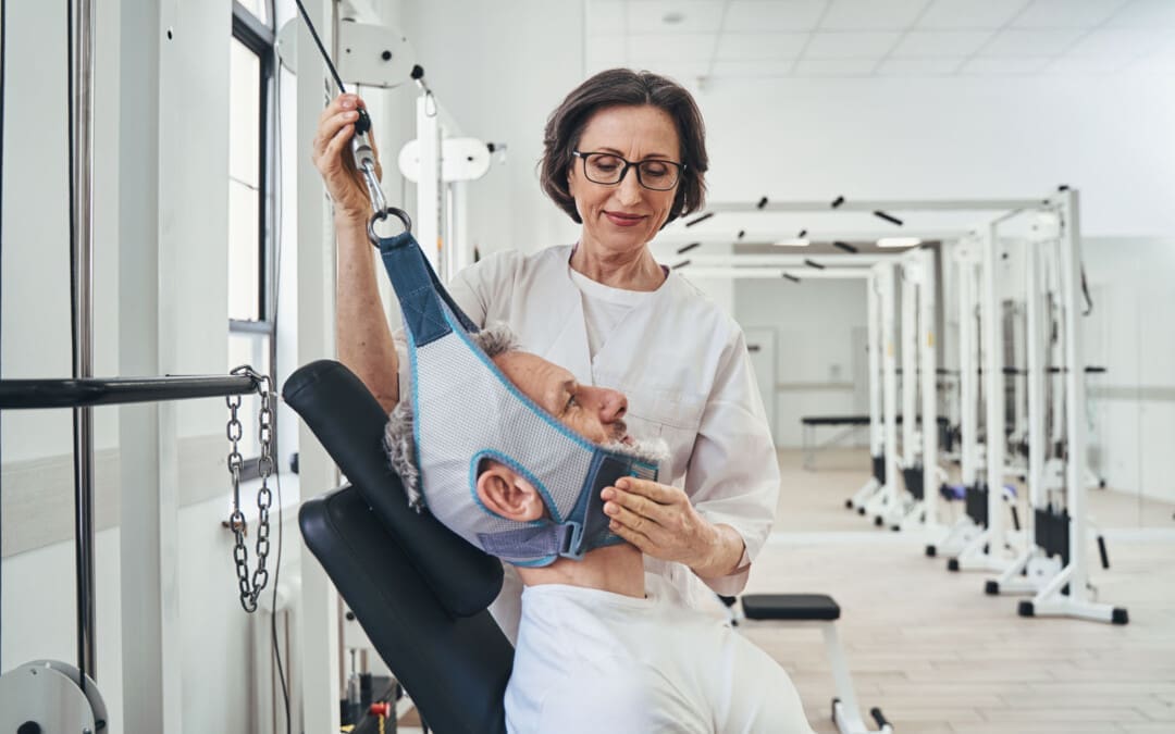
A physical therapist may use various treatments and modalities to treat the individual’s condition. Cervical traction is one treatment to help relieve neck pain and can help:
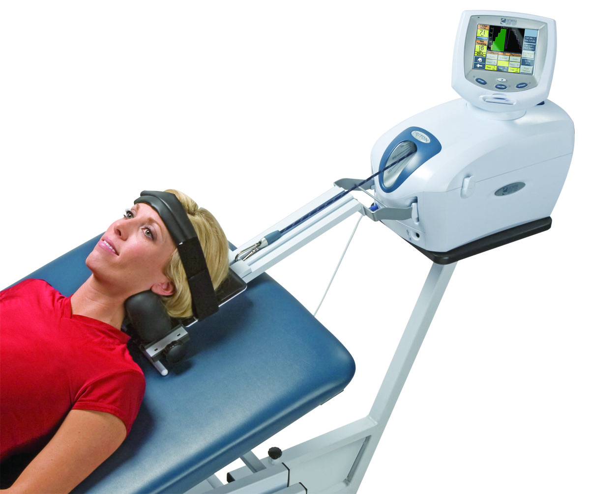
There are different neck traction techniques. The therapist may use a mechanical traction device that requires the individual to strap their head and neck to a machine that gently pulls the neck, providing relief. (Romeo, A. et al., 2018) The physical therapist may also use manual traction with their hands to help the process. This type allows the therapist to easily adjust the amount of traction force and the direction of the pull on the neck. (Romeo, A. et al., 2018)
Home Traction Devices
Cervical traction that works for individuals at the clinic may also benefit from using a traction device at home to maintain their gains. Over-the-door cervical traction may be an effective, safe, and simple way to administer neck traction in the comfort of one’s home to help decrease pain. (Fritz, J. M. et al., 2014) Medical supply stores and pharmacies carry over-the-door traction devices, which can also be found online. If finding an over-the-door traction unit is difficult, the therapy clinic can help order one from a medical supplier.
Setting Up The Device
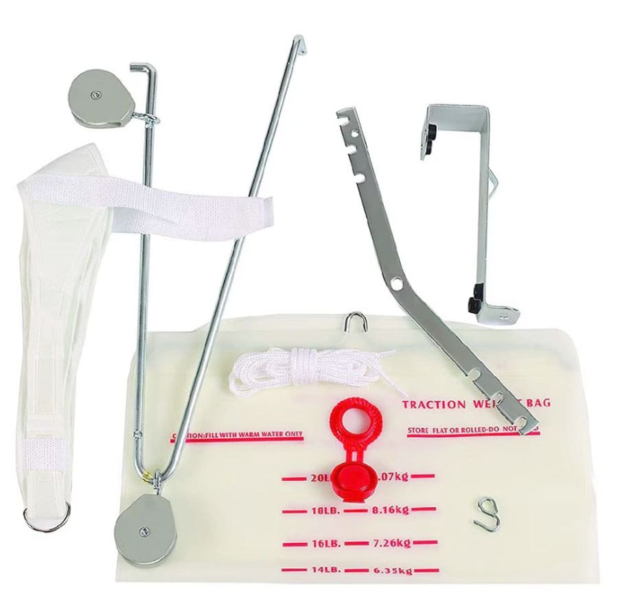
Before using a neck pain traction device, consult a physical therapist or doctor to ensure neck traction is safe and on usage. Over-the-door traction units may be built differently but comprise the main parts:
A hook-and-pulley apparatus that hangs over the door.
A nylon cord that’s fed through the pulley.
A weight that could be a water bag that hangs on the end of the cord to provide traction force.
A harness to wear on your head.
The traction unit must be hung over a door secured to the top and closed.
It is recommended that the door be a closet door so no one will open it while using the device.
If a closet door is not available, be sure to lock the door so that no one can open it while attached to the device.
A small hook with a pulley attached to it hangs on the door.
The harness attaches to the end of the nylon cord opposite the weighted water bag.
The harness goes on the head, and the chin strap should fit snugly under the chin and be secured by the hook-and-loop fasteners.
The two straps on either side of your head should be hooked to the end of the nylon cord, which is fed through the pulley system.
After strapping on the head harness, fill the water bag using the markings on the bag to indicate its weight and hang it on one end of the cord.
Fill the bag until the water reaches the desired mark, usually 8–15 pounds.
After filling the water bag, sit in a chair facing the door and hang the bag on the other end of the cord not attached to the head harness.
Do not drop the bag; this could cause a sudden forceful traction motion on your neck.
How Long to Use
Individuals should generally use the traction device for about 15 to 20 minutes each session and can perform several sessions per day. (American Physical Therapy Association. 2020) While using the over-the-door traction device, there should be a gentle pulling sensation in the neck, relieving the neck pain, and if there is arm pain or tingling, it should also decrease. Traction does not replace neck exercises or postural correction in treating neck pain. Be sure to follow the therapist’s prescribed exercises. Active engagement is essential to treating and preventing neck pain from returning. If the pain increases while using the traction device, stop using it and consult a physical therapist or doctor. Ask a physical therapist or other neuromusculoskeletal medical professional if self-care traction is appropriate for specific conditions.
Injury Medical Chiropractic and Functional Medicine Clinic works with primary healthcare providers and specialists to develop an optimal health and wellness solution that helps individuals return to normal. Our providers create personalized care plans for each patient, including Functional Medicine, Acupuncture, Electro-Acupuncture, and Sports Medicine principles through an integrated approach to treat injuries and chronic pain syndromes to improve ability through flexibility, mobility, and agility programs to relieve pain. If other treatment is needed, Dr. Jimenez has teamed up with top surgeons, clinical specialists, medical researchers, and rehabilitation providers to provide the most effective treatments.
Neck Injuries
References
Alshami, A. M., & Bamhair, D. A. (2021). Effect of manual therapy with exercise in patients with chronic cervical radiculopathy: a randomized clinical trial. Trials, 22(1), 716. https://doi.org/10.1186/s13063-021-05690-y
Madson, T. J., & Hollman, J. H. (2017). Cervical Traction for Managing Neck Pain: A Survey of Physical Therapists in the United States. The Journal of orthopaedic and sports physical therapy, 47(3), 200–208. https://doi.org/10.2519/jospt.2017.6914
Romeo, A., Vanti, C., Boldrini, V., Ruggeri, M., Guccione, A. A., Pillastrini, P., & Bertozzi, L. (2018). Cervical Radiculopathy: Effectiveness of Adding Traction to Physical Therapy-A Systematic Review and Meta-Analysis of Randomized Controlled Trials. Physical therapy, 98(4), 231–242. https://doi.org/10.1093/physth/pzy001
Fritz, J. M., Thackeray, A., Brennan, G. P., & Childs, J. D. (2014). Exercise only, exercise with mechanical traction, or exercise with over-door traction for patients with cervical radiculopathy, with or without consideration of status on a previously described subgrouping rule: a randomized clinical trial. The Journal of orthopaedic and sports physical therapy, 44(2), 45–57. https://doi.org/10.2519/jospt.2014.5065
American Physical Therapy Association. (2020). Physical therapy guide to cervical radiculopathy. https://www.choosept.com/guide/physical-therapy-guide-cervical-radiculopathy
Can understanding the location of the funny bone and how pain can be managed after injury help expedite recovery and prevention for individuals who have hit their funny bone?
Elbow Funny Bone Nerve Injury
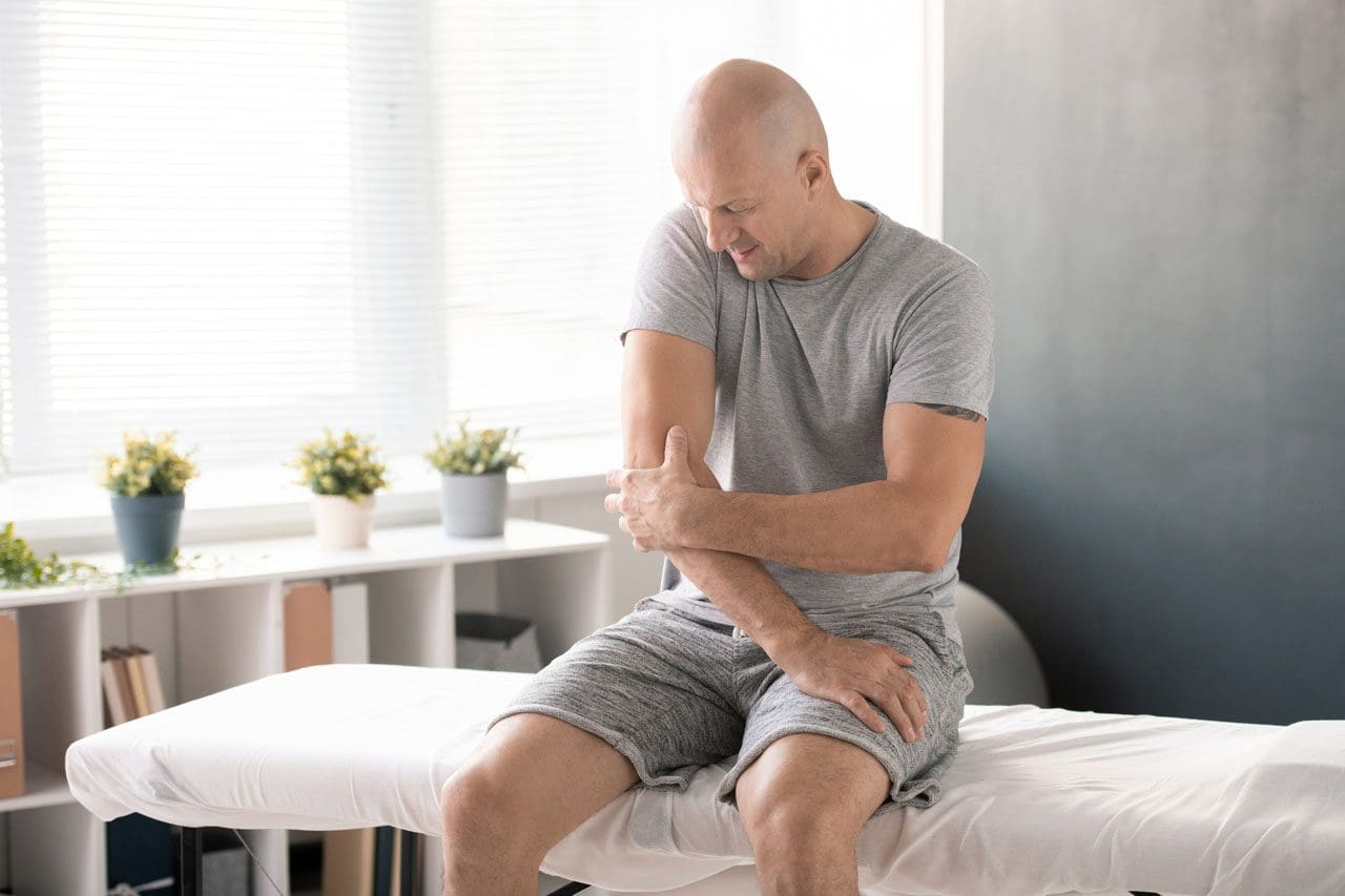
Behind the elbow is an area known as the “funny bone,” where the ulnar nerve has less tissue and bone protection. This is where part of the ulnar nerve passes around the back of the elbow. Because less tissue and bone protect the nerve in this area, taking a hit like bumping into something can cause an electric shock-like pain and a tingling sensation down the arm and to the outside fingers typical of an irritated nerve. Most injuries to the funny bone resolve quickly, and the pain disappears after a few seconds or minutes, but sometimes, an ulnar nerve injury can lead to more persistent symptoms.
Anatomy
The funny bone is not a bone but the ulnar nerve. The nerve runs down the arm, passing around the back of the elbow. (Dimitrova, A. et al., 2019) Because the ulnar nerve is on top of the elbow and there is very little fatty cushion, lightly bumping this spot can cause pain and tingling sensations down the forearm. Three bones comprise the junction of the elbow that include:
Humerus – arm bone
Ulna and radius – forearm bones
The humerus has a groove that protects and holds the ulnar nerve as it passes behind the joint. This is where the nerve can be injured or irritated when the nerve is hit or pinched against the end of the bone, causing the funny bone pain.
Electrical Pain Sensation
When hitting the ulnar nerve or funny bone where the ulnar nerve provides sensation, pain, and electrical/tingling sensations are experienced from the forearm to the outside fingers. This part of the arm and hand is called the ulnar nerve distribution. (American Academy of Orthopaedic Surgeons. 2024) The ulnar nerve provides sensation into most of the pinky finger and about half of the ring finger. Other nerves, including the median and radial nerve, supply sensation to the rest of the hand.
Treatment
Usually, a sharp jolt to the elbow quickly resolves. Some recommendations to help symptoms improve faster include:
Shaking the forearm and hand out.
Straightening out and bending the elbow to stretch the nerve.
Decreasing mobility of the elbow.
Applying ice to the area.
Taking anti-inflammatory medications.
Treating Long-Lasting Pain
In rare circumstances, injuries to the ulnar nerve can cause more persistent symptoms, a condition known as cubital tunnel syndrome. Cubital tunnel syndrome can happen after an injury or from elbow overuse. Individuals with cubital tunnel syndrome may benefit from wearing a splint at night. Standard-sized splints can be ordered online, but most are fabricated by an occupational or hand therapist. If symptoms become more long-lasting, surgery may be recommended to relieve pressure and tension on the ulnar nerve (American Academy of Orthopaedic Surgeons, 2024). The procedure decompresses the nerve by relieving any tight constrictions around it and releasing them. In severe cases, the nerve is repositioned to an area that doesn’t place as much pressure on the nerve, known as an ulnar nerve transposition.
Injury Medical Chiropractic and Functional Medicine Clinic works with primary healthcare providers and specialists to develop an optimal health and wellness solution that helps individuals return to normal. Our providers create personalized care plans for each patient, including Functional Medicine, Acupuncture, Electro-Acupuncture, and Sports Medicine principles through an integrated approach to treat injuries and chronic pain syndromes to improve ability through flexibility, mobility, and agility programs to relieve pain. If other treatment is needed, Dr. Jimenez has teamed up with top surgeons, clinical specialists, medical researchers, and rehabilitation providers to provide the most effective treatments.
Chiropractic Treatment For Carpal Tunnel Syndrome
References
Dimitrova, A., Murchison, C., & Oken, B. (2019). Local effects of acupuncture on the median and ulnar nerves in patients with carpal tunnel syndrome: a pilot mechanistic study protocol. Trials, 20(1), 8. https://doi.org/10.1186/s13063-018-3094-5
American Academy of Orthopaedic Surgeons. (2024). Ulnar nerve entrapment at the elbow. https://orthoinfo.aaos.org/en/diseases–conditions/ulnar-nerve-entrapment-at-the-elbow-cubital-tunnel-syndrome/
For individuals who sit at work for long hours, can years of practicing unhealthy posture be corrected through a step-by-step approach to ensure optimal body position while sitting?
Sitting Posture
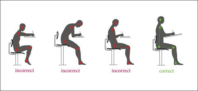
Sitting up straight with a healthy posture requires the conscious alignment of the hips, pelvis, lower back, upper back, shoulders, neck, and head. Learning or retraining oneself to maintain correct sitting posture can relieve lower back pain, improve respiration and digestion, and reduce tension in the neck and shoulders. (Albarrati, A. et al., 2018) It starts by paying attention to posture throughout the day and correcting it whenever forward head posture, leaning, or slouching develops. Targeted exercises can also help build upper-body strength, and stretching can stabilize and strengthen the core muscles, lower back, and pelvic joints. (Albarrati, A. et al., 2018)
Sit Up Straight Guide
Sitting up straight can be uncomfortable because it is not a natural position for the body to be in for an extended time. Nowadays, work, school, appointments, and other activities require us to sit much longer than intended. The muscles also have to work against gravity, leading to muscle exhaustion, slouching, and slumping, which can cause chronic back, leg, neck, and/or shoulder pain. (Jung, K. S. et al., 2020)
Sitting up straight may seem simple, but the focus tends to be on straightening just the lower/lumbar spine. This posture is unsustainable and exhausts and stresses the upper and lower back. (Jung, K. S. et al., 2020) The whole body needs to be considered when protecting the stability and balance of the spine. Learning and maintaining the ability to sit up straight is a process that requires practice. Find a comfortable chair to sit in, and follow these steps to achieve the optimal postural alignment (Canadian Centre for Occupational Health and Safety, 2022)
Knee Spacing
The hips should be at a roughly 90-degree angle.
Knee Position
The knees should be at a 90-degree angle level with the hips.
Use a pillow to achieve the right knee position if the seat is too low.
Keep the Feet Flat on the Floor
If feet don’t reach the floor, place a footstool, box, book, or other flat object underneath them.
Sitting Bones
Also known as the ischial tuberosities, these are two knobby bones on the underside of the pelvis.
Feel around to find them.
Pelvis Adjustment
Shift the body so that the sitting bones are directly under the pelvis rather than situated too far back, stressing the lower back or too far forward, leading to slumping.
Spine Check
There should be a slight spinal curve, and one should be able to slip a hand between the lower back and the back of the chair.
Shoulder Check
The shoulders should be level and vertically aligned with the hips.
If the shoulder blades are pulled back or the shoulders are lifted or curled forward, relax them into a neutral position.
Head Positioning
The head tends to tilt too far forward while sitting as work and the day progresses.
Adjust the head position to align the neck with the upper spine.
The head should be slightly tilted forward, with the ears aligned with the shoulders.
Check for Pain and Discomfort
Pain may be due to structural imbalances of the spine, pelvis, or hips.
Use a lumbar chair support or place a rolled-up towel or cushion at the lower back to keep the back straight.
Added Tips
Tools and tricks to help prevent and avoid back, hip, and neck pain.
Chairs
All the bells and whistles for an ergonomic desk chair are unnecessary.
Focus on features like adjustable seat height and lumbar support. The correct seat depth recommendations are deeper if tall and shallower if short. (van Niekerk, S. M. et al., 2012)
Cushions
If sitting on a cushion or using one to bolster the back or hips, recommendations are not to go too soft.
Cushions that are too soft allow the ability to shift from one hip to the next, often without realizing it.
They usually eventually flatten and lose support.
Monitor Position
There is no point in sitting straight if the monitor is too high or too low.
The monitor should be at eye level to maintain the proper head and shoulder alignment.
If the monitor is too low, place a box or book underneath it.
If it is too high, raise the chair’s height and place a footrest under the feet to keep them flat.
Avoid Crossing Legs or Feet
Crossing the legs or feet places stress on the opposite hip, thigh, and knee and wears the body out faster.
If the hips or legs are tiring prematurely, the individual is not sitting correctly or in the wrong chair.
Use Comfortable Footwear
Maintaining flat feet on the floor while sitting is imperative.
This is not possible in high heels or platform shoes.
Change into a comfortable pair of flat shoes while sitting.
Take Regular breaks
Even with an ergonomic desk chair, the body is not meant to be sitting for hours and hours.
Get up at least every hour, walking and stretching to reactivate the muscles and circulation.
Sitting up straight requires body alignment awareness, stable core muscles, and balanced pelvis, hips, spine, shoulders, neck, and head positioning. It may take some time before these steps become normal, but they will become second nature with perseverance and practice. Injury Medical Chiropractic and Functional Medicine Clinic works with primary healthcare providers and specialists to develop an optimal health and wellness solution that fully benefits the individual to get back to normal. Using an integrated approach to treat injuries and chronic pain syndromes to improve ability through flexibility, mobility, and agility programs to relieve pain. Our providers create personalized care plans for each patient, including Functional Medicine, Acupuncture, Electro-Acupuncture, and Sports Medicine principles. If other treatment is needed, Dr. Jimenez has teamed up with top surgeons, clinical specialists, medical researchers, and rehabilitation providers to provide the most effective treatments.
Posture and Mobility
References
Albarrati, A., Zafar, H., Alghadir, A. H., & Anwer, S. (2018). Effect of Upright and Slouched Sitting Postures on the Respiratory Muscle Strength in Healthy Young Males. BioMed research international, 2018, 3058970. https://doi.org/10.1155/2018/3058970
Jung, K. S., Jung, J. H., In, T. S., & Cho, H. Y. (2020). Effects of Prolonged Sitting with Slumped Posture on Trunk Muscular Fatigue in Adolescents with and without Chronic Lower Back Pain. Medicina (Kaunas, Lithuania), 57(1), 3. https://doi.org/10.3390/medicina57010003
Canadian Centre for Occupational Health and Safety. (2022). Working in a sitting position – good body position. Retrieved from https://www.ccohs.ca/oshanswers/ergonomics/sitting/sitting_position.html
van Niekerk, S. M., Louw, Q. A., & Hillier, S. (2012). The effectiveness of a chair intervention in the workplace to reduce musculoskeletal symptoms. A systematic review. BMC musculoskeletal disorders, 13, 145. https://doi.org/10.1186/1471-2474-13-145
For individuals wanting to try Pilates for back pain and exercise, can learning how to find their neutral spine help improve flexibility and increase the range of motion in the joints?
Pilates Neutral Spine
Pilates is a functional exercise modality emphasizing core stability, which is fundamental to developing a balanced body. The exercises strengthen the muscles, improve flexibility, and increase the range of motion in the joints. (Kloubec J. 2011) It is considered a functional fitness method because its principles work to establish more graceful, efficient movements from everyday life, such as improving posture. Pilates has shown its effectiveness in that it is often used in physical therapy and rehabilitation settings. (Byrnes, K., Wu, P. J., and Whillier, S. 2018) However, knowing how to find the neutral spine is essential for performing various Pilates exercises correctly. (Barbosa, A. C. et al., 2018) This subtle adjustment during practice may help prevent injury and increase overall performance. A neutral spine is the natural position of the spine when all three curves:
Cervical (neck)
Thoracic (middle)
Lumbar (lower)
Are active and in healthy alignment.
This is the strongest position for the spine when standing or sitting, allowing the body to move more naturally.
Alignment
The following exercise can help find the Pilates neutral spine.
Basic Position
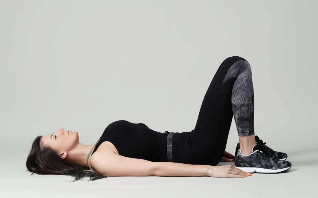
Lie on the back with knees bent and feet flat on the floor.
Ensure the legs are parallel to the hips, knees, heels, and toes.
Let the arms rest at your sides.
Relax
Relax the body, including the shoulders, neck, and jaw.
Allow the back to melt into the floor.
The rib cage will drop when the lower ribs are released to the floor.
Breathe Deep
Inhale all the way into the body, allowing it to move into the back and sides of the rib cage and all the way to the pelvis.
Pelvic Tilt
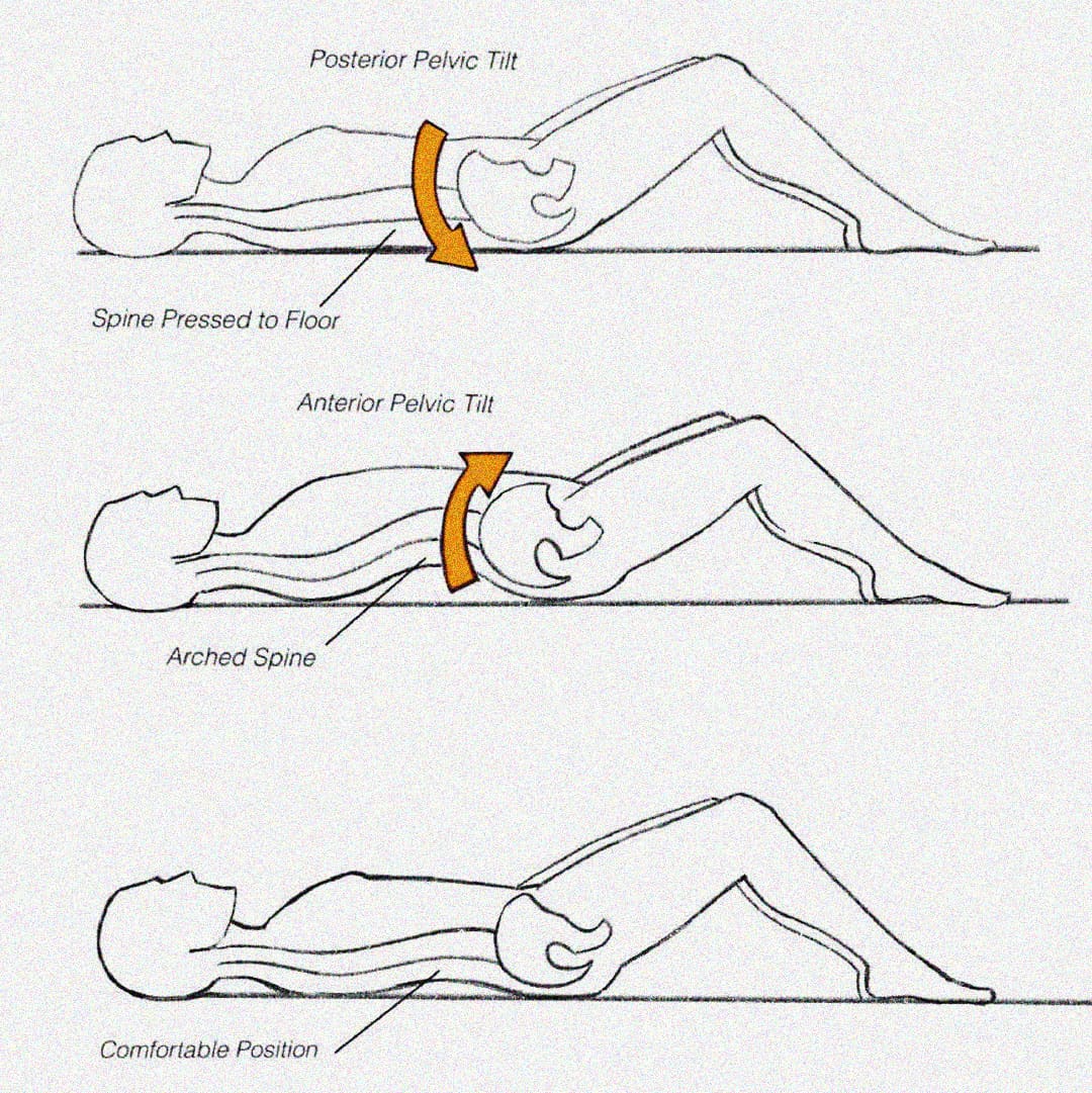
Exhale and use the abdominals to press the lower spine into the floor in a pelvic tuck. (Eickmeyer S. M. 2017)
Inhale to release.
Exhale and pull the lower spine off the floor, creating a pelvic tilt.
Inhale to release.
Pilates exercises don’t use excess energy or tension. Proper alignment and a neutral spine position can ensure that tension is released and excess energy is not exerted. (Byrnes, K., Wu, P. J., and Whillier, S. 2018) When performing the exercise, ensure that the shoulders, neck, and legs are relaxed and not involved in the movement.
When Exercising
Once a neutral spine is achieved, the goal is to maintain this spinal position during the exercises and when changing positions.
Start by lifting the right leg and placing it back down without letting the hips move.
Then, repeat the motion with the left leg.
Engage the abdominal muscles to help stabilize the pelvis, keeping it from moving and maintaining a neutral spine.
Repeat this process with each leg.
Once each leg can be lifted easily, test with both legs.
Exhale deeply and lift the legs while keeping the core and pelvis stable.
Then, lower them back down.
When performing this progression, there may be a want to release the abs and let the back arch.
This will cause a tuck and tilt position away from the neutral spine position.
If this progression is difficult, keep practicing until you can maintain a neutral spine.
Once this basic progression feels easy, try additional progressions and positioning.
Visualization Tips
Most people have their spines in one of two positions: tucked or tilted. A neutral spine requires individuals to be in between, with the lower abdominals flat and the lower spine’s natural curve slightly off the floor. The following visualization can help establish a neutral spine.
Balanced Pelvic Placement
Imagine a cup of water sitting on the lower abdomen, a couple of inches below the belly button.
Allow the abdominal muscles to drop toward the spine, flattening the belly.
You don’t want the water to spill, so the pelvis cannot be tipped forward or tucked under.
Body Scan Meditation
Once the body is relaxed with a balanced alignment on the floor.
Allow breathing to become deep and full and the abdominals to drop toward the floor.
The natural neck and lower spine curves should be off the floor.
Ensure the lower spine is not pressed into the floor, as this indicates a pelvic tilt.
If there is any discomfort or pain when working to increase endurance, seek advice from a healthcare professional. Injury Medical Chiropractic and Functional Medicine Clinic uses an integrated approach to treating injuries and chronic pain syndromes. It offers personalized care plans that improve ability through flexibility, mobility, and agility programs to relieve pain. Our providers use an integrated approach to create personalized care plans for each patient, including Functional Medicine, Acupuncture, Electro-Acupuncture, and Sports Medicine principles. Our goal is to relieve pain naturally by restoring health and function to the body. If other treatment is needed, Dr. Jimenez has teamed up with top surgeons, clinical specialists, medical researchers, and rehabilitation providers to provide the most effective treatments.
Is Motion Key to Healing?
References
Kloubec J. (2011). Pilates: how does it work and who needs it?. Muscles, ligaments and tendons journal, 1(2), 61–66.
Byrnes, K., Wu, P. J., & Whillier, S. (2018). Is Pilates an effective rehabilitation tool? A systematic review. Journal of bodywork and movement therapies, 22(1), 192–202. https://doi.org/10.1016/j.jbmt.2017.04.008
Barbosa, A. C., Vieira, E. R., Silva, A. F., Coelho, A. C., Martins, F. M., Fonseca, D. S., Barbosa, M. A., & Bordachar, D. (2018). Pilates experience vs. muscle activation during abdominal drawing-in maneuver. Journal of bodywork and movement therapies, 22(2), 467–470. https://doi.org/10.1016/j.jbmt.2017.05.002
Eickmeyer S. M. (2017). Anatomy and Physiology of the Pelvic Floor. Physical medicine and rehabilitation clinics of North America, 28(3), 455–460. https://doi.org/10.1016/j.pmr.2017.03.003
Can understanding the nucleus pulposus help in body positioning and prevention for individuals wanting to practice spinal hygiene and protect their discs from injury?
Nucleus Pulposus
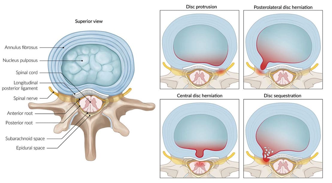
The spinal discs are located between the spine’s vertebrae and are the body’s natural impact and shock absorbers. Within the disc is the nucleus pulposus, which plays a major role in providing the spine with shock absorption during movement. (Zhou Z. et al., 2014) The discs have a tough outer portion and a soft inner core. They are the:
It forms the tough circular exterior and comprises concentric sheets of collagen fibers or lamellae surrounding the inner core.
It has cartilaginous endplates that firmly attach to the vertebrae above and below.
Nucleus Pulposus
The nucleus pulposus is the inner core soft filling of the discs.
It contains a network of fibers suspended in a mucoprotein gel with a water base to maintain strength and pliability.
The near-liquid consistency makes it responsive to movement to handle the body’s axial load.
It helps maintain spinal suspension to prevent pressure on the bones and prevent bone-to-bone contact, reducing the potential for injuries and pain.
Shock Absorber
Each intervertebral disc is a shock-absorbing cushion, with the nucleus pulposus providing shock-absorbing properties (Zhou Z. et al., 2014). The intervertebral discs move as the body moves. For example, when arching the back, the disc moves forward slightly, and when twisting, the disc twists as well.
Spinal Action
The intervertebral disc supports spinal movements. When bending, twisting, arching, or tilting the spine, the nucleus pulposus swivels to accommodate these actions. These repeated spinal actions, which occur throughout the day and night, contribute to shifting positions while sitting, working, playing sports, carrying groceries, performing house chores, etc. An example is bending forward to pick something up. This action involves forward spinal flexion, which is bending the spine forward, flattening, or rounding. When bending using flexion, the spinal bones come closer together, pushing the nucleus pulposus toward the back.
Injuries
The disc can be pushed too far back with persistent or excessive spinal flexion. If the fibers of the annulus fibrosus become weak, they can tear, causing the nucleus pulposus to leak out and disc herniation. Generally, the nucleus pulposus will leak to the side and back; however, this corresponds to the location of the very sensitive nerve root/s with which it can come into contact, causing pain and other symptoms. The most common causes of disc herniation are degenerative wear and tear changes of the disc and trauma. Disc degeneration occurs as the body ages; it weakens the annulus fibers, allowing the nucleus pulposus to distend, bulge, or herniate.
Aging
Disc degeneration occurs with age but can also occur with injuries to the area. In young individuals, the nucleus pulposus is mostly water. For this age group, a herniation from trauma is more likely than in older individuals. (Ucar, D. et al., 2021) But as the body ages, the discs, especially the nucleus pulposus, begin to dry out. This dehydration leads to a significant loss of disc height. (UCLA Health, 2024) By age 60 or 70, the discs may be composed entirely of fiber, which can cause the shock absorption function not to work and disappear.
Chiropractic therapy is among the more conservative treatment options for a herniated disc and may be tried first before proceeding with more invasive treatments. Injury Medical Chiropractic and Functional Medicine Clinic works with primary healthcare providers and specialists to develop an optimal health and wellness solution that fully benefits the individual to get back to normal.
The Science of Functional Healing
References
Zhou, Z., Gao, M., Wei, F., Liang, J., Deng, W., Dai, X., Zhou, G., & Zou, X. (2014). Shock absorbing function study on denucleated intervertebral disc with or without hydrogel injection through static and dynamic biomechanical tests in vitro. BioMed research international, 2014, 461724. https://doi.org/10.1155/2014/461724
Nosikova, Y. S., Santerre, J. P., Grynpas, M., Gibson, G., & Kandel, R. A. (2012). Characterization of the annulus fibrosus-vertebral body interface: identification of new structural features. Journal of anatomy, 221(6), 577–589. https://doi.org/10.1111/j.1469-7580.2012.01537.x
Ucar, D., Duman, S., Bayram, Y., & Ucar, B. Y. (2021). Extruded disc herniations are experienced earlier by inactive young people in the high-tech gaming era. Journal of medicine and life, 14(3), 402–407. https://doi.org/10.25122/jml-2021-1059
Can physical therapies help relieve muscle contractures in individuals who have endured prolonged bed rest, inactivity, or lack of use of certain muscle groups?
Muscle Contracture
A muscle contracture, or contracture deformity, is caused when a muscle loses elasticity. This causes permanent shortening and tightening of muscle fibers, which reduces flexibility and makes movement difficult. Muscles that cannot move and stretch cause the surrounding joints to lose mobility and develop pain symptoms. When trying to stretch the contracted muscle, the individual will feel the muscle become very rigid, which can increase pain. (Lieber, R. L., and Fridén, J. 2019) Delaying treatment can potentially cause irreversible and chronic symptoms.
Commonly Affected Muscles
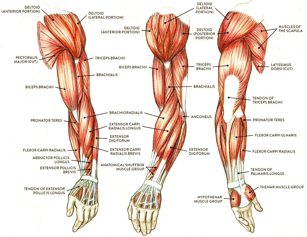
Flexor muscles bend the joints and are those most affected by contractures. The stiffening and tightening prevent the body parts from moving out and away. The most common include:
Wrist and Finger Flexors
Muscle groups that bend the wrist and fingers.
Biceps
The primary elbow flexor that bends the arm.
Gastrocnemius and Soleus
The calf muscles which allow the ankle to point the foot/plantarflexion.
Hamstrings
A group of three muscles behind the thigh that bend the knee.
Causes
The permanent shortening of muscle fibers and changes in muscle structure cause muscle contractures or stiffer-than-normal tissue that is difficult to stretch. Sarcomeres are structural units of muscles that cause fibers to contract.
With contractures, the sarcomeres overly lengthen when the muscle fibers tighten. This increase in sarcomere length prevents the muscle from contracting normally, resulting in weakness. Muscle fibers are encased in an extracellular matrix, a mesh composed of collagen and other proteins that help transmit force and provide muscle contraction. Muscle contractures cause the amount of collagen within the extracellular matrix to increase, causing a stiffening of fibers that restricts movement. (Lieber, R. L., and Fridén, J. 2019)
Muscle contractures also form from decreased satellite cells. Satellite cells are specialized stem cells that can rebuild muscle and are necessary for muscle regeneration and repair. Without the proper amount of satellite cells, other cells like fibroblasts significantly increase in the muscle tissue, causing the fibers to become stiff and fibrotic or more fibrous. These changes to the sarcomeres, collagen within the extracellular matrix, and decreased satellite cells all result from conditions in which neurological input to the brain and spinal cord muscles becomes reduced. This is caused by lack of use, injury, or neurological and neuromuscular conditions. (Lieber, R. L., and Fridén, J. 2019)
Cerebral Palsy
Contractures often occur from upper motor neuron lesions, which prevent signals from the brain and spinal cord from reaching the motor neurons that control muscle contraction. When these signals are weakened or blocked, muscles become stiff and weak from lack of stimulation. (Lieber, R. L., and Fridén, J. 2019)
Cerebral palsy is a group of disorders affecting mobility caused by an upper motor neuron lesion that is present at birth and is the most common motor disability in children. It causes:
Cognitive impairment
Decreased muscle strength
Problems with movement, coordination, and functional motions.
Because cerebral palsy prevents the muscles of the legs from being sufficiently stimulated, contractures commonly develop in the hips, knees, and ankles. Individuals can have a 75% decrease in satellite cells to repair muscle tissue and prevent muscle fibrosis or stiffening. Specific genes linked to collagen production are also altered, causing irregular changes to the extracellular matrix of muscles. (Lieber, R. L., and Fridén, J. 2019)
Muscular Dystrophy
Muscular dystrophy is a group of inherited neuromuscular disorders characterized by muscle weakness and wasting. Deficient nerve supply to muscles causes them to become stiff and tight, inhibiting the functional range of motion needed to move joints and activate muscles to move. Clinical research suggests that individuals with muscular dystrophy have decreased levels of satellite cells to repair, increasing the risk of developing muscle contracture. (Lieber, R. L., and Fridén, J. 2019)
Disuse-induced Muscle Wasting or Disuse Atrophy
When muscles are not used for some time because of hospitalization, prolonged bed rest, or immobilization from wearing braces, splints, or casts after injuries, the blood circulation and electrical signaling from nerves to muscles decreases. This results in weakness, increased muscle tightness and stiffness, and muscle wasting/atrophy. Over time, stiff and tight muscles can progress to contractures that become extremely difficult to stretch.
Trauma or Injury
Muscle or tendon injuries can cause contractures as scar tissue develops, joining muscle fibers and joints together. This can significantly restrict movement. Large burns can also cause skin, muscles, and joint contractures. The range of motion can become significantly limited, and the changes can become irreversible if not aggressively treated.
Other Causes
Other forms of upper motor neuron lesions that can cause contractures because of weak or blocked electrical input to muscles as a result of brain or spinal cord damage include:
Neuromuscular disorders like spinal muscular atrophy – SMA.
Conditions that cause inflammation and joint stiffening, like juvenile rheumatoid arthritis.
A history of diabetes also increases the risk of developing contractures affecting finger flexors, like Dupuytren’s contractures and stenosing tenosynovitis
or trigger finger. (Lieber, R. L., and Fridén, J. 2019)
Symptoms
Symptoms include:
Extremely stiff and tight muscles resistant to stretching.
Pain from the inability to stretch.
Loss of range of motion.
Impaired joint mobility.
Severe contractures can interfere with the functional range of motion needed to move joints to complete normal tasks and movements, such as standing up from a chair and walking.
Treatment
Physical Therapy
Physical therapies can help reduce the severity through stretching and soft tissue mobilization to decrease tightness. (Lieber, R. L., and Fridén, J. 2019)
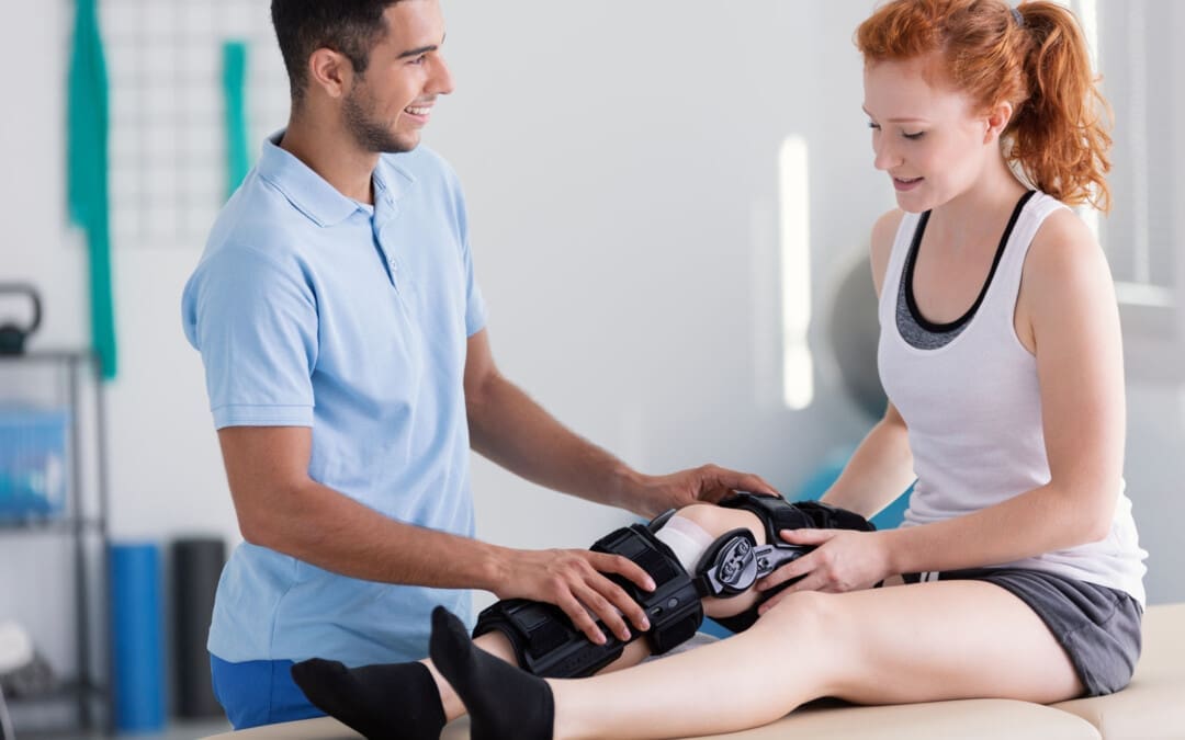
Specialized braces or splints can be custom-made to fit different body parts.
These provide a prolonged low-intensity stretch over a period of time to increase muscle length.
Once the muscle has stretched, a new brace or splint may be needed to adjust to the increased range of motion. (Lieber, R. L., and Fridén, J. 2019)
Surgery
In severe cases where muscle contractures limit the functional range of motion needed for activities of daily living or ADLs, surgical release of the contracted tissue may be recommended. This surgery can improve functional movements like walking, getting in and out of bed, and standing up from chairs. The tight muscles can be surgically cut, and the tendons can be lengthened to allow more mobility. (Lieber, R. L., and Fridén, J. 2019)
The causes of muscle contracture are not always avoidable, but various treatment options are available to help loosen up tight muscles and preserve or restore the range of motion. It’s important to move daily and stretch common areas like the fingers, arms, and legs to reduce the risk of muscle tightness and prevent contractures from developing. It is imperative to seek medical treatment for severe contractures resulting from neuromuscular disorders, including physical and occupational therapy, to prevent contractures from worsening and regaining as much functional range as possible.
Injury Medical Chiropractic and Functional Medicine Clinic uses an integrated approach personalized to the individual that focuses on what works for them and treats injuries and chronic pain syndromes through personalized care plans that improve ability through flexibility, mobility, and agility programs to relieve pain. Our providers use an integrated approach to create personalized care plans for each patient, including Functional Medicine, Acupuncture, Electro-Acupuncture, and Sports Medicine principles. Our goal is to relieve pain naturally by restoring health and function to the body. If other treatment is needed, Dr. Jimenez has teamed up with top surgeons, clinical specialists, medical researchers, and rehabilitation providers to provide the most effective treatments.
Chiropractic Treatment for Cerebral Palsy
References
Lieber, R. L., & Fridén, J. (2019). Muscle contracture and passive mechanics in cerebral palsy. Journal of applied physiology (Bethesda, Md. : 1985), 126(5), 1492–1501. https://doi.org/10.1152/japplphysiol.00278.2018
Can the thoracolumbar fascia cause or contribute to lower back pain and inflammation?
Thoracolumbar Fascia
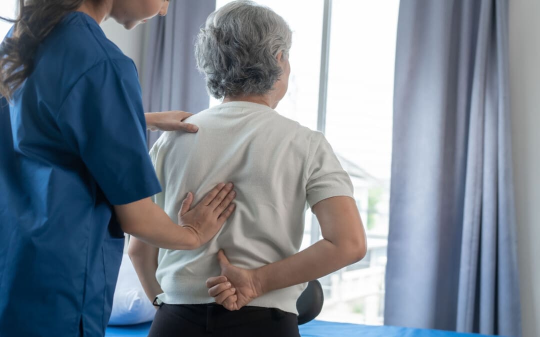
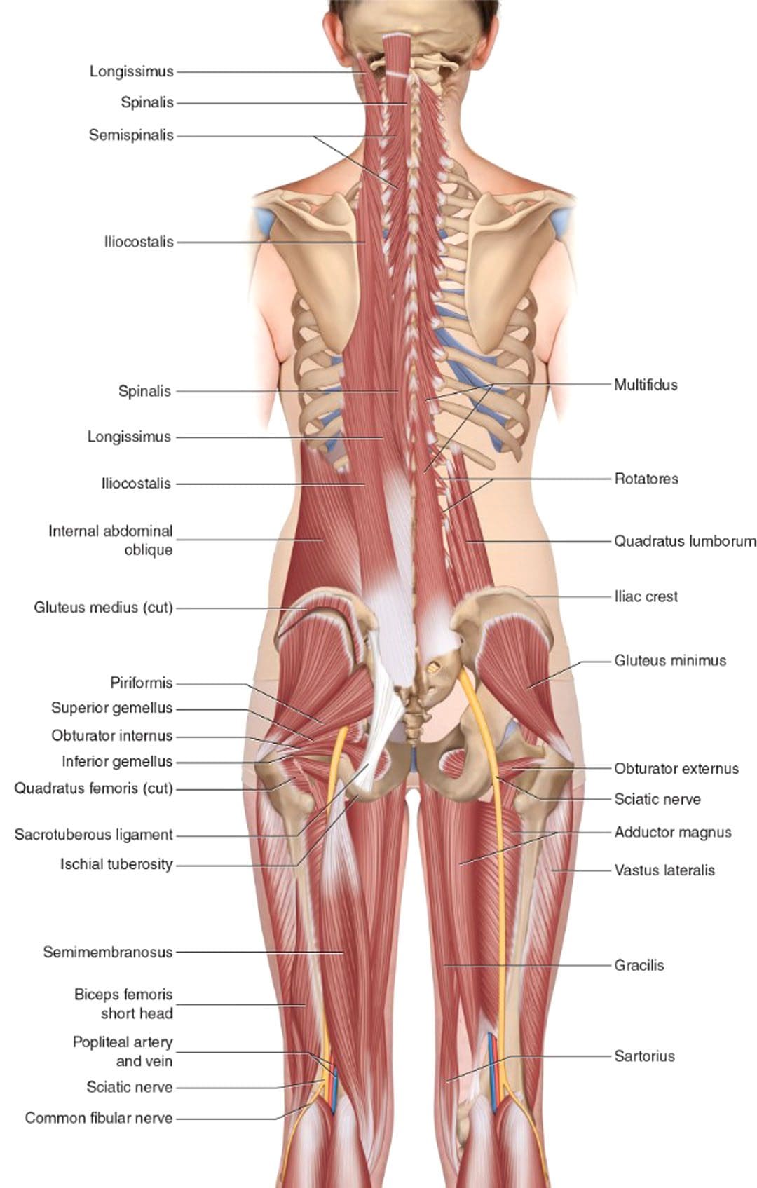
Tissue behind the spinal column, positioned at both the lower back and mid-back levels, is connected to the thoracolumbar fascia, also called the lumbodorsal fascia or LF. The fascia is a thick connective tissue that covers and supports all the body’s muscles, bones, tendons, ligaments, and organs. The fascia also contains nociceptive nerve endings, also known as free nerve endings, that arise from the central nervous system, i.e., the brain and spinal cord, which may be responsible for some forms of back pain and stiffness caused by injury or inflammation.
Anatomy
The thoracolumbar fascia is divided into three layers:
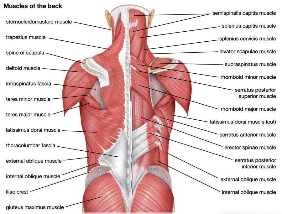
Many of the back muscles attach to the thoracolumbar fascia. The erector spinae muscle group, known as the paraspinals, runs longitudinally down the spine. They are attached to the thoracolumbar fascia and the bony spine. The lumbar part of the posterior layer of the thoracolumbar fascia extends from the lowest rib to the top of the hip bone or the iliac crest. On the same path, it connects with the transverse abdominal muscle. The thoracolumbar fascia connections help bridge the back muscles to the abdominal wall muscles. The latissimus dorsi, a large back muscle that bears and moves the body’s weight with the arms and shoulders, is also connected to the thoracolumbar fascia, with the fibers extending outward from the fascia. The front part of the thoracolumbar fascia, or anterior layer, covers a muscle called the quadratus lumborum. This muscle bends the trunk to the side, helps maintain a healthy posture, and is often focused on muscle-related lower back pain.
What the Fascia Does
The thoracolumbar fascia, examined from the back of an anatomical drawing or diagram, is diamond-shaped. Its shape, large size, and central location uniquely position it to unify and synchronize the upper body’s movements with the lower body’s. The fascia’s fibers are very strong, enabling the tissue sheath to lend support (Willard, F. H. et al., 2012) . The tissue is also flexible, enabling it to help circulate forces of movement and contralateral movements as the back muscles contract and relax. An example is walking.
Back Pain
Scientists and doctors don’t know for sure, but it’s possible that the thoracolumbar fascia may contribute to lower back pain. A study found that the fascia may generate back pain based on: (Wilke, J. et al., 2017)
Sustaining micro-injuries and/or inflammation, which are often related, may cause signal changes in the free nerve endings in the fascia. Nerve endings acquire information from the outer areas of the body, like skin and other fascia, and relay it back to the central nervous system. The theory is that when the fascia close to the skin becomes injured, damaged, and/or backed up with inflammatory chemicals and substances, it is communicated as pain and other sensations back to the brain and spinal cord.
After a back injury, tissues tighten and stiffen. Some studies of patients with back pain noted alterations in their thoracolumbar fascia.
Injuries tend to stimulate nerves, which can lead to increased sensitivity.
Injury Medical Chiropractic and Functional Medicine Clinic focuses on and treats injuries and chronic pain syndromes through personalized care plans that improve ability through flexibility, mobility, and agility programs to relieve pain. Our providers use an integrated approach to create personalized care plans for each patient, including Functional Medicine, Acupuncture, Electro-Acupuncture, and Sports Medicine principles. Our goal is to relieve pain naturally by restoring health and function to the body. If other treatment is needed, Dr. Jimenez has teamed up with top surgeons, clinical specialists, medical researchers, and rehabilitation providers to provide the most effective treatments.
Sciatica, Causes, Symptoms, and Tips
References
Willard, F. H., Vleeming, A., Schuenke, M. D., Danneels, L., & Schleip, R. (2012). The thoracolumbar fascia: anatomy, function and clinical considerations. Journal of anatomy, 221(6), 507–536. https://doi.org/10.1111/j.1469-7580.2012.01511.x
Wilke, J., Schleip, R., Klingler, W., & Stecco, C. (2017). The Lumbodorsal Fascia as a Potential Source of Low Back Pain: A Narrative Review. BioMed research international, 2017, 5349620. https://doi.org/10.1155/2017/5349620
IFM's Find A Practitioner tool is the largest referral network in Functional Medicine, created to help patients locate Functional Medicine practitioners anywhere in the world. IFM Certified Practitioners are listed first in the search results, given their extensive education in Functional Medicine

 The traction unit must be hung over a door secured to the top and closed.

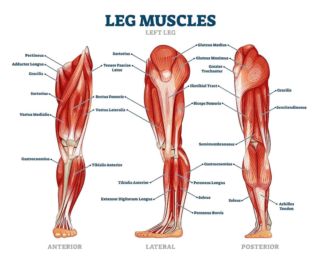
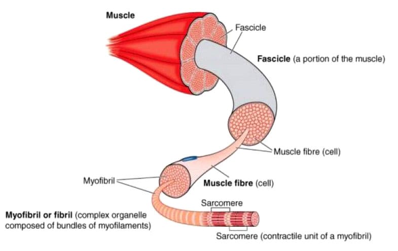 With contractures, the sarcomeres overly lengthen when the muscle fibers tighten. This increase in sarcomere length prevents the muscle from contracting normally, resulting in weakness. Muscle fibers are encased in an extracellular matrix, a mesh composed of collagen and other proteins that help transmit force and provide muscle contraction. Muscle contractures cause the amount of collagen within the extracellular matrix to increase, causing a stiffening of fibers that restricts movement. (
 What the Fascia Does









