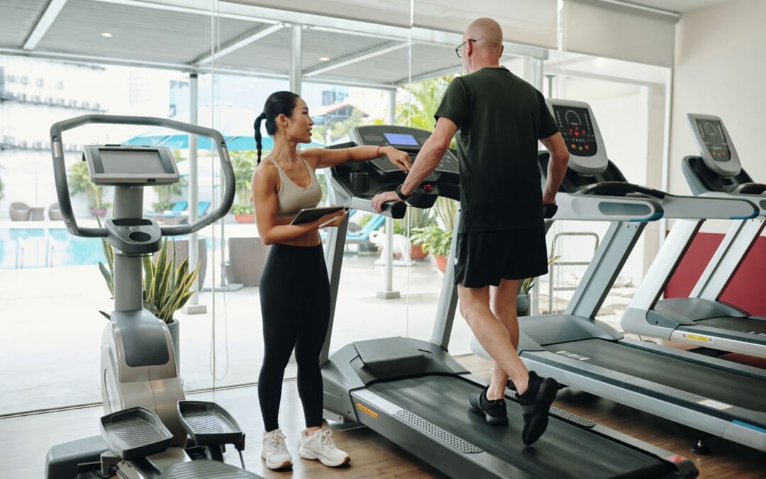
Can walking backward on a treadmill aid in treating conditions and injuries, improving strength, gait, and mobility in the lower extremities and knee, hip, and ankle range of motion (ROM)?
Walking Backward On A Treadmill
Walking backward is often used in physical therapy to treat multiple conditions and injuries and help improve strength, gait, balance, and mobility in the lower extremities. It is also used to improve knee, hip, and ankle range of motion and lower back pain and burn calories.
The Difference Backward vs. Forward
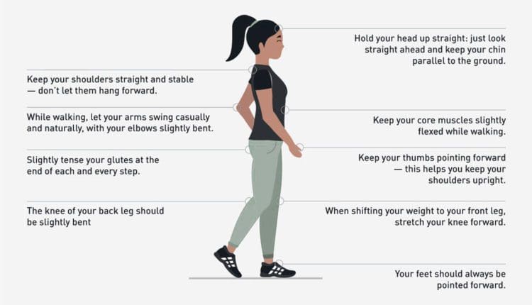
The difference from forward walking is that certain motions are normal gait characteristics.
First, the leg swings through the air, and the heel lands on the ground.
Then, the straight knee bends slightly as you roll from heel to toe.
As this occurs, the opposite leg rolls from the toes and up into the air.
This heel-to-toe motion repeats, and normal walking takes place.
Reverse walking creates the opposite gait process.
The leg swings through the air and reaches backward with a bent knee.
The toes contact the ground, and the bent knee straightens as you roll from toe to heel.
Then, the heel leaves the ground with the knee straight, repeating the process.
Reverse walking can help increase knee extension range of motion. (Zhang M. et al., 2022)
Individuals with knee injuries, surgeries, or arthritis may lose their knee extension or the knee’s ability to straighten.
The bent knee straightens fully when walking backward, moving from the toes onto the heel.
This helps to improve the range of motion into knee extension.
Quadriceps Function
Reverse walking can improve the function of the quadriceps muscles in the upper thighs. (Alghadir A. H. et al., 2019)
The quads are active when straightening the knee.
Exercises like quad sets, short arc quads, and straight leg raises increase quad strength.
Hamstring Flexibility
The hamstrings are located in the back of the upper thighs and work to bend the knees and extend the hips.
For individuals with tight hamstrings, a therapist may have you walk backward on the treadmill to improve the flexibility of the muscle group. (BBC, 2023)
When walking backward, the hamstring contracts to bend the knee as it swings through the air.
Then, the hamstring is stretched from the toe to heel movement, and the knee straightens.
Walking backward can help reset one’s gait and improve one’s ability to walk forward.
A study on stroke patients found that backward walking training was superior to standing balance training for improving balance and walking speed. (Rose, D. K. et al., 2018)
Burns Calories
Backward walking has been found to burn more calories than forward walking.
As a result, it improves heart and lung health better.
Conditions That Benefit
Individuals with a lower extremity impairment that results in loss of normal walking mobility may benefit from reverse walking. Common conditions that may help include: (BBC, 2023) (DelMastro H. M. et al., 2023)
Hamstring strain
Sciatica with tightness in hamstrings
Hip, knee or ankle surgery
Lower extremity fracture
Cerebral palsy
Multiple sclerosis
Parkinson’s disease
Stroke
A physical therapist will decide if an individual’s condition warrants reverse walking as part of a rehabilitation program.
Injury Medical Chiropractic and Functional Medicine Clinic
Injury Medical Chiropractic and Functional Medicine Clinic works with primary healthcare providers and specialists to develop an optimal health and wellness solution. We focus on what works for you to relieve pain, restore function, and prevent injury. Regarding musculoskeletal pain, specialists like chiropractors, acupuncturists, and massage therapists can help mitigate the pain through spinal adjustments that help the body realign itself. They can also work with other medical professionals to integrate a treatment plan to resolve musculoskeletal issues.
Control Foot Motion and Posture
References
Cha, H. G., Kim, T. H., & Kim, M. K. (2016). Therapeutic efficacy of walking backward and forward on a slope in normal adults. Journal of Physical Therapy Science, 28(6), 1901–1903. https://doi.org/10.1589/jpts.28.1901
Zhang, M., Pang, J., Lu, J., Kang, M., Chen, B., Jones, R. K., Zhan, H., & Liu, A. (2022). The Immediate Effect of Backward Walking on External Knee Adduction Moment in Healthy Individuals. Journal of Healthcare Engineering, 2022, 4232990. https://doi.org/10.1155/2022/4232990
Alghadir, A. H., Anwer, S., Sarkar, B., Paul, A. K., & Anwar, D. (2019). Effect of 6-week retro or forward walking program on pain, functional disability, quadriceps muscle strength, and performance in individuals with knee osteoarthritis: a randomized controlled trial (retro-walking trial). BMC musculoskeletal disorders, 20(1), 159. https://doi.org/10.1186/s12891-019-2537-9
BBC. (2023). Why walking backwards can be good for your health and brain. https://www.bbc.com/future/article/20231110-why-walking-backwards-can-be-good-for-your-health-and-brain
Balasukumaran, T., Olivier, B., & Ntsiea, M. V. (2019). The effectiveness of backward walking as a treatment for people with gait impairments: a systematic review and meta-analysis. Clinical Rehabilitation, 33(2), 171–182. https://doi.org/10.1177/0269215518801430
Rose, D. K., DeMark, L., Fox, E. J., Clark, D. J., & Wludyka, P. (2018). A Backward Walking Training Program to Improve Balance and Mobility in Acute Stroke: A Pilot Randomized Controlled Trial. Journal of Neurologic Physical Therapy: JNPT, 42(1), 12–21. https://doi.org/10.1097/NPT.0000000000000210
Thomas, K. S., Hammond, M., & Magal, M. (2018). Graded forward and backward walking at a matched intensity on cardiorespiratory responses and postural control. Gait & posture, 65, 20–25. https://doi.org/10.1016/j.gaitpost.2018.06.168
DelMastro, H. M., Ruiz, J. A., Simaitis, L. B., Gromisch, E. S., Neto, L. O., Cohen, E. T., Wong, E., Krug, R. J., & Lo, A. C. (2023). Effect of Backward and Forward Walking on Lower Limb Strength, Balance, and Gait in Multiple Sclerosis: A Randomized Feasibility Trial. International journal of MS care, 25(2), 45–50. https://doi.org/10.7224/1537-2073.2022-010
Can the straight leg test help find the cause of back or hamstring pain in individuals experiencing it?
Straight Leg Test
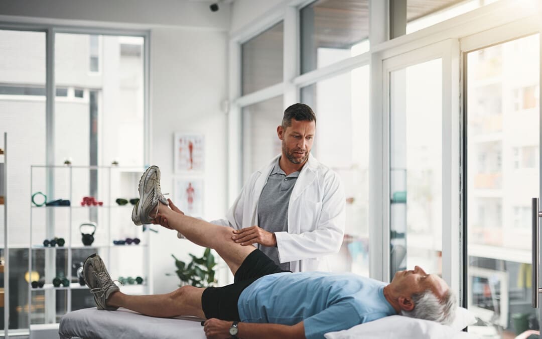
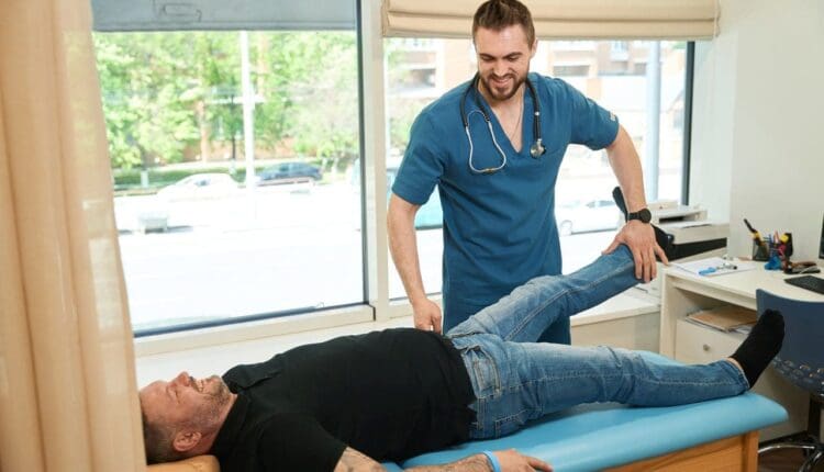
The straight leg raise test is often used to diagnose sciatica/radiculopathy, herniated discs, and other spinal problems. The healthcare provider giving the test performs most of the movement as they assess what’s causing the leg and/or back pain. The patient lies on their back with the legs straight. The provider will have the patient perform specific movements and inform them of how it feels. Then, they’ll raise the leg to see if and at what point symptoms begin to show. Providers often use this test alongside imaging studies.
Some studies suggest the straight leg test helps diagnose sciatica and other causes. (Pesonen J. et al., 2021)
Purpose
The straight leg raise is one of the most common manual tests done during physical exams. The straight leg raise test seeks to reproduce the pain or other symptoms in a controlled fashion to provide clues to what’s happening. It is a manual exam, and the healthcare provider will:
Position the patient
Moves the patient
Create pressure to see how well the patient can resist it
This is often used alongside imaging tests, such as an X-ray or CT scan. (Allegri M. et al., 2016) Its goal is to check for nerve movement and sensitivity of nerve tissue to compression. The straight leg lift test is neurodynamic because it uses movement to diagnose nerve problems. (Baselgia L.T. et al., 2017)
During the Test
Expect to feel some pain during the test, as the whole point is to see what aggravates the symptoms. They may be caused by:
Most of the tests are passive, with the provider doing the lifting. The patient can help achieve the most accurate result by staying as relaxed as possible and being clear about what is felt. (Pande K. 2015) The procedure:
The patient lies on their back with their legs straight.
The provider will ask the patient to turn one of the legs in.
This tells them what hip position affects the lower back symptoms.
They’ll then ask you to bring the leg toward the body’s center.
Then, they’ll lift the straight leg until the patient experiences symptoms.
Pain suggests a herniated disc.
If there is no pain, this also provides valuable information.
The procedure is repeated with the other leg.
Modifications
It’s important to let the examiner know about any limitations. The straight leg raise test has modifications if the patient cannot lift their leg while it’s straight or if they have difficulty lying on their back, which can also help avoid an injury during the test.
Variations
The healthcare provider may repeat the test with the ankle in a dorsiflexed position/raising the foot. Then, they’ll have the patient do it with their chin tucked into their chest. (Young R. et al., 2013) These variations can help check for nerve involvement in specific locations, such as the spinal cord or the dura mater, the membrane covering the brain and spinal cord. (Venne G. et al., 2017) The spinal cord nerves are likely involved and affected if the usual pain is in the back or leg but not the chin, neck, or foot. (Camino Willhuber GO, Piuzzi NS. 2023)
Injury Medical Chiropractic and Functional Medicine Clinic
Injury Medical Chiropractic and Functional Medicine Clinic works with primary healthcare providers and specialists to build optimal health and wellness solutions. We focus on what works for you to relieve pain, restore function, prevent injury, and mitigate issues through adjustments that help the body realign itself. The clinic can also work with other medical professionals to integrate a treatment plan to resolve musculoskeletal problems.
From Injury to Recovery with Chiropractic Care
References
Casiano, V. E., Sarwan, G., Dydyk, A. M., & Varacallo, M. A. (2025). Back Pain. In StatPearls. https://www.ncbi.nlm.nih.gov/pubmed/30844200
Pesonen, J., Shacklock, M., Suomalainen, J. S., Karttunen, L., Mäki, J., Airaksinen, O., & Rade, M. (2021). Extending the straight leg raise test for improved clinical evaluation of sciatica: validity and diagnostic performance with reference to the magnetic resonance imaging. BMC musculoskeletal disorders, 22(1), 808. https://doi.org/10.1186/s12891-021-04649-z
Allegri, M., Montella, S., Salici, F., Valente, A., Marchesini, M., Compagnone, C., Baciarello, M., Manferdini, M. E., & Fanelli, G. (2016). Mechanisms of low back pain: a guide for diagnosis and therapy. F1000Research, 5, F1000 Faculty Rev-1530. https://doi.org/10.12688/f1000research.8105.2
Baselgia, L. T., Bennett, D. L., Silbiger, R. M., & Schmid, A. B. (2017). Negative Neurodynamic Tests Do Not Exclude Neural Dysfunction in Patients With Entrapment Neuropathies. Archives of physical medicine and rehabilitation, 98(3), 480–486. https://doi.org/10.1016/j.apmr.2016.06.019
Pande K. (2015). The Use of Passive Straight Leg Raising Test: A Survey of Clinicians. Malaysian Orthopaedic Journal, 9(3), 44–48. https://doi.org/10.5704/MOJ.1511.012
Young, R., Nix, S., Wholohan, A., Bradhurst, R., & Reed, L. (2013). Interventions for increasing ankle joint dorsiflexion: a systematic review and meta-analysis. Journal of foot and ankle research, 6(1), 46. https://doi.org/10.1186/1757-1146-6-46
Venne, G., Rasquinha, B. J., Kunz, M., & Ellis, R. E. (2017). Rectus Capitis Posterior Minor: Histological and Biomechanical Links to the Spinal Dura Mater. Spine, 42(8), E466–E473. https://doi.org/10.1097/BRS.0000000000001867
Camino Willhuber, G. O., & Piuzzi, N. S. (2025). Straight Leg Raise Test. In StatPearls. https://www.ncbi.nlm.nih.gov/pubmed/30969539
For individuals looking to improve sleep, could weight training be the answer to increase healthy sleep?
Weight Resistance Training
Researchers found that resistance weight training improves sleep quality, among other methods like diet and sleep hygiene practices. Resistance training may improve sleep by reducing anxiety and stress, benefiting heart health, and promoting muscle recovery. Researchers examined non-medication methods of improving sleep in younger and middle-aged adults, comparing exercise, diet, and sleep hygiene practices. The findings showed that for adults under 65, resistance training is the best non-pharmacological way to improve sleep quality. (Hirohama, K. et al., 2024) These findings reinforce that sleep and exercise lead to improved restful sleep. Resistance training is highly effective compared to aerobic exercise (running or cycling) in improving sleep quality. The research shows that resistance exercise has more profound benefits than aerobic training alone, particularly in enhancing sleep quality and reducing insomnia symptoms.
Resistance Training and Improved Sleep
Resistance training, also called strength training or weight training, is intended to build muscle and strength. This can include:
Your body weight
Resistance bands
Free weights
Weight-lifting machines
The mechanisms by which exercise alters sleep are unknown. However, the researchers point out that weight training may improve sleep by improving mental health issues such as anxiety and depression. (Cunningham J. E. A., & Shapiro C. M. 2018) (Carbone E. A. et al., 2023) Other research on resistance training and sleep found that resistance training helped decrease anxiety and stress. (Alley J. R. et al., 2015) Resistance training can benefit heart health by increasing blood circulation. This means the heart’s vessels don’t have to work as hard during sleep, when blood pressure should naturally drop, a phenomenon known as nocturnal dipping, which is beneficial for individuals with high blood pressure.
Another reason this training may help with sleep is its impact on the body. Previous research on resistance training and sleep suggests that, because weight training stresses muscle tissue, it may signal the brain that sleep is needed to repair the damage (Iowa State University, 2022). The hormone adenosine, which helps promote sleep, is a key factor in why exercise increases adenosine levels and helps the body doze off. (Roig M. et al., 2022)
How Much Resistance Training Is Needed?
Sleep doctors advise patients to perform some form of daily movement or exercise to improve sleep. The amount of resistance training an individual needs depends on their circumstances, such as underlying conditions, injuries, fitness level, physical ability, and time.
The guidelines for physical activity from the Department of Health and Human Services recommend that adults do a muscle-strengthening activity at least 2 days each week, in addition to at least 150 to 300 minutes of moderate-intensity aerobic activity.
A meta-analysis found that a 55-minute resistance training session, consisting of three sets of 10-12 exercises, three times a week, positively affected sleep quality.
They added that higher-intensity and more frequent training could have a greater effect on sleep.
In other research on exercise and sleep, study participants were grouped into resistance training, aerobic exercise, and combination workouts (both resistance and aerobic exercise). Each group participated in 60-minute training sessions three times a week, with the combination group doing 30 minutes of resistance training and 30 minutes of aerobic exercise per session. (Iowa State University, 2022) Resistance training increased sleep by an average of 40 minutes compared to about 23 minutes for those who did an hour of aerobic exercise. A combination of aerobic and resistance increased sleep by about 17 minutes. (American Heart Association, 2022)
Previous research on resistance training and sleep found that exercise at any time can improve sleep quality compared with no strength training. However, evening sessions tend to help individuals sleep a little better, particularly for those who struggle with insomnia or have irregular sleep patterns. (Alley J. R. et al., 2015) Resistance exercise may offer benefits regarding the ability to fall asleep and stay asleep for those with osteoporosis, sarcopenia, anxiety, or depression, as it can improve overall physical health and reduce symptoms associated with these conditions, leading to better sleep quality.
Strength Workouts
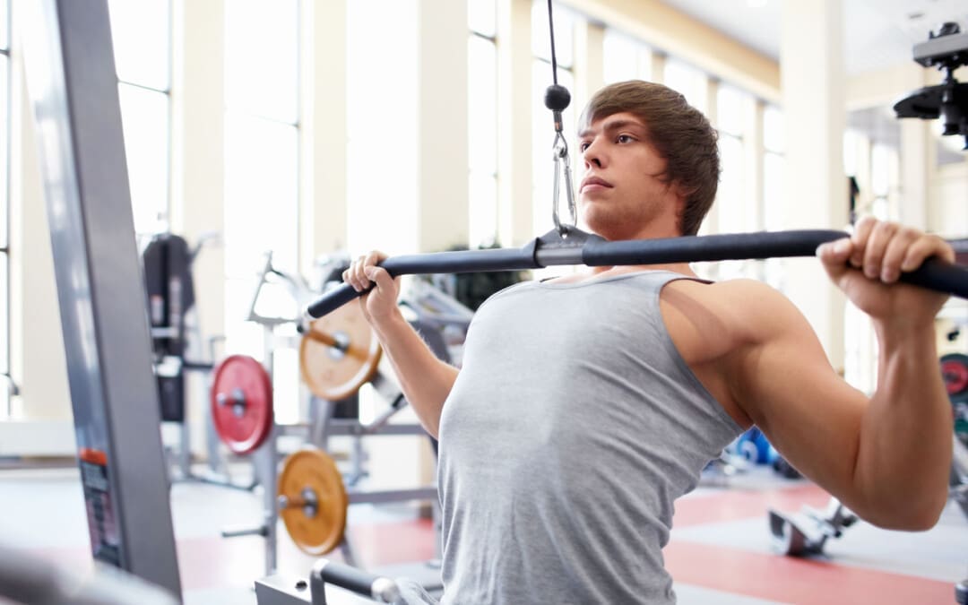
The goal is resistance training that targets all major muscle groups. The Iowa State University research had participants use 12 resistance machines to perform exercises, including:
Leg presses
Chest presses
Lat pulldowns
Bicep curls
Abdominal crunches
Participants performed three sets of 8-12 reps at 50%-80% of their one-rep max. (Iowa State University, 2022)
In one study, people worked out for 30 minutes on nine different resistance machines, doing three sets of 10 reps on each machine. (Alley J. R. et al., 2015)
However, individuals can also use dumbbells for bicep curls or chest presses, barbells for deadlifts or back squats, resistance bands for lateral shoulder raises or leg extensions, or their body weight for movements such as chin-ups or push-ups.
Other Ways to Improve Sleep Without Meds
Other non-drug methods to improve sleep include a healthy diet, proper hydration, dietary changes, and stress management, such as meditation. Aerobic exercise is known to improve both sleep quantity and quality. (Kovacevic A. et al., 2018) Lifestyle behaviors outside of exercise are also important for sleep. Sleep hygiene refers to having healthy sleep habits and behaviors at night. This includes optimizing pre-bedtime routines to prepare the mind and body for sleep and improving bedroom environments to ensure sleep support, meaning it is cool, dark, quiet, and free from distractions like phones. Another important method of improving sleep is to listen to your body and not force yourself to stay up late when exhausted. Pushing the brain and body to stay up later leads to chronic sleep debt. Eating sugary and high-fat foods before bed can hinder sleep patterns. Drinking caffeine after dinner or close to bedtime can disrupt the sleep cycle, and alcohol consumption before bed can hurt sleep quality.
Injury Medical Chiropractic and Functional Medicine Clinic
Individuals need rest time to adjust to working out, so consult a doctor on the balance of rest and training. Injury Medical Chiropractic and Functional Medicine Clinic works with primary healthcare providers and specialists to develop an optimal health and wellness solution. We focus on what works for you to relieve pain, restore function, and prevent injury. Regarding musculoskeletal pain, specialists such as chiropractors, acupuncturists, and massage therapists can help mitigate it through spinal adjustments that help the body realign itself. They can also work with other medical professionals, such as physical therapists and orthopedic doctors, to integrate a comprehensive treatment plan that addresses the underlying causes and symptoms to effectively resolve musculoskeletal issues.
Weight Loss Techniques
References
Hirohama, K., Imura, T., Hori, T., Deguchi, N., Mitsutake, T., & Tanaka, R. (2024). The effects of nonpharmacological sleep hygiene on sleep quality in nonelderly individuals: A systematic review and network meta-analysis of randomized controlled trials. PloS one, 19(6), e0301616. https://doi.org/10.1371/journal.pone.0301616
Cunningham, J. E. A., & Shapiro, C. M. (2018). Cognitive Behavioural Therapy for Insomnia (CBT-I) to treat depression: A systematic review. Journal of psychosomatic research, 106, 1–12. https://doi.org/10.1016/j.jpsychores.2017.12.012
Carbone, E. A., Menculini, G., de Filippis, R., D’Angelo, M., De Fazio, P., Tortorella, A., & Steardo, L., Jr (2023). Sleep Disturbances in Generalized Anxiety Disorder: The Role of Calcium Homeostasis Imbalance. International journal of environmental research and public health, 20(5), 4431. https://doi.org/10.3390/ijerph20054431
Alley, J. R., Mazzochi, J. W., Smith, C. J., Morris, D. M., & Collier, S. R. (2015). Effects of resistance exercise timing on sleep architecture and nocturnal blood pressure. Journal of strength and conditioning research, 29(5), 1378–1385. https://doi.org/10.1519/JSC.0000000000000750
Iowa State University. (2022). Pumping iron may improve sleep more than cardio workouts. https://www.news.iastate.edu/news/2022/03/03/resistance-exercise-sleep
Roig, M., Cristini, J., Parwanta, Z., Ayotte, B., Rodrigues, L., de Las Heras, B., Nepveu, J. F., Huber, R., Carrier, J., Steib, S., Youngstedt, S. D., & Wright, D. L. (2022). Exercising the Sleepy-ing Brain: Exercise, Sleep, and Sleep Loss on Memory. Exercise and Sport Sciences Reviews, 50(1), 38–48. https://doi.org/10.1249/JES.0000000000000273
American Heart Association. (2022). Resistance exercise may improve sleep more than aerobic exercise. https://www.heart.org/en/news/2022/03/03/resistance-exercise-may-improve-sleep-more-than-aerobic-exercise#:~:text=The%20results%20are%20considered%20preliminary,at%20night%2C%22%20Brellenthin%20said.
Kovacevic, A., Mavros, Y., Heisz, J. J., & Fiatarone Singh, M. A. (2018). The effect of resistance exercise on sleep: A systematic review of randomized controlled trials. Sleep medicine reviews, 39, 52–68. https://doi.org/10.1016/j.smrv.2017.07.002
Can physical therapy help individuals with a pinched nerve in the neck?
Pinched Nerve In The Neck
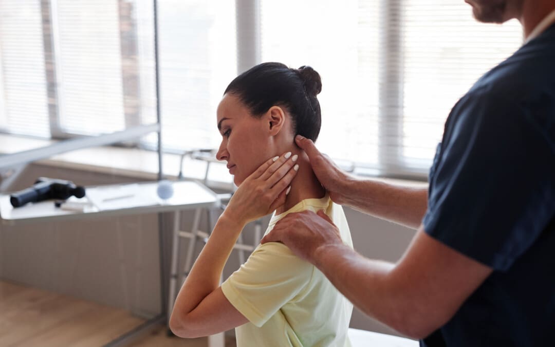
A pinched nerve in the neck can cause pain, numbness, and weakness that extends through the neck, shoulder, and arm. (American Academy of Orthopaedic Surgeons, 2024) Over-the-counter pain medications, resting the muscles, and gentle stretching can help. However, if you’re still in pain after a few days, see a healthcare provider. Common treatments include:
Rest
Over-the-counter (OTC) pain meds
Physical therapy
Steroid injections
Wearing a neck collar
Surgery is rarely needed, but it can provide relief if other treatments don’t help. Most of the time, a pinched nerve resolves within days or weeks.
Sharp pain that extends into the shoulder and arm.
The pain worsens, or there is a shooting sensation when turning the head.
Tingling or feeling of pins-and-needles in the fingers or hand.
Weakness in the arm, shoulder, or hand.
Numbness or loss of feeling.
Often, these symptoms occur only on one side. Some find their pain less when they lift their hand onto their head, which can relieve pressure on the nerve.
Causes
The cervical spine is the spinal cord area around the neck. It’s made up of seven vertebrae. Nerves branch off the spinal cords in the spaces between the vertebrae. Nerve compression occurs when the space between two vertebrae is reduced, putting pressure on the nerve, pinching it, and causing pain. Pinched nerves develop from age because the spinal discs between the vertebrae become compressed over time. Age causes about 70% to 80% of nerve compression. Other factors that cause pinched nerves include: (Harvard Health Publishing, 2021)
Degenerative disc disease
A herniated disc
Injuries like car accidents falls, or other trauma to the spine
Find a comfortable position and try to allow the muscles in your neck to relax and rest.
Heat or Ice
Warmth and coolness can relieve pain and inflammation.
Use a warm or cool compress for 15 minutes at a time.
Over-The-Counter Pain Medications
Pain medications, including nonsteroidal anti-inflammatory drugs (NSAIDs), can help bring relief.
Treatment for Severe Symptoms
If pain doesn’t resolve within a few days, or if it is so bad that you can’t go about daily activities, it’s recommended to see a healthcare provider. They can diagnose a pinched nerve after a physical exam and may also recommend imaging, including an X-ray, CT scan, MRI, or EMG, to reveal what’s causing the symptoms. After diagnosing the condition, the healthcare provider will develop a personalized treatment plan, which may include the following (Harvard Health Publishing, 2021)
Physical Therapy
Physical therapy can help build strength and flexibility in the neck.
This is especially important if there is frequent nerve pain in the same spot.
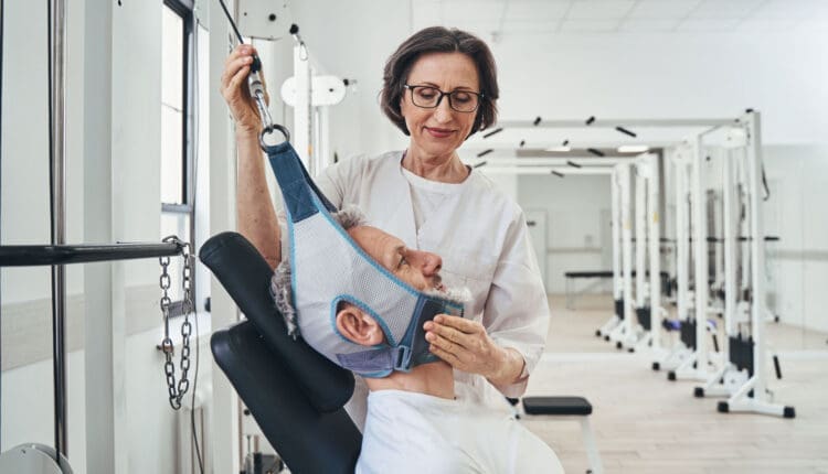
Cervical Collar
A soft cervical collar is a brace that fits around the neck.
It supports your head so the neck muscles can relax, facilitating healing.
The collar can also keep the head from turning in painful ways.
Oral Corticosteroids
Oral steroids like prednisone can help reduce inflammation.
If inflammation or swelling in the neck puts more pressure on the nerve, they can help.
Steroid Injections
Steroid shots right into the painful tissue reduce inflammation right away.
Muscle Relaxers
These medications keep the muscles in the neck from seizing up.
As the muscles relax, this brings pain relief.
Narcotic Pain Medications
Narcotic pain medications can be used short-term by individuals who have severe pain.
A healthcare provider will inform the patient of the benefits and drawbacks of these medications, which include opiates.
Hold for 20 seconds, then return to a neutral position.
Do this five times.
Eyes to Sky
Lean your head back and look toward the sky.
Hold for 20 seconds, then return to your starting position.
Do this five times.
Side to Side
Turn your head to the right as far as possible, bringing your chin in line with your shoulder.
Hold for 20 seconds, then turn as far as possible toward the left.
Repeat four times.
Ear to Shoulder
Bring your ear down toward your shoulder.
Hold for 20 seconds, then repeat the exercise on the other side.
Alternate between the right and left, stretching each side five times.
While it’s normal for exercises to hurt from stretching the muscles, they should never hurt more than a five on a pain scale of 1 to 10. If they do, stop exercising (National Health Service, 2025)
Healing Time
Healing and recovery depend on the severity of the injury. Some individuals find that the pain from a pinched nerve goes away in days, while for others, it can last for weeks. The pain goes away and then returns. If pain doesn’t go away with the conservative treatments or lasts more than a few days, talk with a healthcare provider or return for a second visit. Rarely do individuals need surgery to bring pain relief. The healthcare provider will discuss whether surgery is the best option and what to expect regarding pain relief. (American Academy of Orthopaedic Surgeons, 2024)
Injury Medical Chiropractic and Functional Medicine Clinic
Injury Medical Chiropractic and Functional Medicine Clinic works with primary healthcare providers and specialists to build optimal health and wellness solutions. We focus on what works for you to relieve pain, restore function, prevent injury, and mitigate issues through adjustments that help the body realign itself. The clinic can also work with other medical professionals to integrate a treatment plan to resolve musculoskeletal problems.
Revitalize and Rebuild with Chiropractic
References
American Academy of Orthopaedic Surgeons. OrthoInfo. (2024). Cervical radiculopathy (pinched nerve). https://orthoinfo.aaos.org/en/diseases–conditions/cervical-radiculopathy-pinched-nerve/
Harvard Health Publishing. Publishing, H. H. (2021). Treating a pinched nerve. https://www.health.harvard.edu/pain/treating-a-pinched-nerve
National Institute of Neurological Disorders and Stroke. (2025). Pinched Nerve Definition. Retrieved from https://www.ninds.nih.gov/health-information/disorders/glossary-neurological-terms#-P-
National Health Service. Service, N. H. (2025). Exercises for neck problems. https://www.nhsinform.scot/illnesses-and-conditions/muscle-bone-and-joints/neck-and-back-problems-and-conditions/exercises-for-neck-problems
Individuals who have fractured their scaphoid bone may experience pain and swelling in the wrist just below the thumb. Can immobilization with a cast and physical therapy help?
Scaphoid Fracture
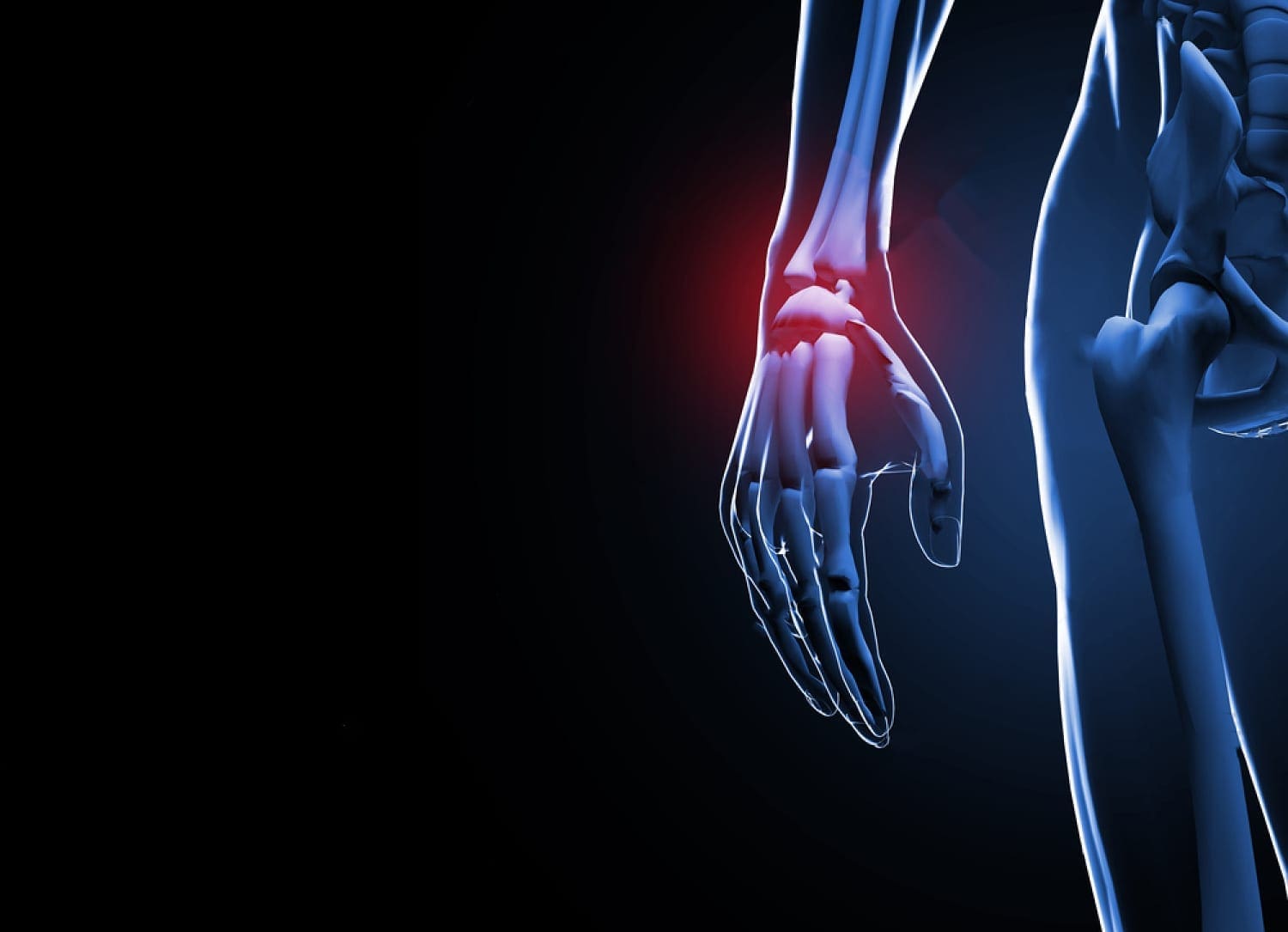
A scaphoid fracture is a break in one of the wrist’s small or carpal bones. This type of fracture occurs most often after a fall onto an outstretched hand. Symptoms typically include swelling and pain in the wrist just below the base of the thumb. These fractures can be difficult to diagnose since they don’t always appear on an X-ray. If the X-ray is negative and the healthcare provider suspects a scaphoid fracture, an MRI may be necessary. Surgery may be required in more severe cases or when the injury is not healing correctly. (American Academy of Orthopaedic Surgeons, 2023)
A Break In The – Navicular Bone
The scaphoid is one of eight carpal bones in the wrist. It is located just below the thumb’s base and is shaped like a kidney bean. This bone can be identified by holding a thumbs-up position and feeling for the hollow between the two tendons below your thumb. The scaphoid is located at the base of the hollow. A break in the scaphoid bone most commonly occurs in the middle of the bone but can also happen at either end. A scaphoid fracture can be displaced or non-displaced (American Academy of Orthopaedic Surgeons, 2023)
Displaced Fracture
It is when the bone fragments have moved out of alignment.
Non-displaced Fracture
It is when the fragments are still in their normal location in the hand.
The scaphoid’s blood supply comes from a small vessel that enters the most distant part of the bone and flows back through the bone. Because of this one small blood supply, a fracture in the center can stop the circulation to the proximal portion of the bone. Because of this, scaphoid fractures need immediate diagnosis and treatment.
Symptoms
Pain or deep aching on the thumb-side of the wrist, typically after a fall on an outstretched arm, could be a scaphoid fracture. Other symptoms experienced include: (American Academy of Orthopaedic Surgeons, 2023)
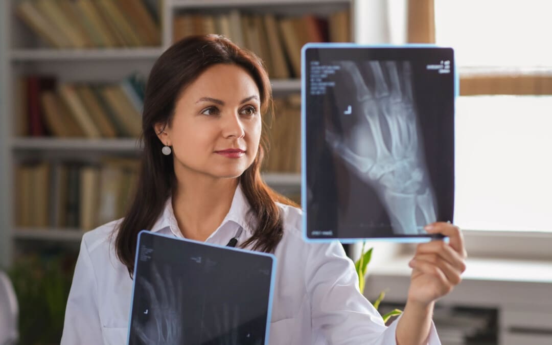
A healthcare provider will evaluate the hand for tenderness and pain in the hollow and/or the bone. If a break is suspected, they will order an X-ray. (Clementson M., Björkman A., & Thomsen N. O. B. 2020) Many patients are diagnosed with a wrist sprain when they have a fracture. Diagnosis can be difficult because the fracture often doesn’t appear on X-rays until weeks after the healing process starts. Physicians commonly treat a wrist injury as a scaphoid fracture initially and then repeat X-rays within two weeks. (American Academy of Orthopaedic Surgeons, 2023) If the injury doesn’t show on an X-ray, the provider may order an MRI, as these fractures can be easier to see on an MRI. An MRI can help ensure appropriate treatment immediately. (Wong S. B. S., & Peh W. C. G. 2019)
Treatment
If a wrist fracture is diagnosed, the wrist will be immobilized in a cast. However, a healthcare provider may also put the wrist in a cast if the X-ray is negative but they suspect a fracture. This will stabilize the injury until an MRI can be performed. With immobilization and follow-up treatment, scaphoid fractures often heal without surgery. Repeat X-rays are taken over several weeks or months so the provider can make sure the injury is healing correctly. If it is not healing correctly, surgery may be recommended. (Clementson M., Björkman A., & Thomsen N. O. B. 2020) If the fracture is displaced, healing correctly may be a challenge. In this case, a physician may recommend initial surgery to reposition the bones. (Clementson M., Björkman A., & Thomsen N. O. B. 2020) This type of surgery involves pinning the bone in place with screws.
Rehabilitation is an important part of healing because immobilization takes a long time. Wrist range-of-motion exercises can be started, followed by strengthening exercises for the wrist flexors and extensors. Supination, pronation, and grip exercises are also part of physical therapy.
This condition causes degeneration of the cartilage in the joint.
Avascular Necrosis
This is when the blood supply to the bone is reduced or cut off, causing the bone to die.
Injury Medical Chiropractic and Functional Medicine Clinic
Injury Medical Chiropractic and Functional Medicine Clinic works with primary healthcare providers and specialists to build optimal health and wellness solutions. We focus on what works for you to relieve pain, restore function, prevent injury, and mitigate issues through adjustments that help the body realign itself. The clinic can also work with other medical professionals to integrate a treatment plan to resolve musculoskeletal problems.
Skateboarding Injury Treatment
References
American Academy of Orthopaedic Surgeons. (2023). Scaphoid fracture of the wrist. https://orthoinfo.aaos.org/en/diseases–conditions/scaphoid-fracture-of-the-wrist
Clementson, M., Björkman, A., & Thomsen, N. O. B. (2020). Acute scaphoid fractures: guidelines for diagnosis and treatment. EFORT open reviews, 5(2), 96–103. https://doi.org/10.1302/2058-5241.5.190025
Wong, S. B. S., & Peh, W. C. G. (2019). The role of magnetic resonance imaging in the evaluation of scaphoid fractures. Journal of Medical Radiation Sciences, 66(1), 3–4. https://doi.org/10.1002/jmrs.316
Almigdad, A., Al-Zoubi, A., Mustafa, A., Al-Qasaimeh, M., Azzam, E., Mestarihi, S., Khair, Y., & Almanasier, G. (2024). A review of scaphoid fracture, treatment outcomes, and consequences. International orthopaedics, 48(2), 529–536. https://doi.org/10.1007/s00264-023-06014-2
Are individuals with high foot arches or participating in sports involving repetitive ankle motion at risk for developing peroneal tendon injuries?
Peroneal Tendon Injuries
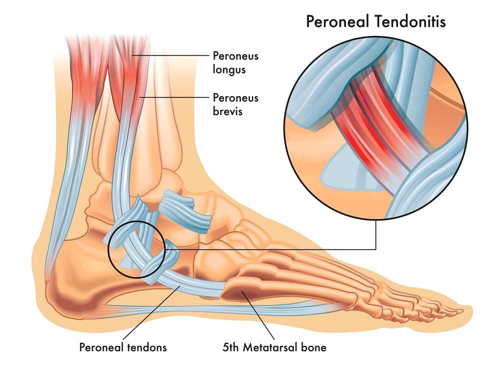
The peroneal tendons connect the muscles of the outer side of the lower leg to the foot. They may be acute—occurring suddenly—or chronic—developing over time. The basic types of peroneal tendon injuries are tendonitis, tears, and subluxation.
Anatomy and Function
The two major peroneal muscles (peroneus longus and peroneus brevis) are outside the lower leg, next to the calf muscles. The peroneal tendons run along the outer side of the ankle and attach to the foot, connecting these muscles to bone. They help stabilize the ankle joint, point the foot downward (plantarflexion), and turn the foot outward (eversion). In normal gait, the motion of the peroneal muscles is balanced by the muscles that invert the foot or rock the foot inward from the ankle. The two peroneal tendons sit one on top of the other right behind the fibula (the smaller lower leg bone). This closeness can contribute to problems with the peroneal tendons, as they rub together behind the ankle.
Tendonitis
The most common problem is inflammation or tendonitis. The tendons are usually inflamed just behind the fibula bone at the ankle joint. This part of the fibula is the bump on the outside of the ankle, and the peroneal tendons are located just behind that bony prominence. Tendonitis can either result from repetitive overuse or an acute injury. Common symptoms of tendonitis include:
Pain behind the ankle
Swelling over the peroneal tendons
Tenderness of the tendons
Pain usually worsens if the foot is pulled down and inwards, stretching the peroneal tendons.
Typical treatment of peroneal tendonitis is accomplished by:
Ice application
Applying ice to the area can help reduce swelling and control pain.
Rest
Resting is important to allow the tendon to heal.
A supportive device (walking boot or ankle brace) or crutches can help in severe cases.
Braces and boots provide support, reduce tendons’ stress, and allow rest and inflammation to subside.
Anti-inflammatory Medications
Motrin or Aleve are anti-inflammatory and can reduce the swelling around the tendon.
Physical Therapy
Physical therapy can help restore normal ankle joint mechanics, help with swelling and pain relief, and correct strength imbalances.
Cortisone Injections
Cortisone injections are low-risk if administered to the area around the tendon and not more often than every three months.
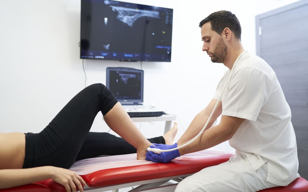
Ultrasound guidance can help ensure the medication is injected into the correct area. (Walt J. & Massey P. 2023)
Tendon Tears
Tears can occur and are more likely to happen in the peroneus brevis tendon. Tears are believed to be the result of two issues with the tendon. One is the blood supply. Tears of the peroneus brevis tendon almost always occur in the area where the blood supply and nutrition of the tendon are the poorest.
The second issue is the closeness between the two tendons, causing the peroneus brevis tendon to be wedged between the peroneus longus tendon and the bone. (Saxena A., & Bareither D. 2001) Tears of the peroneus brevis tendon are often treated with the same treatments for tendonitis. About half of the tears diagnosed by imaging are found to be asymptomatic. For individuals who don’t find lasting relief from symptoms, surgery may be necessary. Surgical options for peroneal tendon tears (Dombek M. F. et al., 2001)
Tendon Debridement and Repair
During a tendon debridement, the damaged tendon and the surrounding inflammatory tissue are removed.
The tear can be repaired, and the tendon can be tubularized to restore its normal shape.
Tenodesis
A tenodesis is a procedure where the damaged area of the tendon is sewn to the normal tendon.
In this case, the damaged segment of the peroneal tendon is removed, and the ends left behind are sewn to the adjacent remaining peroneal tendon.
Tenodesis is often recommended for tears involving more than 50% of the tendon. (Castilho R. S. et al., 2024)
Depending on the surgical procedure, Recovery after surgery can take several weeks of restricted weight-bearing and immobilization. Following immobilization, therapy can begin. Recovery is usually six to 12 weeks, depending on the surgery, but a full return to sports and activities may take several months. Risks of surgery include infection, stiffness, skin numbness near the incision, persistent swelling, and persistent pain.
Injury Medical Chiropractic and Functional Medicine Clinic
Injury Medical Chiropractic and Functional Medicine Clinic works with primary healthcare providers and specialists to build optimal health and wellness solutions. We focus on what works for you to relieve pain, restore function, prevent injury, and help mitigate issues through adjustments that help the body realign itself. They can also work with other medical professionals to integrate a treatment plan to resolve musculoskeletal problems.
The Difference of Using Custom Foot Orthotics
References
Walt, J., & Massey, P. (2025). Peroneal Tendon Syndromes. In StatPearls. https://www.ncbi.nlm.nih.gov/pubmed/31335074
Saxena, A., & Bareither, D. (2001). Magnetic resonance and cadaveric findings of the “watershed band” of the Achilles tendon. The Journal of foot and ankle surgery: official publication of the American College of Foot and Ankle Surgeons, 40(3), 132–136. https://doi.org/10.1016/s1067-2516(01)80078-8
Dombek, M. F., Orsini, R., Mendicino, R. W., & Saltrick, K. (2001). Peroneus brevis tendon tears. Clinics in podiatric medicine and surgery, 18(3), 409–427.
Castilho, R. S., Magalhães, J. M. B., Veríssimo, B. P. M., Perisano, C., Greco, T., & Zambelli, R. (2024). Minimally Invasive Peroneal Tenodesis Assisted by Peroneal Tendoscopy: Technique and Preliminary Results. Medicina (Kaunas, Lithuania), 60(1), 104. https://doi.org/10.3390/medicina60010104
Triceps tendonitis can be frustrating, especially when you need to rest from normal activities. Can knowing the causes, symptoms, and diagnosis help individuals understand the treatment and prevention of the condition?
Triceps Tendonitis
Tendons are connective tissue that attach muscles to bones, enabling the joints to move. Triceps tendonitis is inflammation of the tendon at the back of the elbow. It is most often caused by overuse of the triceps muscles. The condition can cause pain, swelling, and loss of function. It’s common in jobs that require regular hammering, shoveling, and overhead reaching, as well as in weightlifters, gymnasts, and boxers.
Causes
The triceps muscle runs along the back of the upper arm and is attached to the point of the elbow by a tendon. This muscle straightens the elbow. Tendonitis develops from repeatedly straightening the elbow against resistance. This causes tiny tears in the tissue, which leads to inflammation. Triceps tendonitis commonly occurs from repetitive manual labor tasks or sports activities like throwing sports, gymnastics, or boxing. Exercises that target the triceps muscle can cause tendonitis to develop, including kickbacks, dips, push-ups, and bench presses. For this reason, triceps tendonitis is also known as weightlifter’s elbow. (Orthopedic & Spine Center, N.D.)
Symptoms
Inflammation leads to a series of chemical reactions in the area of the injury. There are five main symptoms of inflammation:
Redness
Warmth
Swelling
Pain
Loss of function
With triceps tendonitis, the pain and stiffness are felt at the back of the elbow. Weakness may also present when using the affected arm. Tendonitis usually causes more pain with movement than when resting. Pain can occur when straightening the elbow, which puts tension on the triceps, or bending the elbow, which stretches the tendon.
Diagnosis
A healthcare provider will ask questions and perform a physical exam to diagnose tendonitis. (Harvard Health Publishing, 2014) A healthcare provider will ask you to describe the pain and the activities that make it better or worse. The elbow will be assessed for movement issues, swelling, or deformities that could indicate a worse injury, like a tendon tear. A healthcare provider will order X-rays or other imaging, such as MRI, to assess for a bone fracture or more extensive damage to the tendon.
Treatment
Several types of treatments include self-care, physical therapy, and medications.
Self Care
Triceps tendonitis can be treated at home if symptoms are addressed early. These include:
Rest
Rest the triceps tendon by avoiding activities that cause pain for a few days.
Ice
Apply ice to the triceps tendon for 15 to 20 minutes, two to three times daily.
Massage the area with an ice cube for several minutes.
Range of Motion Exercises
Decrease stiffness in the elbow with gentle range of motion exercises.
Slowly bend and straighten the elbow in a pain-free range, 10 times.
Repeat several times per day.
Physical Therapy
Physical therapists use various therapies and tools when treating tendonitis, including (Prall J. & Ross M. 2019)
PT Modalities
Physical therapy modalities are used to decrease pain, inflammation, and stiffness.
Examples include ultrasound, electrical stimulation, and light therapy.
Manual Therapy
Manual techniques for tendonitis include soft tissue massage, friction massage, stretching, and joint mobilization.
Exercise
Stretching and strengthening exercises that target the triceps muscle and any other muscle weakness that might have contributed to the condition.
Activity Modification
A therapist will look at the activities that led to the tendonitis and ensure you use the correct form and proper body mechanics.
Sometimes, a physical therapist can make on-site changes in the work environment to help prevent further injury.
Nonsteroidal anti-inflammatory medications are often used to treat tendonitis.
Many are available over-the-counter, including Aleve, Bayer, and Advil.
These medications can also be prescribed in higher doses by a healthcare provider.
Pain-Relievers
Additional over-the-counter medications such as Tylenol can help decrease pain.
Oral Steroids
These medications might be prescribed for short-term use to decrease inflammation if over-the-counter medications are ineffective in treating symptoms.
Tendonitis is frequently treated with an injection of steroid medication to decrease inflammation.
However, having multiple injections in the same area can eventually cause tendons to weaken more. (NYU Langone Health, 2024)
Platelet-Rich Plasma
PRP is made by taking a small amount of blood and separating the platelets or cells that release growth factors to promote healing.
The liquid is then injected into the tendon.
PRP is controversial; some studies support its use, while others do not.
While PRP may show some promise, insurance may not cover PRP treatments because of the ambiguity in the clinical data.
Injury Medical Chiropractic and Functional Medicine Clinic
Prevention is key. Warming up before a workout, stretching, and using the proper form can help prevent triceps tendonitis. Temporarily halting activities to address symptoms can prevent more serious injury. Injury Medical Chiropractic and Functional Medicine Clinic works with primary healthcare providers and specialists to build optimal health and wellness solutions. We focus on what works for you to relieve pain, restore function, prevent injury, and help mitigate issues through adjustments that help the body realign itself. They can also work with other medical professionals to integrate a treatment plan to resolve musculoskeletal problems.
Harvard Health Publishing. (2014). Tendonitis. https://www.health.harvard.edu/diseases-and-conditions/tendonitis
Prall, J., & Ross, M. (2019). The management of work-related musculoskeletal injuries in an occupational health setting: the role of the physical therapist. Journal of Exercise Rehabilitation, 15(2), 193–199. https://doi.org/10.12965/jer.1836636.318
Aurora Health Care. (2025). Tendonitis symptoms & treatment. https://www.aurorahealthcare.org/services/orthopedics/conditions/tendonitis
Spoendlin, J., Meier, C., Jick, S. S., & Meier, C. R. (2015). Oral and inhaled glucocorticoid use and risk of Achilles or biceps tendon rupture: a population-based case-control study. Annals of Medicine, 47(6), 492–498. https://doi.org/10.3109/07853890.2015.1074272
NYU Langone Health. (2024). Therapeutic injections for bursitis & tendinitis in adults. https://nyulangone.org/conditions/bursitis-tendinitis/treatments/therapeutic-injections-for-bursitis-tendinitis
IFM's Find A Practitioner tool is the largest referral network in Functional Medicine, created to help patients locate Functional Medicine practitioners anywhere in the world. IFM Certified Practitioners are listed first in the search results, given their extensive education in Functional Medicine