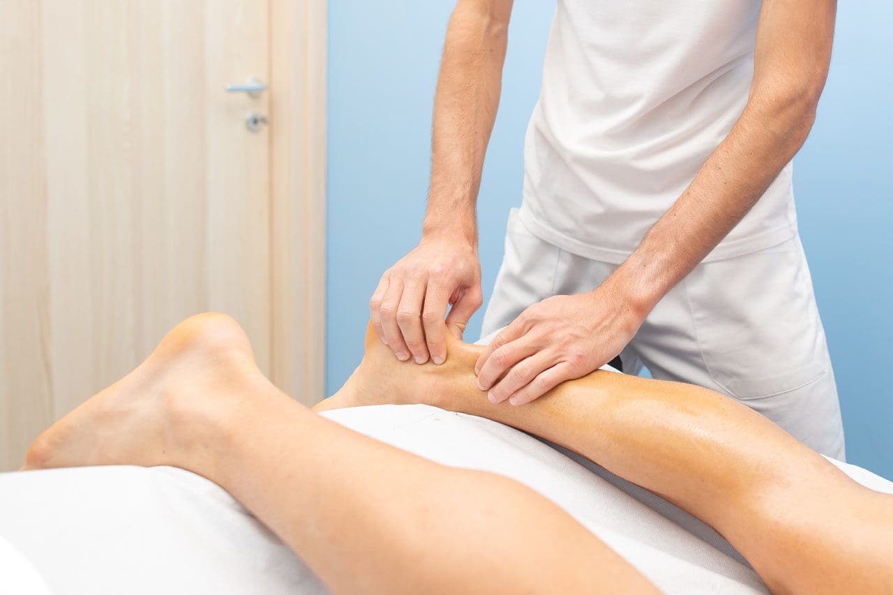
Individuals who participate in physical and sports activities could suffer an Achilles tendon tear. Can understanding the symptoms and risks help in treatment and return the individual back to their sports activity sooner?
Achilles Tendon
This is a common injury that occurs when the tendon attaching the calf muscle to the heel gets torn.
About the Tendon
The Achilles tendon is the largest tendon in the body.
In sports and physical activities, intense explosive movements like running, sprinting, quickly shifting positions, and jumping are exerted on the Achilles.
The injury often occurs without any contact or collision but rather the running, starting, stopping, and pulling actions placed on the feet.
Certain antibiotics and cortisone shots can increase the likelihood of Achilles tear injuries.
A specific antibiotic, fluoroquinolones, has been shown to increase the risk of Achilles tendon problems.
Cortisone shots are also associated with Achilles tears, which is why many healthcare providers don’t recommend cortisone for Achilles tendonitis. (Anne L. Stephenson et al., 2013)
Symptoms
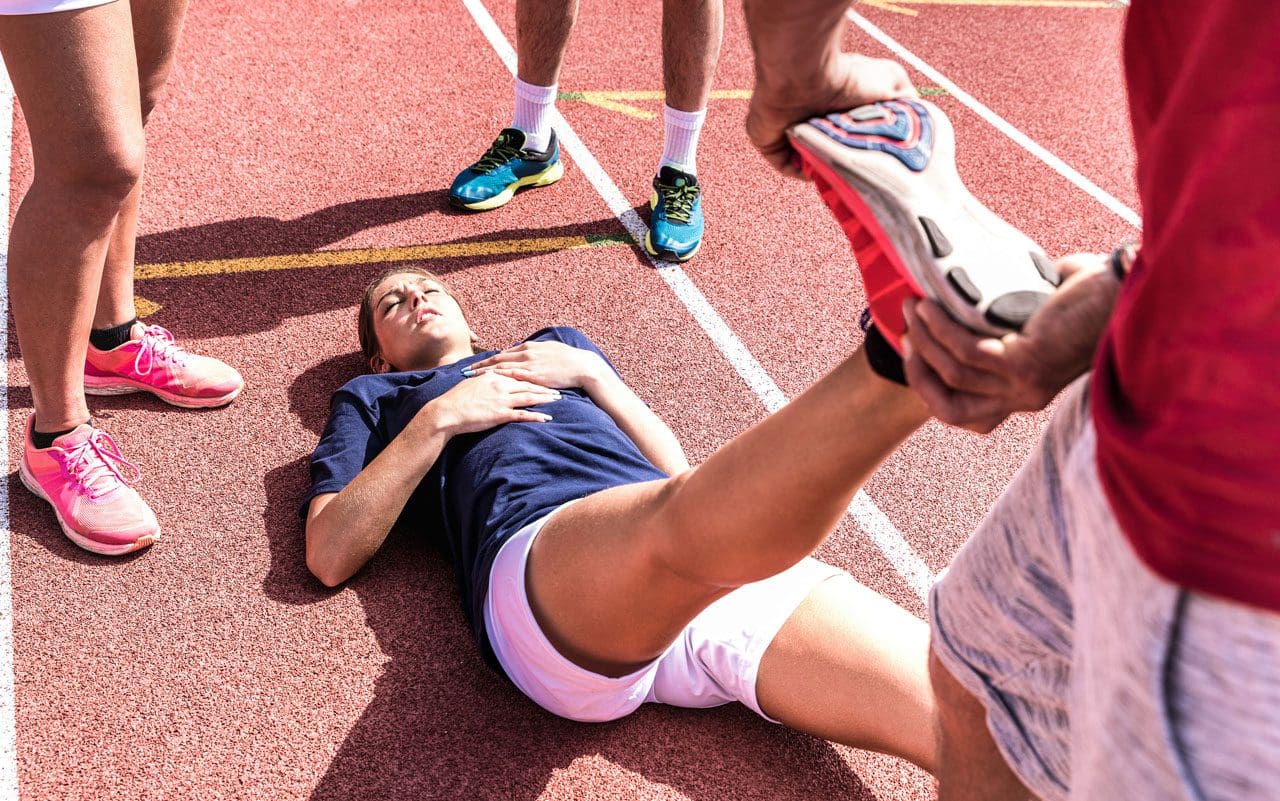
A tendon tear or rupture causes sudden pain behind the ankle.
Individuals may hear a pop or a snap and often report the feeling as being kicked in the calf or heel.
Individuals have difficulty pointing their toes downward.
Individuals may have swelling and bruising around the tendon.
A healthcare provider will examine the ankle for continuity of the tendon.
Squeezing the calf muscle is supposed to cause the foot to point downwards, but in individuals with a tear, the foot will not move, resulting in positive results on the Thompson test.
A defect in the tendon can usually be felt after a tear.
X-rays may be used to rule out other conditions, including ankle fracture or ankle arthritis.
Fluoroquinolone antibiotics are commonly used for the treatment of respiratory infections, urinary tract infections, and bacterial infections. These antibiotics are associated with Achilles tendon rupture, but further research is needed to determine how they affect the Achilles tendon. Individuals taking these medications are advised to consider an alternative medication if Achilles tendon problems begin to develop. (Anne L. Stephenson et al., 2013)
Treatment
Depending on the severity of the injury, treatment can consist of non-surgical techniques or surgery.
The benefit of surgery is there is usually less immobilization.
Individuals can often return to sports activities sooner, and there is less chance of re-rupturing the tendon.
Thevendran, G., Sarraf, K. M., Patel, N. K., Sadri, A., & Rosenfeld, P. (2013). The ruptured Achilles tendon: a current overview from biology of rupture to treatment. Musculoskeletal surgery, 97(1), 9–20. https://doi.org/10.1007/s12306-013-0251-6
Stephenson, A. L., Wu, W., Cortes, D., & Rochon, P. A. (2013). Tendon Injury and Fluoroquinolone Use: A Systematic Review. Drug safety, 36(9), 709–721. https://doi.org/10.1007/s40264-013-0089-8
Pedowitz, D., & Kirwan, G. (2013). Achilles tendon ruptures. Current reviews in musculoskeletal medicine, 6(4), 285–293. https://doi.org/10.1007/s12178-013-9185-8
Yasui, Y., Tonogai, I., Rosenbaum, A. J., Shimozono, Y., Kawano, H., & Kennedy, J. G. (2017). The Risk of Achilles Tendon Rupture in the Patients with Achilles Tendinopathy: Healthcare Database Analysis in the United States. BioMed research international, 2017, 7021862. https://doi.org/10.1155/2017/7021862
Individuals that engage in heavy exercise can develop heat cramps from overexertion. Can knowing the causes and symptoms help prevent future episodes from happening?
Heat Cramps
Heat cramps can develop during exercise from overexertion or prolonged exposure to high temperatures. The muscle cramps, spasms, and pain can range from mild to severe.
Electrolytes like sodium, calcium, and magnesium are important for properly functioning muscles, including the heart. The primary role of sweating is to regulate the body’s temperature. (MedlinePlus. 2015) Sweat is mostly water, electrolytes, and sodium. Excessive sweating from physical activity and exertion or a hot environment can cause electrolyte imbalances that lead to cramps, spasms, and other symptoms.
Causes and Activities
Heat cramps most commonly affect individuals who sweat excessively during strenuous activity or are exposed to hot temperatures for prolonged periods. The body and organs need to cool down, which causes sweat production. However, too much sweating can lead to dehydration and electrolyte depletion. (Centers for Disease Control and Prevention. 2022)
Age – Children and adults 65 years and older have the highest risk.
Excessive sweating.
Low sodium diet.
Preexisting Medical Conditions – heart disease, diabetes mellitus, and obesity are conditions that can increase the risk of muscle cramping.
Medications – blood pressure, diuretics, and antidepressants can affect electrolyte balance and hydration.
Alcohol consumption.
Self-Care
If heat cramps begin, immediately stop the activity and look for a cool environment. Rehydrate the body to replenish the fluid loss. Staying hydrated and drinking fluids regularly during intense activity or in a hot environment can help prevent the body from cramping. examples of beverages that increase electrolytes include:
Gently applying pressure and massaging affected muscles can help reduce pain and spasms. As symptoms resolve, it is recommended to not return to strenuous activity too soon because additional exertion can progressively lead to heatstroke or heat exhaustion. (Centers for Disease Control and Prevention. 2021) Heatstroke and heat exhaustion are two heat-related illnesses. (Centers for Disease Control and Prevention. 2022)
Heatstroke is when the body loses the ability to regulate temperature and can cause dangerously high temperatures.
Heat exhaustion is the body’s response to excessive fluid and electrolyte loss.
The majority of heat cramps develop during activities because of the exertion and sweating, causing more electrolytes to be lost and the body to become more dehydrated.
Symptoms can also develop minutes to hours after activity has ceased.
Duration
Most heat-related muscle cramps will resolve with rest and hydration within 30–60 minutes.
If muscle cramping or spasms do not subside within one hour, seek professional medical attention.
For individuals with heart conditions or on a low-sodium diet who develop heat cramps, regardless of duration, medical help is necessary to ensure there are no complications.
Drink plenty of fluids before and during physical activities.
Avoid alcohol and caffeinated beverages.
Avoid exercising or exposure to extreme heat during peak sunlight hours.
Avoid tight and dark-colored clothing.
Assessing Patients In A Chiropractic Setting
References
Gauer, R., & Meyers, B. K. (2019). Heat-Related Illnesses. American family physician, 99(8), 482–489.
Centers for Disease Control and Prevention. (2022). Heat stress — heat related illness. The National Institute for Occupational Safety and Health (NIOSH) Retrieved from https://www.cdc.gov/niosh/topics/heatstress/heatrelillness.html#cramps
MedlinePlus. (2015). Sweat. Retrieved from https://medlineplus.gov/sweat.html#cat_47
FoodData Central. (2019). Nuts, coconut water (liquid from coconuts). Retrieved from https://fdc.nal.usda.gov/fdc-app.html#/food-details/170174/nutrients
FoodData Central. (2019). Milk, nonfat, fluid, with added vitamin A and vitamin D (fat free or skim). Retrieved from https://fdc.nal.usda.gov/fdc-app.html#/food-details/746776/nutrients
Centers for Disease Control and Prevention. (2012). Frequently asked questions (FAQ) about extreme heat. Retrieved from https://www.cdc.gov/disasters/extremeheat/faq.html
For individuals trying to maintain a healthy spine, can understanding the causes and prevention of rotated vertebrae help protect the spine from harmful rotation of vertebrae?
Spinal Rotation
Healthy spine rotation is an important aspect of injury prevention, and rotated vertebrae or a twisted spine can result from spine, nerve, or muscle disease or certain movements.
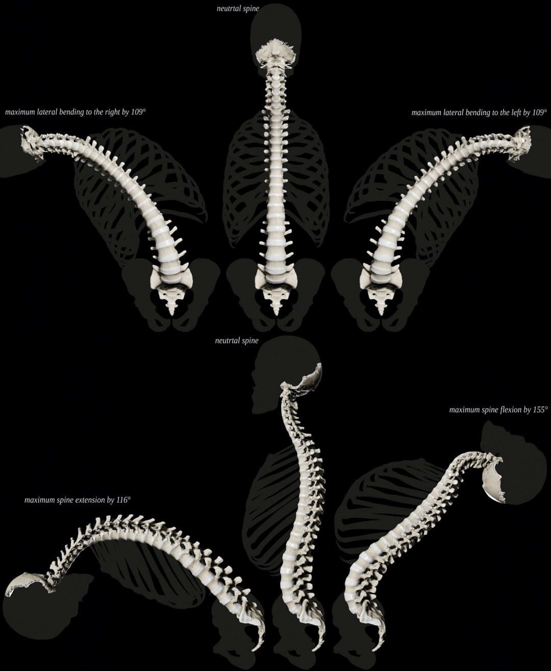
Normal Spine Twisting Capability
The spine can move in several ways. Spine movements include:
Bending – Rounding forward
Extending – Arching backward
Tilting sideways is powered by muscles that aid in twisting.
Although the spine can move in many directions, there are limits to how far it can and should go. (Xinhai Shan et al., 2013). This is especially true with twisting. The spinal column is made of 26 interconnected bones called vertebrae. When moving, each vertebrae bone moves accordingly. Rotated or twisted vertebrae, especially when bending forward like lifting heavy objects, are associated with a risk of back injuries like strain and herniated discs.
How Rotation Works
Rotation is a basic movement in which individuals can turn their spinal column. When twisting, the spine also bends to the side. The muscles involved in spine rotation include:
The internal oblique abdominals and the external oblique abdominals don’t directly attach to the spine but are the primary muscles responsible for powering spinal rotation in the lower back.
Intrinsic muscles, including the multifidus and longissimus, contribute to twisting movement as well.
The multifidus helps the spine twist when one side is contracted/activated and extends the lumbar spine when both sides contract.
The multifidus helps control the movement, and the longissimus provides the movement with some extension.
Age and The Spine
As individuals age, the body accumulates tension and/or weakness in the oblique abdominal and other trunk muscles. Sedentary habits primarily bring on these changes. (Pooriput Waongenngarm et al., 2016)
Chronically tight back and abdominal muscles impair the range of motion of the trunk, as well as twisting ability.
Muscle weakness and tightness affect spinal movements.
Weakened muscles can decrease support for spinal movement and decrease overall trunk stability.
Spinal Rotation and Scoliosis
Scoliosis is a common condition that causes a lateral curve of the spine. Some of the vertebrae become displaced to the side. Often, abnormal vertebral rotation underlies this displacement. Treatment often focuses on controlling vertebral rotation with medical guidance and physical therapy. (John P. Horne et al., 2014)
Over-Rotating The Spine
Many individuals over-rotate their spines with manual work, which can increase the risk of back injuries. (National Institutes of Health. 2020). Over-rotation can happen with activities like digging or shoveling.
Exercise For A Healthy Spine
A recommended way to achieve optimal rotation of the spine is with daily back exercises. (National Spine Health Foundation. 2015). An effective back exercise program will consist of movements in every direction.
Yoga is recommended because it places emphasis on developing flexibility and strength in all directions.
Pilates does the same.
An injury prevention exercise program will work the hip and pelvic muscles as well.
Individuals with a spine condition should consult their healthcare provider or physical therapist about how to exercise the spine safely, as rotation exercises could worsen back problems like bulging or herniated discs.
Core Strength For A Pain-Free Back
References
Shan, X., Ning, X., Chen, Z., Ding, M., Shi, W., & Yang, S. (2013). Low back pain development response to sustained trunk axial twisting. European spine journal : official publication of the European Spine Society, the European Spinal Deformity Society, and the European Section of the Cervical Spine Research Society, 22(9), 1972–1978. https://doi.org/10.1007/s00586-013-2784-7
Waongenngarm, P., Rajaratnam, B. S., & Janwantanakul, P. (2016). Internal Oblique and Transversus Abdominis Muscle Fatigue Induced by Slumped Sitting Posture after 1 Hour of Sitting in Office Workers. Safety and health at work, 7(1), 49–54. https://doi.org/10.1016/j.shaw.2015.08.001
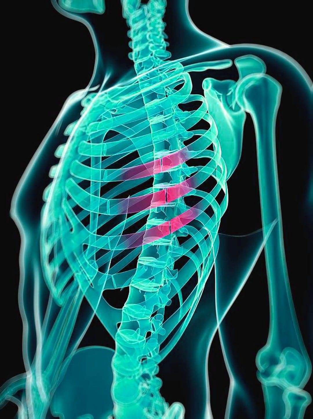
Individuals may not realize they have a cracked rib until symptoms like pain when taking in a deep breath begin to present. Can knowing the symptoms and causes of cracked or broken ribs help in diagnosis and treatment?
Cracked Rib
A broken/fractured rib describes any break in the bone. A cracked rib is a type of rib fracture and is more a description than a medical diagnosis of a rib that has been partially fractured. Any blunt impact to the chest or back can cause a cracked rib, including:
Falling
Vehicle collision
Sports injury
Violent coughing
The main symptom is pain when inhaling.
The injury typically heals within six weeks.
Symptoms
Cracked ribs are usually caused by a fall, trauma to the chest, or intense violent coughing. Symptoms include:
Swelling or tenderness around the injured area.
Chest pain when breathing/inhaling, sneezing, laughing, or coughing.
Chest pain with movement or when lying down in certain positions.
Possible bruising.
Although rare, a cracked rib can cause complications like pneumonia.
See a healthcare provider immediately if experiencing difficulty breathing, severe chest pain, or a persistent cough with mucus, high fever, and/or chills.
Types
In most cases, a rib usually gets broken in one area, causing an incomplete fracture, which means a crack or break that does not go through the bone. Other types of rib fractures include:
Displaced and Nondisplaced Fractures
Completely broken ribs may or may not shift out of place.
If the rib does move, this is known as a displaced rib fracture and is more likely to puncture lungs or damage other tissues and organs. (Yale Medicine. 2024)
A rib that stays in place usually means the rib is not completely broken in half and is known as a nondisplaced rib fracture.
Flail Chest
A section of the ribcage can break away from the surrounding bone and muscle, although this is rare.
If this happens, the ribcage will lose stability, and the bone will move freely as the individual inhales or exhales.
This broken ribcage section is called a flail segment.
This is dangerous as it can puncture the lungs and cause other serious complications, like pneumonia.
Causes
Common causes of cracked ribs include:
Vehicle collisions
Pedestrian accidents
Falls
Impact injuries from sports
Overuse/Repetitive stress brought on by work or sports
Severe coughing
Older individuals can experience a fracture from a minor injury due to the progressive loss of bone minerals. (Christian Liebsch et al., 2019)
The Commonality of Rib Fractures
Rib fractures are the most common type of bone fracture.
They account for 10% to 20% of all blunt trauma injuries seen in emergency rooms.
In cases where an individual seeks care for a blunt injury to the chest, 60% to 80% involve a broken rib. (Christian Liebsch et al., 2019)
Diagnosis
A cracked rib is diagnosed with a physical exam and imaging tests. During the examination, a healthcare provider will listen to the lungs, press gently on the ribs, and watch as the rib cage moves. The imaging test options include: (Sarah Majercik, Fredric M. Pieracci 2017)
X-rays – These are for detecting recently cracked or broken ribs.
CT Scan – This imaging test comprises multiple X-rays and can detect smaller cracks.
MRI – This imaging test is for soft tissues and can often detect smaller breaks or cartilage damage.
Bone Scan – This imaging test uses a radioactive tracer to visualize the structure of bones and can show smaller stress fractures.
Treatment
In the past, treatment used to involve wrapping the chest with a band known as a rib belt. These are rarely used today as they can restrict breathing, increasing the risk of pneumonia or even a partial lung collapse. (L. May, C. Hillermann, S. Patil 2016). A cracked rib is a simple fracture that requires the following:
Rest
Over-the-counter or prescription medications can help manage pain symptoms.
Nonsteroidal anti-inflammatory drugs – NSAIDs like ibuprofen or naproxen are recommended.
If the break is extensive, individuals may be prescribed stronger pain medication depending on the severity and underlying conditions.
Physical therapy can expedite the healing process and help maintain the range of motion of the chest wall.
For patients who are frail and elderly individuals, physical therapy can help the patient walk and normalize certain functions.
A physical therapist can train the individual to transfer between bed and chairs safely while maintaining awareness of any movements or positioning that make the pain worse.
A physical therapist will prescribe exercises to keep the body as strong and limber as possible.
For example, lateral twists can help improve the range of motion in the thoracic spine.
During the early stages of recovery, it is recommended to sleep in an upright position.
Lying down can add pressure, causing pain and possibly worsen the injury.
Use pillows and bolsters to help support sitting up in bed.
What may feel like a cracked rib may be a similar condition, which is why it’s important to get checked out. Other possible symptom causes can include:
Bruised ribs – This occurs when the ribs are not cracked, but the smaller blood vessels around the region burst and leak into surrounding tissues. (Sarah Majercik, Fredric M. Pieracci 2017)
Pulled muscle – A muscle strain, or pulled muscle, occurs when the muscle gets overstretched, which can lead to a tear. The ribs are not affected, but it can feel like they are. (Sarah Majercik, Fredric M. Pieracci 2017)
Emergency
The most common complication is being unable to take a deep breath because of the pain. When the lungs cannot breathe deeply enough, mucous and moisture can build up and lead to an infection like pneumonia. (L. May, C. Hillermann, S. Patil 2016). Displaced rib fractures can also damage other tissues or organs, increasing the risk of a collapsed lung/pneumothorax or internal bleeding. It is recommended to seek immediate medical attention if symptoms develop like:
Shortness of breath
Difficulty breathing
A bluish color of the skin caused by lack of oxygen
A persistent cough with mucus
Chest pain when breathing in and out
Fever, sweating, and chills
Rapid heart rate
The Power of Chiropractic Care In Injury Rehabilitation
Liebsch, C., Seiffert, T., Vlcek, M., Beer, M., Huber-Lang, M., & Wilke, H. J. (2019). Patterns of serial rib fractures after blunt chest trauma: An analysis of 380 cases. PloS one, 14(12), e0224105. https://doi.org/10.1371/journal.pone.0224105
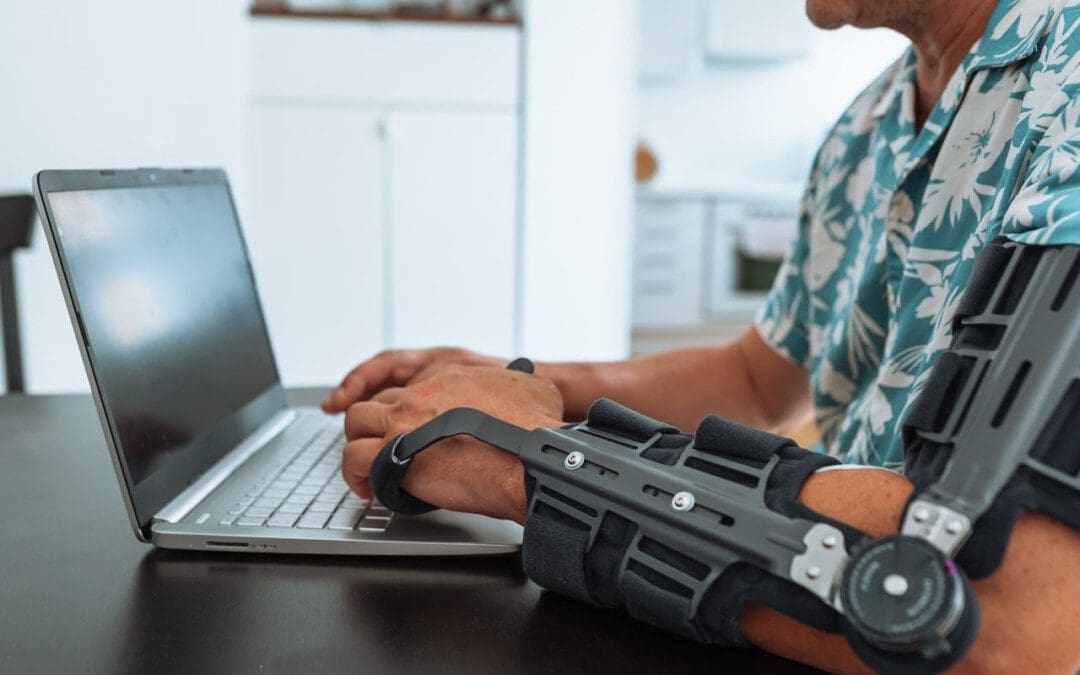
A dislocated elbow is a common injury in adults and children and often happens in tandem with bone fractures and nerve and tissue damage. Can physical therapy help to support recovery and ensure range of motion?
Dislocated Elbow Injury
Elbow dislocations are generally caused by trauma when the elbow bones no longer connect. Individuals falling onto an outstretched hand is the most common cause of the injury. (James Layson, Ben J. Best 2023) Healthcare providers will try to relocate the elbow using a closed reduction. Individuals may require surgery if they cannot relocate the elbow using closed reduction.
The hinge function allows the bending and straightening of the arm.
Ball-and-socket joint
The ball-and-socket function allows you to rotate the palm of your hand to face up or face down.
A dislocated elbow injury can damage bones, muscles, ligaments, and tissues. (American Academy of Orthopaedic Surgeons. 2021) The longer the elbow remains out of the joint, the more damage can occur. Elbow dislocations rarely reset into their joints on their own and are recommended to be evaluated by a qualified healthcare provider to prevent permanent damage to nerves or function.
It is not recommended to try to reset the elbow on your own.
A healthcare provider will work to restore the joint and ensure proper alignment.
Before the reset, they will perform a physical examination to assess blood circulation and any nerve damage.
A closed reduction means that the elbow can be relocated without surgery.
Before the closed reduction, a healthcare provider will administer medications to help relax the individual and address the pain. (Medline Plus. 2022)
Once relocated into the correct position, a healthcare provider applies a splint (usually at a 90-degree angle of flexion) to keep the elbow in place. (James Layson, Ben J. Best 2023)
The objective is to prevent elbow extension, which can cause re-dislocation.
Progress can be challenging for individuals in post total ankle replacement surgery. How can physical therapy help in recovery and restoring leg function?
Total Ankle Replacement Post Surgery Physical Therapy
Total ankle replacement surgery is a major procedure that takes time to recover. A total ankle replacement surgery or arthroplasty can benefit individuals with chronic ankle pain or disability. This procedure can significantly improve an individual’s overall pain and function with time. Physical therapy is essential to regaining movement in the ankle and restoring full mobility. A physical therapist will work with the individual to control pain and swelling, restore the ankle’s range of motion, train on walking gait and balance, and rebuild strength in the leg. This will help maximize the chances of a successful outcome after surgery.
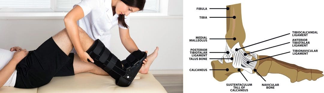
Total Ankle Replacement
The ankle joint is the section of the lower leg where the shinbone/tibia meets the talus bone on the top of the foot. What can happen is the slippery surface/articular cartilage that coats the ends of these bones begins to thin or deteriorate. As the deterioration progresses, it can lead to significant pain, disability, and difficulty walking. (Cleveland Clinic. 2021) This is where a specialist may recommend total ankle replacement for the best results. Various conditions can be helped by this procedure, including:
During an ankle replacement procedure, an orthopedic surgeon removes the damaged ends of the tibia and talus bones and replaces them with an artificial covering. A polyethylene component is also secured between the two structures to support the smooth movement of the new joint endings. (Massachusetts General Hospital. N.D.) Following the procedure, individuals are typically placed in a protective boot or splint. The healthcare provider will recommend staying off the leg for 4 to 8 weeks to allow healing.
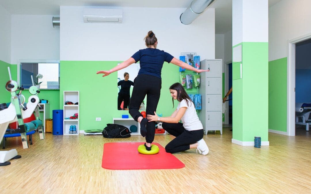
Physical Therapy
Outpatient physical therapy is usually initiated several weeks after the ankle operation. (UW Health Orthopedics and Rehabilitation. 2018) Physical therapy can last for five months or more, depending on the severity of the condition and injury. The physical therapist will focus on different areas to get the best results. (Cort D. Lawton et al., 2017)
Pain and Swelling Control
Post-operative pain and swelling are normal after a total ankle replacement. It is not unusual for an ankle to be swollen for even six to 12 months after the operation. (UW Health Orthopedics and Rehabilitation. 2018) The surgeon will normally prescribe medication to help manage discomfort early on, and physical therapy also plays an important role in addressing the symptoms. Treatments used can include:
Electrical stimulation – mild electrical pulses applied to the muscles.
Ice
Vasopneumatic compression, where an inflatable sleeve is used to create pressure around the area, is commonly utilized at the beginning of physical therapy to reduce pain or swelling.
Other modalities, such as stretching and targeted exercises, are combined with other treatments.
Range of Motion
Early after the procedure, the ankle will be very stiff and tight. This is due to several factors, including the inflammation and swelling after surgery and the time spent immobilized in a boot.
The physical therapist will employ various techniques to improve the ankle joint’s range of motion to rotate and flex.
The physical therapist may employ passive stretching induced by an outside force such as the therapist or a resistance band) to help improve mobility.
After multiple weeks of reduced movement and lack of bearing any weight on the ankle, the muscles that surround the ankle have often atrophied/weakened, which can impact balance.
When the individual can begin placing weight on the leg, the therapist will apply proprioceptive/sense of body position training to improve overall stability. (UW Health Orthopedics and Rehabilitation. 2018)
Balance exercises will be added to the home program and will progress from week to week.
Strength
The muscles in the leg, ankle, and foot become weak from the surgery and the time spent in a splint or boot. These structures have a significant role in balance, the ability to stand, walk, and go up or down the stairs.
Regaining the strength and power of these muscles is a critical goal of rehabilitation.
In the first weeks, the physical therapist will focus on gentle strengthening exercises.
Isometrics lightly activate the muscles but avoid irritating the surgical site.
As time passes and weight-bearing is allowed, these gentle moves are replaced with more challenging ones, like resistance bands and standing exercises, to accelerate strength gains.
Lawton, C. D., Butler, B. A., Dekker, R. G., 2nd, Prescott, A., & Kadakia, A. R. (2017). Total ankle arthroplasty versus ankle arthrodesis-a comparison of outcomes over the last decade. Journal of orthopaedic surgery and research, 12(1), 76. https://doi.org/10.1186/s13018-017-0576-1
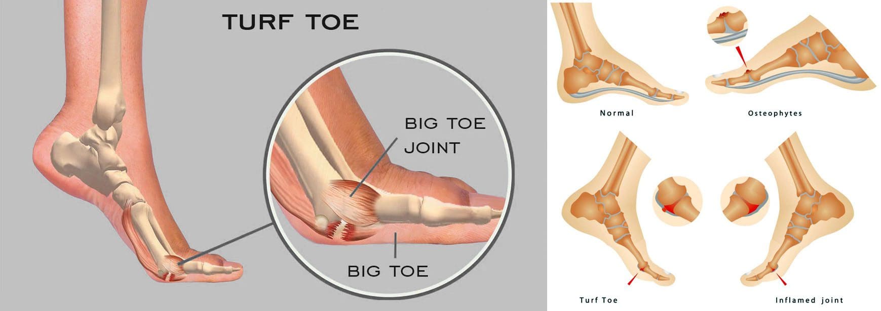
For individuals experiencing a turf toe injury, can knowing the symptoms help athletes and non-athletes with treatment, recovery time, and returning to activities?
Turf Toe Injury
A turf toe injury affects the soft tissue ligaments and tendons at the base of the big toe under the foot. This condition usually occurs when the toe is hyperextended/forced upward, such as when the ball of the foot is on the ground and the heel is lifted. (American Academy of Orthopaedic Surgeons. 2021) The injury is common among athletes who play sports on artificial turf, which is how the injury got its name. However, it can also affect non-athletes, like individuals working on their feet all day.
Recovery time after turf toe injury depends on the severity and the type of activities the individual plans to return to.
Returning to high-level sports activities after a severe injury can take six months.
These injuries vary in severity but usually improve with conservative treatment. In severe cases, surgery could be required.
Pain is the primary issue that stops physical activities after a grade 1 injury, while grades 2 and 3 can take weeks to months to heal completely.
Meaning
A turf toe injury refers to a metatarsophalangeal joint strain. This joint comprises ligaments that connect the bone on the sole of the foot, below the big toe/proximal phalanx, to the bones that connect the toes to the larger bones in the feet/metatarsals. The injury is usually caused by hyperextension that often results from a pushing-off motion, like running or jumping.
If experiencing turf toe symptoms, see a healthcare provider for a proper diagnosis so they can develop a personalized treatment plan. They will perform a physical exam to assess pain, swelling, and range of motion. (American Academy of Orthopaedic Surgeons. 2021) If the healthcare provider suspects tissue damage, they may recommend imaging with X-rays and (MRI) to grade the injury and determine the proper course of action.
Grades 2 and 3 come with partial or complete tissue tearing, severe pain, and swelling. Treatments for more severe turf toe can include: (Ali-Asgar Najefi et al., 2018)
Limited weight bearing
Using assistive devices like crutches, a walking boot, or a cast.
Physical therapy also includes proprioception and agility training exercises, orthotics, and wearing recommended shoes for specific physical activities. (Lisa Chinn, Jay Hertel. 2010)
A physical therapist can also help ensure that the individual does not return to physical activities before the injury is fully healed and prevent the risk of re-injury.
Grade 1 – Subjective as it varies depending on the individual’s pain tolerance.
Grade 2 – Four to six weeks of immobilization.
Grade 3 – Eight weeks minimum of immobilization.
It can take up to six months to return to normal function.
Returning To Normal Activities
After a grade 1 turf toe injury, individuals can return to normal activities once the pain is under control. Grades 2 and 3 take longer to heal. Returning to sports activities after a grade 2 injury can take around two or three months, while grade 3 injuries and cases that require surgery can take up to six months. (Ali-Asgar Najefi et al., 2018)
Sports Chiropractic Treatment
References
American Academy of Orthopaedic Surgeons. (2021). Turf toe.
American College of Foot and Ankle Surgeons. Foot Health Facts. (2023). RICE protocol.
Najefi, A. A., Jeyaseelan, L., & Welck, M. (2018). Turf toe: A clinical update. EFORT open reviews, 3(9), 501–506. https://doi.org/10.1302/2058-5241.3.180012
Pinter, Z. W., Farnell, C. G., Huntley, S., Patel, H. A., Peng, J., McMurtrie, J., Ray, J. L., Naranje, S., & Shah, A. B. (2020). Outcomes of Chronic Turf Toe Repair in Non-athlete Population: A Retrospective Study. Indian journal of orthopaedics, 54(1), 43–48. https://doi.org/10.1007/s43465-019-00010-8
Chinn, L., & Hertel, J. (2010). Rehabilitation of ankle and foot injuries in athletes. Clinics in sports medicine, 29(1), 157–167. https://doi.org/10.1016/j.csm.2009.09.006
IFM's Find A Practitioner tool is the largest referral network in Functional Medicine, created to help patients locate Functional Medicine practitioners anywhere in the world. IFM Certified Practitioners are listed first in the search results, given their extensive education in Functional Medicine