Individuals who have sustained trauma to the knee area from work, physical activity, or a motor vehicle collision can experience significant pain and mobility impairment. Can physical therapy help heal and strengthen the PLC?
Posterolateral Corner Knee Injury
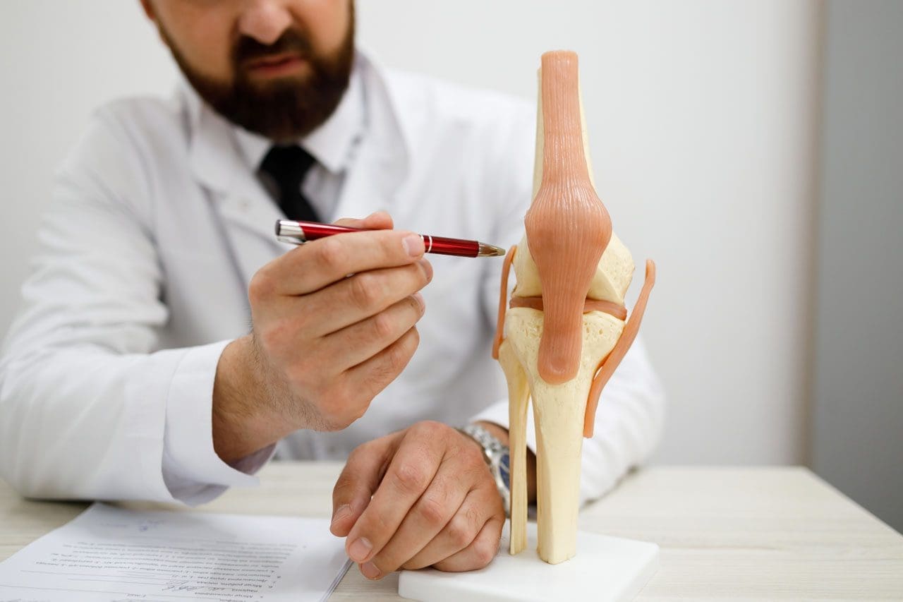
The posterolateral corner, or PLC, comprises muscles, tendons, and ligaments in the back of the knee that help support and stabilize the outside region. The primary role of the PLC is to prevent the knee from excessive amounts of rotation or bowing/turning outward. (Chahla J. et al., 2016) Posterolateral corner injuries can cause significant pain and can dramatically impact an individual’s ability to walk, work, or maintain independence. Treatment options will depend on the severity of the injury.
The Posterolateral Corner
The posterolateral corner comprises multiple structures that support and stabilize the outside of the knee. The structures are subdivided into primary and secondary stabilizers. The primary group includes:
The primary role is to prevent the knee from excessively turning outward, so the grouping provides secondary assistance in preventing the lower leg bone/tibia from shifting forward or backward on the thighbone/femur. Occasionally, one or several posterolateral corner structures can be sprained, strained, or torn.
How Injury Occurs
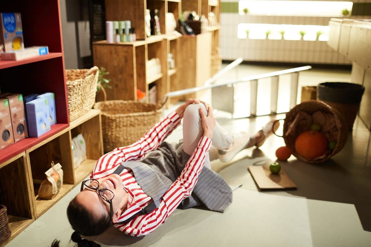
An injury occurs when a direct blow to the inner portion of the front of the knee causes the leg to bow outward. A posterolateral corner injury may also be sustained without contact, for example, if the knee hyperextends or buckles away from the other leg into a varus/bow leg position. Because the knee usually moves during a PLC, concurrent sprains or tears to the anterior cruciate ligament/ACL or posterior cruciate ligament/PCL are also common. (Chahla J. et al., 2016) Other situations that can also cause PLC injuries include automobile crashes and falls from elevated surfaces. (Shon O. J. et al., 2017) When this type of trauma causes a posterolateral corner injury, bone fractures are also common.
Symptoms
Depending on the severity of the injury, multiple symptoms may be present, including:
For individuals who suspect that they have sustained a PLC injury or have any of the symptoms listed, it is critical to be seen by an orthopedic specialist or emergency room physician. A healthcare provider will properly evaluate the leg and develop the appropriate treatment.
Diagnosis
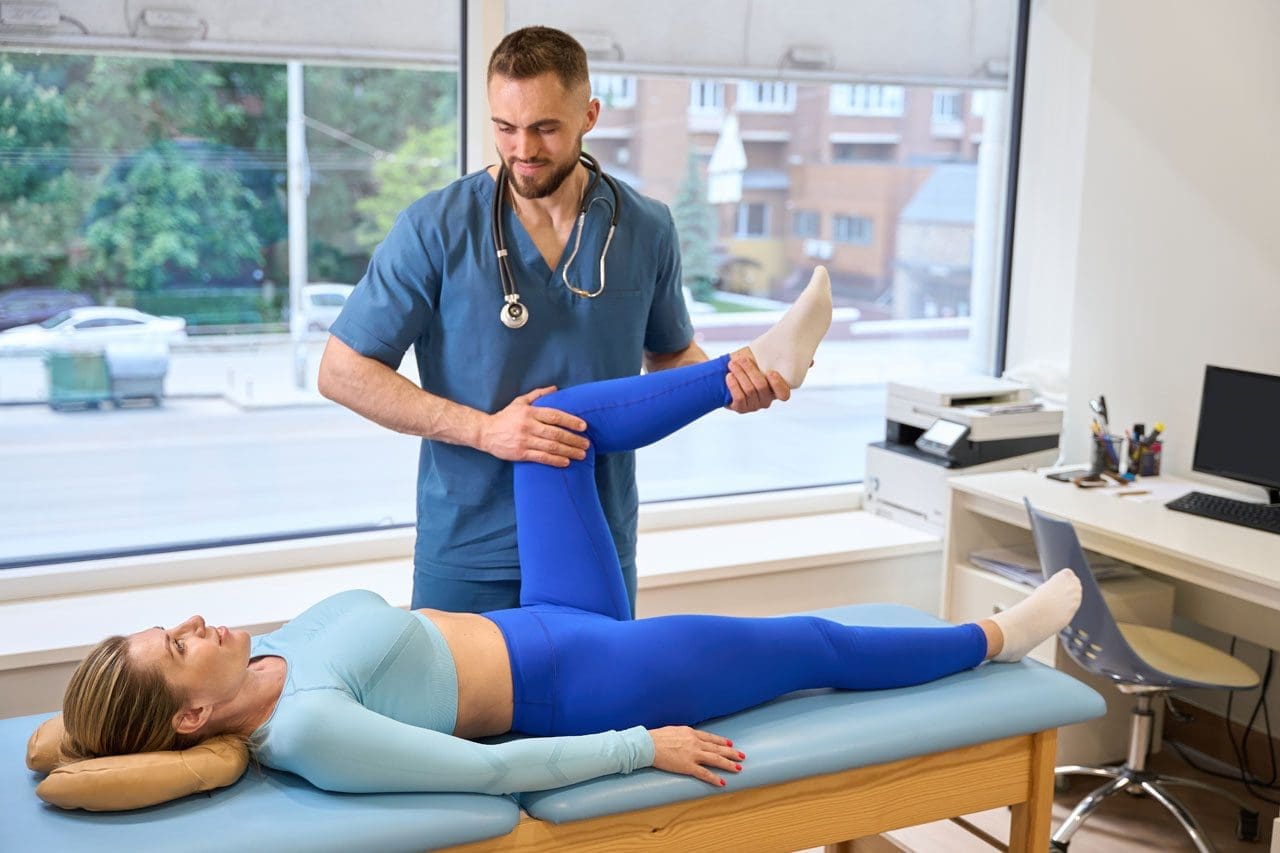
Diagnosis begins with a comprehensive examination. In addition to looking for the symptoms noted, a healthcare provider will move the legs in different directions to assess for any instability. The dial test may be performed, which involves having the patient lie on their stomach while the healthcare professional assesses the side-to-side rotation in the leg to check for excessive motion. (Shon O. J. et al., 2017) Imaging is frequently ordered to determine which anatomical structures are affected more accurately. X-rays can help rule out concurrent fractures and check for excessive laxity in the knee area. MRIs are also useful for visualizing the various tendons and ligaments, helping the healthcare provider look closely at any sprains or tears that may have occurred. However, MRIs may be less accurate in diagnosing PLC injuries after 12 weeks, so they should be obtained as soon as possible. Based on this evaluation, the injury may be classified using the following system (Shon O. J. et al., 2017)
Grade 1
0 to 5 degrees of rotational or varus/bowing instability.
Incompletely torn posterolateral corner.
Grade 2
6 to 10 degrees of rotational or varus/bowing instability.
Incompletely torn posterolateral corner.
Grade 3
Eleven or more degrees of rotational or varus/bowing instability.
Completely torn posterolateral corner.
Treatment
The care received after a posterolateral corner injury can vary depending on the structures involved and the overall severity.
Nonsurgical
Nonsurgical treatment is typically reserved for isolated grade 1 or 2 PLC injuries. (Shon O. J. et al., 2017) Depending on which structures are affected, a stabilizing brace may be worn, and crutches are often needed to decrease the strain on the knee. Physical therapy is also commonly prescribed and focuses on the following goals:
Gradually reintroducing specific movements like running and jumping.
Surgery
Non-surgical treatment tends not to work with grade 3 injuries. If surgery is not performed, individuals may also suffer from chronic knee instability or develop long-term osteoarthritis. (Chahla J. et al., 2019) Surgical treatment is often recommended for grade 3 injuries. The damaged primary stabilizers are surgically reconstructed using a graft from another body region. Surgical repairs may also be performed on any secondary stabilizers to improve stability. (Chahla J. et al., 2019) Any other ligament injuries, such as ACL, PCL, or concurrent fractures, will also be addressed. Following the procedure, individuals immobilize their knee with a brace and do not place weight on the affected leg to protect the surgical area. Depending on the surgeon’s recommendations, this can last six weeks or more. Physical therapy is also initiated after a surgical procedure. Though rehabilitation progresses slowly, the goals are often the same as when treating milder PLC injuries. Returning to work, sports, and/or physical activity after surgery may take six months of therapy or more. (Shon O. J. et al., 2017)
Injury Medical Chiropractic and Functional Medicine Clinic works with primary healthcare providers and specialists to develop a personalized treatment program through an integrated approach to treat injuries and chronic pain syndromes, improve flexibility, mobility, and agility, relieve pain, and help individuals return to normal activities. If other treatments are needed, Dr. Jimenez has teamed up with top surgeons, clinical specialists, medical researchers, and rehabilitation providers to provide the most effective treatments.
Knee Injury Rehabilitation
References
Chahla, J., Moatshe, G., Dean, C. S., & LaPrade, R. F. (2016). Posterolateral Corner of the Knee: Current Concepts. The archives of bone and joint surgery, 4(2), 97–103.
Shon, O. J., Park, J. W., & Kim, B. J. (2017). Current Concepts of Posterolateral Corner Injuries of the Knee. Knee surgery & related research, 29(4), 256–268. https://doi.org/10.5792/ksrr.16.029
Chahla, J., Murray, I. R., Robinson, J., Lagae, K., Margheritini, F., Fritsch, B., Leyes, M., Barenius, B., Pujol, N., Engebretsen, L., Lind, M., Cohen, M., Maestu, R., Getgood, A., Ferrer, G., Villascusa, S., Uchida, S., Levy, B. A., Von Bormann, R., Brown, C., … Gelber, P. E. (2019). Posterolateral corner of the knee: an expert consensus statement on diagnosis, classification, treatment, and rehabilitation. Knee surgery, sports traumatology, arthroscopy : official journal of the ESSKA, 27(8), 2520–2529. https://doi.org/10.1007/s00167-018-5260-4
The levator scapulae muscles could be overstretched for individuals experiencing upper back, shoulder, and neck pain. Can physical therapies help relieve pain and retrain the muscles to function properly?
Levator Scapulae Muscles
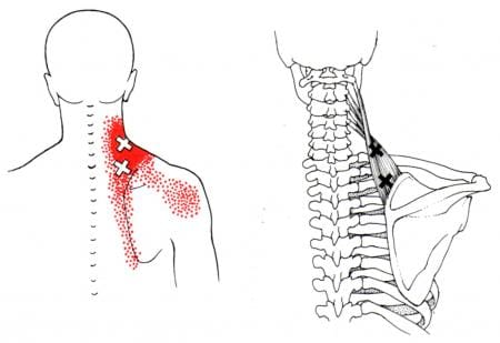
The levator scapulae muscles stretch along the spine across the upper back and neck. They originate from the cervical vertebrae C1 to C4 and attach to the inside top edge of the scapula. (Chotai P. N. et al., 2015) They elevate the shoulder blades, tilt the head, and work with other muscles to stabilize the spine and influence neck motion and upper back posture. The shoulder blade/scapula is the flat, triangular-shaped bone that sits on top of the upper part of the rib cage. Where the levator scapulae attaches, the muscles raise the shoulder blade, a movement called elevation, and indirectly rotates the scapula’s bottom tip downward towards the spine. These shoulder blade movements are part of the shoulder joint’s larger flexion and abduction movements.
Flexion occurs when moving the arm forward and up, and abduction occurs when moving the arm out to the side.
The levator scapulae muscles contract during flexion and/or abduction,
The muscles also contract when moving the neck in side bending, lateral flexion, rotation, or twisting.
Symptoms
Neck and shoulder pain are common problems for office workers, truck drivers, and others who sit most of the day for their jobs. It can worsen when there is no spinal support for maintaining correct alignment. Poorly designed work chairs may contribute to chronic muscle tension and spasms, plus they may contribute to muscle weakness. One of the primary functions of the levator scapulae muscles is to keep the shoulder blade in a position that supports a vertical alignment of the head and to prevent forward head posture. (Yoo W. G. 2018)
However, the shoulder blade is an extremely movable bone. Maintaining stability and correct neck posture is not easy. The levator scapulae muscles may not be strong enough to keep the shoulder blade where it’s supposed to be for healthy posture, and instead, they become overstretched. When muscles are overstretched, they often become taut to try to maintain some form of stability. It can feel like muscle tension because the muscles shorten, but there is a difference. The situation can worsen if one slouches, has no lumbar support, and/or the desk or steering wheel is too high or too low, which may force the shoulder blade upward or downward.
Pain Relief
The levator scapulae muscles are two of several shoulder muscles that may contribute when neck pain presents. This is why a qualified, licensed health professional should diagnose any pain or dysfunction in the area. Physical therapy can help get the body back on track. (Yoo W. G. 2018) Treatment may consist of restoring muscle strength and flexibility in the shoulder, neck, and upper back muscles and developing better posture habits. Physical therapy exercises, particularly those that address upper back posture, can help reduce the stress placed on the levator scapulae muscles and can help decrease kyphosis, often a precursor to forward head posture. Stretching, strengthening, and posture lessons will help relieve pain and increase the physical functioning of the upper body. Other tips for avoiding and relieving muscle pain include:
Stretching regularly by gently rotating the neck and shoulders.
Applying heat to promote relaxation.
Regular massage therapy.
Adjusting chairs and/or monitor height to align with a straight line of sight.
Not carrying heavy bags, especially on one shoulder.
Avoiding side sleeping without proper head support.
Injury Medical Chiropractic Clinic
Visiting a chiropractic and physical therapy team can help treat injuries and chronic pain syndromes, relieve pain, resolve musculoskeletal issues, and prevent future symptoms. Injury Medical Chiropractic and Functional Medicine Clinic works with primary healthcare providers and specialists to develop a personalized care program for each patient through an integrated approach to help return to normal and optimal function. If other treatments are needed, Dr. Jimenez has teamed up with top surgeons, clinical specialists, medical researchers, and rehabilitation providers to provide the most effective treatments.
The Natural Way to Heal: Non-Surgical Chiropractic Care for Pain Relief
References
Chotai, P. N., Loukas, M., & Tubbs, R. S. (2015). Unusual origin of the levator scapulae muscle from mastoid process. Surgical and radiologic anatomy : SRA, 37(10), 1277–1281. https://doi.org/10.1007/s00276-015-1508-6
Yoo W. G. (2018). Effects of thoracic posture correction exercises on scapular position. Journal of physical therapy science, 30(3), 411–412. https://doi.org/10.1589/jpts.30.411
For individuals experiencing piriformis syndrome. Can knowing the causes and what it feels like help in diagnosis and treatment?
Managing Piriformis Syndrome
Piriformis syndrome is where spasms occur in the piriformis muscle located in the buttocks. These muscle spasms can cause pain and numbness in the buttocks and the back of the leg. (Cedars Sinai, 2022)
What Is It?
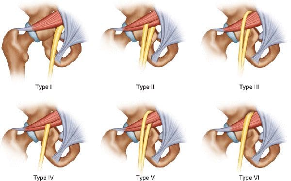
Piriformis syndrome is an irritation of the sciatic nerve from the piriformis muscle. Although the piriformis muscle is small compared to other muscles around the hip and thigh, it supports the hip joint’s external rotation or turning out. The sciatic nerve supplies the lower extremities with motor and sensory functions. The piriformis tendon and sciatic nerve cross each other behind the hip joint in the deep buttock. Both are about one centimeter in diameter. The piriformis muscle spasms can irritate the sciatic nerve, causing sciatica symptoms. (Cedars Sinai, 2022)
Triggers
A piriformis syndrome diagnosis means the piriformis tendon binds or spasms around the sciatic nerve, causing irritation and symptoms. Many doctors and specialists support the theory that when the piriformis muscle and its tendon tighten, this can cause compression and pinch the nerve. This can decrease blood circulation and irritate the nerve due to pressure. (Cass S. P. 2015) Many doctors also believe that piriformis syndrome occurs from anatomic variation of the muscle and tendon. It is thought this muscle-tendon variation irritates the nerve in some, leading to sciatica symptoms.
How It Feels
Common signs and symptoms experienced include (Cass S. P. 2015)
Pain in the buttocks.
Pain behind the hip.
Electric shock pains traveling down the back of the lower extremity.
Numbness in the lower extremity.
Tenderness with pressure that often causes pain when sitting.
Some develop symptoms abruptly, while others gradually increase in symptoms in the back of their thighs.
Most who are diagnosed with piriformis syndrome are generally active individuals who experience increasing difficulty with certain types of physical activity.
Testing
There are no specific tests that accurately diagnose piriformis syndrome. Doctors will order tests, including MRI and nerve conduction studies. Because it can be difficult to diagnose, there are likely many misdiagnosis cases. This means that some with the condition don’t have a piriformis diagnosis. In addition, some with vague hip pain may receive this diagnosis even if they don’t have the condition. (Cass S. P. 2015) An injection is often administered into the piriformis muscle when the diagnosis is uncertain. (Jankovic D. et al., 2013) Performing an injection can help determine the specific location of the discomfort. When an injection is given into the piriformis muscle or tendon, it is administered by ultrasound guidance to ensure the needle delivers medication to the correct location. (Bardowski E. A., and Byrd J. W. T. 2019)
Differential Diagnosis
Some other conditions with buttock pain can have similar symptoms. Other causes can include:
Radiculopathy/Sciatica
Herniated discs
Hip bursitis
Spinal stenosis
The diagnosis of piriformis syndrome is given when these diagnoses are eliminated as possible causes of pain.
Treatment
Managing piriformis syndrome is quite general, and it is often difficult to recover from. Common treatment and management suggestions include the following. (Jankovic D. et al., 2013)
Rest
Avoiding activities that cause symptoms for at least a few weeks.
Physical Therapy
Focuses on stretching and strengthening the hip rotator muscles.
Anti-inflammatory Medication
To decrease inflammation around the tendon.
Deep Massage
Used to relax the piriformis muscle and help release the compressed nerve.
Cortisone Injections
It can help decrease inflammation and swelling.
Botulinum Toxin Injection
It can paralyze the piriformis muscle to reduce pain and discomfort.
In severe cases, surgery can be performed to loosen the piriformis tendon, known as a piriformis release (Cass S. P. 2015). This surgical procedure is recommended when conservative treatments have been tried for at least six months and other causes of pain have been evaluated and ruled out. Recovery takes several months.
The goal of managing piriformis syndrome is to improve the range of motion and flexibility around the hip and diminish inflammation around the sciatic nerve. Working with a professional chiropractic team can help relieve pain, return individuals to normal function, and expedite healing. Injury Medical Chiropractic and Functional Medicine Clinic works with primary healthcare providers and specialists to develop a customized treatment program through an integrated approach to treating injuries and chronic pain syndromes, improving flexibility, mobility, and agility. If other treatments are needed, Dr. Jimenez has teamed up with top surgeons, clinical specialists, medical researchers, and rehabilitation providers to provide the most effective treatments.
Piriformis Syndrome Treatment Chiropractor
References
Cedars Sinai. Sinai, C. (2022). Piriformis syndrome. https://www.cedars-sinai.org/health-library/diseases-and-conditions/p/piriformis-syndrome.html
Cass S. P. (2015). Piriformis syndrome: a cause of nondiscogenic sciatica. Current sports medicine reports, 14(1), 41–44. https://doi.org/10.1249/JSR.0000000000000110
Jankovic, D., Peng, P., & van Zundert, A. (2013). Brief review: piriformis syndrome: etiology, diagnosis, and management. Canadian journal of anaesthesia = Journal canadien d’anesthesie, 60(10), 1003–1012. https://doi.org/10.1007/s12630-013-0009-5
Bardowski, E. A., & Byrd, J. W. T. (2019). Piriformis Injection: An Ultrasound-Guided Technique. Arthroscopy techniques, 8(12), e1457–e1461. https://doi.org/10.1016/j.eats.2019.07.033
An annular fissure is caused by age-related changes to the spine, which often do not cause symptoms but can cause back pain. Can understanding the causes help individuals manage lower back pain and help healthcare providers develop an effective treatment program?
Annular Fissure
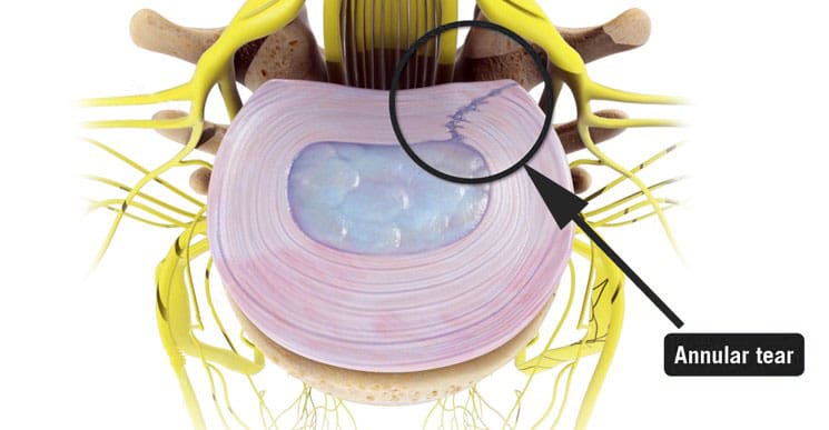
An annular fissure is a discogenic condition that affects the spine and can cause lower back pain. Also called an annular tear, it’s usually a wear-and-tear condition that happens over time rather than a condition caused by trauma. It usually happens when the fibers that make up the annulus or the tough outer covering of the intervertebral disc break or separate. To manage it, healthcare providers may recommend:
Making lifestyle changes.
Staying aware of how you go about daily activities and take steps to make adjustments, such as being mindful of unhealthy posture.
Start doing exercises that help make the back stronger.
Medical care if pain and other symptoms need to be managed.
Symptoms
Lower back pain may be a sign of an annular fissure, or there may be no symptoms. Symptoms can include:
Pain
Weakness
Numbness
Electrical sensations travel down one leg or arm if a cervical/neck tear is present.
Numbness and weakness may be caused by the nerves getting irritated or compressed near an annular tear. (Stadnik, T. W. et al., 1998)
These symptoms can also be similar to a herniated disc, which can be a complication of an annular fissure.
However, studies have shown that annular tears and herniated discs often go unnoticed because they have few obvious symptoms. (Jarvik, J. G. et al., 2005)
Annulus Function
The annulus comprises several layers of tough fibers/fibrocartilage that surround, contain, and protect the soft, liquid nucleus inside the disc. The layers of the annulus fibrosus crisscross to provide support. The nucleus is a shock absorber cushions the body’s weight on the spinal joints when sitting, standing, or moving. Its strength also allows the disc to buffer the jolts and jars it experiences. It also helps maintain the integrity of the intervertebral joint by supporting the space between the two vertebrae. When an annular fissure occurs, the fibers separate or tear off from insertion on the nearby spinal bone. A fissure can also be a break in the fibers of one or more layers. (Jarvik, J. G. et al., 2005)
Causes
An annular tear is not the standard term medical professionals use to describe or diagnose a fissure because the word tear suggests that trauma has led to the separation or break in the fibers. While an injury can cause an annular fissure, it’s usually caused by long-term wear and tear. (Guterl, C. C. et al., 2013) The tears are typically caused by age-related degenerative changes in the disc, which can also lead to degeneration in other areas of the spine. Wear and tear are caused by annular fissures due to an individual’s daily living habits, such as sitting, standing, walking, climbing stairs, and performing other routine movements.
Treatment
While a large annular fissure is not likely to improve without treatment, a small one could heal independently. However, once an area has torn, it becomes more likely to continue tearing. (Virginia Spine Institute, N.D.) Conservative treatment is usually enough to control pain and symptoms. Physical therapy and anti-inflammatory medication are the first line of treatment. (Cheng, J. et al., 2019) Medication can be over-the-counter or prescription. Physical therapy treatment includes exercises, traction, and other therapies. If these do not help with the symptoms, the provider may suggest a steroid injection to reduce inflammation and pain. It can take three to six months to recover from degenerative disc problems if doing a standard treatment plan that includes rest, low-impact therapy exercises, and anti-inflammatory treatments. (Cheng, J. et al., 2019)
In severe cases, surgery may be recommended, including disc replacement surgery. An annular tear is not a reason to have disc replacement surgery alone; it is only when there are degenerative changes in the vertebral disc that surgery might be necessary. (Yue, J. J. et al., 2012)
Improving Body Alignment
Not paying attention and being aware of how the body performs everyday activities can, over time, set the stage for an annular fissure and other musculoskeletal injuries. However, fixing daily movement and posture habits to prevent injuries can be done through simple adjustments. For example, strengthening the core and back muscles can reduce pressure on the spine and help prevent injuries. (Camp, C. L. et al., 2016) The idea is to improve joint and overall body alignment. Activities can include:
Strength training
Walking
Pilates classes
Yoga
Tai chi
Somatic exercises
These activities help with muscle balance and joint alignment, which are recommended prevention strategies that physical therapists use when working with individuals who need help with spinal problems.
Visiting a chiropractic and physical therapy team can help treat injuries and chronic pain syndromes, relieve pain, resolve musculoskeletal issues, and prevent future symptoms. Injury Medical Chiropractic and Functional Medicine Clinic works with primary healthcare providers and specialists to develop a personalized care program for each patient through an integrated approach to treating injuries, improving flexibility, mobility, and agility to help return to normal and optimal function. If other treatments are needed, Dr. Jimenez has teamed up with top surgeons, clinical specialists, medical researchers, and rehabilitation providers to provide the most effective treatments.
Back Pain Specialist
References
Stadnik, T. W., Lee, R. R., Coen, H. L., Neirynck, E. C., Buisseret, T. S., & Osteaux, M. J. (1998). Annular tears and disk herniation: prevalence and contrast enhancement on MR images in the absence of low back pain or sciatica. Radiology, 206(1), 49–55. https://doi.org/10.1148/radiology.206.1.9423651
Jarvik, J. G., Hollingworth, W., Heagerty, P. J., Haynor, D. R., Boyko, E. J., & Deyo, R. A. (2005). Three-year incidence of low back pain in an initially asymptomatic cohort: clinical and imaging risk factors. Spine, 30(13), 1541–1549. https://doi.org/10.1097/01.brs.0000167536.60002.87
Guterl, C. C., See, E. Y., Blanquer, S. B., Pandit, A., Ferguson, S. J., Benneker, L. M., Grijpma, D. W., Sakai, D., Eglin, D., Alini, M., Iatridis, J. C., & Grad, S. (2013). Challenges and strategies in the repair of ruptured annulus fibrosus. European cells & materials, 25, 1–21. https://doi.org/10.22203/ecm.v025a01
Virginia Spine Institute. (N.D.). Annular disc tear Understanding the Symptoms, Causes, and Treatments. https://www.spinemd.com/conditions/annular-disc-tear/
Cheng, J., Santiago, K. A., Nguyen, J. T., Solomon, J. L., & Lutz, G. E. (2019). Treatment of symptomatic degenerative intervertebral discs with autologous platelet-rich plasma: follow-up at 5-9 years. Regenerative medicine, 14(9), 831–840. https://doi.org/10.2217/rme-2019-0040
Yue, J. J., Telles, C., Schlösser, T. P., Hermenau, S., Ramachandran, R., & Long, W. D., 3rd (2012). Do presence and location of annular tear influence clinical outcome after lumbar total disc arthroplasty? A prospective 1-year follow-up study. International journal of spine surgery, 6, 13–17. https://doi.org/10.1016/j.ijsp.2011.09.001
Camp, C. L., Conti, M. S., Sgroi, T., Cammisa, F. P., & Dines, J. S. (2016). Epidemiology, Treatment, and Prevention of Lumbar Spine Injuries in Major League Baseball Players. American journal of orthopedics (Belle Mead, N.J.), 45(3), 137–143.
For individuals who are feeling back pain, neck pain, or shoulder pain, can using postural techniques help decrease pain and develop strategies to maintain correct posture?
Maintaining Correct Posture
Exercise and postural correction are two of the most important strategies to manage pain. (Robin McKenzie, the great spine guru PT, (May S. and Donelson R. 2008) However, it is not easy to maintain correct posture as the day progresses. While working or driving, most of us forget to be mindful of our posture; we slip back into unhealthy habits of sitting slouched, to the side, forward, etc, which often leads to continued pain and movement limitations. Studies on postural correction and its effect on overall back or neck pain levels are limited. (Lederman E. 2011) However, some studies show that a forward-head posture increases stress and load on the neck. (Hansraj K. K. 2014) If an individual has pain that improves with sitting upright, perhaps checking posture as the day progresses could be beneficial. There are ways to start changing posture and maintain the change. First, visit a physical therapist, spine specialist, or chiropractor to learn how to sit or stand to manage posture problems. They can teach individuals how to attain healthy posture and manage their condition. These strategies can then be used to maintain correct posture.
Lumbar Support
A lumbar roll/support is one of the simplest ways to attain and maintain a healthy sitting posture. These specialized pillows can help maintain the forward curve in the lower back. Maintaining the lumbar lordosis curve is essential for keeping stress off the back muscles, joints, and discs. Lumbar support pillows can be found in various stores, office supply stores, or purchased online. A physical therapist PT can help train individuals to use a lumbar roll properly.
Utilize the Slouch Overcorrect Exercise
The slouch overcorrect exercise is a simple maneuver for training the body to find and maintain its neutral position. It involves slowly shifting from a slouched position to an overcorrected postural position. Once posture is overcorrected, a slight relaxation from a fully erect posture will return to sitting properly. Practicing this exercise daily can help train the body to feel the muscles, listen to the body, and maintain proper posture. The muscles have memory, and the more often the body is placed in optimal posture, the muscles memorize their healthy, pain-free positioning.
Kinesiology Tape
Kinesiology tape is a flexible cotton adhesive that facilitates muscle contractions and inhibits muscle spasms and pain. (Han J. T. et al., 2015) Using the tape is a simple way to help facilitate the postural support muscles. The tape can be applied to the middle trapezius and rhomboid muscles to help support the shoulder blades and spine. Kinesiology tape for postural control also gently pulls on the muscles when slouching to remind the individual to sit up or stand straight.
Scapular Stabilization Exercises
A physical therapist or chiropractor can help improve posture through targeted exercises. Strengthening the muscles attached to the shoulder blades can help individuals better control their posture. (Shiravi S. et al., 2019) Exercises like the prone T, I, or Y can help improve the muscles’ neuromuscular recruitment to maintain correct posture.
Use an Alarm
Setting an alarm while working can help train the body to maintain correct posture. Most of us sit at a computer, desk, or workstation with proper posture, but depending on what we’re working on, we eventually slouch, lean to the side, or have a forward head posture. Pretty much all computers and phones have an alarm setting or app. Use the alarm or timer to go off every 20 to 30 minutes. When the alarm rings, look at your posture to recognize what you’re doing, get up and move around to stretch the body, sit and readjust, reset the alarm, and continue working. As you get better at maintaining appropriate posture, you won’t need the alarm anymore, and readjusting will become automatic.
Chiropractic Team
Sitting and standing with unhealthy posture can significantly cause back, neck, and shoulder pain. By visiting a chiropractic and physical therapy team and learning to attain and maintain correct posture, you can quickly relieve pain, resolve musculoskeletal issues, and prevent future pain symptoms. Injury Medical Chiropractic and Functional Medicine Clinic works with primary healthcare providers and specialists to develop a personalized care plan for each patient through an integrated approach to treating injuries and chronic pain syndromes, improving flexibility, mobility, and agility programs to relieve pain and help return to normal and optimal function. If other treatments are needed, Dr. Jimenez has teamed up with top surgeons, clinical specialists, medical researchers, and rehabilitation providers to provide the most effective treatments.
Scapular Winging In-Depth
References
May, S., & Donelson, R. (2008). Evidence-informed management of chronic low back pain with the McKenzie method. The spine journal : official journal of the North American Spine Society, 8(1), 134–141. https://doi.org/10.1016/j.spinee.2007.10.017
Lederman E. (2011). The fall of the postural-structural-biomechanical model in manual and physical therapies: exemplified by lower back pain. Journal of bodywork and movement therapies, 15(2), 131–138. https://doi.org/10.1016/j.jbmt.2011.01.011
Hansraj K. K. (2014). Assessment of stresses in the cervical spine caused by posture and position of the head. Surgical technology international, 25, 277–279.
Han, J. T., Lee, J. H., & Yoon, C. H. (2015). The mechanical effect of kinesiology tape on rounded shoulder posture in seated male workers: a single-blinded randomized controlled pilot study. Physiotherapy theory and practice, 31(2), 120–125. https://doi.org/10.3109/09593985.2014.960054
Shiravi, S., Letafatkar, A., Bertozzi, L., Pillastrini, P., & Khaleghi Tazji, M. (2019). Efficacy of Abdominal Control Feedback and Scapula Stabilization Exercises in Participants With Forward Head, Round Shoulder Postures and Neck Movement Impairment. Sports health, 11(3), 272–279. https://doi.org/10.1177/1941738119835223
For individuals dealing with headaches, can utilizing self-care techniques help ease symptoms?
Headaches Self Care
There are different self-pain-relieving approaches to ease headache-related pain that use non-medication options. While headache self-care pain-relieving techniques can help temporarily, they are recommended to help ease headache symptoms until professional treatment is available. Individuals are encouraged to try multiple strategies to find the right treatment regimen that works for them, which is usually a process of trial and error. Self-care headache relief maneuvers can include temple massages, breathing exercises, exercise, or applying a cold pack to the head.
Self-Care Relief
Commonly used for tension headaches or migraine, a few examples of self-care techniques include (Espí-López G. V. et al., 2016)
Massaging the temples, neck, or scalp with the hand, fingers, or a massage tool.
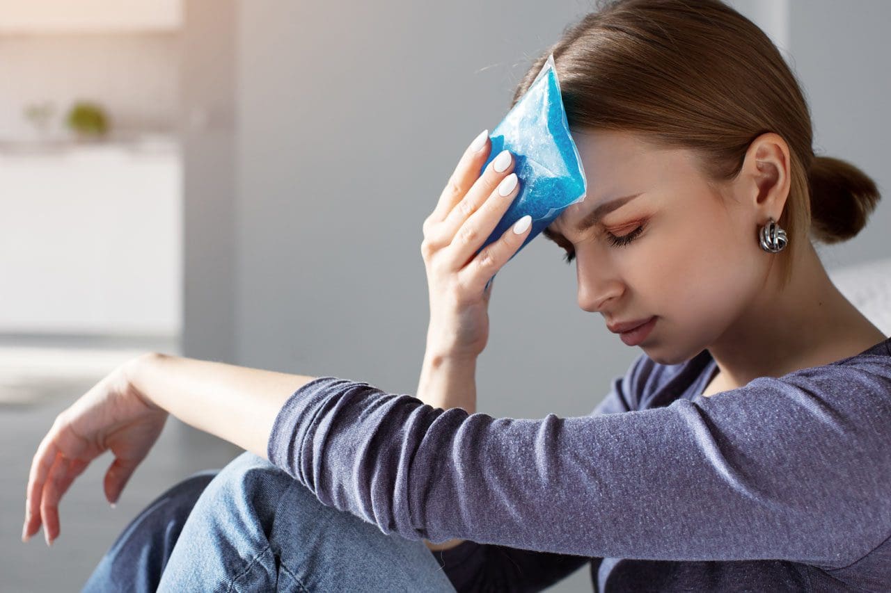
Applying a cold pack to the affected area.
Heat can be used on the affected area, like a headband or a hot shower.
Compression such as a handkerchief or belt wrapped tightly around the head or pressing firmly on the painful area.
Compression is more commonly utilized in migraineurs versus scalp massage in people with tension headaches. This is often due to the throbbing sensation caused by a migraine versus a tension headache, which feels like a tight grip or band around the head.
Individuals with cluster headaches are more likely to utilize unique approaches, such as covering one ear, rotating the head, shallow breathing, moving around, or closing the nostril on the same side as the head pain.
Various scientific studies support the potential benefit of these complementary therapies. However, individuals should talk with a healthcare provider to choose the best strategy for their headache type and their unique needs/goals. Working with a chiropractic team can help expedite healing. Injury Medical Chiropractic and Functional Medicine Clinic works with primary healthcare providers and specialists to develop a customized treatment plan through an integrated approach to treating injuries and chronic pain syndromes, improving flexibility, mobility, and agility, relieving pain, and helping individuals return to normal activities. If other treatments are needed, Dr. Jimenez has teamed up with top surgeons, clinical specialists, medical researchers, and rehabilitation providers to provide the most effective treatments.
Tension Headaches and Chiropractic Care
References
Espí-López, G. V., Zurriaga-Llorens, R., Monzani, L., & Falla, D. (2016). The effect of manipulation plus massage therapy versus massage therapy alone in people with tension-type headache. A randomized controlled clinical trial. European journal of physical and rehabilitation medicine, 52(5), 606–617.
Millstine, D., Chen, C. Y., & Bauer, B. (2017). Complementary and integrative medicine in the management of headache. BMJ (Clinical research ed.), 357, j1805. https://doi.org/10.1136/bmj.j1805
Can incorporating stretches and exercises help relieve pain and provide more support for individuals with knee pain?
Knee Strengthening
The knee is the largest joint connecting the shin and thigh bones. Its cartilage, muscles, ligaments, and nerves all play a role in maintaining the joint’s health and movement. Individuals of all ages can experience knee pain, but it is more common in those with labor-intensive jobs, arthritis, and athletes. A healthcare provider will prescribe targeted stretches and exercises for individuals with pain, inflammation, or a health disorder that affects the strength of their knees to relieve knee pain while strengthening the muscles around the joint. Knee stretches and exercises are great for those with sore knees and knee pain or stiffness, but they can also be used as preventative medicine.
Exercise Benefits
Doing knee-strengthening exercises can significantly improve joint health. The benefits associated with exercises include (Zeng C. Y. et al., 2021)
Avoid curving the spine to get closer to the thigh during the stretch. The key is not how far you can bend down but getting a deep stretch. Individuals not yet flexible enough to go all the way down should bend until comfortable but can still feel the stretch in their hamstrings. With practice and time, flexibility will improve. This stretch should be felt in the hamstrings.
Step Exercises
Step exercises, or step-ups, involve using a platform at least 6 inches high. To perform the exercise:
Put one foot onto the platform and use that leg to lift the other foot off the floor.
Hold the position with the foot hanging loosely for 3 to 5 seconds, then place the foot back on the floor.
Avoid locking the knees when stepping up on the platform. Locking the knees switches off the muscles that need strengthening (American Academy of Orthopaedic Surgeons, 2009). Step exercises work out various areas of the leg. Individuals should feel this exercise in the thigh, hip, and buttocks.
Post-Exercise Stretching
Post-exercise stretching is vital in physical rehabilitation and strengthening. Stretching after exercise, individuals can (Afonso J. Clemente. et al., 2021)
Relieve and reduce soreness.
Expedite recovery times.
Increase range of motion.
Strengthen the knees more quickly and effectively.
Post-exercise stretches for knee pain and weakness can include:
Quadricep Stretch
This stretch targets the quad muscles located in the thighs. To perform this stretch:
Stand with the feet planted flat on the floor, spaced the same width as the shoulders.
Bending the left knee, grab the foot with the left hand and pull the heel toward the buttocks.
Hold for 5 to 10 seconds.
Repeat on both sides.
What to Avoid
There is no need to pull the heel too far into the buttocks. The goal is to feel a deep stretch. Pushing it too far can lead to injury. (Lee J. H. et al., 2021)
Toe Touches
Toe touches will help stretch calve muscles and hamstrings. To perform this stretch:
Sit on the floor with the legs straight out in front.
Bend the body over the legs at the hips, reaching for the toes in front while maintaining a straight back.
Relax the shoulders and keep them away from the ears.
Once touching the toes, hold the stretch for 15 seconds.
What To Avoid
Individuals who cannot yet touch their toes should perform the same steps and reach only as far as is comfortably possible. Over time, flexibility will improve. (Lee J. H. et al., 2021)
Injury Medical Chiropractic Clinic
These are examples of exercises and stretches that a physical therapy and chiropractic team may prescribe. Each case is different and requires reviewing individual medical history and physical examination to determine the proper treatment program. Injury Medical Chiropractic and Functional Medicine Clinic works with primary healthcare providers and specialists to develop a personalized treatment program through an integrated approach to treating injuries and chronic pain syndromes, improving flexibility, mobility, and agility to relieve pain and help individuals return to normal activities. If other treatments are needed, Dr. Jimenez has teamed up with top surgeons, clinical specialists, medical researchers, and rehabilitation providers to provide the most effective treatments.
Knee Pain Rehabilitation
References
Zeng, C. Y., Zhang, Z. R., Tang, Z. M., & Hua, F. Z. (2021). Benefits and Mechanisms of Exercise Training for Knee Osteoarthritis. Frontiers in physiology, 12, 794062. https://doi.org/10.3389/fphys.2021.794062
American Academy of Orthopaedic Surgeons. OrthoInfo. (2009). Knee exercises. https://orthoinfo.aaos.org/en/staying-healthy/knee-exercises/
Kothawale S. and Rao K. (2018). Effectiveness of positional release technique versus active release technique on hamstrings tightness. Int J Physiother Res., 6(1), 2619-2622. https://doi.org/https://dx.doi.org/10.16965/ijpr.2017.265
Wang, H., Ji, Z., Jiang, G., Liu, W., & Jiao, X. (2016). Correlation among proprioception, muscle strength, and balance. Journal of physical therapy science, 28(12), 3468–3472. https://doi.org/10.1589/jpts.28.3468
Afonso, J., Clemente, F. M., Nakamura, F. Y., Morouço, P., Sarmento, H., Inman, R. A., & Ramirez-Campillo, R. (2021). The Effectiveness of Post-exercise Stretching in Short-Term and Delayed Recovery of Strength, Range of Motion and Delayed Onset Muscle Soreness: A Systematic Review and Meta-Analysis of Randomized Controlled Trials. Frontiers in physiology, 12, 677581. https://doi.org/10.3389/fphys.2021.677581
Lee, J. H., Jang, K. M., Kim, E., Rhim, H. C., & Kim, H. D. (2021). Effects of Static and Dynamic Stretching With Strengthening Exercises in Patients With Patellofemoral Pain Who Have Inflexible Hamstrings: A Randomized Controlled Trial. Sports health, 13(1), 49–56. https://doi.org/10.1177/1941738120932911
IFM's Find A Practitioner tool is the largest referral network in Functional Medicine, created to help patients locate Functional Medicine practitioners anywhere in the world. IFM Certified Practitioners are listed first in the search results, given their extensive education in Functional Medicine