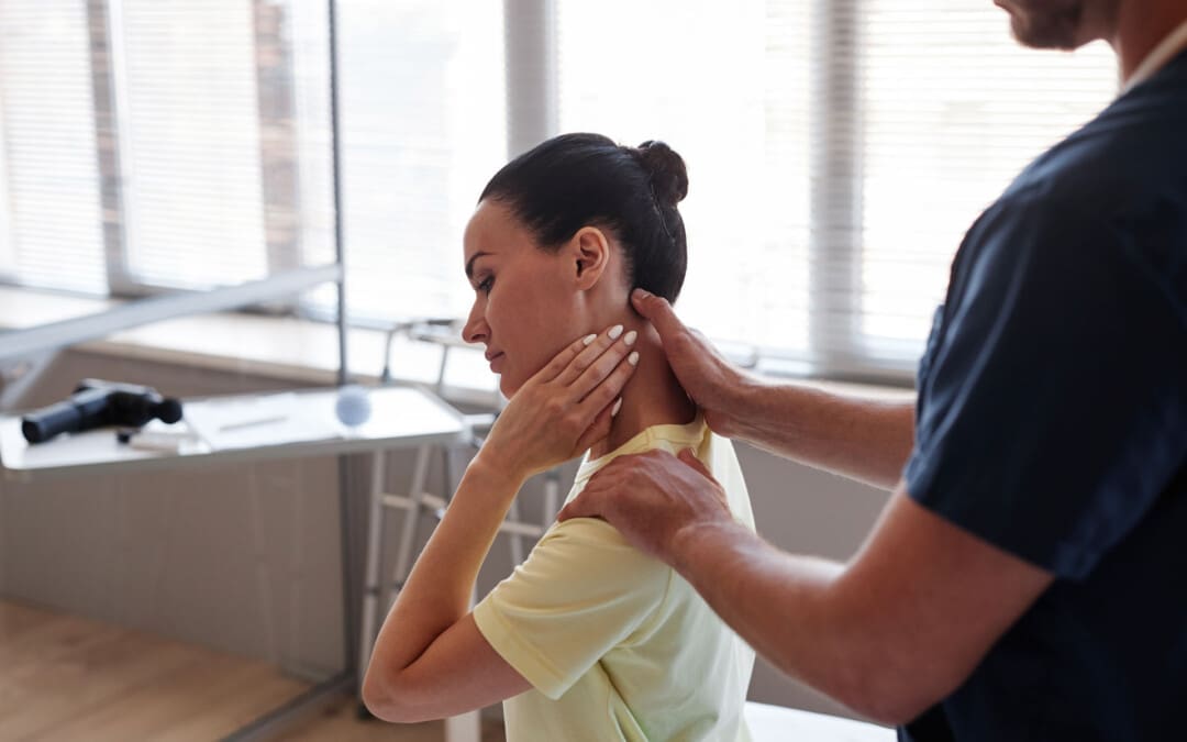
Can physical therapy help individuals with a pinched nerve in the neck?
Pinched Nerve In The Neck
A pinched nerve in the neck can cause pain, numbness, and weakness that extends through the neck, shoulder, and arm. (American Academy of Orthopaedic Surgeons, 2024) Over-the-counter pain medications, resting the muscles, and gentle stretching can help. However, if you’re still in pain after a few days, see a healthcare provider. Common treatments include:
Rest
Over-the-counter (OTC) pain meds
Physical therapy
Steroid injections
Wearing a neck collar
Surgery is rarely needed, but it can provide relief if other treatments don’t help. Most of the time, a pinched nerve resolves within days or weeks.
Sharp pain that extends into the shoulder and arm.
The pain worsens, or there is a shooting sensation when turning the head.
Tingling or feeling of pins-and-needles in the fingers or hand.
Weakness in the arm, shoulder, or hand.
Numbness or loss of feeling.
Often, these symptoms occur only on one side. Some find their pain less when they lift their hand onto their head, which can relieve pressure on the nerve.
Causes
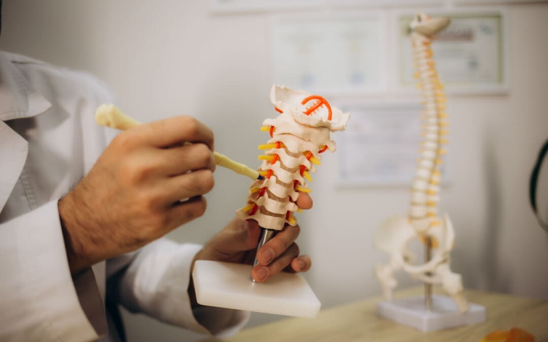
The cervical spine is the spinal cord area around the neck. It’s made up of seven vertebrae. Nerves branch off the spinal cords in the spaces between the vertebrae. Nerve compression occurs when the space between two vertebrae is reduced, putting pressure on the nerve, pinching it, and causing pain. Pinched nerves develop from age because the spinal discs between the vertebrae become compressed over time. Age causes about 70% to 80% of nerve compression. Other factors that cause pinched nerves include: (Harvard Health Publishing, 2021)
Degenerative disc disease
A herniated disc
Injuries like car accidents falls, or other trauma to the spine
Find a comfortable position and try to allow the muscles in your neck to relax and rest.
Heat or Ice
Warmth and coolness can relieve pain and inflammation.
Use a warm or cool compress for 15 minutes at a time.
Over-The-Counter Pain Medications
Pain medications, including nonsteroidal anti-inflammatory drugs (NSAIDs), can help bring relief.
Treatment for Severe Symptoms
If pain doesn’t resolve within a few days, or if it is so bad that you can’t go about daily activities, it’s recommended to see a healthcare provider. They can diagnose a pinched nerve after a physical exam and may also recommend imaging, including an X-ray, CT scan, MRI, or EMG, to reveal what’s causing the symptoms. After diagnosing the condition, the healthcare provider will develop a personalized treatment plan, which may include the following (Harvard Health Publishing, 2021)
Physical Therapy
Physical therapy can help build strength and flexibility in the neck.
This is especially important if there is frequent nerve pain in the same spot.
Cervical Collar
A soft cervical collar is a brace that fits around the neck.
It supports your head so the neck muscles can relax, facilitating healing.
The collar can also keep the head from turning in painful ways.
Oral Corticosteroids
Oral steroids like prednisone can help reduce inflammation.
If inflammation or swelling in the neck puts more pressure on the nerve, they can help.
Steroid Injections
Steroid shots right into the painful tissue reduce inflammation right away.
Muscle Relaxers
These medications keep the muscles in the neck from seizing up.
As the muscles relax, this brings pain relief.
Narcotic Pain Medications
Narcotic pain medications can be used short-term by individuals who have severe pain.
A healthcare provider will inform the patient of the benefits and drawbacks of these medications, which include opiates.
Hold for 20 seconds, then return to a neutral position.
Do this five times.
Eyes to Sky
Lean your head back and look toward the sky.
Hold for 20 seconds, then return to your starting position.
Do this five times.
Side to Side
Turn your head to the right as far as possible, bringing your chin in line with your shoulder.
Hold for 20 seconds, then turn as far as possible toward the left.
Repeat four times.
Ear to Shoulder
Bring your ear down toward your shoulder.
Hold for 20 seconds, then repeat the exercise on the other side.
Alternate between the right and left, stretching each side five times.
While it’s normal for exercises to hurt from stretching the muscles, they should never hurt more than a five on a pain scale of 1 to 10. If they do, stop exercising (National Health Service, 2025)
Healing Time
Healing and recovery depend on the severity of the injury. Some individuals find that the pain from a pinched nerve goes away in days, while for others, it can last for weeks. The pain goes away and then returns. If pain doesn’t go away with the conservative treatments or lasts more than a few days, talk with a healthcare provider or return for a second visit. Rarely do individuals need surgery to bring pain relief. The healthcare provider will discuss whether surgery is the best option and what to expect regarding pain relief. (American Academy of Orthopaedic Surgeons, 2024)
Injury Medical Chiropractic and Functional Medicine Clinic
Injury Medical Chiropractic and Functional Medicine Clinic works with primary healthcare providers and specialists to build optimal health and wellness solutions. We focus on what works for you to relieve pain, restore function, prevent injury, and mitigate issues through adjustments that help the body realign itself. The clinic can also work with other medical professionals to integrate a treatment plan to resolve musculoskeletal problems.
Revitalize and Rebuild with Chiropractic
References
American Academy of Orthopaedic Surgeons. OrthoInfo. (2024). Cervical radiculopathy (pinched nerve). https://orthoinfo.aaos.org/en/diseases–conditions/cervical-radiculopathy-pinched-nerve/
Harvard Health Publishing. Publishing, H. H. (2021). Treating a pinched nerve. https://www.health.harvard.edu/pain/treating-a-pinched-nerve
National Institute of Neurological Disorders and Stroke. (2025). Pinched Nerve Definition. Retrieved from https://www.ninds.nih.gov/health-information/disorders/glossary-neurological-terms#-P-
National Health Service. Service, N. H. (2025). Exercises for neck problems. https://www.nhsinform.scot/illnesses-and-conditions/muscle-bone-and-joints/neck-and-back-problems-and-conditions/exercises-for-neck-problems
Individuals experiencing persistent pain, weakness, numbness, and tingling in the back could be suffering from nerve root encroachment. Could surgery ease nerve compression and improve symptoms for persistent and severe cases?
Surgical Decompression
The pain, weakness, numbness, and tingling associated with nerve root encroachment are usually first treated with non-surgical therapies that include:
Anti-inflammatory medications
Physical therapy
This can be enough to address the irritation of the spinal nerve root. But when cases become severe, surgical decompression may be recommended and necessary. It can be done in a couple of different ways.
Causes and Symptoms
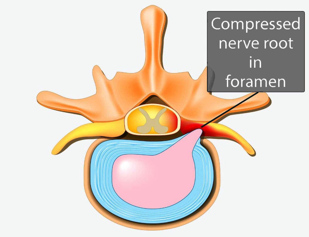
Vertebrae are bones in the spine. Small openings called foramina allow a spinal nerve root to pass through on each side of the vertebra. When nerve root encroachment is present, the spinal nerve root gets compressed, pinched, and trapped, which can cause peripheral symptoms such as numbness, tingling, pain, or weakness to develop. Nerve root encroachment is typically caused by normal aging degenerative wear and tear changes in the vertebrae. (Choi Y. K. 2019) These degenerative changes can include:
Facet joint hypertrophy
Ligament and bone hypertrophy
Disc disorders
Formation of bone spurs or osteophytes.
If these degenerative changes progress, they can encroach and compress a nerve root, leading to peripheral symptoms. (Choi Y. K. 2019)
When Surgery Is Recommended
When symptoms occur, initial treatment will involve:
Physical therapy
Chiropractic realignment
Massage therapies
Rest
Lifestyle adjustments
Nonsteroidal anti-inflammatories – NSAIDs
Corticosteroid injections into the spine
If conservative therapies don’t fully heal or improve symptoms or there are neurological problems like difficulty with balance or walking, then surgery may be recommended. Severe pain that limits normal function is an indication for surgery, and rapidly progressive weakness of the arms and/or legs or signs of cauda equina syndrome are indications for emergency surgery.
Surgery Options
Different types of spinal surgery may be performed. A neurosurgeon will decide the best procedure for each patient based on their case, age, medical conditions, and other factors. Specific spinal surgical decompression depends on what is causing the nerve compression. In most cases, it involves removing bone or tissue to relieve nerve pressure or provide support to stabilize the joint. The most common types of surgical decompression include: (Mayo Clinic Health System, 2022)
Maintain the stability and alignment of the spine.
Improve the stability and alignment of the spine.
Anterior Surgery
The anterior approach to surgery means that the spine is accessed through the anterior/front of the spine. In this surgery, one or more discs and bone spurs may be removed through an incision in the front of the neck. (American Association of Neurological Surgeons, 2024) For example, an anterior cervical discectomy may alleviate pressure on one or more nerve roots in the neck. With an anterior lumbar interbody fusion, a surgeon removes a degenerative disc in the lower spinal area by going through a patient’s lower abdomen. (American Association of Neurological Surgeons, 2024) After the disc is removed, a structural device, usually made of bone, fills the space where it once was. This device encourages bone healing and helps the vertebrae’s bodies fuse.
Posterior Surgery
Posterior surgery means the spine is accessed through the posterior/back of the spine. An example is removing a thickened ligament, bone spur, or disc material in the neck. To do this, a small incision in the back of the neck may be made to remove part of the back of the vertebrae called the lamina. This is called a posterior cervical laminectomy. (American Association of Neurological Surgeons, 2024) A posterior lumbar interbody fusion removes a degenerative disc by going through the back. (American Association of Neurological Surgeons, 2024) Like the anterior approach, a structural device often contains bone to fill the space where the disc once was to fuse the bones.
Potential Risks
As with any surgery, it’s important that the individual and their healthcare provider carefully discuss the benefits and risks. Spinal surgical decompression includes: (Proietti L. et al., 2013)
Bleeding
Blood clots
Surgical site infection
Urinary tract infection
Lung infection
Intestinal blockage
There are also specific risks to the area of the spine being operated on and how it is surgically approached. For example, a cervical anterior procedure may injure the esophagus, trachea, or carotid artery. Likewise, damage to the C5 nerve root/C5 palsy can occur from cervical spinal decompressive surgery. This complication causes weakness, numbness, and pain in the shoulders. (Thompson S. E. et al., 2017) The spinal cord may also be injured during surgery and result in paralysis, although this is rare. (American Association of Neurological Surgeons, 2024)
Injury Medical Chiropractic and Functional Medicine Clinic
Injury Medical Chiropractic and Functional Medicine Clinic works with primary healthcare providers and specialists to develop an optimal health and wellness solution. We focus on what works for you to relieve pain, restore function, and prevent injury. Regarding musculoskeletal pain, specialists like chiropractors, acupuncturists, and massage therapists can help mitigate the pain through spinal adjustments that help the body realign itself. They can also work with other medical professionals to integrate a treatment plan to resolve musculoskeletal issues.
The Non-Surgical Solution
References
Choi Y. K. (2019). Lumbar foraminal neuropathy: an update on non-surgical management. The Korean journal of pain, 32(3), 147–159. https://doi.org/10.3344/kjp.2019.32.3.147
Mayo Clinic Health System. (2022). Decompress and stabilize: understanding types of back surgery. Speaking of Health. https://www.mayoclinichealthsystem.org/hometown-health/speaking-of-health/understanding-types-of-back-surgery
American Association of Neurological Surgeons. (2024). Cervical spine. https://www.aans.org/patients/conditions-treatments/cervical-spine/
American Association of Neurological Surgeons. (2024). Lumbar spinal stenosis. https://www.aans.org/patients/conditions-treatments/lumbar-spinal-stenosis/
Proietti, L., Scaramuzzo, L., Schiro’, G. R., Sessa, S., & Logroscino, C. A. (2013). Complications in lumbar spine surgery: A retrospective analysis. Indian journal of orthopaedics, 47(4), 340–345. https://doi.org/10.4103/0019-5413.114909
Thompson, S. E., Smith, Z. A., Hsu, W. K., Nassr, A., Mroz, T. E., Fish, D. E., Wang, J. C., Fehlings, M. G., Tannoury, C. A., Tannoury, T., Tortolani, P. J., Traynelis, V. C., Gokaslan, Z., Hilibrand, A. S., Isaacs, R. E., Mummaneni, P. V., Chou, D., Qureshi, S. A., Cho, S. K., Baird, E. O., … Riew, K. D. (2017). C5 Palsy After Cervical Spine Surgery: A Multicenter Retrospective Review of 59 Cases. Global spine journal, 7(1 Suppl), 64S–70S. https://doi.org/10.1177/2192568216688189
Are there blood tests for individuals with chronic and severe back pain symptoms that can help healthcare providers diagnose?
Blood Tests To Help Diagnose Back Pain
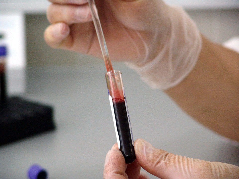
If a healthcare provider suspects an infection or inflammatory arthritis is the cause of back pain, blood tests may be used to diagnose. When trying to find the cause of back pain, a healthcare provider will examine the patient’s medical history, perform a physical examination, and, if necessary, order diagnostic tests. (Dansie E. J. and Turk D. C. 2013) For example, the National Institute of Arthritis and Musculoskeletal and Skin Diseases says that MRIs can reveal abnormalities in the spine. Still, a person may not feel pain or experience any other symptoms. The NIAMS also says healthy, pain-free individuals can have elevated SED levels. A high sedimentation rate or sed rate, also known as an erythrocyte sedimentation rate (ESR) test, can indicate inflammation in the body. (National Institute of Arthritis and Musculoskeletal and Skin Diseases, 2023)
Commonly Used Tests
Blood tests that can help diagnose back pain include:
Complete Blood Count – CBC
This test can indicate inflammation or infections.
Sed Rate or Erythrocyte Sedimentation Rate
This test measures inflammation by analyzing how red blood cells settle through plasma.
If the SED rate indicates that inflammation is present, the possibility of an underlying cause may be some form of arthritis or a tumor, which is rare.
A genetic marker in the blood that is more common in individuals with ankylosing spondylitis and reactive arthritis. (McMichael A. and Bowness P. 2002)
This marker may be tested if the healthcare provider suspects either disease.
Ankylosing spondylitis is an inflammatory arthritis affecting the sacroiliac joints, hips, and spine. (Sieper J. et al., 2002)
Injury Medical Chiropractic and Functional Medicine Clinic
At Injury Medical Chiropractic and Functional Medicine Clinic, we focus on what works for you to relieve pain and restore function. Regarding musculoskeletal pain, specialists like chiropractors, acupuncturists, and massage therapists can help mitigate the pain through spinal adjustments that help the body realign itself. Our areas of practice include Chronic Pain, Personal Injury, Auto Accident Care, Work Injuries, Back Injury, Low Back Pain, Neck Pain, Migraine Headaches, Sports Injuries, severe sciatica, Scoliosis, Complex Herniated Discs, Fibromyalgia, Chronic Pain, Complex Injuries, Stress Management, Wellness and nutrition, Functional Medicine Treatments, and in-scope care protocols. They can also work with other associated medical professionals to develop a personalized treatment plan to help relieve muscle pain, improve the body’s flexibility and mobility, resolve musculoskeletal issues, and prevent future pain symptoms from reoccurring.
Integrative Medicine Approach
References
Dansie, E. J., & Turk, D. C. (2013). Assessment of patients with chronic pain. British journal of anaesthesia, 111(1), 19–25. https://doi.org/10.1093/bja/aet124
National Institute of Arthritis and Musculoskeletal and Skin Diseases. (2023). Back Pain. Retrieved from https://www.niams.nih.gov/health-topics/back-pain
Harrison M. (2015). Erythrocyte sedimentation rate and C-reactive protein. Australian prescriber, 38(3), 93–94. https://doi.org/10.18773/austprescr.2015.034
Sproston, N. R., & Ashworth, J. J. (2018). Role of C-Reactive Protein at Sites of Inflammation and Infection. Frontiers in immunology, 9, 754. https://doi.org/10.3389/fimmu.2018.00754
McMichael, A., & Bowness, P. (2002). HLA-B27: natural function and pathogenic role in spondyloarthritis. Arthritis research, 4 Suppl 3(Suppl 3), S153–S158. https://doi.org/10.1186/ar571
Sieper, J., Braun, J., Rudwaleit, M., Boonen, A., & Zink, A. (2002). Ankylosing spondylitis: an overview. Annals of the rheumatic diseases, 61 Suppl 3(Suppl 3), iii8–iii18. https://doi.org/10.1136/ard.61.suppl_3.iii8
Hamdulay, S. S., Glynne, S. J., & Keat, A. (2006). When is arthritis reactive?. Postgraduate medical journal, 82(969), 446–453. https://doi.org/10.1136/pgmj.2005.044057
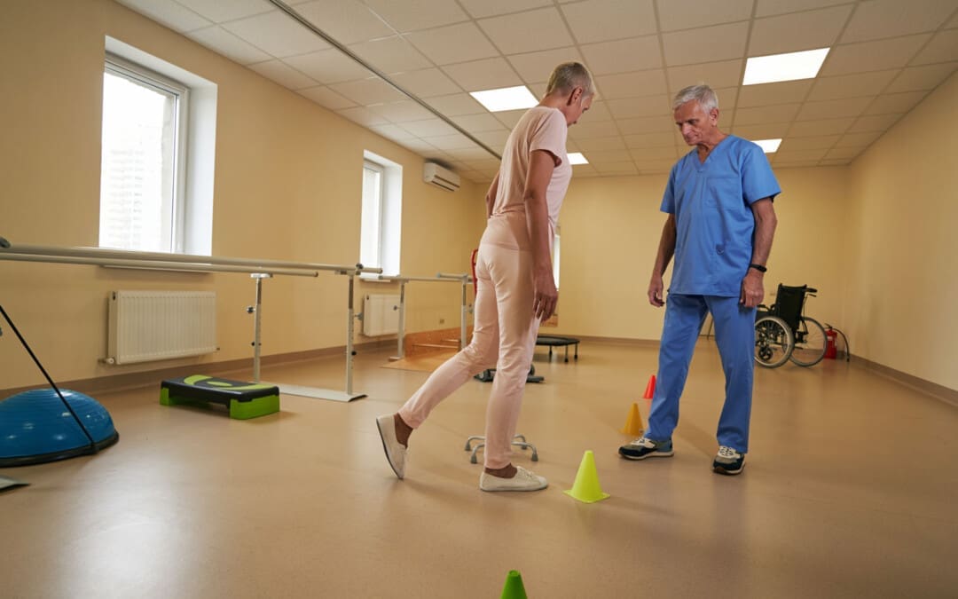
Individuals walking with a limp that results in pain could have an antalgic gait, an abnormal walking pattern commonly seen in emergency clinics and primary care offices. Can recognizing the symptoms help healthcare providers develop an effective treatment for the underlying cause?
Antalgic Gait
Limping and having an antalgic gait usually indicate a larger issue within the leg or lower back. It is the most common type of abnormal gait. There are various causes of antalgic gait, including acute injuries and gradually progressing medical conditions. The most common causes include osteoarthritis in one of the leg’s joints, lumbar radiculopathy, or an injury to a ligament or tendon. Paying attention to when the limping occurs, and any accompanying symptoms can help determine its origins.
Walking
When limping, the stance phase during walking is shorter than the swing phase. Individuals may widen their legs apart to provide a support base to compensate for the imbalance. In severe cases, an individual may swing their leg irregularly or take several side steps.
Causes and Symptoms
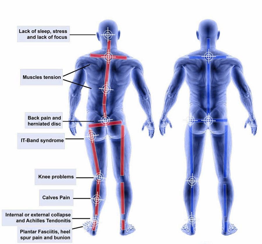
Antalgic gait can be caused by pain in any part of the lower extremity. Limping when walking may be a primary concern but is rarely the only complaint. Other associated symptoms may also be present, depending on the cause. These include:
Limited range of motion
Joint stiffness
Muscular weakness
Numbness and tingling
Pain
Swelling
Leg instability or buckling
Clicking or popping
Common Causes include:
Hip, knee, and/or Foot problems
When the hip, knee, ankle, or foot joints are injured or have some issue, walking can be painful and lead to a limp.
Sprains, Strains, or Soft-tissue Injuries
Sprains, strains, and soft-tissue injuries can result from acute injury or chronic, repetitive activities over time. (Pirker W. and Katzenschlager R. 2017) Sprains affect the body’s ligaments, while strains impact muscle tendons. However, sprains and strains occur when the impacted structure is overstretched or partially torn. The damage can lead to pain and antalgic gait. An injury to several other soft-tissue structures, including a bursa or fluid-filled sacs that reduce friction, meniscus, or fat pad, can also lead to limping. Symptoms typically include swelling, pain, and limited range of motion. More severe injuries can also make the leg feel unstable and cause it to give way when walking. Sometimes, bruising can also occur in the area of the injury. (American Academy of Orthopaedic Surgeons, 2020)
Osteoarthritis
Osteoarthritis occurs when the smooth, articular cartilage that lines the ends of bones begins to thin and deteriorate. This can alter the normal movement of a joint and lead to pain. Osteoarthritis symptoms gradually progress, affect individuals over 50, and worsen after periods of sedentary activity (Arthritis Foundation, Osteoarthritis, N.D.) Typically, it results in pain, stiffness, clicking, and occasionally swelling in the affected joint. These symptoms are usually worse in the morning and at the end of a long activity day. Moving around and warming the joint improves osteoarthritis symptoms. (Arthritis Foundation, Osteoarthritis, N.D.)
Lower Back Radiculopathy
Lumbar radiculopathy is when the nerve roots branching off the spine’s lower region become compressed or inflamed. This can occur because of disc issues like bulging, degeneration, herniation, bone spurring, or, rarely, a growth or tumor. (Johns Hopkins Medicine, 2024) Because these nerves control movement, sensation, and strength in the legs and feet, irritation in one can lead to limping. (Yokogawa N. et al., 2015) The antalgic gait from this condition frequently comes on suddenly and is commonly accompanied by back pain symptoms. This can include shooting pain and paresthesia in the leg. Depending on which nerve is involved, individuals may also experience muscular weakness in certain areas of the lower extremity. Sometimes, the affected leg feels like it will buckle while standing or walking. (Johns Hopkins Medicine, 2024)
Other causes include:
Broken bones
Tumors
Infections
Blood clots
Vascular issues
Treatment
Treatment for antalgic gait depends on the underlying cause but can include:
Rest, ice, and elevation are important for injuries. Individuals can control their initial symptoms by icing, elevating the leg, and resting from irritating activities.
Activity modifications
Antibiotics for infections
Pain relievers
Anti-inflammatories
Physical therapy is also frequently initiated to strengthen the core and alleviate walking symptoms.
A spinal injection or surgery can reduce the pressure on the nerve root if conservative interventions fail to improve antalgic gait patterns. (Johns Hopkins Medicine, 2024)
Crutches, canes, walkers, or assistive devices can reduce pressure traveling through an affected joint and improve overall walking quality. A study found that using a cane for two months helped reduce pain and improve function in individuals with knee osteoarthritis. (Fang M. A. et al., 2015)
Injury Medical Chiropractic and Functional Medicine Clinic
Though it can be tempting to ignore the limp and push through it, discussing the condition with a healthcare provider is important. A thorough medical exam and diagnostic tests, such as X-ray, MRI, or EMG, can help determine the underlying causes of a limp, help discover the cause of the problem, and help improve the quality of walking. Injury Medical Chiropractic and Functional Medicine Clinic works with primary healthcare providers and specialists to develop personalized treatment programs. Using an integrated approach to treating injuries and chronic pain syndromes to improve flexibility, mobility, and agility and help individuals return to normal activities. Dr. Jimenez has teamed up with top surgeons, clinical specialists, medical researchers, and rehabilitation providers if other treatments are needed.
Chiropractic and Integrative Healthcare
References
Pirker, W., & Katzenschlager, R. (2017). Gait disorders in adults and the elderly : A clinical guide. Wiener klinische Wochenschrift, 129(3-4), 81–95. https://doi.org/10.1007/s00508-016-1096-4
American Academy of Orthopaedic Surgeons. (2020). Sprains, strains, and other soft-tissue injuries. https://orthoinfo.aaos.org/en/diseases–conditions/sprains-strains-and-other-soft-tissue-injuries/
Yokogawa, N., Toribatake, Y., Murakami, H., Hayashi, H., Yoneyama, T., Watanabe, T., & Tsuchiya, H. (2015). Differences in Gait Characteristics of Patients with Lumbar Spinal Canal Stenosis (L4 Radiculopathy) and Those with Osteoarthritis of the Hip. PloS one, 10(4), e0124745. https://doi.org/10.1371/journal.pone.0124745
Fang, M. A., Heiney, C., Yentes, J. M., Harada, N. D., Masih, S., & Perell-Gerson, K. L. (2015). Effects of contralateral versus ipsilateral cane use on gait in people with knee osteoarthritis. PM & R : the journal of injury, function, and rehabilitation, 7(4), 400–406. https://doi.org/10.1016/j.pmrj.2014.09.018
Can eating white rice as a primary carbohydrate source provide quick energy and glycogen replenishment for athletes and bodybuilders who require high volumes of carbohydrates for fuel and muscle recovery?
Sports Nutrition White Rice
Athletes and bodybuilders often eat white rice to restore mass amounts of glycogen after an intense workout, race, or game. Brown rice is nutritious and recommended for overall health, but athletes and bodybuilders incorporate further nutritional guidelines for added fuel and performance. White rice is a starchy grain used by more than half the world’s population because of its versatility, availability, and ability to adapt to various flavors and seasonings. Its chewiness and soft texture add substance to and complement many meals.
Bodybuilders
Athletes and bodybuilders need macronutrients to fuel extreme training and replenish depleted glycogen stores. Carbohydrate-rich foods like white rice are high on the glycemic index (a score for how foods affect blood sugar and insulin levels) and provide a readily available source of carbohydrates for muscle glycogen synthesis. (Thomas, D. E. et al., 1991) White rice is considered excellent sports nutrition for athletes. (Melin, A. et al., 2016) Although white rice is considered less nutritious than brown rice, athletes and weightlifters consume it as part of their specifically developed nutrition plans. One of the most popular meals is a bowl of white rice combined with grilled chicken breast, providing lean protein. Endurance runners often load up on carbohydrates like white rice before marathons. Strenuous workouts deplete sugar/glycogen in the muscles. Eating the right carbohydrates is important to replenish those stores. Knowing how to improve carbohydrate availability during prolonged exercise is essential for athletes. (Burke, L. M. et al., 2011)
Fuel and Muscle Recovery
The high glycemic value of white rice provides quick fuel for hard workouts and expedites muscle recovery. It does not have the negative effects of potential gastrointestinal issues, allergy symptoms, or blocking the ability to absorb micronutrients. Brown rice contains phytic acid(located in the grain’s bran), an antinutrient that binds to essential minerals like iron, zinc, calcium, and magnesium and prevents the body from absorbing them. The milling process to change brown rice to white removes the phytate. Research is ongoing on degrading phytic acid in brown rice and whole grains, and some studies have found antioxidant benefits in phytate. This could increase the amount of carbohydrates that are safe for athletes. (Liang, J. et al., 2008)
Safe Carbohydrate
Athletes and bodybuilders with food sensitivity may have issues eating whole grains, as brown rice has more fiber. Extreme exercise requires a surplus of carbohydrates. For prolonged exercise lasting more than two hours, athletes should consume 60 grams per hour of carbohydrates. White rice is considered safe to consume before exercise as it is easy on the stomach and has been shown to meet sports nutrition recommendations.
Workouts
Rice contains more carbohydrates than potatoes for the same serving size. Parboiled, converted, and instant white rice can be eaten pre- and post-workout meals, ensuring the body is thoroughly fueled for training, competition, and recovery. However, white rice may not be the best option for sedentary individuals.
Brown Rice
Brown rice is healthier for bodybuilders who train less than four days per week or have a metabolic disease.
Brown rice is a nutrient-dense food recommended for everyday active individuals who can tolerate whole grains and is a rich source of fiber and nutrients essential for a well-balanced healthy diet.
Injury Medical Chiropractic and Functional Medicine Clinic
Injury Medical Chiropractic and Functional Medicine Clinic works with primary healthcare providers and specialists to develop personalized programs. We focus on what works for you to enhance fitness and improve the body through research methods and total wellness programs. These programs strive to achieve improvement goals, and athletes can condition themselves to excel through proper fitness and nutrition. Our providers use Functional Medicine, Acupuncture, Electro-Acupuncture, and Sports Medicine principles. An integrated approach improves flexibility, mobility, and agility. If further training or treatments are needed, Dr. Jimenez has teamed up with top surgeons, clinical specialists, trainers, medical researchers, and rehabilitation providers.
Sports Nutrition and Dietitian
References
Thomas, D. E., Brotherhood, J. R., & Brand, J. C. (1991). Carbohydrate feeding before exercise: effect of glycemic index. International journal of sports medicine, 12(2), 180–186. https://doi.org/10.1055/s-2007-1024664
Melin, A., Tornberg, Å. B., Skouby, S., Møller, S. S., Faber, J., Sundgot-Borgen, J., & Sjödin, A. (2016). Low-energy density and high fiber intake are dietary concerns in female endurance athletes. Scandinavian journal of medicine & science in sports, 26(9), 1060–1071. https://doi.org/10.1111/sms.12516
Burke, L. M., Hawley, J. A., Wong, S. H., & Jeukendrup, A. E. (2011). Carbohydrates for training and competition. Journal of sports sciences, 29 Suppl 1, S17–S27. https://doi.org/10.1080/02640414.2011.585473
Liang, J., Han, B. Z., Nout, M. J., & Hamer, R. J. (2008). Effects of soaking, germination and fermentation on phytic acid, total and in vitro soluble zinc in brown rice. Food chemistry, 110(4), 821–828. https://doi.org/10.1016/j.foodchem.2008.02.064
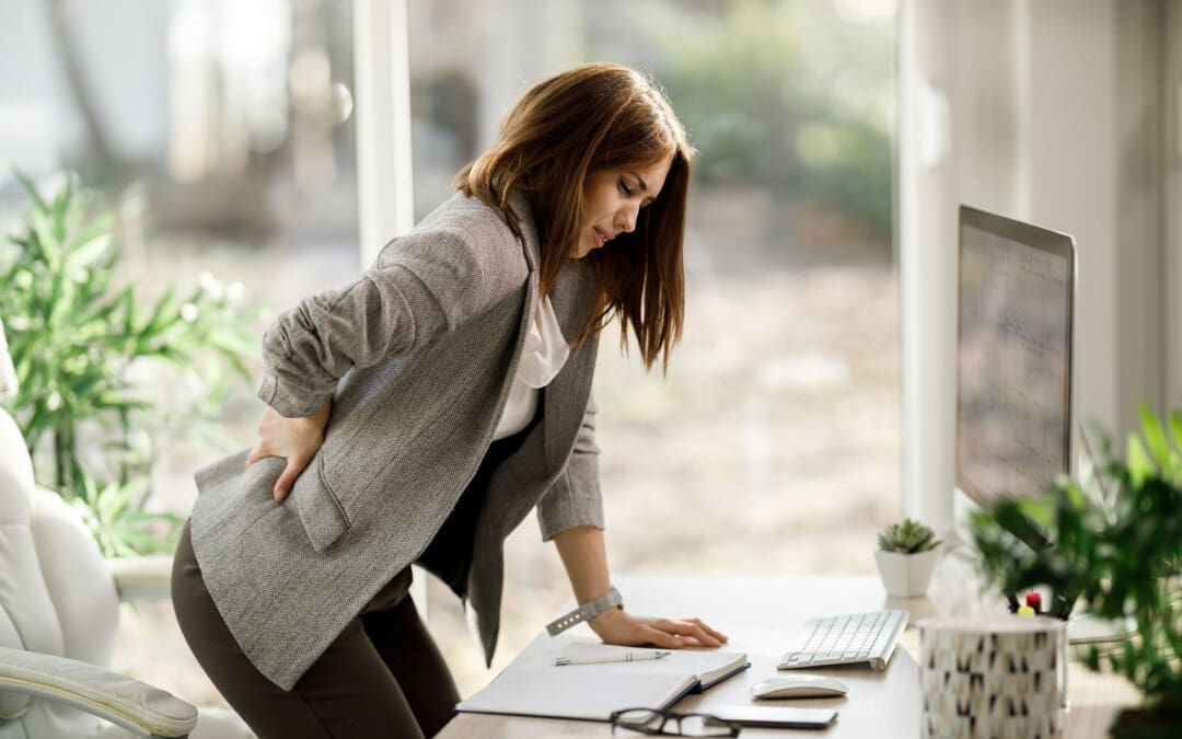
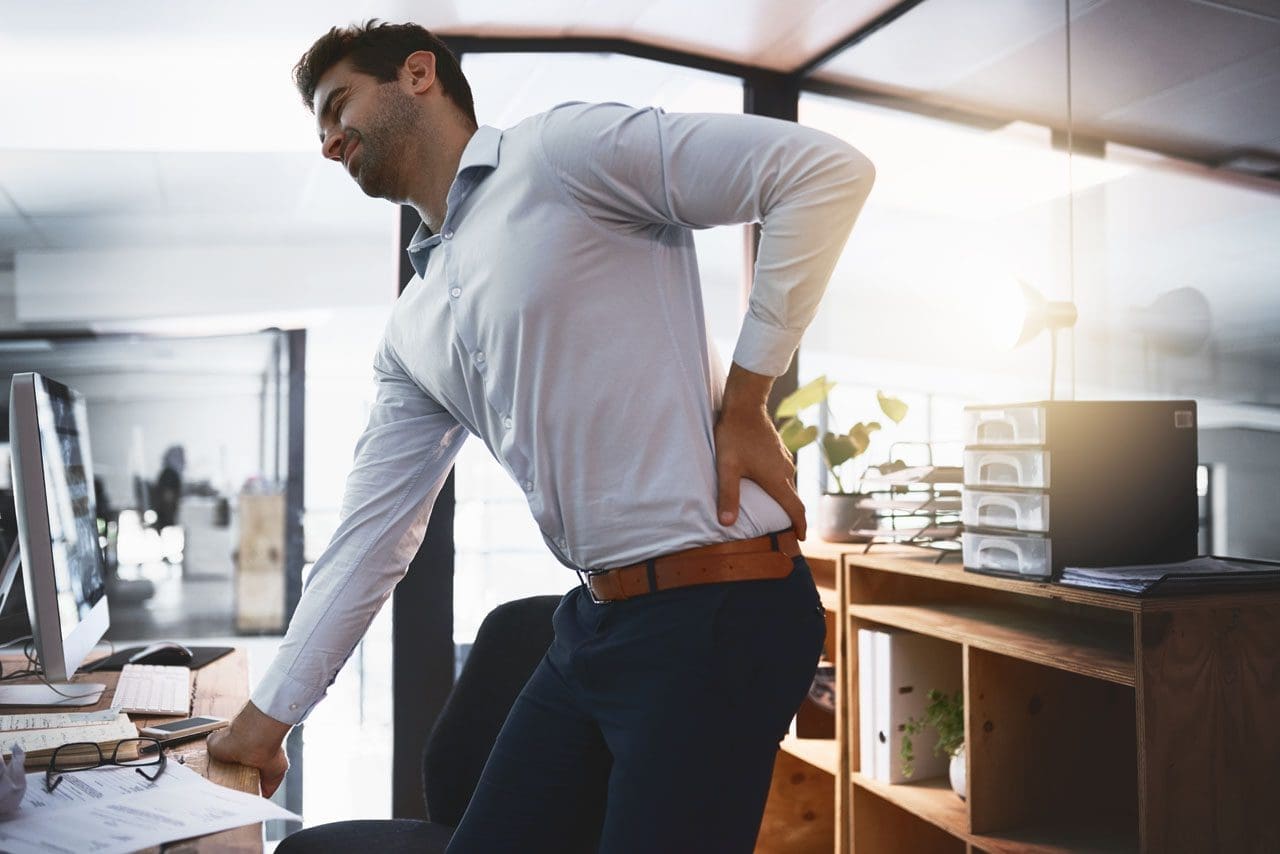
Individuals who sit in an office chair for a long period naturally tend to slouch over or slouch down into the chair. This posture can overstretch the spinal ligaments, strain the discs and surrounding structures in the spine, and contribute to or worsen back pain. Can the right office chair settings help relieve and prevent future injuries?
Work Office Chair
Sitting in an office chair for prolonged periods can cause lower back pain or worsen an existing back problem. The main reason behind this is that sitting is a static posture that increases stress in the back, shoulders, arms, and legs, putting pressure on the back muscles and spinal discs. Over time, incorrect sitting posture can damage the spinal structures. Most office chair customers base their decisions on availability, color, style, and price. However, it’s recommended that individuals learn about fitting seating equipment to their height, frame, or any condition, such as back or hip pain. The controls on or under the armrests and at the bottom of the chair seat, including the levers, paddles, and knobs, are there for a reason. Here are some recommendations for adjusting their work office chair to decrease back and hip pain.
Height Adjustment
The chair’s height affects the quadriceps, psoas, and hamstring muscles, which are important in posture-related back pain. Height adjustment is the primary way to change the angle of the hip joint while sitting. This angle affects the position of the pelvis and the degree of curve in the lower back, which can alter the spine’s normal alignment. (De Carvalho D. et al., 2017) Adjusting the chair’s height can provide a reference for other chair and workstation adjustments.
Back Angle
The hip angle is how close (increased hip flexion) or how far away (less hip flexion) the trunk is to the top of the thigh when sitting. Adjusting chair height can control the angle of the hip joint. When adjusting the height, adjust the degree of flexion at the hip joint to ensure ideal alignment for the individual body frame. A recent study measured the load that sitting has on simulated spinal discs. Researchers concluded that pressure on the spine can be relieved with a more open angle between the trunk and the thigh, that is, the hip joint angle. (Rohlmann A. et al., 2011) The backrest, seat tilt, and lumbar support features help maintain a pain-free back and relieve pressure and tension in the lower back and should be utilized.
Seat Depth
A kitchen chair, for example, may have no back supports or armrests to assist with posture and angle. (Holzgreve F. et al., 2022) When sitting, the individual’s back should be against the back of the work office chair for the best support and a healthy posture. Individuals will want to check and see how concave the seat surface is. If there is a prominent curve, this will cause the back to be rounded when sitting, which can become uncomfortable and lead to back pain. A chair with built-in support or a pillow can help adjust the depth. This means that chair size matters, as different people will need seats of varying depths to match their musculoskeletal structure.
Seat Height
Another way to understand the hip joint angle is to compare the height of the knees to the height of the hips. This is usually the easiest way to assess whether the chair height is right while adjusting. When the seat is right, the feet will be flat on the floor. The feet should reach the floor without causing pressure on the back of the thighs. Individuals with dangling feet, which may be because of their height, should place a footrest or thick book under them. The knees should be approximately level with or lower than the hips. In this case, the level is a 90-degree angle between the hip and trunk, which is stress-free on the hips and back.
Risks
Chair Too High
For individuals who can’t reach their feet to the floor, the chair is probably too high. The Occupational Safety and Health Administration (OSHA) says this is potentially hazardous because it can lead to scooting forward and forgoing the backrest’s support (United States Department of Labor, N.D.) Sitting like this is considered an awkward posture and a risk factor for work-related musculoskeletal disorders (MSD). Musculoskeletal disorders and symptoms caused by awkward sitting posture include muscle fatigue, swelling, pain, numbness, or decreased circulation. (Ng, P. K., Jee, K. S. and Lim, S. Y. 2016)
Chair Too Low
If the knees are higher than the hips, the chair is probably too low, causing extreme flexion in the hip joints. Most individuals’ backs can’t handle this well because their hip muscles are not flexible enough. If sitting with knees higher than hips, the position can cause lower back pain.
Taking Breaks
No matter how comfortable a work office chair is, prolonged static posture is unhealthy for the spine and is a common contributor to back problems and muscle strain. Remember to stand, stretch, and walk for at least a minute or two every half hour to prevent the back from staying in one position for a long period. A quick stretch or minimal movement, like a quick walk, will help. A longer walk will help even more, promoting blood circulation to supply nutrients to all the spinal structures.
Moving and stretching regularly throughout the day will help keep the joints, ligaments, muscles, and tendons loose and promote comfort, relaxation, and the ability to focus productively.
Injury Medical Chiropractic and Functional Medicine Clinic works with primary healthcare providers and specialists to develop personalized treatment programs. An integrated approach to treating injuries and chronic pain syndromes improves flexibility, mobility, and agility, relieving pain and helping individuals return to normal activities. If other treatments are needed, Dr. Jimenez has teamed up with top surgeons, clinical specialists, medical researchers, and rehabilitation providers.
Low Back Pain: Impact and Chiropractic Solutions
References
De Carvalho, D., Grondin, D., & Callaghan, J. (2017). The impact of office chair features on lumbar lordosis, intervertebral joint and sacral tilt angles: a radiographic assessment. Ergonomics, 60(10), 1393–1404. https://doi.org/10.1080/00140139.2016.1265670
Rohlmann, A., Zander, T., Graichen, F., Dreischarf, M., & Bergmann, G. (2011). Measured loads on a vertebral body replacement during sitting. The spine journal : official journal of the North American Spine Society, 11(9), 870–875. https://doi.org/10.1016/j.spinee.2011.06.017
Holzgreve, F., Maurer-Grubinger, C., Fraeulin, L., Bausch, J., Groneberg, D. A., & Ohlendorf, D. (2022). Home office versus ergonomic workstation – is the ergonomic risk increased when working at the dining table? An inertial motion capture based pilot study. BMC musculoskeletal disorders, 23(1), 745. https://doi.org/10.1186/s12891-022-05704-z
United States Department of Labor. (N.D.). Computer workstations eTool. Retrieved from https://www.osha.gov/etools/computer-workstations
Ng, P. K., Jee, K. S. & Lim, S. Y. (2016). Development of Ergonomics Guidelines for Improved Sitting Postures in the Classroom among Malaysian University Students. American Journal of Applied Sciences, 13(8), 907-912. https://doi.org/10.3844/ajassp.2016.907.912
IFM's Find A Practitioner tool is the largest referral network in Functional Medicine, created to help patients locate Functional Medicine practitioners anywhere in the world. IFM Certified Practitioners are listed first in the search results, given their extensive education in Functional Medicine