Spinal disc deterioration from aging is normal, but health issues or injuries can advance the degenerative process. Disc protrusions are related to herniated discs but are the mildest form of the condition and are a common form of spinal disc deterioration that can cause neck and back issues. However, individuals may have a small protruding disc that can go undetected unless it irritates or compresses the surrounding nerves. Chiropractic care, decompression, and massage therapy can realign the disc back into position, relieving discomfort and pain.
Disc Protrusion
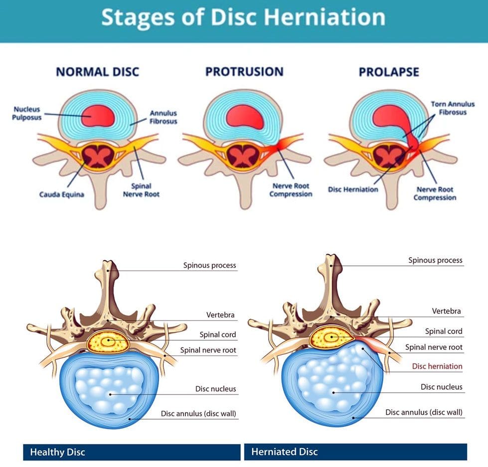
A disc is like a sturdy soft rubber shock absorber/cushion with added gel inside. The gel acts as a shock absorber. When the gel begins to protrude out slightly, this is a disc protrusion. Once a protruding disc begins to develop, it usually remains in that position. The disc can sometimes reabsorb on its own and realign back into position, but there is no way of knowing that will happen or how long it will take. With age and/or injuries, the body’s parts change. The spine’s discs dehydrate and lose elasticity weakening the discs and making them more vulnerable to herniation stages:
First Stage
Following natural weakening can be classified as a disc protrusion when the disc’s core begins pushing into the spinal column.
Disc protrusions can be tiny or push out an entire side of the disc.
Second Stage
Disc deterioration often consists of a bulging disc when the core pushes out farther around the circumference beyond the disc’s outer layer, called the annulus fibrosus, creating the telltale bulge.
A bulging disc involves more than 180 degrees of the disc’s circumference.
Third Stage
The third stage is a herniated disc, meaning the disc’s outer wall has torn, allowing the inner gel to leak out, usually irritating the surrounding nerves.
Fourth Stage
The fourth stage is sequestration, a herniated disc in which a piece of the nucleus breaks free of the vertebral disc fragments and falls into the spinal canal.
Types
A disc protrusion is one type of disc herniation that pushes out but remains connected. Different types compress and irritate the discs differently and produce various symptoms, including:
Paracentral
This is the most common, where the disc protrusion jams the space between the central canal and the foramen.
Central
This is where the disc protrusion impinges into the spinal canal, with or without spinal cord compression.
Foraminal
The disc intrudes into the foramen, the space through which nerve roots branch off the spinal cord and exit the vertebrae.
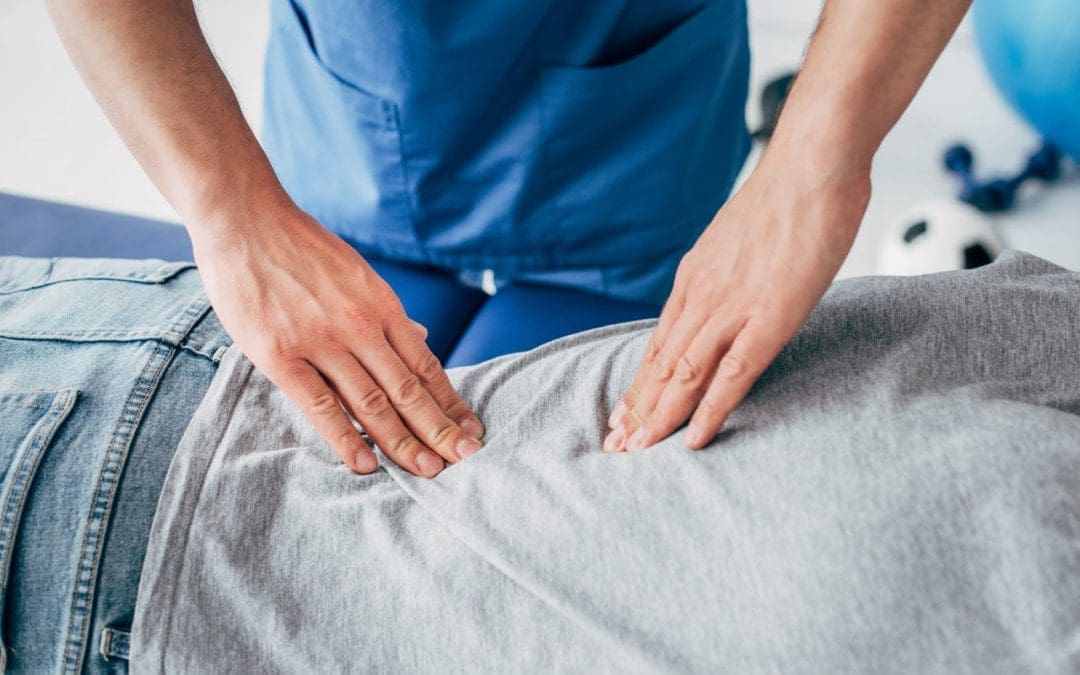
Symptoms, Diagnosis, and Chiropractic Care
Individuals with a disc protrusion can have symptoms similar to sciatica, which includes back, buttock, and leg discomfort, numbness, and pain sensations.
Treatment for disc protrusion will be based on the individual’s symptoms.
A chiropractor will take a detailed medical history and perform a physical examination.
A spinal MRI test could be ordered depending on the injury or condition.
A customized treatment plan will be developed to fit the individual’s medical needs.
Most disc protrusions improve after a few weeks of rest, avoiding strenuous activities, activity modification, an anti-inflammatory diet, and gentle exercises that the chiropractic team will provide.
True Spinal Decompression
References
Fardon, David F et al. “Lumbar disc nomenclature: version 2.0: Recommendations of the combined task forces of the North American Spine Society, the American Society of Spine Radiology and the American Society of Neuroradiology.” The spine journal: official journal of the North American Spine Society vol. 14,11 (2014): 2525-45. doi:10.1016/j.spinee.2014.04.022
Mysliwiec, Lawrence Walter, et al. “MSU classification for herniated lumbar discs on MRI: toward developing objective criteria for surgical selection.” The European spine journal: official publication of the European Spine Society, the European Spinal Deformity Society, and the European Section of the Cervical Spine Research Society vol. 19,7 (2010): 1087-93. doi:10.1007/s00586-009-1274-4
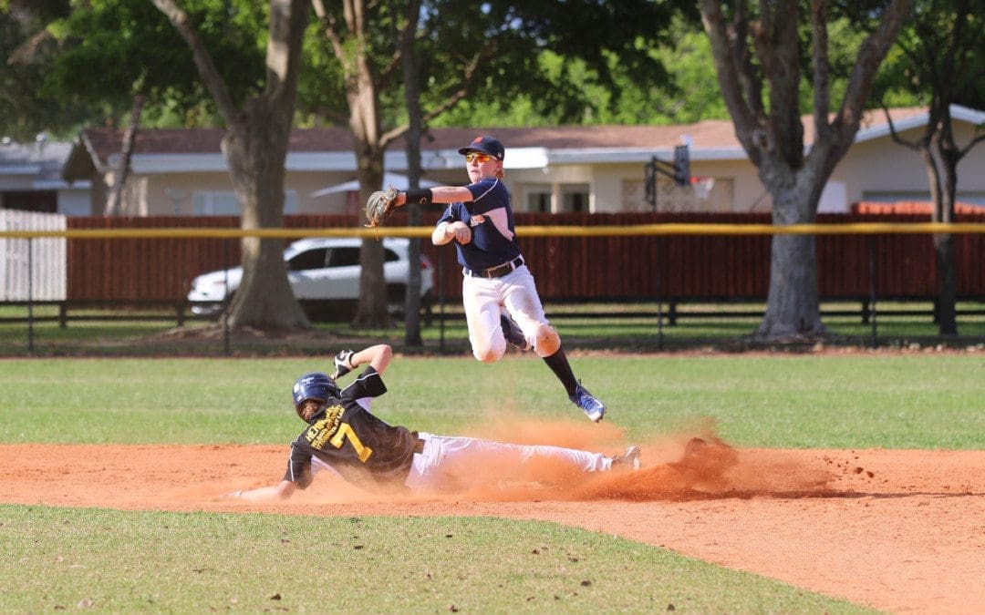
The game of baseball takes a toll on the body, especially when players advance from little league to high school, college, minor league, and the pros. The most common baseball injuries can range from mild to severe, from normal wear and tear on the joints and muscles to repetitive stress injuries, collisions with other players, getting hit with the ball, or bodily trauma. A chiropractor can provide ideal treatment for players of all ages and levels with decreased downtime and expedited healing and recovery.
Baseball Injuries
Although there have been a lot of advances in player safety and health, from helmets with face guards to shin and arm padding, the equipment lessens the impact and risks of injury. The game still involves running, sliding, twisting, and jumping, causing the body to maneuver awkwardly. Players often report sliding into first, feeling a pop or twisting to catch a fly ball, and feeling something snap. The most common injuries include:
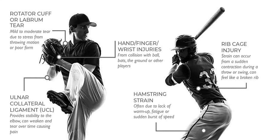
Torn Labrum
Cartilage surrounding the shoulder joint socket, known as the labrum, often gets torn.
The soft tissue keeps the bones in place and provides stability.
Pitching and throwing motions stress the labrum.
With time, the cartilage begins to overstretch and tear, leading to swelling, shoulder pain, weakness, and overall instability.
Rotator Cuff Tears
The rotator cuff structure involves a complex set of tendons and muscles that stabilize the shoulder.
Pitchers are the most vulnerable, but all players are susceptible.
Cases are caused by not warming up and stretching correctly and repetitive/overuse movements.
Swelling and pain are the most common symptoms.
With a severe tear, a player will lose the ability to rotate the shoulder correctly.
Shoulder Instability or Dead Arm
This is when the shoulder muscles become overly fatigued, and the joint becomes unstable, losing the ability to throw precisely.
The condition is called dead arm by players and trainers.
This type of injury is caused by overuse and repeated stress.
Healing involves letting the shoulder rest for an extended period, but treatment, like chiropractic or physical therapy, could be recommended depending on the severity.
Pitchers Elbow
A pitcher’s elbow injury is caused by overuse and sustained/repeated damage to the tendons that rotate the wrist.
Pain and swelling occur along the inside of the elbow and forearm.
Wrist Tendonitis and Trauma
Wrist Tendonitis or tenosynovitis happens when the ligaments and tendons become tender, swollen, ruptured, or torn.
This causes inflammation, pain, and weakness.
Trauma injuries can result from collisions with another player, the ground, or a ball.
Knee Tears and Trauma
Knee injuries can be caused by normal wear and tear, overuse, or traumatic impact.
The fibrous bands are what stabilize and cushion the knee.
Overuse and any awkward movement can cause the tearing of the various ligaments.
The bands can develop micro-tears or complete ruptures, causing inflammation, pain, and instability.
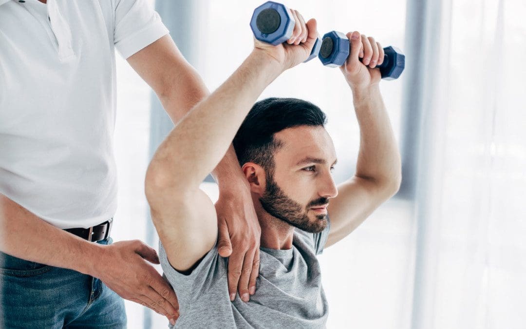
Chiropractic Care and Rehabilitation
Chiropractic treatment and physical therapy have been found to help athletes maintain flexibility and range of motion, rehabilitate the body after an injury, and prevent new injuries or worsening of current injuries.
Chiropractic helps stretch and flex the muscles to stay limber and less prone to injury.
Chiropractic is a natural pain reliever for sore muscles and joint pain.
Physical therapy can strengthen an injured area during recovery and educate on proper form and techniques.
Taping and strapping can help support the elbows, wrists, ankles, and knees, reducing stress.
A combination of treatment approaches can help decrease recovery time so players can get back on the field.
Shoulder Adjustment Baseball Injuries
References
Bullock, Garrett S et al. “Shoulder Range of Motion and Baseball Arm Injuries: A Systematic Review and Meta-Analysis.” Journal of athletic training vol. 53,12 (2018): 1190-1199. doi:10.4085/1062-6050-439-17
Lyman, Stephen, and Glenn S Fleisig. “Baseball injuries.” Medicine and sport science vol. 49 (2005): 9-30. doi:10.1159/000085340
Matsel, Kyle A et al. “Current Concepts in Arm Care Exercise Programs and Injury Risk Reduction in Adolescent Baseball Players: A Clinical Review.” Sports health vol. 13,3 (2021): 245-250. doi:10.1177/1941738120976384
Shitara, Hitoshi, et al. “Shoulder Stretching Intervention Reduces the Incidence of Shoulder and Elbow Injuries in High School Baseball Players: a Time-to-Event Analysis.” Scientific reports vol. 7 45304. 27 Mar. 2017, doi:10.1038/srep45304
Wilk, Kevin E, and Christopher A Arrigo. “Rehabilitation of Elbow Injuries: Nonoperative and Operative.” Clinics in sports medicine vol. 39,3 (2020): 687-715. doi:10.1016/j.csm.2020.02.010
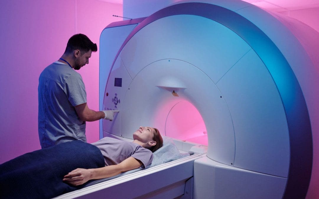
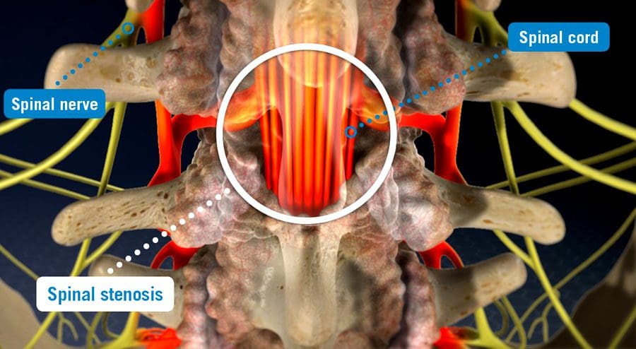
Spinal stenosis is when space somewhere along or within the spine begins to narrow, closing off the ability of normal/comfortable movement and nerve circulation. It can affect different areas, including the cervical/neck, lumbar/low back, and, less commonly, the thoracic/upper or mid-back regions causing tingling, numbness, cramping, pain, muscle weakness, or a combination in the back, leg/s, thighs, and buttocks. There can be various factors causing the stenosis; correct diagnosing is the first step, and where a spinal stenosis MRI comes in.
Spinal Stenosis MRI
Stenosis can be challenging to diagnose as it is more of a symptom/complication than a condition, often caused by herniated discs, bone spurs, a congenital condition, post-surgery, or after an infection. Magnetic resonance imaging/MRI is a common test used in diagnosis.
Diagnosis
A healthcare professional, like a chiropractor, physical therapist, spine specialist, or physician, will begin with understanding symptoms and medical history.
A physical exam will be conducted to learn more about the location, duration, positions, or activities that decrease or worsen the symptoms.
Additional tests include muscle strength, gain analysis, and balance testing to help better understand where the pain is coming from.
To confirm a diagnosis, imaging will be required to see what is going on.
An MRI uses computer-generated imaging to produce images that show bone and soft tissues, like muscles, nerves, and tendons, and if they are compressed or irritated.
A healthcare professional and MRI technician will go over the safety requirements before the imaging.
Because the machine uses powerful magnets, there can be no metal on or in the body, like implanted prostheses or devices that include:
A different imaging test may be used if an individual cannot have an MRI like a CT scan.
An MRI can range from several minutes to an hour or longer, depending on how many positions are necessary to isolate the injured area and get a clear image. The test is painless, but sometimes individuals are asked to maintain a specific position that could be uncomfortable. The technician/s will ask if there is discomfort and offer any help to make the experience as easy as possible.
Treatment
Not all cases of stenosis cause symptoms, but there are treatment options that a healthcare professional can recommend.
Conservative care is the first recommendation that includes chiropractic, decompression, traction, and physical therapy.
Treatment increases muscle strength, improves range of motion, improves posture and balance, decreases discomfort symptoms, and incorporates strategies to prevent and manage symptoms.
Prescription medications could be part of a larger treatment plan.
Surgery could become an option in more severe cases where conservative care is not working.
Spinal Stenosis
References
Database of Abstracts of Reviews of Effects (DARE): Quality-assessed Reviews [Internet]. York (UK): Centre for Reviews and Dissemination (UK); 1995-. Diagnosis of lumbar spinal stenosis: an updated systematic review of the accuracy of diagnostic tests. 2013. Available from: https://www.ncbi.nlm.nih.gov/books/NBK142906/
Ghadimi M, Sapra A. Magnetic Resonance Imaging Contraindications. [Updated 2022 May 8]. In: StatPearls [Internet]. Treasure Island (FL): StatPearls Publishing; 2022 Jan-. Available from: https://www.ncbi.nlm.nih.gov/books/NBK551669/
Gofur EM, Singh P. Anatomy, Back, Vertebral Canal Blood Supply. [Updated 2021 Jul 26]. In: StatPearls [Internet]. Treasure Island (FL): StatPearls Publishing; 2022 Jan-. Available from: https://www.ncbi.nlm.nih.gov/books/NBK541083/
Lurie, Jon, and Christy Tomkins-Lane. “Management of lumbar spinal stenosis.” BMJ (Clinical research ed.) vol. 352 h6234. 4 Jan. 2016, doi:10.1136/bmj.h6234
Stuber, Kent, et al. “Chiropractic treatment of lumbar spinal stenosis: a review of the literature.” Journal of chiropractic medicine vol. 8,2 (2009): 77-85. doi:10.1016/j.jcm.2009.02.001
Football season is here, and the sport demands healthy, strong bodies. It is explosive, with high-intensity plays lasting between 2-15 seconds. Strength and power are put out in a few moments then the player rests up and does it again. A football training chiropractor can take players to another level with therapeutic massage, body strengthening, and rehabilitation so that players can enjoy a healthy injury-free season.
Football Training
Stretching and Warm-Up
Stretching and a dynamic warm-up are essential to strengthening the body and preventing injury. Stretching is necessary to increase the range of motion in the muscles, especially when the body is in an awkward position; it can adapt. A dynamic warm-up increases the core temperature of the muscles and prepares the muscles, joints, and nervous system for the physical event. The critical muscles are the hip flexors, hamstrings, and calves. The warm-up consists of a series of progressive movement drills that include:
Proper stretching, followed by a dynamic warm-up, will maximize performance.
Cardio, Aerobic, and Anaerobic Fitness
Cardio training increases oxygen and blood circulation to play for a long time without getting tired.
Aerobic fitness increases oxygen and provides endurance to break through or enhance tackles, sustained effort, and strength.
Anaerobic fitness utilizes high-intensity exercises to challenge the body without using a lot of oxygen like cardio and aerobics do.
All are important, especially for players playing the whole or most of the game.
Core Strength
The core is where power and strength come from. It refers to the muscles around the trunk and pelvis, including the diaphragm, abdominal wall, low back, and hips. Reinforcing the core will enhance balance, stability, and efficiency and reduce the risk of injury. The core muscles under the washboard abs link upper-body power with lower-body torque. In-season strength training provides a progressive buildup to optimal fitness and performance. The focus is on the following:
Speed maintenance.
Aerobic and anaerobic fitness.
Strength and power.
Emphasis on injury prevention training stabilizer muscles for balance and agility.
It is recommended to allow at least two days between training sessions and games. Avoid strength training on the same day as working out on the field.
Rest entirely from strength training for one week in five.
Light workouts are fine.
Hydration
Football players have unique hydration needs due to the exposure to extreme heat or cold while wearing heavy equipment. Top-rated athletic trainers monitor weather conditions, length and time of day at practice and games, and the hydration levels of each player. Hydration recommendations include:
Hydrate before, during, and after practices and games.
Two to three hours before the game, drink 17 to 20 fluid ounces of water or a sports drink.
Ten to 20 minutes before the game, drink seven to 10 fluid ounces of water or a sports drink.
During practices, drink seven to 10 fluid ounces of water or sports drink every 10 to 20 minutes with the helmet off.
Post-practice/game, correct any fluid loss, ideally within two hours.
The hydration should contain water to restore hydration, carbohydrates to replenish glycogen stores, and electrolytes to speed the recovery process.
Football Training Chiropractic
Chiropractic care has become integral to NFL players’ health and training programs. All 32 teams have a chiropractor, and according to the Professional Football Chiropractic Society, the average NFL team chiropractor gives 30-50 treatments a week. Chiropractic treats conditions like neuromusculoskeletal strain injuries, neck pain, back pain, strains to the hamstring and quadriceps, and injuries caused by whiplash-like movements. Benefits include:
Iaia, F Marcello, et al. “High-intensity training in football.” International journal of sports physiology and performance vol. 4,3 (2009): 291-306. doi:10.1123/ijspp.4.3.291
Lorenz, Daniel, and Scot Morrison. “CURRENT CONCEPTS IN PERIODIZATION OF STRENGTH AND CONDITIONING FOR THE SPORTS PHYSICAL THERAPIST.” International journal of sports physical therapy vol. 10,6 (2015): 734-47.
Robbins, Daniel W. The Normalization of Explosive Functional Movements in a Diverse Population of Elite American Football Players. Journal of Strength and Conditioning Research: April 2012 – Volume 26 – Issue 4 – p 995-1000
doi: 10.1519/JSC.0b013e31822d53b7
Stump, John L, and Daniel Redwood. “The use and role of sports chiropractors in the national football league: a short report.” Journal of manipulative and physiological therapeutics vol. 25,3 (2002): E2. doi:10.1067/mmt.2002.122326
Zein MI, Saryono S, Laily I, Garcia-Jimenez JV. The effect of high-intensity circuit training-modified FIFA 11+ program on physical fitness among young football players. J Sports Med Phys Fitness 2020;60:11-6. DOI: 10.23736/S0022-4707.19.09813-X
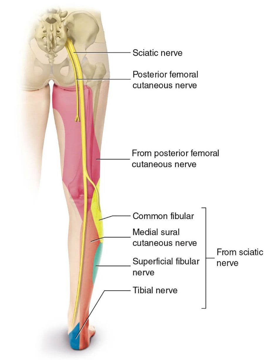
Sciatica pain can radiate to the knee. Individuals with sciatica do report unique/unusual knee pain that was never there and no past or recent physical injuries. Sciatica is the culprit, as the knee muscles are powered and controlled by nerves in the lower spine. Irritation or compression of these nerves can cause symptoms that can include: random back pain, hamstring tightness, weakness in the hips or quadriceps, the development of bunions, and knee pain and/or weakness. Chiropractic treatment can release the compression, heal the sciatic nerve, and alleviate knee problems.
Sciatica Pain Can Radiate To The Knee
Spine conditions that can cause sciatica include:
Disc herniation – Where the inside of the discs leak out and compress and/or irritate surrounding nerves.
Spinal stenosis – The spinal canal begins to narrow, not allowing enough space for the nerves to rest comfortably, resulting in compressed nerves.
Spondylolisthesis – A condition that occurs when a vertebrae slips forward onto the vertebrae below it.
Any can cause irritation, inflammation, or compression of the sciatic nerve leading to painful sensations that extends from the lower back down through the leg.
Symptoms
Common knee symptoms that may be experienced with sciatica include:
A dull ache, warm sensation, or sharp pain around the knee.
Bunions form from weakened stabilizing muscles that affect walking, running, and standing posture.
As sciatica pain can radiate to the knee, individuals will usually also experience pain in their buttocks, thigh, calf, and/or foot. The nerve sensations and other symptoms in the knee can be felt through a branch of the sciatic nerve known as the peroneal nerve.
Duration
The knee pain will last as long as sciatica does, depending on the type of sciatica, whether it is acute or chronic.
An acute sciatic episode usually resolves after a few weeks, with possible future flare-ups.
Chronic sciatica is a long-term condition that does not resolve independently and necessitates intervention by a specialist.
Chiropractic Treatment Plan
Depending on the diagnosis, a chiropractor will develop a personalized treatment plan to address the root cause and heal the injury. The treatment plan will include therapeutic massage, posture training, and at-home self-care to help heal and prevent future sciatica.
Massage Rehabilitation
Physical therapy and therapeutic massage will loosen and relax the muscles, nerves, tendons, and ligaments.
Heat and ice, exercises, and stretches will prepare the muscles and nerves for chiropractic decompression adjustments.
Posture Training
Training will be provided to maintain the back, hips, knees, and feet in proper alignment.
Training on removing pressure from the lower back and restabilizing the rest of the body.
Training on proper body mechanics, safe lifting techniques, and injury prevention.
Self-Pain Management
Training on self-care habits that include healthy weight, core strengthening exercises and stretches for the back muscles, and proper rest for a full recovery.
An anti-inflammatory diet to reduce/eliminate inflammation and achieve a healthy weight and a nutrition plan to maintain overall health.
Surgery
Surgery is the final option when conservative treatments are not working.
Treating Severe & Complex Sciatica Syndromes
References
Dydyk AM, Khan MZ, Singh P. Radicular Back Pain. [Updated 2021 Nov 2]. In: StatPearls [Internet]. Treasure Island (FL): StatPearls Publishing; 2022 Jan-. Available from: https://www.ncbi.nlm.nih.gov/books/NBK546593/
Hirabayashi, Hiroki, et al. “Characteristics of L3 nerve root radiculopathy.” Surgical neurology vol. 72,1 (2009): 36-40; discussion 40. doi:10.1016/j.surneu.2008.08.073
Jandre Reis, Felipe Jose, and Adriana Ribeiro Macedo. “Influence of Hamstring Tightness in Pelvic, Lumbar and Trunk Range of Motion in Low Back Pain and Asymptomatic Volunteers during forwarding Bending.” Asian spine journal vol. 9,4 (2015): 535-40. doi:10.4184/asj.2015.9.4.535
Jeong, Ui-Cheol, et al. “The effects of self-mobilization techniques for the sciatic nerves on physical functions and health of low back pain patients with lower limb radiating pain.” Journal of physical therapy science vol. 28,1 (2016): 46-50. doi:10.1589/jpts.28.46
Toxin overload is the condition of having an excessive amount of toxins in the body. Harmful substances can come from water, food, cleaning products, and environmental sources that individuals are exposed to regularly. Toxins are also produced in the body by poor gut health through autointoxication. Considering the number of toxins from food additives, preservatives, and perfumes to cleaning products, cosmetic products, and plastic water bottles, much of everyday life include exposure to chemicals that are not healthy. That’s why it’s recommended to undergo regular detoxes to ensure optimal body function and disease prevention.
Toxin Overload
One of the main ways toxins damage the body is they poison the enzymes, which prevents the body from functioning correctly. The body relies on enzymes for every physiological function. When toxins damage the enzymes, the production of hemoglobin in the blood is prevented, which can accelerate aging and lead to the failure of energy production and lower protection against oxidated stress. The failure of normal body functions increases the risk of diseases that include:
Proper waste elimination is essential to optimal health.
80% of the immune system is in the gut, and with a compromised digestive system, toxins can begin to accumulate.
Fatigue
When the body efficiently delivers nutrients to the cells and eliminates waste, there should be balanced energy throughout the day.
Toxin overload can cause individuals to experience fatigue, even in individuals that eat healthily and exercise, which could be an indicator of accumulation.
Chronic fatigue and viral infections could present from a weakened immune system.
Muscle Joint Aches and Pains
When gut health is compromised, undigested food particles can cause tears in the lining of the intestinal wall leading to a leaky gut.
The food particles enter the bloodstream and can cause an inflammatory response.
They can lodge themselves in weak areas of the joints, causing pain and increased muscle soreness.
Proper digestion and detoxification help eliminate toxins from the joints and muscles and heal the damaged lining.
Insomnia
Sleep is when the body detoxes, repairs, and rejuvenates itself.
Sleep problems could be a sign that the body is struggling to detoxify.
Chronic Headaches
Chronic headaches often result from imbalances in the body resulting from toxin overload and obstructed/blocked detoxification pathways.
Fluid Retention and Congestion
The lymphatic system is part of the circulatory system. The primary function is to transport lymph, a clear fluid that contains white blood cells essential for regulating inflammation.
Diet, hormone imbalances, sedentary lifestyle, medications, and genetics can contribute to fluid retention and congestion, causing stagnation of the lymphatic system.
If the system becomes congested, it can cause pain and swelling.
Unusual Weight Loss or Gain
Increased belly/visceral fat is the fat stored within the abdominal cavity. This is the most dangerous fat because of its proximity to vital organs like the liver, pancreas, and stomach.
Visceral fat or active fat influences how hormones function in the body. Stress, lack of exercise, and an unhealthy diet contribute to excess visceral fat.
Individuals trying to lose weight unsuccessfully could be a sign of having excessive toxins in the body.
Skin Problems
The skin reveals what’s happening inside the body.
Acne, rosacea, eczema, or other chronic skin issues, could indicate toxins are traveling through the skin.
When waste is not eliminated thoroughly through sweat, urine, and feces, the body could try to expel it through the skin.
Improving the body’s digestive and detoxification processes can help heal the root problem.
Giannini, Edoardo G et al. “Liver enzyme alteration: a guide for clinicians.” CMAJ : Canadian Medical Association journal = journal de l’Association medicale canadienne vol. 172,3 (2005): 367-79. doi:10.1503/cmaj.1040752
Grant, D M. “Detoxification pathways in the liver.” Journal of inherited metabolic disease vol. 14,4 (1991): 421-30. doi:10.1007/BF01797915
Lala V, Goyal A, Minter DA. Liver Function Tests. [Updated 2022 Mar 19]. In: StatPearls [Internet]. Treasure Island (FL): StatPearls Publishing; 2022 Jan-. Available from: https://www.ncbi.nlm.nih.gov/books/NBK482489/
Mattick, R P, and W Hall. “Are detoxification programmes effective?.” Lancet (London, England) vol. 347,8994 (1996): 97-100. doi:10.1016/s0140-6736(96)90215-9
Seaman, David R. “Toxins, Toxicity, and Endotoxemia: A Historical and Clinical Perspective for Chiropractors.” Journal of chiropractic humanities vol. 23,1 68-76. 3 Sep. 2016, doi:10.1016/j.echu.2016.07.003
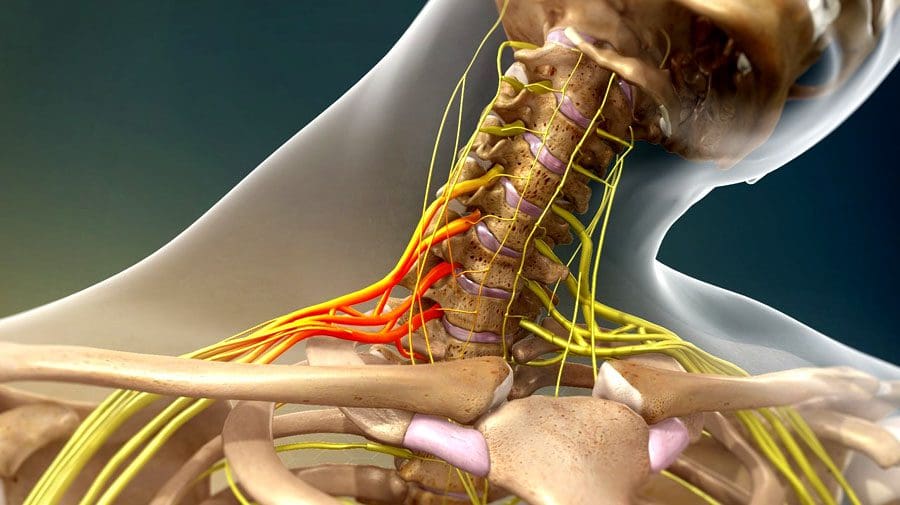
The body’s nerves are the communication system that carries messages between the brain and the rest of the body. Some nerves transmit messages from the brain to muscles to make the body move, while others relay pain, pressure, or temperature signals. Tiny fibers bundled inside each nerve carry the messages with an outer layer/sheathing that insulates and protects the nerves. The brachial plexus is a network of nerves that send signals from the spinal cord to the shoulders, arms, and hands. A brachial plexus nerve injury occurs when the nerves are over-stretched, compressed, torn, cut, or ripped from the spinal cord.
Brachial Plexus Nerve Injury
The injury involves the head or neck hitting or getting hit and shifting to one side while the shoulder is stretched/pulled in the opposite direction.
Minor brachial plexus injuries are commonly known as stingers or burners and are common in sports like football, wrestling, hockey, soccer, and basketball.
Severe brachial plexus injuries can cause arm paralysis and usually result from vehicle or motorcycle accidents.
Other conditions like inflammation or tumors can affect the brachial plexus.
Sometimes babies can sustain brachial plexus injuries during birth.
Pressure and stretching injuries do not physically sever the nerve but can disrupt communication.
Cutting injuries vary depending on the severity of the cut and because the nerves are in a protective canal that can also be fractured or broken. If the canal remains intact, the nerve fibers could grow back with time.
However, surgery is necessary to repair the damage if the canal is broken.
Signs and symptoms of a brachial plexus nerve injury can vary, depending on the severity and location of the injury. Usually, only one arm is affected.
Minor Injuries
Minor damage comes from over-stretching or mild compression.
An electric or burning sensation shoots down the arm.
Numbness and weakness in the arm.
Neck pain.
These symptoms usually last for a few seconds or minutes but can linger for days or longer.
Severe Injuries
More-severe symptoms result from injuries that impact, tear, or rupture the nerves.
The most severe injury occurs when the nerve root is torn from the spinal cord.
Symptoms include:
Intense pain.
Writhing neck pain.
Weakness or inability to use specific shoulder, arm, and/or hand muscles.
Complete lack of movement and feeling in the shoulder, arm, and/or hand.
Symptoms in both arms.
Complications
With time, most brachial plexus injuries in children and adults heal with minimal long-term damage. But some injuries can cause long-lasting problems that include:
Joint Stiffness
The joints can stiffen, making movement difficult.
Healthcare providers often recommend ongoing chiropractic and physical rehabilitation during recovery.
Atrophy
Nerves regrow slowly and can take some time to completely heal after the injury.
During that time, lack of use can cause the muscles to break down.
Chronic Pain
Nerve damage can cause pain signals to be constantly firing.
Numbness
It can occur in the arm or hand, increasing the risk of worsening the injury or causing new injuries.
Disability
Recovery from a severe brachial plexus injury depends on age, damage, location, and severity.
Even with surgery, individuals can experience long-term muscle weakness or paralysis.
Chiropractic Treatment and Rehabilitation
Treatment depends on the severity of the damage. Chiropractic can help realign, rehabilitate, stretch, and strengthen the muscles, nerves, tendons, joints, and ligaments to expedite recovery. For less severe injuries:
Muscle strengthening and posture exercises help maintain motion.
Therapeutic massage will stimulate circulation and keep the muscles loose.
For severe injuries:
Surgery
Continued chiropractic and physical rehabilitation to maintain thorough circulation, range of motion, and relaxed muscles.
The Brachial Plexus
References
Brucker, J et al. “Brachial plexus birth injury.” The Journal of neuroscience nursing: Journal of the American Association of Neuroscience Nurses vol. 23,6 (1991): 374-80. doi:10.1097/01376517-199112000-00006
Gutkowska, Olga, et al. “Brachial plexus injury after shoulder dislocation: a literature review.” Neurosurgical review vol. 43,2 (2020): 407-423. doi:10.1007/s10143-018-1001-x
Joyner, Benny, et al. “Brachial plexus injury.” Pediatrics in review vol. 27,6 (2006): 238-9. doi:10.1542/pir.27-6-238
Noland, Shelley S et al. “Adult Traumatic Brachial Plexus Injuries.” The Journal of the American Academy of Orthopaedic Surgeons vol. 27,19 (2019): 705-716. doi:10.5435/JAAOS-D-18-00433
IFM's Find A Practitioner tool is the largest referral network in Functional Medicine, created to help patients locate Functional Medicine practitioners anywhere in the world. IFM Certified Practitioners are listed first in the search results, given their extensive education in Functional Medicine