Can the short-term potato diet be a solution for individuals trying to lose weight?
Potato Diet
There is no one-size-fits-all approach to a healthy lifestyle diet. A successful nutrition plan needs to be personalized to the individual. Potatoes are an excellent source of fiber, vitamins, and minerals and are the primary source of calories in this diet. The potato diet has variations, but followers eat plain potatoes for several days in the diet’s simplest form.
What Can Be Eaten?
While there are variations, all potato diets are vegan, low in dietary fat, and encourage eating until satisfied. Instead of weighing portions or counting calories, individuals are encouraged to eat until full. The potato diet encourages volume eating, when individuals fill up on foods that are naturally lower in calories. Eating a large volume leaves the body feeling full, though fewer calories are eaten throughout the day.
Potato Types
The variety of potatoes allowed depends on the version of the potato diet being followed. Some require sticking to plain white potatoes, while a more forgiving version allows other varieties, such as yellow, red, and sweet potatoes.
Plant-based Foods, Low-Calorie, and Whole
In its purest form, the diet does not allow for other food besides potatoes, even fruits and vegetables. However, looser forms of the potato diet are designed to be more sustainable in the long term. Depending on the version of the potato diet being followed, individuals may be able to consume unprocessed foods in their whole form. However, potatoes should make up the bulk of the plate even when other foods are permitted. The additional foods are supplements to the potatoes for their nutritional content and include:
Fruits
Grains
Legumes
Vegetables
Condiments and Seasonings
Condiments, sauces, and seasonings are limited to small quantities of low-fat condiments like mustard and homemade ketchup. Salt is allowed to season the potatoes, but not a lot. Generally, sauces and seasonings should be made with fat-free ingredients and kept to a minimum.
Beverages
Water, plain tea, and plain coffee are the only beverages allowed on the potato diet. Staying hydrated is essential, so drinking plenty of water is encouraged.
What Is Restricted?
Depending on the diet version, certain foods are not allowed. In the strictest form, individuals eat only plain potatoes for several days.
Added Fats
Because the potato diet is low-fat, it strictly prohibits animal products and fats such as vegetable oils. Fats are a dense source of calories; even small amounts add up quickly. However, healthy fats can promote the absorption of potato nutrients. Because it is designed for maximum weight loss over a short period, the diet restricts added fats like:
Butter
Vegetable oil
Avocado
Nuts
Seeds
Processed and Refined Foods
The diet encourages eating whole, unprocessed foods during the plan’s duration. Processed foods are usually high in calories, fat, and sodium and are lower in nutrients than whole foods. For example, a baked potato is more nutritious than tater tots, fries, and potato chips, even though all are made with potatoes. Others include:
Bread
Cereal
Crackers
Muffins
Pasta
Doughnuts
Animal Products
All variations of the potato diet are vegan. While following the diet, all animal products are restricted and include:
Eggs
Dairy
Meat
Poultry
Fish
Seafood
Preparation
The diet is designed to be followed short-term for quick weight loss, so there is no meal schedule. Followers can eat breakfast, lunch, dinner, and snacks until full. Typically, individuals participate for two to five days, though some may follow the diet for up to a week. The potato preparation method is as important as the type of potato. It is recommended to use cooking methods that do not require added fat, such as boiling, steaming, baking, and roasting.
How many potatoes to eat in a day depends on the individual. A general recommendation is to eat approximately two to five pounds daily. Consuming enough calories in this diet is important since it is restrictive, and followers may not get enough nutrients if they eat too few calories. An extremely low-fat or fat-free diet is not sustainable. (Harvard Health Publishing, Harvard Medical School, 2021)
Pros
The potato diet can help with short-term weight loss and reduce fat and sodium intake, as well as a few other health benefits and include the following:
Weight Loss
The diet is designed for weight loss.
It is effective because it is naturally low in fat and calories.
Short-term
Individuals who prefer shorter diets for quick results may appreciate that the diet lasts two to five days.
Improve Digestion
Some like the diet for digestive benefits.
Potatoes are easy to digest, making this food gentle on the gastrointestinal tract.
Potatoes contain a rich source of fiber, which keeps food moving smoothly through the digestive system.
Easy to Follow
Mono diets are the easiest to follow.
It is easy to understand what is allowed and what to avoid.
Individuals who struggle with complicated rules will appreciate the simplicity.
Cons
The potato diet can come with health risks that include the following:
Sustainability
Eating only one food, even a root vegetable, is not sustainable.
Unbalanced
Many nutrient-rich foods are not allowed.
Though potatoes are nutritious, they lack some essential nutrients.
The diet can result in nutritional deficiencies.
Short-term Weight Loss
The weight loss may not be sustainable since the diet is followed for a few days.
Any weight lost during the diet may not be kept off long-term.
This may be water weight, which is different from losing body fat.
Once the individual returns to their normal lifestyle, they may regain some or all of the weight lost and possibly gain weight.
Discourages Healthy Eating
A nutrient-dense diet is rich in various foods.
The potato diet is nutritionally unbalanced and discourages healthy eating habits.
Injury Medical Chiropractic and Functional Medicine Clinic
This is a restrictive diet that is not meant to be followed long-term. Before starting a new diet plan, consult a healthcare provider or a registered dietitian, especially if underlying health conditions exist. Injury Medical Chiropractic and Functional Medicine Clinic works with primary healthcare providers and specialists to develop a personalized treatment plan through an integrated approach to treating injuries and chronic pain syndromes, improving flexibility, mobility, and agility programs to relieve pain and help individuals return to optimal function. Our providers use an integrated approach to create personalized care plans for each patient and restore health and function to the body through nutrition and wellness, functional medicine, acupuncture, Electroacupuncture, and sports medicine protocols. If the individual needs other treatment, they will be referred to a clinic or physician best suited for them. Dr. Jimenez has teamed up with top surgeons, clinical specialists, medical researchers, nutritionists, and health coaches to provide the most effective clinical treatments.
Functional Nutrition and Lifestyle Change
References
Harvard Health Publishing, Harvard Medical School. (2021). Know the facts about fats. https://www.health.harvard.edu/staying-healthy/know-the-facts-about-fats
Schaumberg, K., & Anderson, D. (2016). Dietary restraint and weight loss as risk factors for eating pathology. Eating behaviors, 23, 97–103. https://doi.org/10.1016/j.eatbeh.2016.08.009
U.S. Department of Health and Human Services and U.S. Department of Agriculture. (2020). 2020–2025 Dietary Guidelines for Americans. Retrieved from https://www.dietaryguidelines.gov/sites/default/files/2020-12/Dietary_Guidelines_for_Americans_2020-2025.pdf
Can understanding what knee tests are used help a healthcare provider diagnose the cause of individuals experiencing knee pain?
Knee Pain Tests
A knee examination is the first step in determining the cause of knee pain. Different knee tests may be performed during the exam to help the healthcare provider find the cause and develop an optimal treatment plan. These tests evaluate knee function and range of motion and look for conditions and injuries such as arthritis, meniscus tears, ACL tears, other ligament injuries, and kneecap issues.
Checking If There is Fluid in the Knee
Many individuals know if their knee is swollen, as they can see or feel the swelling. However, if there is excess fluid in the knee joint, the healthcare provider may compress the joint to feel for excess fluid. Fluid is often visible above the kneecap and can be compressed in this area. Fluid may also be detected in the back of the knee, referred to as a Baker’s cyst if the fluid has collected into a cluster. (Frush T. J., & Noyes F. R. 2015)
Arthritis Tests
Certain characteristic findings can detect knee arthritis:
Crepitus
Crepitus is the sensation when rough cartilage or exposed bone is rubbing when the knee is bent. (Lo G. H. et al., 2018)
The examiner will feel and listen for grinding as the knee is bent back and forth.
Deformity
As knee cartilage wears away, the knees can become progressively knock-kneed or bow-legged.
Limited Motion
If arthritis, bone spurs, and swelling prevent normal mobility, the knee’s range of motion often becomes limited.
Torn Meniscus Tests
Tests used to determine if there is a meniscus tear include:
Joint Line Tenderness
Joint line tenderness is a non-specific test in which the area of the meniscus is felt. It is considered a positive test when there is pain in this area.
McMurray’s test
This test is performed with the patient lying flat. The examiner bends the knee and rotates the shin bone.
This test is performed with the patient squatting.
The test is performed with the leg fully externally rotated or internally rotated, depending on whether the lateral or medial meniscus is being tested.
A click is heard or felt over the area of the tear.
ACL Tear Tests
These knee pain tests are for an anterior cruciate ligament (ACL) tear:
Lachman Test
The Lachman test is one of the most reliable to diagnose an ACL tear.
With the knee slightly bent, the examiner stabilizes the thigh while pulling the shin forward.
The shin shifts too far forward with a torn ACL.
Anterior Drawer Test
This test is performed with the patient lying flat.
The knee is bent 90 degrees, and then the shin is pulled forward to check the stability of the ACL.
Pivot Shift Test
The pivot shift test can be difficult, especially if the patient is experiencing discomfort and cannot relax the knee.
This test places stress on the knee joint and assesses the rotational stability of the ACL.
Other Ligament Injuries
For a suspected injury to other ligaments, including the posterior cruciate ligament (PCL), medial collateral ligament (MCL), and lateral collateral ligament (LCL), the following tests may be used:
Posterior Drawer Test
The posterior drawer is performed similarly to the anterior drawer test, in which the patient lies flat.
The knee is bent 90 degrees; the shin is pushed backward to check stability and function and detect if the posterior cruciate ligament (PCL) has been injured.
Collateral Ligament Stability
Side-to-side stability of the knee detects problems with the MCL and LCL.
The shin is shifted to each side, with the patient lying flat and the knee slightly bent.
The LCL or MCL damage causes the knee to open up too much, a condition known as varus (LCL) or valgus (MCL) instability. (Ohori T. et al., 2017)
Kneecap Tests
Tests for kneecap issues include:
Patellar Grind
In this test, also called Clarke’s sign, the patient lies on their back with the leg extended.
The examiner pushes the kneecap down to reproduce the knee pain while the patient flexes the thigh muscles.
Damaged cartilage can cause a grinding sensation/crepitus.
Patellar Tenderness
The examiner can slightly lift the kneecap and place direct pressure on parts of the underside.
The examiner looks for regions of sensitivity or pain.
Patellar Apprehension
This test indicates an unstable kneecap.
The examiner places pressure on the kneecap in a certain direction, and the patient may feel like the kneecap is going to pop out.
Injury Medical Chiropractic and Functional Medicine Clinic
Knee pain tests typically check the range of motion, discomfort symptoms, and sounds that could indicate a specific type of knee injury. Injury Medical Chiropractic and Functional Medicine Clinic works with primary healthcare providers and specialists to develop an optimal health and wellness solution. We focus on what works for you to relieve pain, restore function, and prevent injury. Regarding musculoskeletal pain, specialists like chiropractors, acupuncturists, and massage therapists can help mitigate the pain through spinal adjustments that help the body realign itself. They can also work with other medical professionals to integrate a treatment plan to resolve musculoskeletal issues.
Overcoming an ACL Injury
References
Frush, T. J., & Noyes, F. R. (2015). Baker’s Cyst: Diagnostic and Surgical Considerations. Sports health, 7(4), 359–365. https://doi.org/10.1177/1941738113520130
Lo, G. H., Strayhorn, M. T., Driban, J. B., Price, L. L., Eaton, C. B., & Mcalindon, T. E. (2018). Subjective Crepitus as a Risk Factor for Incident Symptomatic Knee Osteoarthritis: Data From the Osteoarthritis Initiative. Arthritis care & research, 70(1), 53–60. https://doi.org/10.1002/acr.23246
Gupta, Y., Mahara, D., & Lamichhane, A. (2016). McMurray’s Test and Joint Line Tenderness for Medial Meniscus Tear: Are They Accurate?. Ethiopian journal of health sciences, 26(6), 567–572. https://doi.org/10.4314/ejhs.v26i6.10
Ohori, T., Mae, T., Shino, K., Tachibana, Y., Fujie, H., Yoshikawa, H., & Nakata, K. (2017). Varus-valgus instability in the anterior cruciate ligament-deficient knee: effect of posterior tibial load. Journal of experimental orthopaedics, 4(1), 24. https://doi.org/10.1186/s40634-017-0087-3
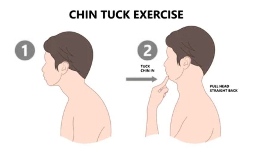
Can cervical retraction be a good addition to a home exercise program for individuals with neck pain, spinal arthritis, or needing to strengthen their neck muscles?
Cervical Retraction
Cervical retraction is a neck exercise that involves gently sliding the head backward while keeping the chin tucked. It can help with:
Neck pain
Stretching and loosening the muscles at the back of the neck.
Headaches
Tightness
Improve posture
Improve flexibility and mobility
Preparation
Basic steps for performing a cervical retraction exercise:
Sit upright with good posture.
Tuck your chin as far as you can comfortably.
Look up while keeping your chin tucked.
Rotate the neck 1-2 inches to each side.
Return to the starting position.
Stop the exercise if you experience pain.
You want to get good at this without loading the joints while learning the movement. Gently and accurately moving your head in alignment with the neck will help you find the correct head action as it moves. Then, it can be performed as a full exercise. Performing the movement correctly requires focus. This is why cervical retraction is done while sitting in a chair with proper posture. Individuals can also stand, but it is more complicated for the body to coordinate than sitting, but it can be done once the individual has practiced.
Sitting or Standing
Gently tuck your chin down toward your neck.
The focus is alignment.
Keeping your chin where it is, press your head back.
There will be soreness, especially for those with pain symptoms, but the neck should feel better.
Be mindful of any intense or severe pain resulting from cervical retraction.
Individuals with cervical spondylosis (neck arthritis) stop if pain presents. (Cleveland Clinic, 2023)
Other Neck Exercises
Another good neck-strengthening exercise is the isometric neck press. In this exercise, you move your head forward, backward, and to each side while your hand provides resistance. This develops flexibility and is recommended for those with arthritis in this area. (Sadeghi, A. et al., 2022) Other exercises include: (Pain Consultants of West Florida, 2019)
Neck extensions: Backward bending can help relieve nerve compression and ease the strain on the cervical spine.
Side rotation: This exercise can improve neck mobility.
Shoulder rolls: This exercise can help keep the neck and shoulder joints fluid.
Injury Medical Chiropractic and Functional Medicine Clinic
If you have a neck condition or radiculopathy that causes pain or other symptoms going down the arm or are unsure how to do it, check with a healthcare provider or physical therapist before trying the exercise. Injury Medical Chiropractic and Functional Medicine Clinic works with primary healthcare providers and specialists to develop an optimal health and wellness solution. We focus on what works for you to relieve pain, restore function, and prevent injury. Regarding musculoskeletal pain, specialists like chiropractors, acupuncturists, and massage therapists can help mitigate the pain through spinal adjustments that help the body realign itself. They can also work with other medical professionals to integrate a treatment plan to resolve musculoskeletal issues.
Neck Injuries
References
North American Spine Society. (2012). Cervical exercise: The Backbone of Spine Treatment. https://www.spine.org/KnowYourBack/Prevention/Exercise/Cervical-Exercise
Cleveland Clinic. (2023). Could your neck pain actually be neck arthritis? https://my.clevelandclinic.org/health/diseases/17685-cervical-spondylosis
Pain Consultants of West Florida. (2019). Chronic Neck Pain: How Core Exercises Can Help. Our Blog. https://pcwfl.com/chronic-neck-pain-how-core-exercises-can-help/
Sadeghi, A., Rostami, M., Ameri, S., Karimi Moghaddam, A., Karimi Moghaddam, Z., & Zeraatchi, A. (2022). Effectiveness of isometric exercises on disability and pain of cervical spondylosis: a randomized controlled trial. BMC sports science, medicine & rehabilitation, 14(1), 108. https://doi.org/10.1186/s13102-022-00500-7
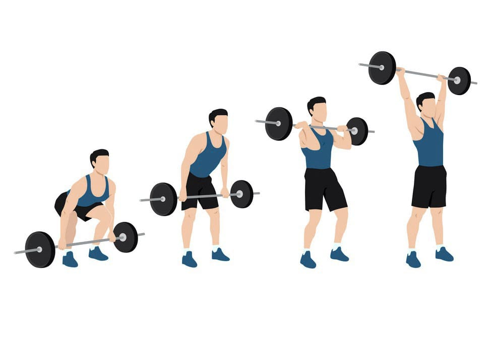
Is the clean and press recommended for intermediate-level weightlifters looking to increase strength and power?
Clean and Press
The clean and press is a power exercise that builds cardiovascular and muscular endurance, stamina, and strength. It focuses on strength and speed. (Soriano M. A., Suchomel T. J., & Comfort P. 2019) Individuals new to the clean and press should start light to learn the proper form. Once they master the technique, they should add weight until they reach the point where six to eight repetitions cause breathlessness. It is a great exercise to include in a circuit as part of a regular strength training program.
The Benefits
The clean and press work out several muscle groups.
The lower half of the movement strengthens the hips, glutes, and hamstrings.
The upper half targets the shoulders, chest, back, and arms.
Power training is important for athletes who need quick bursts in their sport, like sprinters or jumpers. (Sarabia J. M. et al., 2017) However, anyone at an intermediate lifting level can use power exercises to increase their heart rate to anaerobic levels, generating an increased calorie burn in their overall workout. Individuals must regularly pick up objects off the floor and place them in cabinets or shelves. The clean press can train the body to use the correct form.
Step-by-Step
Start with feet shoulder-width apart and hold the barbell around 2 inches from the shins.
Push the hips back and grab the barbell so the palms face the body and hands are shoulder-width apart.
Keep the hips down, chest lifted, eyes forward, and arms long.
Maintain core engagement and drive through the heels to pull the bar quickly up to the chest, just in front of the collarbone.
Keep the spine tall.
Be explosive and fast in the movement when pulling the bar, keeping it as close to the body as possible.
To pull the bar underneath the shoulders, shrug the shoulders up and point the elbows forward.
As soon as the bar reaches the chest, drive through the heels, press overhead, and straighten the arms and legs.
Keep the core tight.
Return to the starting position in a controlled manner.
Common Errors
Avoid the following errors to get the most from the exercise and prevent strain and injury.
Shifting Weight Forward
The weight should always remain on the heels during the cleaning and press.
Rounding the Back
The upper back should be straight and not rounded when lifting.
Grip Position
The grip should be no more than 2 inches wider than the shoulders.
If it is too wide, there is an increased risk of wrist pain, and if it is too narrow, shoulder joint strain.
Modifications and Variations
The clean and press can be practiced differently to meet an individual’s fitness level, which will also determine how much weight to lift.
Modification
Beginners can practice with an empty bar.
If possible, exercise in a room with a mirror to ensure the body is in the correct form.
Variation
The exercise can be performed with dumbbells or a barbell.
The barbell allows going a little heavier and provides stability.
The dumbbells encourage each side to work individually rather than the stronger side taking over for the weaker side.
A single-arm clean and press can be done with a dumbbell, adding a stability and balance challenge.
A clean press can be combined with leg exercises, such as squats or lunges, to superset the lower body.
A clean and press can also be used in upper body workouts to increase the heart rate.
For example, it can be used in a circuit-style workout:
Four minutes on the treadmill or elliptical.
Eight repetitions of clean and presses.
Four minutes on the treadmill or elliptical.
Eight repetitions of clean and press.
Perform for 15 to 20 minutes for a solid, complete workout.
Safety
It is recommended that individuals consult a doctor or physical therapist if they have issues with their ankles, knees, hips, wrists, shoulders, neck, or back, as the exercise involves multiple joints. It is not recommended during pregnancy.
Injury Medical Chiropractic and Functional Medicine Clinic
Injury Medical Chiropractic and Functional Medicine Clinic works with primary healthcare providers and specialists to develop an optimal health and wellness solution. We focus on what works for you to relieve pain, restore function, and prevent injury. Regarding musculoskeletal pain, specialists like chiropractors, acupuncturists, and massage therapists can help mitigate the pain through spinal adjustments that help the body realign itself. They can also work with other medical professionals to integrate a treatment plan to resolve musculoskeletal issues.
Exercise Prescription
References
Soriano, M. A., Suchomel, T. J., & Comfort, P. (2019). Weightlifting Overhead Pressing Derivatives: A Review of the Literature. Sports medicine (Auckland, N.Z.), 49(6), 867–885. https://doi.org/10.1007/s40279-019-01096-8
Calatayud, J., Colado, J. C., Martin, F., Casaña, J., Jakobsen, M. D., & Andersen, L. L. (2015). CORE MUSCLE ACTIVITY DURING THE CLEAN AND JERK LIFT WITH BARBELL VERSUS SANDBAGS AND WATER BAGS. International journal of sports physical therapy, 10(6), 803–810.
Sarabia, J. M., Moya-Ramón, M., Hernández-Davó, J. L., Fernandez-Fernandez, J., & Sabido, R. (2017). The effects of training with loads that maximise power output and individualised repetitions vs. traditional power training. PloS one, 12(10), e0186601. https://doi.org/10.1371/journal.pone.0186601
For individuals going through post surgery, injury rehabilitation, illness and/or chronic condition management, can physical therapy isometric exercises help?
Isometric Exercise
Isometric exercises are used in physical therapy to help build muscle endurance, improve range of motion, relieve pain, and reduce blood pressure more effectively than other types of exercise. Because they don’t involve joint movement, they are a solid starting point for rehabilitation and are suitable for individuals with a limited range of motion. They can be performed by pushing against an immovable object, like a wall, or by having a therapist provide resistance. Examples of isometric exercises include:
A physical therapist/PT may have a patient perform isometric exercises after injury or illness. During an isometric contraction, the muscle does not change in length, and there is no motion around the joint surrounding the muscle/s. (Rhyu H. S. et al., 2015)
When To Use
Isometric muscular contractions can be used at any time during physical rehabilitation and strengthening or a home exercise program and are regularly used with the following (Rhyu H. S. et al., 2015)
Post-surgery
When muscles cannot contract forcefully enough to move the joint it surrounds.
To help increase neuromuscular input to a specific muscle/s.
When injury or condition frailty makes other forms of exercise dangerous and not beneficial.
A healthcare provider or physical therapist should be consulted first if isometrics are used in a rehabilitation program.
Benefits
The benefits of using isometric exercise after injury or surgery may include the following:
No special equipment is necessary to perform isometric exercises.
The ability to safely contract a muscle while protecting a surgical incision or scar tissue.
The muscles can be strengthened in a specific range of motion around a joint. (NikolaidouO. et al., 2017)
A physical therapist can help determine whether isometric exercise benefits the specific condition.
Effectiveness
Isometric exercise is very effective after injury or surgery. However, when a muscle is contracted isometrically, it gains strength in a very small area and with a short range of motion. For example, an isometric shoulder external rotation performed with the arm at the side will only strengthen the rotator cuff muscles in the specific position that the arm is in. (NikolaidouO. et al., 2017).
Strength gains are specific to the joint’s position during the exercise.
Individuals who want to strengthen their gluteal muscles in their hip using isometrics would have to contract their glute muscles in one specific position for several reps.
Once several reps of the exercise in one position have been performed, the individual moves their hip joint into a new position and repeats the gluteal contractions in the new position.
This makes the exercise time-consuming, but it is perfect for injury rehabilitation, preventing and avoiding worsening or further injuries.
How to Perform
To perform isometric exercises, all that is needed is something stable to push against. (Rhyu H. S. et al., 2015) For example, to strengthen the shoulder muscles:
Stand next to a wall and try to lift an arm out to the side.
Allow the hand to press against the wall so no motion occurs at the shoulder joint.
Once pressed against the wall, hold the contraction for 5 to 6 seconds and slowly release it.
Perform 6 to 10 repetitions of the exercise.
This could be one set of completed isometric exercises for the shoulder muscles.
Elastic resistance bands or tubing can also be used to perform isometric exercises. Hold the tubing in a specific position and then move the body away from the anchor point instead of moving the joint. The muscles will contract against the increased resistance of the elastic tubing, and no motion will occur at the joint. A physical therapist can show and train on how to perform isometric exercises with the bands.
Neuromuscular Stimulation
Isometric exercise can strengthen muscles and help improve the neuromuscular recruitment of the muscles being trained. This enhances muscle contraction and expedites gains in muscle recruitment while protecting the joint. Isometric exercise can also be used during physical therapy using neuromuscular electrical stimulation (NMES). (Fouré A. et al., 2014) For example, a PT may use NMES to improve muscular function for individuals who have difficulty contracting their quadriceps after knee surgery and may be instructed to perform isometric quad-setting exercises during the session.
Injury Medical Chiropractic and Functional Medicine Clinic
A physical therapist can use isometric exercises to help individuals injured or have had surgery and are experiencing difficulty with normal functional mobility by improving their strength during recovery. The exercises can safely enhance the function and stability of the muscles and return individuals to the previous level of activity and function. Injury Medical Chiropractic and Functional Medicine Clinic works with primary healthcare providers and specialists to develop an optimal health and wellness solution. We focus on what works for you to relieve pain, restore function, and prevent injury. Regarding musculoskeletal pain, specialists like chiropractors, acupuncturists, and massage therapists can help mitigate the pain through spinal adjustments that help the body realign itself. They can also work with other medical professionals to integrate a treatment plan to resolve musculoskeletal issues.
Personal Injury Rehabilitation
References
Rhyu, H. S., Park, H. K., Park, J. S., & Park, H. S. (2015). The effects of isometric exercise types on pain and muscle activity in patients with low back pain. Journal of Exercise Rehabilitation, 11(4), 211–214. https://doi.org/10.12965/jer.150224
Nikolaidou, O., Migkou, S., & Karampalis, C. (2017). Rehabilitation after Rotator Cuff Repair. The Open Orthopaedics Journal, 11, 154–162. https://doi.org/10.2174/1874325001711010154
Fouré, A., Nosaka, K., Wegrzyk, J., Duhamel, G., Le Troter, A., Boudinet, H., Mattei, J. P., Vilmen, C., Jubeau, M., Bendahan, D., & Gondin, J. (2014). Time course of central and peripheral alterations after isometric neuromuscular electrical stimulation-induced muscle damage. PloS one, 9(9), e107298. https://doi.org/10.1371/journal.pone.0107298
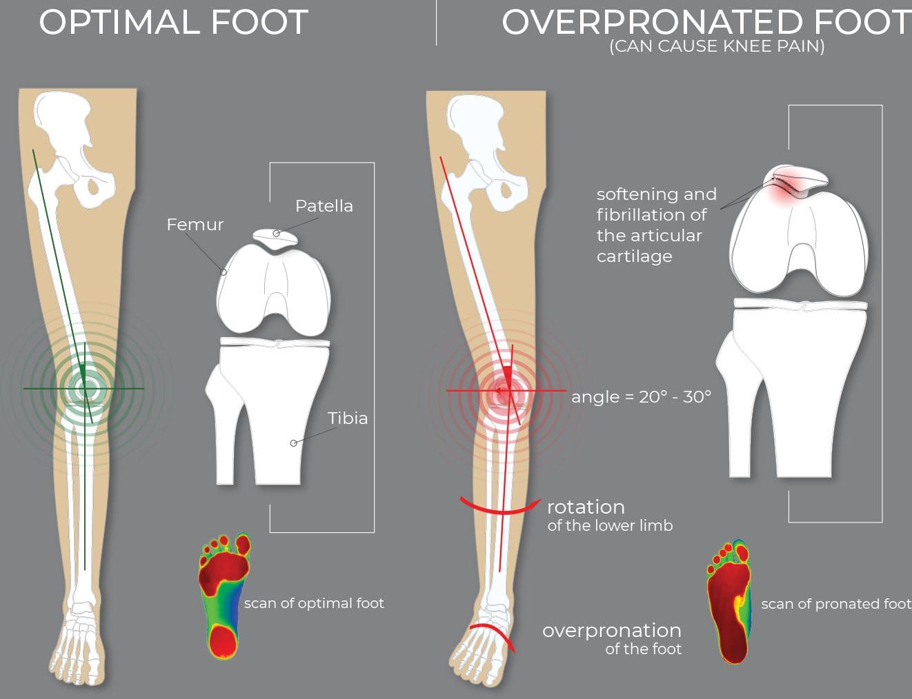
What are the treatment options for individuals dealing with foot overpronation when the foot and ankle move too much downward and inward?
Overpronation
Pronation is the normal foot and ankle movement when taking a step and is usually associated with flat feet. Overpronation is a movement pattern in which the ankle rolls inward and downward, causing the foot’s arch to collapse with each stride. Overpronation can strain the muscles and ligaments in the feet and legs, leading to heel pain, ankle pain, shin splints, and low-back pain. (Pedorthic Association of Canada, 2023) Orthotic inserts for shoes, prescribed stretches, ankle braces, physical therapy, and surgery can all help alleviate the impact of overpronation. (Sánchez-Rodríguez, R. et al., 2020)
Signs and Symptoms
Some individuals with overpronation can have no symptoms at all. (Pedorthic Association of Canada, 2023) while others experience pain or other symptoms in their legs and feet. Overpronation is not a medical condition but a movement pattern that, if left untreated, can increase the risk for certain medical conditions because it strains the feet and leg muscles, joints, and ligaments. (Sánchez-Rodríguez, R. et al., 2020) Certain medical conditions can be a sign of overpronation and include: (Pedorthic Association of Canada, 2023)
Bunions
Heel pain
Plantar fasciitis
Achilles tendon pain
Posterior tibial tendonitis
Shin splints
Knee pain, including patellofemoral pain syndrome
Pain in the iliotibial or IT band
Lower back pain
Arthritis in foot and ankle joints
Stress fractures
Individuals may also experience pain in the midfoot or hips, which can be a symptom of flat feet.
Underpronation
Pronation refers to the normal movement of the foot and ankle while walking. Overpronation and underpronation are both abnormal movement patterns.
Overpronation – when the ankle rolls too much inward and downward.
Underpronation – occurs when an individual’s foot lacks flexibility and moves too little. This condition is called supination and is often associated with a high-arch foot type.
Overpronation can be caused or worsened by flatfeet. However, some individuals have overpronation because their feet and ankles are very flexible, so they tend to move more. Risk factors for flatfeet can also increase the chances of developing overpronation and include:
Age, especially individuals over 40.
Wearing shoes like high heels and shoes with a narrow-toe box.
Women are more prone to overpronate because of the various shoes and high heels worn.
Being overweight
Doing repetitive, impactful movements like running.
Correction and Treatment
Treating overpronation focuses on alleviating strain on muscles in the foot, ankle, and leg to relieve symptoms in the heel, ankle, knees, hips, or back. Common treatments are wearing supportive shoes and/or using foot orthotics. Exercises and stretches are also recommended to maintain flexibility and strength. Surgery is rare, but correcting flat feet that can cause overpronation may be recommended. (Sánchez-Rodríguez, R. et al., 2020) Individuals with overpronation are advised to see a podiatrist who can explain the best treatment options.
Supportive Shoes
The first course of treatment is to wear added supportive footwear. This can include specialized shoes or inserts that support the foot and reduce ankle movement. Individuals are advised to use shoes with firm heel and midfoot support to help prevent disproportionate movement. (Pedorthic Association of Canada, 2023)
Orthotics
A healthcare provider can recommend orthotics for individuals with moderate overpronation. These are meant to support the foot, especially the arch, and reduce overpronation. (Naderi A. Degens H. and Sakinepoor A. 2019) Individuals can purchase orthotics from shoe stores and elsewhere, but those with severe overpronation may need custom orthotics molded to the foot to provide individualized support.
Exercises and Stretches
Exercises and stretches can also help. A study found that exercises targeting the feet, core, and hips helped correct pronation over nine weeks. The exercises included: (Sánchez-Rodríguez, R. et al., 2020)
Toe pickups in which the individuals grab small objects with their toes and move them from one position to another.
Flexing and pointing the toes using a resistance band placed around the toes.
Hip abduction exercises to target the hip and glutes.
Abdominal and oblique muscle exercises to stabilize the torso.
Short-foot exercise raises the foot arch off the ground, drawing the toes toward the heel. (Sulowska I. et al., 2016)
Surgery
Rarely will surgery be needed to treat flat feet and severe overpronation. But if necessary, reconstruction realigns the bones to support the arch better and reduce overpronation. A metal implant is used for flatfeet to stabilize the area. Surgery can also repair torn tendons or other damage contributing to overpronation. (Healthline, 2020)
Injury Medical Chiropractic and Functional Medicine Clinic
Individuals with overpronation but no symptoms don’t necessarily have to see a healthcare provider since this may be the body’s natural movement pattern. But if the feet, legs, hips, or back begin to present with pain and other symptoms, see a healthcare provider who can evaluate gait and recommend treatment options. Injury Medical Chiropractic and Functional Medicine Clinic works with primary healthcare providers and specialists to develop an optimal health and wellness solution. We focus on what works for you to relieve pain, restore function, and prevent injury. Regarding musculoskeletal pain, specialists like chiropractors, acupuncturists, and massage therapists can help mitigate the pain through spinal adjustments that help the body realign itself. They can also work with other medical professionals to integrate a treatment plan to resolve musculoskeletal issues.
Enhance Performance with Functional Foot Orthotics
References
Pedorthic Association of Canada. (2023). Overpronation and Underpronation Correction. https://pedorthic.ca/services/foot-health/pronation/
Sánchez-Rodríguez, R., Valle-Estévez, S., Fraile-García, P. A., Martínez-Nova, A., Gómez-Martín, B., & Escamilla-Martínez, E. (2020). Modification of Pronated Foot Posture after a Program of Therapeutic Exercises. International journal of environmental research and public health, 17(22), 8406. https://doi.org/10.3390/ijerph17228406
Naderi, A., Degens, H., & Sakinepoor, A. (2019). Arch-support foot orthoses normalize dynamic in-shoe foot pressure distribution in medial tibial stress syndrome. European journal of sport science, 19(2), 247–257. https://doi.org/10.1080/17461391.2018.1503337
Sulowska, I., Oleksy, Ł., Mika, A., Bylina, D., & Sołtan, J. (2016). The Influence of Plantar Short Foot Muscle Exercises on Foot Posture and Fundamental Movement Patterns in Long-Distance Runners, a Non-Randomized, Non-Blinded Clinical Trial. PloS one, 11(6), e0157917. https://doi.org/10.1371/journal.pone.0157917
Healthline. (2020). All About Surgery for Flat Feet: Pros and Cons. https://www.healthline.com/health/flat-feet-surgery
For individuals trying to retrain their body movements for back health improvement, what is the spinal area that helps the body twist, bend, and stand upright?
Lumbosacral Joint L5-S1
The L5-S1, also called the lumbosacral joint, is a term used to describe a part of the spine. It is where the lumbar spine ends and the sacral spine begins, and it connects these bones. The lumbosacral joint is also susceptible to misalignment and injury, such as disc herniation or a spinal disorder called spondylolisthesis.
The spinal column is the structure that allows the body to stand upright and helps you twist, bend, and alter trunk and neck position. Typically, 24 movable bones in the spine connect to the sacrum and the coccyx, or the tailbone. The sacrum and the coccyx each have multiple bones that fuse over time. L5-S1 consists of the last bone in the lumbar spine, called L5, and the triangle-shaped bone under it, known as the sacrum. S1 is at the top of the sacrum and comprises five fused bones.
Risk of Injury
Each area of the spine has a curve that goes in opposite directions. The places where the spinal curve directions change are junctional levels. The risk of injuries may be higher at junctional levels because the body weight shifts direction as the curves shift. The L5-S1 junction is located between the lumbar curve and the sacral curve. The lumbar curve sweeps forward, and the sacral curve goes backward.
The lumbosacral joint L5-S1 junction is highly vulnerable to misalignment, wear and tear, and injury. This is because the top of the sacrum is positioned at an angle for most individuals. Aging and injury increase the vulnerability of the L5-S1 junction even more. Pain coming from L5-S1 is usually treated with:
Heat and/or ice
Over-the-counter anti-inflammatory medications
Prescription pain medications
Muscle relaxers
Physical therapy
Chiropractic adjustments
Epidural steroid injections
If these therapies do not help, surgery may be recommended. L5-S1 is one of the two most common sites for back surgery.
Conditions
Disc herniation at L5-S1 is a common injury and cause of sciatica, which can cause pain and other issues (MedlinePlus, 2024). The L5-S1 junction is often the site of a condition known as spondylolisthesis.
Disc Herniation
Discs separate the vertebrae, cushioning the spinal column and allowing movement between vertebrae. A disc herniation means the disc slips out of place. (MedlinePlus, 2022) A disc herniation at L5-S1 is a common cause of sciatica. Symptoms of sciatica include:
Burning
Numbness
Pain or tingling that radiates from the buttock down the leg to the knee or foot.
Disc herniation can also cause chronic back pain and stiffness and trigger painful muscle spasms. Bowel problems are also possible with disc issues at L5-S1. Research links irritable bowel syndrome to herniated discs in the lower back. (Bertilson BC, Heidermakr A, Stockhaus M. 2015) Additional studies found disc problems at L5-S1 can lead to difficulty with sphincter control. (Akca N. et al., 2014) Initial treatments for disc herniation include rest and pain relievers to reduce inflammation and swelling, then physical therapy. Most recover with conservative interventions, and those who don’t may require a steroid injection or surgery. (MedlinePlus, 2022)
Spondylolisthesis
Spondylolisthesis occurs when a vertebra slips forward relative to the bone below it. The most common form of this condition is degenerative spondylolisthesis, which generally begins when the spine wears down with age. Isthmic spondylolisthesis is another common variation and starts as a tiny fracture in the pars interarticularis, a bone that connects the adjoining parts of the facet joint. (American Academy of Orthopaedic Surgeons, 2020) These fractures often occur before age 15, but symptoms do not develop until adulthood. Degeneration of the spine in later adulthood can further worsen the condition.
The angle of the sacrum can also contribute to spondylolisthesis. This is because the S1 tips down in the front and up in the back rather than being horizontal. Individuals with a greater tilt are usually at a higher risk of spondylolisthesis. (Gong S. et al., 2019) However, individuals with spondylolisthesis may not have any symptoms. Those who do may experience: (American Academy of Orthopaedic Surgeons, 2020)
Back stiffness
Standing difficulties
Walking difficulties
Lower back pain
Hamstring tightness
Spondylolisthesis is typically treated with non-surgical interventions that can include:
Pain medications
Heat and/or ice application
Physical therapy
Epidural steroid injections
Usually, non-surgical care is tried for at least six months. If pain and symptoms persist, surgery may be an option. Spinal fusion surgery can be effective but requires a long recovery time and can have additional risks.
Injury Medical Chiropractic and Functional Medicine Clinic
Injury Medical Chiropractic and Functional Medicine Clinic works with primary healthcare providers and specialists to develop an optimal health and wellness solution. We focus on what works for you to relieve pain, restore function, and prevent injury. Regarding musculoskeletal pain, specialists like chiropractors, acupuncturists, and massage therapists can help mitigate the pain through spinal adjustments that help the body realign itself. They can also work with other medical professionals to integrate a treatment plan to resolve musculoskeletal issues.
Chiropractic Healing After Trauma
References
MedlinePlus. (2024). Sciatica. Retrieved from https://medlineplus.gov/sciatica.html
MedlinePlus. (2022). Herniated disk. Retrieved from https://medlineplus.gov/ency/article/000442.htm
American Association of Neurological Surgeons. (2024). Herniate disc. https://www.aans.org/patients/conditions-treatments/herniated-disc/
Bertilson, B. C., Heidermark, A., & Stockhaus, M. (2015). Irritable Bowel Syndrome–a Neurological Spine Problem. Journal of Advances in Medicine and Medical Research, 4(24), 4154–4168. https://doi.org/10.9734/BJMMR/2014/9746
Akca, N., Ozdemir, B., Kanat, A., Batcik, O. E., Yazar, U., & Zorba, O. U. (2014). Describing a new syndrome in L5-S1 disc herniation: Sexual and sphincter dysfunction without pain and muscle weakness. Journal of craniovertebral junction & spine, 5(4), 146–150. https://doi.org/10.4103/0974-8237.147076
American Academy of Orthopaedic Surgeons. (2020). Spondylolysis and spondylolisthesis. https://orthoinfo.aaos.org/en/diseases–conditions/spondylolysis-and-spondylolisthesis/
Gong, S., Hou, Q., Chu, Y., Huang, X., Yang, W., & Wang, Z. (2019). Anatomical factors and pathological parts of isthmic fissure and degenerative lumbar spondylolisthesis.
IFM's Find A Practitioner tool is the largest referral network in Functional Medicine, created to help patients locate Functional Medicine practitioners anywhere in the world. IFM Certified Practitioners are listed first in the search results, given their extensive education in Functional Medicine