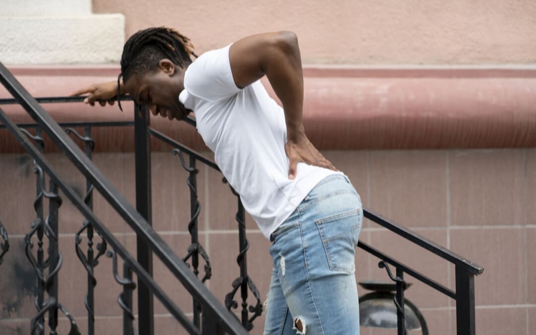
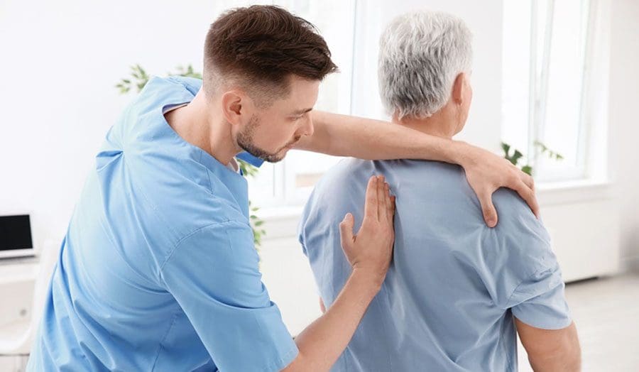
Can understanding how nociceptors function and their role in processing pain signals help individuals who are managing injuries and/or living with chronic pain conditions?
Nociceptors
Nociceptors are nerve endings that detect harmful stimuli, such as extreme temperatures, pressure, and chemicals, and signal pain. They are the body’s first defense against potentially damaging environmental inputs.
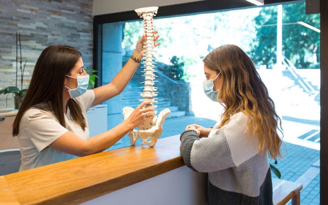
Nociceptors are in the skin, muscles, joints, bones, internal organs, deep tissues, and cornea.
They detect harmful stimuli and convert them into electrical signals.
These signals are sent to the brain’s higher centers.
The brain interprets the signals as pain, which prompts the body to avoid the harmful stimulus.
Nociceptors, often called pain receptors, are free nerve endings all over the body. They play a pivotal role in how the body feels and reacts to pain. The main purpose of a nociceptor is to respond to damage to the body by transmitting signals to the spinal cord and brain. (Purves D, Augustine GJ, Fitzpatrick D, et al., editors. 2001) If you bang your foot, the nociceptors on the skin are activated, sending a signal to the brain via the peripheral nerves to the spinal cord. Pain resulting from any cause is transmitted this way. Pain signals are complex, carrying information about the stimuli’s location and intensity. This causes the brain to fully process the pain and send communication back to block further pain signals.
Thermal nociceptors respond to extreme hot or cold temperatures.
For instance, when touching a hot stove, the nociceptors, which signal pain, are activated immediately, sometimes before you know what you’ve done.
Mechanical
Mechanical nociceptors respond to intense stretching or strain, such as pulling a hamstring or straining a tendon.
The muscles or tendons are stretched beyond their ability, stimulating nociceptors and sending pain signals to the brain.
Chemical
Chemical nociceptors respond to chemicals released from tissue damage.
For example, prostaglandins and substance P or external chemicals like topical capsaicin pain creams.
Silent
Silent nociceptors must be first activated by tissue inflammation before responding to a mechanical, thermal, or chemical stimulus.
Most visceral nociceptors are located on organs in the body.
Polymodal
Polymodal nociceptors respond to mechanical, thermal, and chemical stimuli.
Mechano-thermal
Mechano-thermal nociceptors respond to mechanical and thermal stimuli.
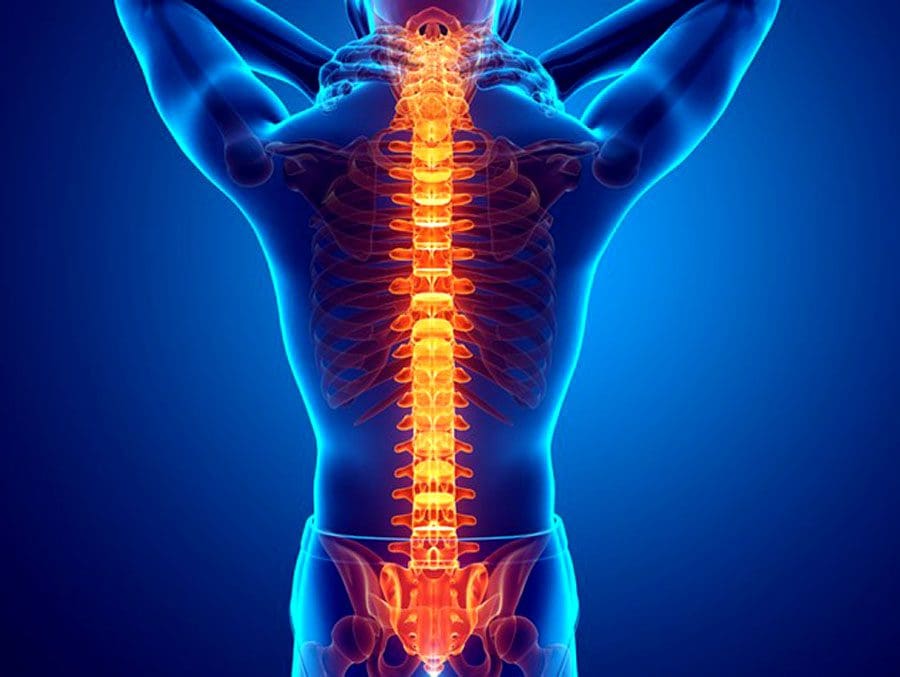
Pain Transmission
Nociceptors are also classified by how fast they transmit pain signals. Transmission speed is determined by the type of nerve fiber known as an axon a nociceptor has. There are two main types.
The first type is A fiber axon, fibers surrounded by a fatty, protective sheath called myelin.
Myelin allows nerve signals/action potentials to travel rapidly.
Because of the difference in transmission speed, the pain signals from the A fibers reach the spinal cord first. As a result, after an acute injury, an individual experiences pain in two phases, one from the A fibers and one from the C fibers. (Ngassapa D. N. 1996)
Pain Perception Phases
When an injury occurs, the stimulated nociceptors activate the A fibers, causing a person to experience sharp, prickling pain.
This is the first phase of pain, known as fast pain, because it is not especially intense but comes right after the stimulus.
During the second phase of pain, the C fibers are activated, causing an intense, burning pain that persists even after the stimulus has stopped.
The fact that the C fibers carry burning pain explains why there is a short delay before feeling the sensation.
The C fibers also carry aching, sore pain caused by organs within the body, such as a sore muscle or stomachache. (Ngassapa D. N. 1996)
Injury Medical Chiropractic and Functional Medicine Clinic
Injury Medical Chiropractic and Functional Medicine Clinic works with primary healthcare providers and specialists to build optimal health and wellness solutions. We focus on what works for you to relieve pain, restore function, prevent injury, and help mitigate issues through adjustments that help the body realign itself. They can also work with other medical professionals to integrate a treatment plan to resolve musculoskeletal problems.
From Injury To Recovery With Chiropractic Care
References
Purves D, A. G., Fitzpatrick D, et al., editors. (2001). Nociceptors. In Neuroscience. 2nd edition. (2nd ed.). Sunderland (MA): Sinauer Associates. https://www.ncbi.nlm.nih.gov/books/NBK10965/
University of Texas McGovern Medical School. (2020). Chapter 6: Pain Principles. https://nba.uth.tmc.edu/neuroscience/m/s2/chapter06.html
Ngassapa D. N. (1996). Comparison of functional characteristics of intradental A- and C-nerve fibres in dental pain. East African medical journal, 73(3), 207–209.
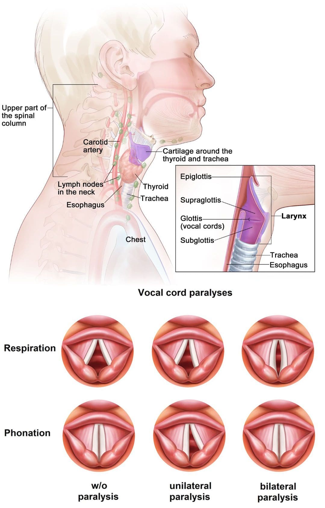
Automobile collisions, work, sports, and personal accidents can cause neck injuries that can affect other areas, leading to long-term health problems. Neck injuries involving soft tissue damage frequently persist after the incident. One of the injuries includes vocal cord damage caused by impact to the larynx. The larynx, or voicebox, is an organ that is behind the Adam’s apple. A neck injury impacting the larynx can affect the ability to speak and breathe and cause vocal cord paralysis. Treatment can involve surgery, voice therapy, physical therapy, and chiropractic.
Vocal Cord Injury
The vocal cords are two flexible bands of muscle tissue at the entrance of the trachea. The vocal cords are normally in a relaxed open position to allow breathing. When talking, the bands combine and vibrate to make a sound. Surgery, viral infections, certain cancers, and neck trauma can cause vocal cord paralysis. In this condition, nerve damage blocks or inhibits impulses from transmitting to the voice box. The muscles, usually one of them, become paralyzed, preventing swallowing and ingesting saliva through the windpipe/trachea. In rare cases, both muscles are unable to move.
Trauma to the neck or chest can injure the voice box nerves.
Infections
Infections like Lyme disease, Epstein-Barr virus, and herpes can cause inflammation and nerve damage.
Tumors
Tumors, cancerous and noncancerous, can grow inside or around the muscles, cartilage, and nerves.
Neurological
Neurological conditions like multiple sclerosis or Parkinson’s disease can lead to vocal cord paralysis.
Surgical Injury
Surgical procedure mistakes or complications on or near the neck or upper chest can result in damage to the voice box nerves.
Surgeries to the thyroid or parathyroid glands, esophagus, neck, and chest have an increased risk.
Stroke
A stroke chokes blood flow to the brain and can damage the region of the brain that transmits messages to the voice box.
Treatment
Treatment is determined by a doctor based on the individual medical condition and diagnostic tests. Treatment can involve:
Speech Therapy
Speech therapy is recommended as the laryngeal muscles are strengthened through various exercises, improving breathing function. A speech therapist will begin working with the individual on exercises targeting the weakened vocal folds by enhancing airflow and blood circulation.
Physical Therapy and Chiropractic
Treatment involves performing gentle exercises that work on the vocal cords gradually and progressively but does not stress them. Chiropractors work with the physical therapist performing high-velocity, low-amplitude manipulation targeted at the lower neck and upper thoracic area, the C3/T1 vertebrae. A treatment plan will also use massage, non-surgical decompression, instrument/tool-assisted soft-tissue mobilization, low laser or ultrasound, and at-home stretches and exercises.
Surgery
Surgery could be necessary for individuals experiencing no improvement despite doing the prescribed speech and physical therapy exercises. Different types of procedures are based on the degree and extent of the paralysis:
Injections – Collagen and fillers are injected into the vocal cords to reposition the affected muscles closer to the larynx.
Phonosurgery – The vocal cords are repositioned through restructuring.
Tracheotomy – If the vocal folds are closing, a surgeon may make an incision in the neck at the opening of the windpipe and insert a breathing tube. This bypasses the air blockage caused by the vocal folds and promotes proper air circulation.
Cervical Spine Instability
References
Chen, Ching-Chang, et al. “Long-term result of vocal cord paralysis after anterior cervical discectomy.” The European spine journal: official publication of the European Spine Society, the European Spinal Deformity Society, and the European Section of the Cervical Spine Research Society vol. 23,3 (2014): 622-6. doi:10.1007/s00586-013-3084-y
Dankbaar JW, et al. Vocal cord paralysis: Anatomy, imaging, and pathology. Insights in Imaging. 2014; doi:10.1007/s13244-014-0364-y.
Fitzpatrick, P C, and R H Miller. “Vocal cord paralysis.” The Journal of the Louisiana State Medical Society: official organ of the Louisiana State Medical Society vol. 150,8 (1998): 340-3.
Kriskovich, M D et al. “Vocal fold paralysis after anterior cervical spine surgery: incidence, mechanism, and prevention of injury.” The Laryngoscope vol. 110,9 (2000): 1467-73. doi:10.1097/00005537-200009000-00011
Vocal fold paralysis. National Institute on Deafness and Other Communication Disorders. https://www.nidcd.nih.gov/health/vocal-fold-paralysis. Accessed May 18, 2022.
Vocal fold paralysis. American Speech-Language-Hearing Association. https://www.asha.org/public/speech/disorders/Vocal-Fold-Paralysis. Accessed May 18, 2022.
Waddell, Roger K. “Chiropractic care for a patient with spasmodic dysphonia associated with cervical spine trauma.” Journal of chiropractic medicine vol. 4,1 (2005): 19-24. doi:10.1016/S0899-3467(07)60108-6
Tremors are extremely rare, but they can result from spinal compression and not necessarily a brain condition like Parkinson’s disease. Tremors are abnormal, involuntary body movements with various causes, most of which are connected to the brain and not the spine. A study reports that more than 75% of individuals with Parkinson’s experienced a resting tremor, and about 60% experience tremors while moving. Sometimes the spine is the contributor caused by compression of the spinal cord.
Spinal Compression Study
A 90-year-old man was hospitalized after having tremors, with Parkinson’s being the initial diagnosis. The tremors progressed to the point where the man could not feed himself or walk without support. The case became the focus of a medical report published by physicians in the Department of Orthopaedic Surgery, Division of the Spine, Singapore Tan Tock Seng Hospital. Along with the tremors, symptoms progressed to:
Difficulty with fine motor skills like buttoning a shirt.
However, it was ruled out because the patient was not presenting with other Parkinson’s symptoms.
For individuals with cervical spondylotic myelopathy tremors, surgery can be used to help the condition. However, with cervical myelopathy, there is often some permanent damage. Individuals have shown that post-surgery and decompression, symptoms still present, maybe not as much, but there will be a need for a symptom management plan.
Prevention
The best way to prevent tremors associated with cervical spondylotic myelopathy is to minimize the strain on the spine that can lead to herniated discs and/or other spinal injuries. The discs in the spine degenerate, dry out and start cracking with age, increasing the risk of rupture. If a tremor develops, contact a doctor, spine specialist, or chiropractor to help diagnose the condition. These doctors can perform physical and neurological tests to determine the cause and treatment options.
Body Composition
Aging Health
Steady weight gain throughout life can lead to adult-onset diabetes. This is partly caused by having more body fat and progressive muscle loss. Loss of skeletal muscle mass is linked to insulin resistance that involves:
The less muscle is available, the less insulin sensitive the body becomes.
As insulin sensitivity decreases, the body becomes more resistant, increasing risk factors for type II diabetes.
This can lead to osteoporosis, where the old bone is reabsorbed more and less new bone is created.
Both men and women can experience decreased muscle mass that can lead to:
Thinner bones
Weaker bones
Increased risk of osteoporosis and severe injury from falls.
To help prevent these issues, it is recommended to:
It is recommended to space out protein intake across meals rather than consuming it all at once. This helps to ensure the proper amount is acquired.
Monitoring body composition regularly can help minimize muscle mass loss and fat mass gain as the body ages.
A regular strength training routine will help strengthen bones muscles and maintain optimal circulation.
References
Heusinkveld, Lauren E et al. “Impact of Tremor on Patients With Early Stage Parkinson’s Disease.” Frontiers in neurology vol. 9 628. 3 Aug. 2018, doi:10.3389/fneur.2018.00628
Jancso, Z et al. “Differences in weight gain in hypertensive and diabetic elderly patients primary care study.” The Journal of nutrition, health & aging vol. 16,6 (2012): 592-6. doi:10.1007/s12603-011-0360-6
Srikanthan, Preethi, and Arun S Karlamangla. “Relative muscle mass is inversely associated with insulin resistance and prediabetes. Findings from the third National Health and Nutrition Examination Survey.” The Journal of clinical endocrinology and metabolism vol. 96,9 (2011): 2898-903. doi:10.1210/jc.2011-0435
Tapia Perez, Jorge Humberto et al. “Treatment of Spinal Myoclonus Due to Degenerative Compression Myelopathy with Cervical Spinal Cord Stimulation: A Report of 2 Cases.” World neurosurgery vol. 136 (2020): 44-48. doi:10.1016/j.wneu.2019.12.170
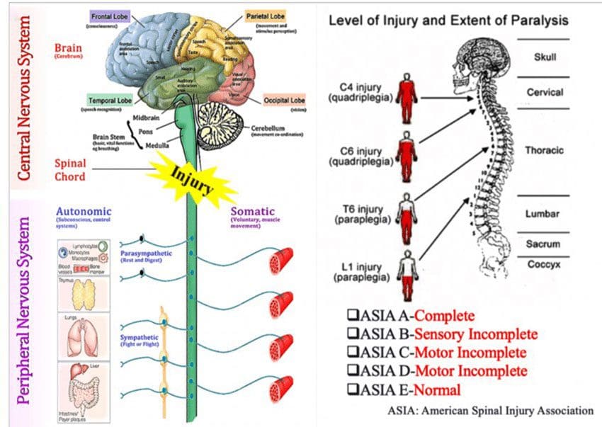
Neuroregenerationcould become an option for spinal cord injury treatments in the future. A spinal cord injury or SCI is when there is damage to the bundle of nerves and cells that send and receive signals from the brain and body. A spinal cord injury can be caused by direct trauma/injury to the cord or damage to the tissue and vertebrae. The damage can result in temporary or permanent changes in:
Sensation
Movement
Strength
Body function/s below the injury site.
There are incomplete and complete injuries. Injuries that cause limited or no cell death can achieve a full recovery. Injuries that are more serious and/or are higher on the spinal cord can cause permanent damage and/or paralysis. Automobile crashes, accidents, and serious falls are the most common causes of spinal cord injuries.
An incomplete injury means the cord can still transmit messages, but there is interference/disturbance.
A complete injury means communication and motor function/voluntary body movement is not transmitting.
Symptoms
Symptoms of a spinal cord injury include:
Unnatural or awkward positioning of the spine or head.
Pain or pressure in the head, neck, or back.
Numbness
Tingling
Loss of or changes in sensation in the hands and feet.
Problems with walking.
Weakness or inability to move parts of the body.
Loss of movement.
Paralysis can occur immediately or develop over time as swelling and bleeding affect the cord.
Loss of bladder and bowel control.
Changes in sexual function.
Difficulty breathing.
SCI Damage Control
A spinal cord injury affects the central nervous system, the body’s central headquarters. Damage can cause complications through what’s called the secondary injury cascade, which is a series of chemical reactions the body activates to help the situation. However, if the chemical response does not stop and stays active, it can worsen the injury. The body recognizes that an emergency has occurred and tries to go into a shut-down mode that kills off some of the cells in the central nervous system. When a spinal injury happens, treatment focuses on stopping the damage as quickly as possible to stop the injury cascade and prevent as much cell death as possible. This act is called neuropreservation, meaning that the team is trying to preserve and save as many nerve cells as possible.
Injury Neuroregeneration Treatment Studies
While current treatment primarily focuses on stopping as much damage as possible then going through physical therapies to maintain spinal alignment and rehabilitate the body, the future of injury treatment is looking towards regrowing and repairing the damaged nerve cells through a process known as neuroregeneration. Repairing nerves that have been damaged could change life for many. Neuroregeneration Treatments being studied include:
Surgery
A study in The Lancet Neurology presents how getting surgery as soon as possible after an injury can provide significant benefits.
The findings could change all of the guidelines for spinal cord injury.
Medication
A study on Riluzole, a medication that has shown promise to slow down nerve cell damage.
A team completed a randomized controlled trial for the medication; soon, the final results will be available.
Scientists are studying ways to grow new nerve cells from an individual’s stem cells without the need for embryonic stem cells.
Specialized stem cells could also be used to help other nerve cells regenerate.
Electrical stimulation
Another approach is using electrical stimulation to restore function in the spinal cord.
Therapy that could help a paralyzed individual walk again.
The Future of Neuroregeneration
Aside from early surgery intervention, most neuroregenerative treatments are not ready or accessible yet. There’s still much more research before it can become a mainstream treatment option. Treatment that involves regenerating nerve cells will take longer than a treatment designed to protect nerve cells. However, more clinical trials are expected to be done in the next few years, with stem cell therapies taking the longest. Some of these therapies could be ready to be used on actual patients in 5-10 years.
Body Composition
The Importance of Measuring Body Composition
Most diet and fitness programs focus on weight loss or gain. However, they tend to overlook that individuals have completely different body compositions. Body composition describes the amount of:
Fat
Bone
Water
Muscle
In the body.
Measuring body composition can tell a body’s unique makeup and help identify areas to work on to improve overall health and wellness. Body composition analysis provides a snapshot of an individual’s health/fitness levels to help achieve health goals from the inside out.
References
Aguilar, Juan et al. “Spinal cord injury immediately changes the state of the brain.” The Journal of neuroscience: the Official Journal of the Society for Neuroscience vol. 30,22 (2010): 7528-37. doi:10.1523/JNEUROSCI.0379-10.2010
Badhiwala, Jetan H; Wilson, Jefferson R; Witiw, Christopher D; et al. (February 2021). The Lancet Neurology Vol. 20, No. 2, P. 117. The Influence of Timing of Surgical Decompression for Acute Spinal Cord Injury: A Pooled Analysis of Individual Patient Data. DOI: 10.1016/S1474-4422(20)30406-3
Chari, Aswin et al. “Surgical Neurostimulation for Spinal Cord Injury.” Brain sciences vol. 7,2 18. 10 Feb. 2017, doi:10.3390/brainsci7020018
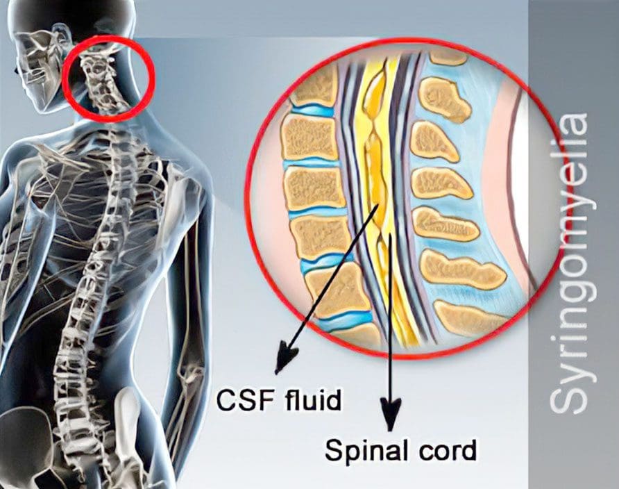
Syringomyelia is a disorder in which afluid-filled cyst/syrinx forms within the spinal cord. It is progressive, meaning that the cyst grows with time causing compression and damage to the spinal cord. The cyst usually begins in the neck/cervical spine but can develop in any area along the spinal cord. There are several possible causes; however, most are associated with a condition known as Chiari malformation. This is where the skull and neck come together, and either the skull is too small or shaped in a way that causes brain tissue to come out and settle in the spinal canal.
Syringomyelia Causes
Syringomyelia can be caused by or from complications of:
Chiari type I malformation develops during the fetal developmental stage and causes the lower part of the brain or cerebellum to stick out from its standard location.
Hemorrhage/bleeding
Inflammation of the spinal cord from virus or bacterial infection like meningitis
Spinal cord injury
Spinal cord tumor
Symptoms
A damaged spinal cord disrupts communication between the brain and the body. Symptoms differ for every individual, but common syringomyelia symptoms include:
Pain, stiffness, or weakness in the neck, arms, back, and/or legs
Symptoms usually develop slowly, but exercise, coughing, or some form of strain can cause sudden onset.
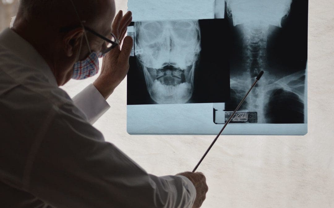
Diagnosis
Physical and neurological exams are performed to determine loss of feeling or inability to move around normally, like walking. Diagnostic tests of the spine will include a CT scan with contrast dye and/or an MRI. Early detection can help before it progresses, causing further damage, and delaying treatment can cause irreversible spinal cord injury. It is recommended at the first sign of symptoms to contact a doctor.
Treatment
Some individuals who have syringomyelia may have no symptoms. These individuals can go about their everyday lives but are recommended to be cautious with neck and back strain. For individuals experiencing symptoms, the primary treatment objectives are to:
Stop or control damage to the spinal cord
Preserve function
Prevent disability
Treatment options include:
Draining the cyst
Surgical removal of the cyst
Chiropractic and physical therapy could be included in the treatment plan to help the individual rebuild lost muscle strength and regain flexibility.
All too often, individuals with this disorder experience treatment delay/s because symptoms can be nonspecific or vague. Education is the key, and individuals can be diagnosed sooner by paying attention to the body’s warning signs.
Body Composition
Does too much protein hurt the kidneys?
While protein restriction can be appropriate for treating existing kidney disease, research shows that high protein intake in healthy individuals does not disrupt or cause damage to the kidneys or kidney function. The amino acids in protein are more likely to be excreted through urine when not being used. However, there are certain risks associated with consuming too much protein, and it is recommended to keep track of protein intake. Eating more protein:
Makes the body feel full longer
Can help curb overeating
Is essential for recovery and growth
When achieving daily caloric goals, maintaining a balance of nutrients like carbohydrates and healthy fats is essential for overall health.
References
Batzdorf, Ulrich. “Primary spinal syringomyelia. Invited submission from the joint section meeting on disorders of the spine and peripheral nerves, March 2005.” Journal of neurosurgery. Spine vol. 3,6 (2005): 429-35. doi:10.3171/spi.2005.3.6.0429
Di Lorenzo, N, and F Cacciola. “Adult syringomyelia. Classification, pathogenesis and therapeutic approaches.” Journal of neurosurgical sciences vol. 49,3 (2005): 65-72.
Fernández, Alfredo Avellaneda et al. “Malformations of the craniocervical junction (Chiari type I and syringomyelia: classification, diagnosis, and treatment).” BMC musculoskeletal disorders vol. 10 Suppl 1, Suppl 1 S1. 17 Dec. 2009, doi:10.1186/1471-2474-10-S1-S1
Naftel, Robert P et al. “Worsening or development of syringomyelia following Chiari I decompression: case report.” Journal of neurosurgery. Pediatrics vol. 12,4 (2013): 351-6. doi:10.3171/2013.7.PEDS12522
Roy, Anil K et al. “Idiopathic syringomyelia: retrospective case series, comprehensive review, and update on management.” Neurosurgical focus vol. 31,6 (2011): E15. doi:10.3171/2011.9.FOCUS11198
Spinal cord injuries or SCI’s don’t just happen from intense force/high-energy trauma like hard falls or auto accidents. Non-traumatic spinal cord injuries are more common. However, traumatic spinal cord injuries tend to get the most attention. This can cause problems as it can delay treatment for individuals with a non-traumatic injury. Awareness is vital because spinal cord disorders tend to not get recognized for their impact on overall health.
Non-traumatic Spinal Cord Injury
Non-traumatic spinal cord injuries is an umbrella term that includes several disorders, like:
Understanding these disorders helps determine a correct diagnosis. Degenerative cervical myelopathy or DCM is the most common form of a non-traumatic spinal cord injury. It is a slow progressive injury that causes continued compression usually brought on by spondylosis or osteoarthritis of the spine’s joints. DCM can have a devastating effect on the quality of life if not diagnosed and treated as it can have a ripple effect by raising the risk of falls, leading to a traumatic spinal cord injury. Being aware can help in preventing the damage.
Other Causes and Complications
Other causes for non-traumatic spinal cord injury include:
One of the most significant risk factors is age. This is due to an increased risk of conditions like osteoarthritis and hypertension as individuals get older, which is why individuals with a non-traumatic spinal cord injury are, on average, older than individuals that suffer a traumatic spinal cord injury. Weakness, instability, and loss of muscle control are common complications that can develop as the non-traumatic spinal cord injury progresses. Other complications that can present include:
Chronic pain
Sleep disturbance
Constipation
Urinary incontinence
Urinary tract infections
Impotence
Pressure ulcers/bed sores if immobilized
Possible blood clots that can lead to deep vein thrombosis
Depression and anxiety
Treatment
With a traumatic spinal cord injury, treatment depends on the severity of the injury. With non-traumatic spinal cord injuries, treatment depends on what type of condition is involved. The primary treatment for non-traumatic spinal cord injuries typically involves various forms of rehabilitation to minimize further damage to the spinal cord. Surgery may be necessary if the spine needs to be decompressed.
Body Composition
Extracellular Water and Intracellular Water
Extracellular Water – ECW
Extracellular is the water located outside the body’s cells.
Allows molecules to be transported to the different organelles inside the cell.
Picks up where the extracellular water leaves off by continuing the pathway for fuel/energy to be transported to the cells.
References
Badhiwala, Jetan H et al. “Degenerative cervical myelopathy – update and future directions.” Nature reviews. Neurology vol. 16,2 (2020): 108-124. doi:10.1038/s41582-019-0303-0
Handbook of Clinical Neurology (2012) “Spinal Cord Injury.” https://www.sciencedirect.com/topics/medicine-and-dentistry/non-traumatic-spinal-cord-injury
Milligan, James et al. “Degenerative cervical myelopathy: Diagnosis and management in primary care.” Canadian family physician Medecin de famille canadien vol. 65,9 (2019): 619-624.
Physical Management in Neurological Rehabilitation (2004) “Spinal cord injury.” https://www.sciencedirect.com/topics/medicine-and-dentistry/non-traumatic-spinal-cord-injury
Spinal meningitis does not just affect the brain. Most think of meningitis as a brain disease, but it can also affect the spine. We will discuss learning how to recognize it and find the right treatment to fix it within the spinal cord. Spinal meningitis can be a potentially deadly infection of the meninges. This is the protective tissue that covers the brain and spinal cord.
It can be caused by viruses, bacteria, or fungi that are transmitted from person to person by sneezing, talking, and sharing food.Viruses and pathogens that cause other infections, like the mumps and measles, can also cause meningitis. The lining around the brain and the spine are connected, which means that infection can travel from one area to another, or remain in the brain or the spine.
�
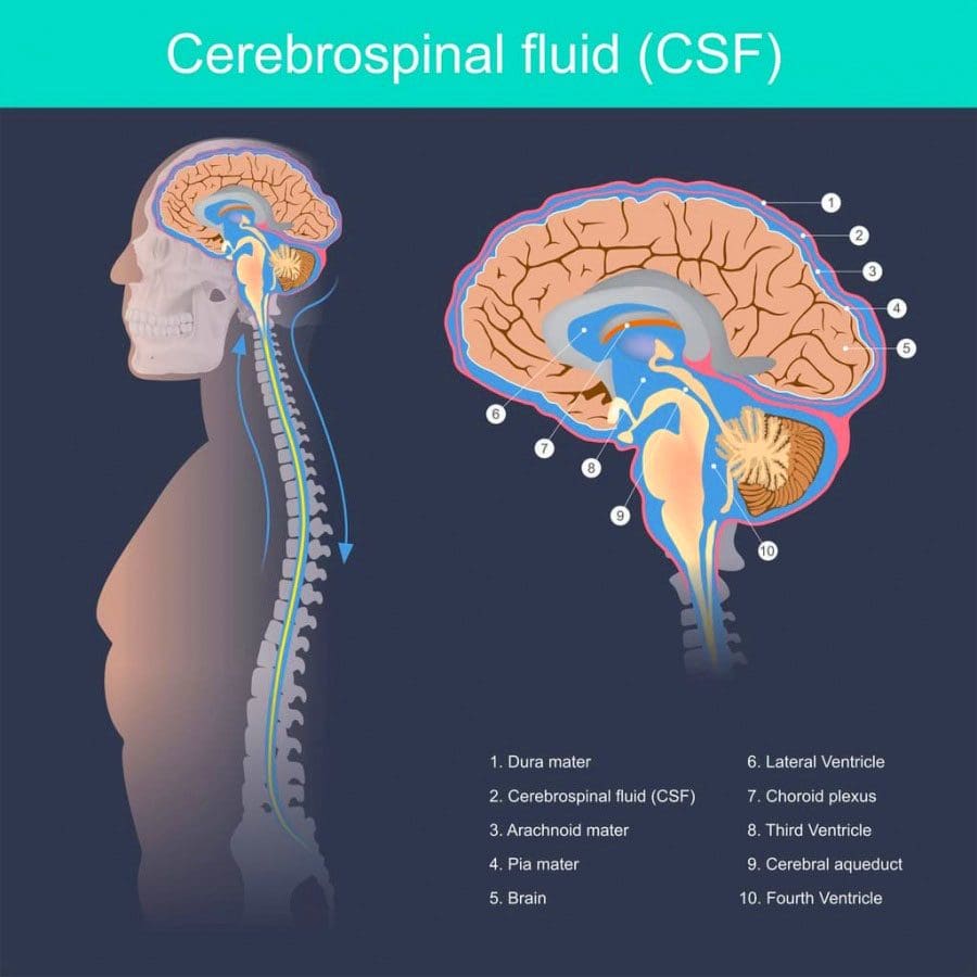
The Meninges
Meninges are the protective membranes that surround the brain and spinal cord. They are made up of three layers:
Dura mater is the thick and tough outer layer
Arachnoid mater is the middle layer made up of strands of connective tissue
Pia mater is the inner layer of cells
Spinal meningitis can develop when a virus, bacteria, or pathogen invade the meninges layers. This causes the immune system to react trying to remove the invading bacteria etc, which causes inflammation. These organisms usually take up residence in the nose and throat and never cause problems. Most individuals that come into contact with these viruses never get sick.
The reason for this is because the body produces fighting antibodies before the pathogens can invade the meninges. Others, possibly from age or underlying conditions, where they are not able to produce enough or any antibodies, makes them vulnerable to the illness. When the brain and spine’s tissue/s get infected with any one of these pathogens, the tissue swells, which constricts proper blood flow to the brain.
Types of Spinal Meningitis
The most common types of spinal meningitis in the United States include:
Viral meningitis
Viral meningitis is caused by enteroviruses, which are common viruses that enter the body through the mouth and travel to the brain and tissues where multiplication ensues. There are other viruses that can also cause meningitis. These include:
Viruses that cause mumps
Herpesviruses – like Epstein-Barr, measles, influenza, West Nile
Lymphocytic choriomeningitis virus from rodents
Any of these viruses can spread to the meninges, causing spinal meningitis to develop. This is a less severe type than bacterial meningitis.
Bacterial meningitis
This is the type where dangerous bacteria invade the meninges. Individuals are at higher risk as this type can be fatal if not treated. Common types of bacterial meningitis include:
Haemophilus influenzae – can cause severe infection/s of the lining of the brain, spinal cord, and the blood.
Pneumococcal meningitis – is caused by the bacterium Streptococcus pneumonia and is the most common form of bacterial meningitis.
Meningococcal meningitis – also known as meningococcal disease, is a less common type. This type is caused by the bacterium Neisseria meningitides. Around 2,600 people in the U.S. are affected yearly.
�
Symptoms
Viral or bacterial spinal meningitis can cause a range of symptoms, including:
Neck and back stiffness
Muscle weakness
Headache
Drowsiness
Fatigue
Fever
Double vision
Sensitivity to light
Nausea
Vomiting
Hearing difficulty
Confusion
Seizures
Rash
Symptoms are often far more pronounced with the bacterial form. This is because it�s associated with more inflammation, compared to the viral type.
Complications
Depending on the type whether viral or bacterial the results can be serious, leading to:
Permanent brain damage
Permanent organ damage
Stroke
Loss of hearing
Loss of limbs
Death
Anyone who experiences symptoms of meningitis should see a doctor immediately for diagnosis and treatment options.
�
Risk for Spinal Meningitis
Getting spinal meningitis depends on various factors like:
Age
Immune system status
If the individual lives in a group environment
Children younger than five
Individuals with weakened immune systems from taking medication/s for other conditions
Recent organ/bone marrow transplants
Babies younger than 1-month-old along with weakened immune systems are more likely to experience severe illness
These are factors that could increase the risk of viral meningitis. Fortunately, most cases are not serious and in children’s cases, most recover in one to two weeks. Meningitis can also occur very rarely after spine surgery where the lining around the dura is torn with an infection happening at the same time.
Diagnosis
Detecting spinal meningitis a doctor will utilize:
Blood tests
Imaging tests
Spinal tap to test the cerebrospinal fluid which surrounds the brain and spinal cord.
The fluid is collected and sent to a lab, where it is analyzed for bacteria or viruses.
Treatment
Antiviral medication can help with certain types of viral meningitis with other meds for treating meningitis symptoms. Doctors recommend bed rest, proper fluids, and medication for fever relief and headache relief. This is for viral meningitis.
Antibiotic medications can treat bacterial spinal meningitis. It is commonly treated with intravenous antibiotics in a hospital setting. Unfortunately, around ten percent of children with bacterial meningitis die from it yearly. Even with immediate antibiotic treatment a child’s body can become overwhelmed by the bacteria/organism. The Meningococcus bacteria can create a toxin that invades the blood. This can be fatal for a child or adolescent within hours. This is why it�s highly recommended to prevent bacterial meningitis than to treat it once it’s active.
Contagious
Proper hygiene like hand washing, not sharing food, beverages, utensils, or body care products like lip salve/balm can help stop the spread of bacterial and viral meningitis.
Neck Pain Chiropractic Care
�
Dr. Alex Jimenez�s Blog Post Disclaimer
The scope of our information is limited to chiropractic, musculoskeletal, physical medicines, wellness, and sensitive health issues and/or functional medicine articles, topics, and discussions. We use functional health & wellness protocols to treat and support care for injuries or disorders of the musculoskeletal system. Our posts, topics, subjects, and insights cover clinical matters, issues, and topics that relate and support directly or indirectly our clinical scope of practice.*
Our office has made a reasonable attempt to provide supportive citations and has identified the relevant research study or studies supporting our posts. We also make copies of supporting research studies available to the board and or the public upon request. We understand that we cover matters that require an additional explanation as to how it may assist in a particular care plan or treatment protocol; therefore, to further discuss the subject matter above, please feel free to ask Dr. Alex Jimenez or contact us at 915-850-0900. The provider(s) Licensed in Texas& New Mexico*
IFM's Find A Practitioner tool is the largest referral network in Functional Medicine, created to help patients locate Functional Medicine practitioners anywhere in the world. IFM Certified Practitioners are listed first in the search results, given their extensive education in Functional Medicine