Can physical therapies help individuals with a Colles’ or wrist fracture?
Colles’ Fracture
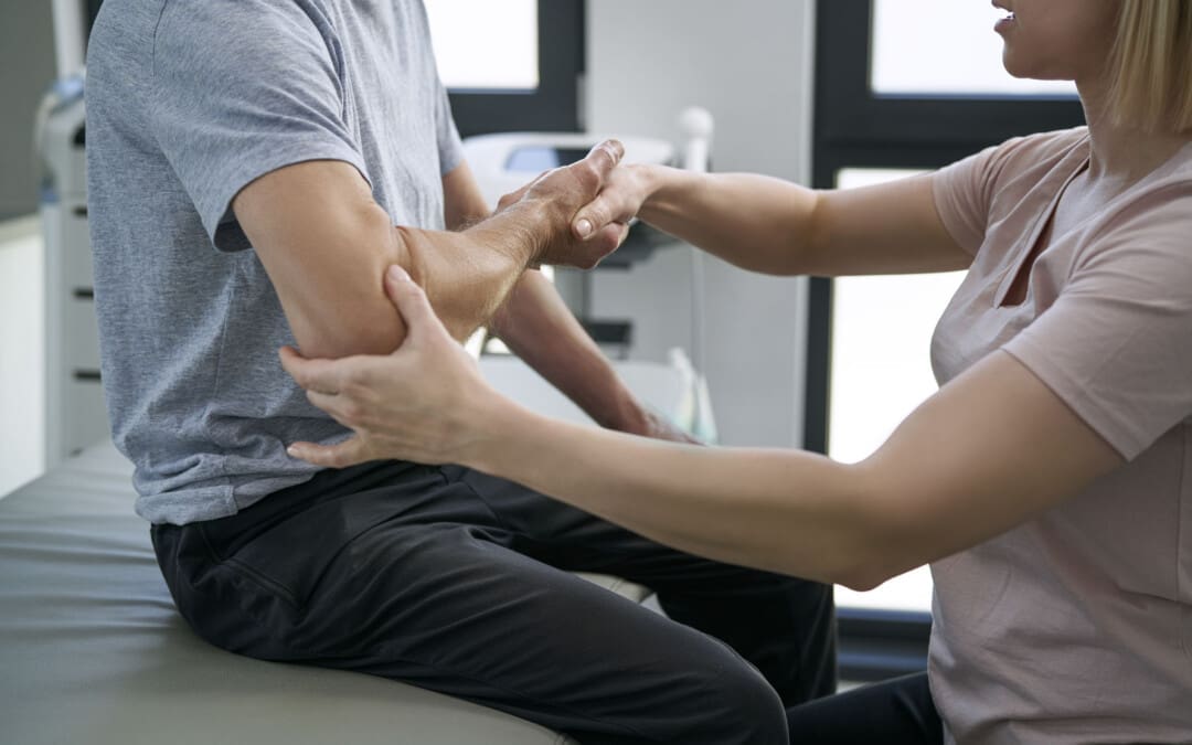
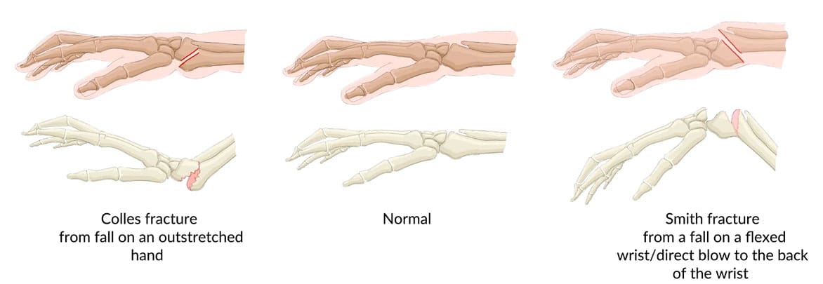
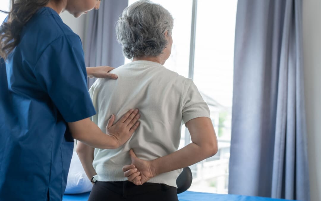
A broken wrist or Colles fracture can be a painful and stressful experience. Individuals may be unable to perform their jobs or engage in recreational activities. A Colles fracture is a break in the radius bone of the forearm that occurs near the wrist, usually about an inch from the end of the bone. It’s a common type of broken wrist often caused by falling on an outstretched hand. (American Academy of Orthopaedic Surgeons, 2022) As the individual lands on their hand, the end of the radius bone breaks off and gets pushed toward the inner wrist. If the wrist is flexed when falling on the hand, the radius may break and move toward the front of the wrist. This is called a Smith’s fracture. (Matsuura, Y. et al., 2017) A physical therapy team can help improve functional mobility to quickly and safely return to normal activity.
Symptoms
Individuals who have suffered trauma to their wrist or have fallen onto their hand or wrist may have a Colles fracture. Common signs and symptoms of a wrist or Colles fracture include: (American Academy of Orthopaedic Surgeons, 2022)
Bruising
Loss of mobility in the wrist.
Swelling in the arm, wrist, or hand.
Pain
Visible deformity or a lump on the backside of the forearm near the wrist.
Initial Treatment
Individuals who have fallen and injured their wrist and hand and suspect a Colles fracture seek immediate medical attention. Call a healthcare provider or report to a local emergency clinic. Left untreated, it can result in complications and permanent loss of arm and hand function. (American Academy of Orthopaedic Surgeons, 2022)
An X-ray will show a wrist fracture.
Because of the pain and swelling, it is recommended that individuals put an ice pack on their wrists and hands until they can get to a healthcare provider or emergency room. The R.I.C.E. principle can help control swelling and lessen pain until a medical professional can provide treatment. The initial treatment is to reduce the fracture. This is where a healthcare provider situates the broken bone or bones back into the correct position to ensure proper healing. This is done manually if the fractured bone is not too far out. If the fracture is severe, a surgical procedure known as an open reduction internal fixation or ORIF may be required to reduce the fracture. (American Academy of Orthopaedic Surgeons, 2022)
Once the fracture has been reduced, it must be immobilized. This is done with a cast or a brace. Individuals may also be required to wear a sling. They may need to visit a physical therapist to learn how to wear the sling properly. It is essential to keep the bones immobilized for proper healing. Consult a healthcare provider for questions about cast, sling, or brace.
Physical Therapy
After four to six weeks of immobilization, a healthcare provider may remove the cast and refer a physical therapist or team. (American Academy of Orthopaedic Surgeons, 2022) A physical therapist may measure and evaluate pain, swelling, range of motion, and strengthening. The physical therapist may assess the surgical scar tissue and analyze the hand, wrist, and arm function of individuals who underwent an ORIF procedure to reduce the fracture. After the initial evaluation, a physical therapist will work with the patient to develop an appropriate plan of care to help improve the impairments and functional limitations. The therapist may prescribe a specific exercise program as well.
Pain and Swelling
Individuals may experience pain and swelling around their wrists and hands.
A physical therapist can provide individuals with various treatments and modalities to help decrease swelling and pain.
Range of Motion
After a Colles’ fracture, individuals may lose hand, wrist, and elbow mobility.
The shoulder may also be tight, especially after wearing a sling.
Range of motion exercises for the hand, wrist, and elbow can be prescribed.
Strength
Loss of strength is common after a Colles’ fracture.
Exercises focusing on hand, wrist, and elbow strength may be prescribed.
At-home exercises and stretches will get the best results from physical therapy.
Scar Tissue
Individuals who have had an ORIF procedure will likely have scar tissue that has developed around the surgical site.
A physical therapist may perform scar tissue massage and mobilization to help improve mobility and can train patients how to self-massage.
Injury Medical Chiropractic and Functional Medicine Clinic
After a few weeks of physical therapy, individuals should notice their mobility and strength improve while pain and swelling decrease. Individuals will find it easier to use their arms and hands to perform functional activities. While the fracture should be fully healed six to eight weeks after injury, individuals may still be limited for potentially 12 to 16 weeks. At Injury Medical Chiropractic and Functional Medicine Clinic, we focus on what works for every patient to restore function. If other treatment is needed, individuals will be referred to a clinic or physician best suited to their injury, condition, or ailment.
Personal Injury Rehabilitation
References
American Academy of Orthopaedic Surgeons. (2022). Distal radius fractures (broken wrist). https://orthoinfo.aaos.org/en/diseases–conditions/distal-radius-fractures-broken-wrist/
Matsuura, Y., Rokkaku, T., Kuniyoshi, K., Takahashi, K., Suzuki, T., Kanazuka, A., Akasaka, T., Hirosawa, N., Iwase, M., Yamazaki, A., Orita, S., & Ohtori, S. (2017). Smith’s fracture generally occurs after falling on the palm of the hand. Journal of orthopaedic research : official publication of the Orthopaedic Research Society, 35(11), 2435–2441. https://doi.org/10.1002/jor.23556
Can learning about comminuted fracture symptoms and repair help individuals and healthcare providers develop effective treatment and rehabilitation programs?
Comminuted Fractures
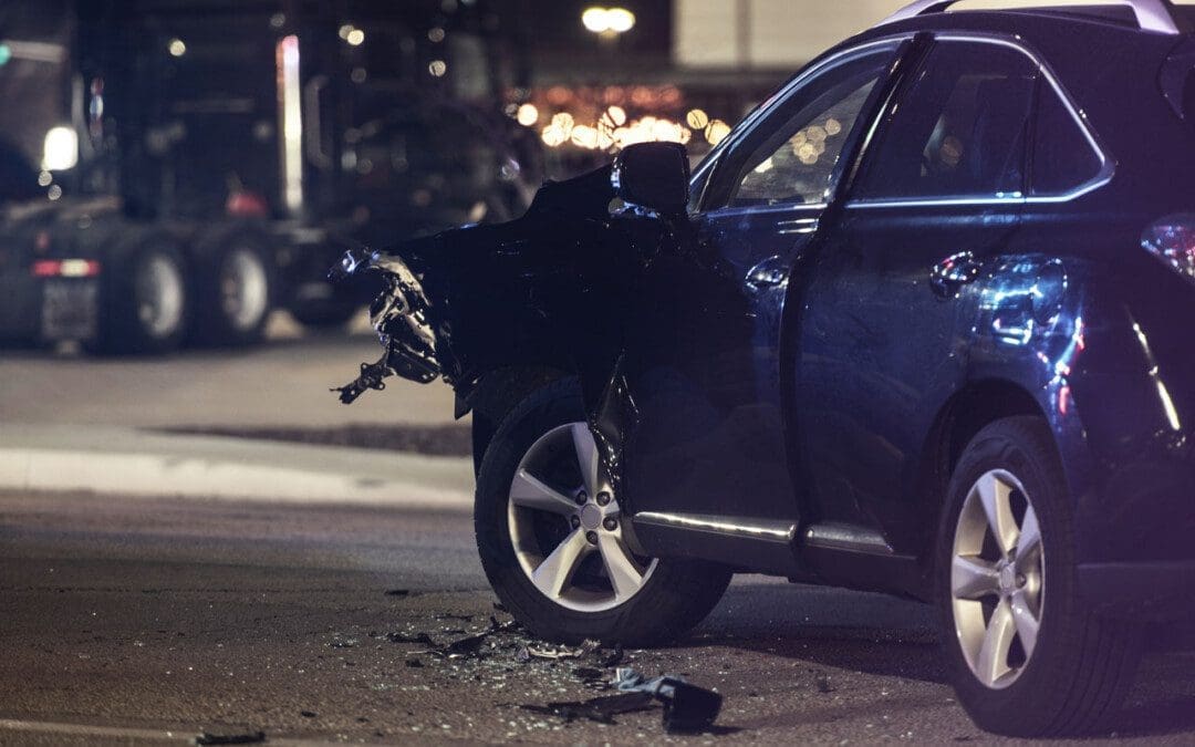
A comminuted fracture is a severe break in which the bone splits into at least three pieces. Comminuted fractures typically happen in the long bones like those in the arms and legs. But they can also happen in other places, including the ribs. (Corinne Tarantino, 2022) They are usually caused by intense impact, like an automobile collision/accident or a severe fall. Depending on the location of the fracture, recovery from a comminuted fracture can take months and often involves:
Surgery – A surgeon will place screws and rods to hold the pieces of the bone in position. Sometimes, the hardware is left in permanently. (American Academy of Orthopaedic Surgeons, 2021)
Wearing a cast for several months.
Physical therapy.
Types
In these types of fractures, the bone is completely broken, not just cracked. The break is a highly comminuted fracture if the bone is broken into four or more pieces. (Corinne Tarantino, 2022)
Symptoms
The symptoms are the same as those of other broken bones, but they can be more intense because the bone is broken in multiple areas, which means there may also be more soft tissue injuries than with a simple fracture. Broken bone symptoms include: (MedlinePlus, 2024)
Persistent pain
Swelling
Bruising
Deformity – the bone looks out of place or is at an odd angle.
Tingling
Numbness
Difficulty moving the limb.
Causes
An intense force causes a comminuted fracture, often a car accident or a hard fall, but it can also result from sports injuries. (Corinne Tarantino, 2022)
Diagnosis
A comminuted fracture is diagnosed by X-ray, which shows where the bone has broken and how many pieces it has split into (MedlinePlus, 2024). Healthcare providers will diagnose any broken bones but also look for other injuries.
Treatment
Typically, broken bones can be treated with casts, braces, or a boot to keep them immobilized. (MedlinePlus, 2024) Because comminuted fractures are more severe, they often need other treatments, including surgery. Sometimes, the bone can be reset instead of surgery using a closed reduction technique, where the healthcare provider resets the bone manually. Surgery may be recommended if that’s not possible or doesn’t work.
Types of Surgery
Surgery allows the healthcare provider to correctly position all the pieces of the bone to heal in a strong, stable formation. The two types commonly used to treat comminuted fractures are: (American Academy of Orthopaedic Surgeons, 2021)
External Fixation
This surgery uses rods and screws outside the body to stabilize the bone.
This external frame is placed during surgery and later removed.
Open Reduction Internal Fixation – ORIF
During this surgery, the bone is stabilized with metal plates, screws, rods, and/or wires placed inside your body.
Sometimes, these are permanent, but in other cases, they are removed later.
An orthopedic surgeon performs these surgeries.
Casting
After surgery, a cast is usually needed to prevent the bone from moving and allow healing. Typically, the cast is worn for six to eight weeks, but it may need to be worn longer with comminuted fractures. Some need a cast for several months (American Academy of Orthopaedic Surgeons, 2021). Sometimes, those with an external fixation must continue wearing a cast after removing the pins and rods, giving the bone more time to stabilize. The healthcare provider will inform the patient how long the cast will need to be worn and which sports activities should be avoided even after the cast comes off.
Physical Therapy
The healthcare provider may recommend physical therapy to help treat the fracture. Usually, when the cast is removed, physical therapy is activated to help rebuild strength and range of motion. (Corinne Tarantino, 2022)
Prognosis
Although these fractures are severe, they are treatable. The prognosis is good for individuals who follow their healthcare provider’s treatment plan. Most don’t have lingering pain after the initial injury and can eventually return to regular activities. (American Academy of Orthopaedic Surgeons, 2021)
Recovery
Healing a comminuted fracture can take months. During that time, it’s helpful to focus on tasks that can be accomplished, like new hobbies that don’t involve physical injury. Talk to the healthcare provider about any problems with the cast or ongoing pain and what to expect during recovery.
Injury Medical Chiropractic and Functional Medicine Clinic works with primary healthcare providers and specialists to develop an optimal health and wellness solution through an integrated approach to treating injuries and chronic pain syndromes, improving flexibility, mobility, and agility programs to relieve pain and help individuals return to normal. Our providers create personalized care plans for each patient. If other treatments are needed, Dr. Jimenez has teamed up with top surgeons, clinical specialists, medical researchers, and rehabilitation providers to provide the most effective treatments.
The Path to Healing Personal Injury
References
Corinne Tarantino, MPH. Osmosis. (2022). Comminuted Fracture: What is it, Examples and More. https://www.osmosis.org/answers/comminuted-fracture
Throckmorton T.W. American Academy of Orthopaedic Surgeons. (2021). Fractures (broken bones). https://orthoinfo.aaos.org/en/diseases–conditions/fractures-broken-bones/
MedlinePlus. National Library of Medicine. (2024). Fractures Also called: Broken bone. Retrieved from https://medlineplus.gov/fractures.html
Can understanding the location of the funny bone and how pain can be managed after injury help expedite recovery and prevention for individuals who have hit their funny bone?
Elbow Funny Bone Nerve Injury
Behind the elbow is an area known as the “funny bone,” where the ulnar nerve has less tissue and bone protection. This is where part of the ulnar nerve passes around the back of the elbow. Because less tissue and bone protect the nerve in this area, taking a hit like bumping into something can cause an electric shock-like pain and a tingling sensation down the arm and to the outside fingers typical of an irritated nerve. Most injuries to the funny bone resolve quickly, and the pain disappears after a few seconds or minutes, but sometimes, an ulnar nerve injury can lead to more persistent symptoms.
Anatomy
The funny bone is not a bone but the ulnar nerve. The nerve runs down the arm, passing around the back of the elbow. (Dimitrova, A. et al., 2019) Because the ulnar nerve is on top of the elbow and there is very little fatty cushion, lightly bumping this spot can cause pain and tingling sensations down the forearm. Three bones comprise the junction of the elbow that include:
Humerus – arm bone
Ulna and radius – forearm bones
The humerus has a groove that protects and holds the ulnar nerve as it passes behind the joint. This is where the nerve can be injured or irritated when the nerve is hit or pinched against the end of the bone, causing the funny bone pain.
Electrical Pain Sensation
When hitting the ulnar nerve or funny bone where the ulnar nerve provides sensation, pain, and electrical/tingling sensations are experienced from the forearm to the outside fingers. This part of the arm and hand is called the ulnar nerve distribution. (American Academy of Orthopaedic Surgeons. 2024) The ulnar nerve provides sensation into most of the pinky finger and about half of the ring finger. Other nerves, including the median and radial nerve, supply sensation to the rest of the hand.
Treatment
Usually, a sharp jolt to the elbow quickly resolves. Some recommendations to help symptoms improve faster include:
Shaking the forearm and hand out.
Straightening out and bending the elbow to stretch the nerve.
Decreasing mobility of the elbow.
Applying ice to the area.
Taking anti-inflammatory medications.
Treating Long-Lasting Pain
In rare circumstances, injuries to the ulnar nerve can cause more persistent symptoms, a condition known as cubital tunnel syndrome. Cubital tunnel syndrome can happen after an injury or from elbow overuse. Individuals with cubital tunnel syndrome may benefit from wearing a splint at night. Standard-sized splints can be ordered online, but most are fabricated by an occupational or hand therapist. If symptoms become more long-lasting, surgery may be recommended to relieve pressure and tension on the ulnar nerve (American Academy of Orthopaedic Surgeons, 2024). The procedure decompresses the nerve by relieving any tight constrictions around it and releasing them. In severe cases, the nerve is repositioned to an area that doesn’t place as much pressure on the nerve, known as an ulnar nerve transposition.
Injury Medical Chiropractic and Functional Medicine Clinic works with primary healthcare providers and specialists to develop an optimal health and wellness solution that helps individuals return to normal. Our providers create personalized care plans for each patient, including Functional Medicine, Acupuncture, Electro-Acupuncture, and Sports Medicine principles through an integrated approach to treat injuries and chronic pain syndromes to improve ability through flexibility, mobility, and agility programs to relieve pain. If other treatment is needed, Dr. Jimenez has teamed up with top surgeons, clinical specialists, medical researchers, and rehabilitation providers to provide the most effective treatments.
Chiropractic Treatment For Carpal Tunnel Syndrome
References
Dimitrova, A., Murchison, C., & Oken, B. (2019). Local effects of acupuncture on the median and ulnar nerves in patients with carpal tunnel syndrome: a pilot mechanistic study protocol. Trials, 20(1), 8. https://doi.org/10.1186/s13063-018-3094-5
American Academy of Orthopaedic Surgeons. (2024). Ulnar nerve entrapment at the elbow. https://orthoinfo.aaos.org/en/diseases–conditions/ulnar-nerve-entrapment-at-the-elbow-cubital-tunnel-syndrome/
Can understanding the nucleus pulposus help in body positioning and prevention for individuals wanting to practice spinal hygiene and protect their discs from injury?
Nucleus Pulposus
The spinal discs are located between the spine’s vertebrae and are the body’s natural impact and shock absorbers. Within the disc is the nucleus pulposus, which plays a major role in providing the spine with shock absorption during movement. (Zhou Z. et al., 2014) The discs have a tough outer portion and a soft inner core. They are the:
It forms the tough circular exterior and comprises concentric sheets of collagen fibers or lamellae surrounding the inner core.
It has cartilaginous endplates that firmly attach to the vertebrae above and below.
Nucleus Pulposus
The nucleus pulposus is the inner core soft filling of the discs.
It contains a network of fibers suspended in a mucoprotein gel with a water base to maintain strength and pliability.
The near-liquid consistency makes it responsive to movement to handle the body’s axial load.
It helps maintain spinal suspension to prevent pressure on the bones and prevent bone-to-bone contact, reducing the potential for injuries and pain.
Shock Absorber
Each intervertebral disc is a shock-absorbing cushion, with the nucleus pulposus providing shock-absorbing properties (Zhou Z. et al., 2014). The intervertebral discs move as the body moves. For example, when arching the back, the disc moves forward slightly, and when twisting, the disc twists as well.
Spinal Action
The intervertebral disc supports spinal movements. When bending, twisting, arching, or tilting the spine, the nucleus pulposus swivels to accommodate these actions. These repeated spinal actions, which occur throughout the day and night, contribute to shifting positions while sitting, working, playing sports, carrying groceries, performing house chores, etc. An example is bending forward to pick something up. This action involves forward spinal flexion, which is bending the spine forward, flattening, or rounding. When bending using flexion, the spinal bones come closer together, pushing the nucleus pulposus toward the back.
Injuries
The disc can be pushed too far back with persistent or excessive spinal flexion. If the fibers of the annulus fibrosus become weak, they can tear, causing the nucleus pulposus to leak out and disc herniation. Generally, the nucleus pulposus will leak to the side and back; however, this corresponds to the location of the very sensitive nerve root/s with which it can come into contact, causing pain and other symptoms. The most common causes of disc herniation are degenerative wear and tear changes of the disc and trauma. Disc degeneration occurs as the body ages; it weakens the annulus fibers, allowing the nucleus pulposus to distend, bulge, or herniate.
Aging
Disc degeneration occurs with age but can also occur with injuries to the area. In young individuals, the nucleus pulposus is mostly water. For this age group, a herniation from trauma is more likely than in older individuals. (Ucar, D. et al., 2021) But as the body ages, the discs, especially the nucleus pulposus, begin to dry out. This dehydration leads to a significant loss of disc height. (UCLA Health, 2024) By age 60 or 70, the discs may be composed entirely of fiber, which can cause the shock absorption function not to work and disappear.
Chiropractic therapy is among the more conservative treatment options for a herniated disc and may be tried first before proceeding with more invasive treatments. Injury Medical Chiropractic and Functional Medicine Clinic works with primary healthcare providers and specialists to develop an optimal health and wellness solution that fully benefits the individual to get back to normal.
The Science of Functional Healing
References
Zhou, Z., Gao, M., Wei, F., Liang, J., Deng, W., Dai, X., Zhou, G., & Zou, X. (2014). Shock absorbing function study on denucleated intervertebral disc with or without hydrogel injection through static and dynamic biomechanical tests in vitro. BioMed research international, 2014, 461724. https://doi.org/10.1155/2014/461724
Nosikova, Y. S., Santerre, J. P., Grynpas, M., Gibson, G., & Kandel, R. A. (2012). Characterization of the annulus fibrosus-vertebral body interface: identification of new structural features. Journal of anatomy, 221(6), 577–589. https://doi.org/10.1111/j.1469-7580.2012.01537.x
Ucar, D., Duman, S., Bayram, Y., & Ucar, B. Y. (2021). Extruded disc herniations are experienced earlier by inactive young people in the high-tech gaming era. Journal of medicine and life, 14(3), 402–407. https://doi.org/10.25122/jml-2021-1059
For individuals who are getting older, can increasing bone strength help prevent fractures and optimize bone health?
Bone Strength
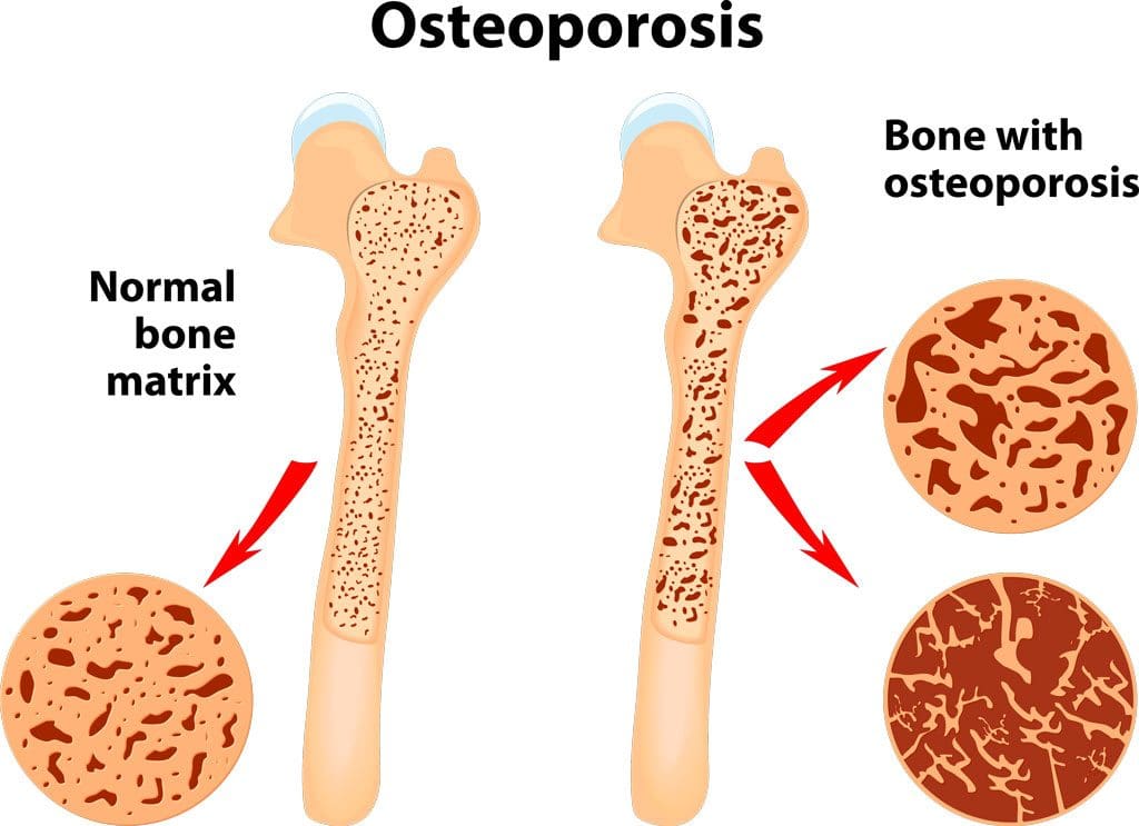
Bone strength is important, as a fractured hip can be serious for older individuals. A study found that for individuals in their 60s who had a hip fracture, 6.5% of women and 9.4% of men died within a year. Among individuals in their 80s, 13.1% of women and 19.6% of men died within a year. (Dimet-Wiley, et al., 2022)
Increasing bone strength can help prevent various issues. A small increase in bone mineral density has been shown to help reduce the risk of fractures, especially hip fractures. A decades-long study found that just a 3% increase in bone strength helps lower the chance of breaking a hip. Researchers enrolled two groups of individuals aged 60 and older, one in 1989 and the second in 1999.
The bone mineral density of each subject’s femoral neck joint at the top of the thigh bone near the hip was measured.
They then followed the subjects for years to see who experienced hip fractures.
While the bone mineral density of the second group was only 3% higher than the first group, these subjects experienced a 46% reduction in hip fractures. (Tran, T. et al., 2023)
Bone Loss
Bone loss is progressive in men and women and increases as the body ages. Osteoporosis is a condition in which bone tissue deteriorates. (Department of Health and Human Services Office of Disease Prevention and Health Promotion. 2020) Bones constantly break down and reform as a normal remodeling process. If the balance of this process is impaired, osteoporosis develops, resulting in more bone breakdown than formation. While men and women experience bone loss, it’s more common in females. (National Institute of Arthritis and Musculoskeletal Diseases. 2022) Menopause is a risk factor because of the decline of estrogen (National Library of Medicine, Medline Plus, 2022). Estrogen reinforces bone strength by protecting against bone breakdown; with estrogen loss, bone breakdown increases. However, anyone of any age or background can experience bone loss due to the following:
While some loss of bone strength is common, several strategies exist to maintain bone health. Exercise, specifically weight-bearing activities, can increase bone strength. When bones and muscles are used to hold a position against gravity, this mechanically stresses the bone, causing it to reform stronger. Movement and physical exercise as medicine and the forces transmitted through the bones generate mechanical signals that tell the cells to increase bone formation relative to breakdown. Exercises focusing on posture, balance, gait, and coordination are recommended for individuals with osteoporosis to strengthen the core, quadriceps, and hip flexors. Different types of exercises can include:
Walking to strengthen the spine and hips.
Walking outside or on a treadmill provides more loading force to the bone.
Planks and push-ups can strengthen the forearm and wrist bones.
Holding a water bottle in each hand and lifting up and down 10 times together or alternating a few times a day.
Side leg lifts can strengthen the hip and forearm bones simultaneously.
Weight training provides the bones with a workout by having them support a weight load.
Any exercise therapy program should be designed by a healthcare provider, physical therapist, and trainer according to the individual’s condition and appropriate for them.
Diet
What goes into the body definitely affects bone health. Calcium and vitamin D are key to bone building, but both are needed as vitamin D is needed to absorb the calcium ingested. Calcium can be found in:
Dairy
Dairy products and non-dairy alternatives are fortified with calcium.
Leafy greens.
Beans.
Almonds.
The recommended daily calcium intake for adults over 50 is 1,200 milligrams.
Vitamin D can come from:
Sunlight
Fish.
Mushrooms.
Fortified milk.
Supplements.
The recommended daily vitamin D intake for adults aged 70 is 15 micrograms and 20 micrograms for individuals over 70.
Studies have found that increasing calcium and vitamin D intake with supplements can help maintain bone health. Talk to a healthcare provider about whether supplements could be beneficial.
Hormone Therapy
Females also naturally produce testosterone, which promotes bone formation. As levels drop with age and negatively impact bone strength, hormone therapy could be recommended. Declining testosterone levels start with women in their 20s and men in their 30s. The typical drop in women is 1% to 3% yearly before menopause and stabilizes somewhat afterward. Female patients at risk of bone loss may be prescribed testosterone in various forms that continuously emit the hormone. The dosage is low, so patients do not experience unwanted hair growth or skin changes. Combined with estrogen, testosterone effectively increases bone growth in female patients. Not everyone is a candidate for hormone therapy, like individuals with a history of breast cancer, heart disease, blood clots, or liver disease. (National Library of Medicine. Medline Plus, 2019)
Making small adjustments can optimize bone health and overall well-being
At Injury Medical Chiropractic and Functional Medicine Clinic, we passionately focus on treating patients’ injuries and chronic pain syndromes to create personalized care plans that improve ability through flexibility, mobility, and agility programs tailored to the individual. Using an integrated approach, our goal is to relieve pain naturally by restoring health and function to the body through Functional Medicine, Acupuncture, Electro-Acupuncture, and Sports Medicine protocols. If the individual needs other treatment, they will be referred to a clinic or physician best suited for them, as Dr. Jimenez has teamed up with the top surgeons, clinical specialists, medical researchers, and premier rehabilitation providers to provide the most effective clinical treatments. We focus on what works for you and strive to better the body through researched methods and total wellness programs.
Chiropractic Care: Movement Medicine
References
Dimet-Wiley, A., Golovko, G., & Watowich, S. J. (2022). One-Year Postfracture Mortality Rate in Older Adults With Hip Fractures Relative to Other Lower Extremity Fractures: Retrospective Cohort Study. JMIR aging, 5(1), e32683. https://doi.org/10.2196/32683
Tran, T. S., Ho-Le, T. P., Bliuc, D., Center, J. R., Blank, R. D., & Nguyen, T. V. (2023). Prevention of Hip Fractures: Trade-off between Minor Benefits to Individuals and Large Benefits to the Community. Journal of bone and mineral research : the official journal of the American Society for Bone and Mineral Research, 38(11), 1594–1602. https://doi.org/10.1002/jbmr.4907
Department of Health and Human Services Office of Disease Prevention and Health Promotion. (2020). Osteoporosis Workgroup. Retrieved from https://health.gov/healthypeople/about/workgroups/osteoporosis-workgroup
National Institute of Arthritis and Musculoskeletal Diseases. (2022). Osteoporosis. Retrieved from https://www.niams.nih.gov/health-topics/osteoporosis
National Library of Medicine. MedlinePlus. (2022). What causes bone loss? Retrieved from https://medlineplus.gov/ency/patientinstructions/000506.htm
National Library of Medicine. MedlinePlus. (2019). Hormone replacement therapy. Retrieved from https://medlineplus.gov/hormonereplacementtherapy.html
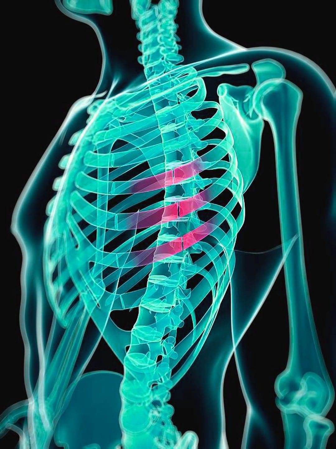
Individuals may not realize they have a cracked rib until symptoms like pain when taking in a deep breath begin to present. Can knowing the symptoms and causes of cracked or broken ribs help in diagnosis and treatment?
Cracked Rib
A broken/fractured rib describes any break in the bone. A cracked rib is a type of rib fracture and is more a description than a medical diagnosis of a rib that has been partially fractured. Any blunt impact to the chest or back can cause a cracked rib, including:
Falling
Vehicle collision
Sports injury
Violent coughing
The main symptom is pain when inhaling.
The injury typically heals within six weeks.
Symptoms
Cracked ribs are usually caused by a fall, trauma to the chest, or intense violent coughing. Symptoms include:
Swelling or tenderness around the injured area.
Chest pain when breathing/inhaling, sneezing, laughing, or coughing.
Chest pain with movement or when lying down in certain positions.
Possible bruising.
Although rare, a cracked rib can cause complications like pneumonia.
See a healthcare provider immediately if experiencing difficulty breathing, severe chest pain, or a persistent cough with mucus, high fever, and/or chills.
Types
In most cases, a rib usually gets broken in one area, causing an incomplete fracture, which means a crack or break that does not go through the bone. Other types of rib fractures include:
Displaced and Nondisplaced Fractures
Completely broken ribs may or may not shift out of place.
If the rib does move, this is known as a displaced rib fracture and is more likely to puncture lungs or damage other tissues and organs. (Yale Medicine. 2024)
A rib that stays in place usually means the rib is not completely broken in half and is known as a nondisplaced rib fracture.
Flail Chest
A section of the ribcage can break away from the surrounding bone and muscle, although this is rare.
If this happens, the ribcage will lose stability, and the bone will move freely as the individual inhales or exhales.
This broken ribcage section is called a flail segment.
This is dangerous as it can puncture the lungs and cause other serious complications, like pneumonia.
Causes
Common causes of cracked ribs include:
Vehicle collisions
Pedestrian accidents
Falls
Impact injuries from sports
Overuse/Repetitive stress brought on by work or sports
Severe coughing
Older individuals can experience a fracture from a minor injury due to the progressive loss of bone minerals. (Christian Liebsch et al., 2019)
The Commonality of Rib Fractures
Rib fractures are the most common type of bone fracture.
They account for 10% to 20% of all blunt trauma injuries seen in emergency rooms.
In cases where an individual seeks care for a blunt injury to the chest, 60% to 80% involve a broken rib. (Christian Liebsch et al., 2019)
Diagnosis
A cracked rib is diagnosed with a physical exam and imaging tests. During the examination, a healthcare provider will listen to the lungs, press gently on the ribs, and watch as the rib cage moves. The imaging test options include: (Sarah Majercik, Fredric M. Pieracci 2017)
X-rays – These are for detecting recently cracked or broken ribs.
CT Scan – This imaging test comprises multiple X-rays and can detect smaller cracks.
MRI – This imaging test is for soft tissues and can often detect smaller breaks or cartilage damage.
Bone Scan – This imaging test uses a radioactive tracer to visualize the structure of bones and can show smaller stress fractures.
Treatment
In the past, treatment used to involve wrapping the chest with a band known as a rib belt. These are rarely used today as they can restrict breathing, increasing the risk of pneumonia or even a partial lung collapse. (L. May, C. Hillermann, S. Patil 2016). A cracked rib is a simple fracture that requires the following:
Rest
Over-the-counter or prescription medications can help manage pain symptoms.
Nonsteroidal anti-inflammatory drugs – NSAIDs like ibuprofen or naproxen are recommended.
If the break is extensive, individuals may be prescribed stronger pain medication depending on the severity and underlying conditions.
Physical therapy can expedite the healing process and help maintain the range of motion of the chest wall.
For patients who are frail and elderly individuals, physical therapy can help the patient walk and normalize certain functions.
A physical therapist can train the individual to transfer between bed and chairs safely while maintaining awareness of any movements or positioning that make the pain worse.
A physical therapist will prescribe exercises to keep the body as strong and limber as possible.
For example, lateral twists can help improve the range of motion in the thoracic spine.
During the early stages of recovery, it is recommended to sleep in an upright position.
Lying down can add pressure, causing pain and possibly worsen the injury.
Use pillows and bolsters to help support sitting up in bed.
What may feel like a cracked rib may be a similar condition, which is why it’s important to get checked out. Other possible symptom causes can include:
Bruised ribs – This occurs when the ribs are not cracked, but the smaller blood vessels around the region burst and leak into surrounding tissues. (Sarah Majercik, Fredric M. Pieracci 2017)
Pulled muscle – A muscle strain, or pulled muscle, occurs when the muscle gets overstretched, which can lead to a tear. The ribs are not affected, but it can feel like they are. (Sarah Majercik, Fredric M. Pieracci 2017)
Emergency
The most common complication is being unable to take a deep breath because of the pain. When the lungs cannot breathe deeply enough, mucous and moisture can build up and lead to an infection like pneumonia. (L. May, C. Hillermann, S. Patil 2016). Displaced rib fractures can also damage other tissues or organs, increasing the risk of a collapsed lung/pneumothorax or internal bleeding. It is recommended to seek immediate medical attention if symptoms develop like:
Shortness of breath
Difficulty breathing
A bluish color of the skin caused by lack of oxygen
A persistent cough with mucus
Chest pain when breathing in and out
Fever, sweating, and chills
Rapid heart rate
The Power of Chiropractic Care In Injury Rehabilitation
Liebsch, C., Seiffert, T., Vlcek, M., Beer, M., Huber-Lang, M., & Wilke, H. J. (2019). Patterns of serial rib fractures after blunt chest trauma: An analysis of 380 cases. PloS one, 14(12), e0224105. https://doi.org/10.1371/journal.pone.0224105
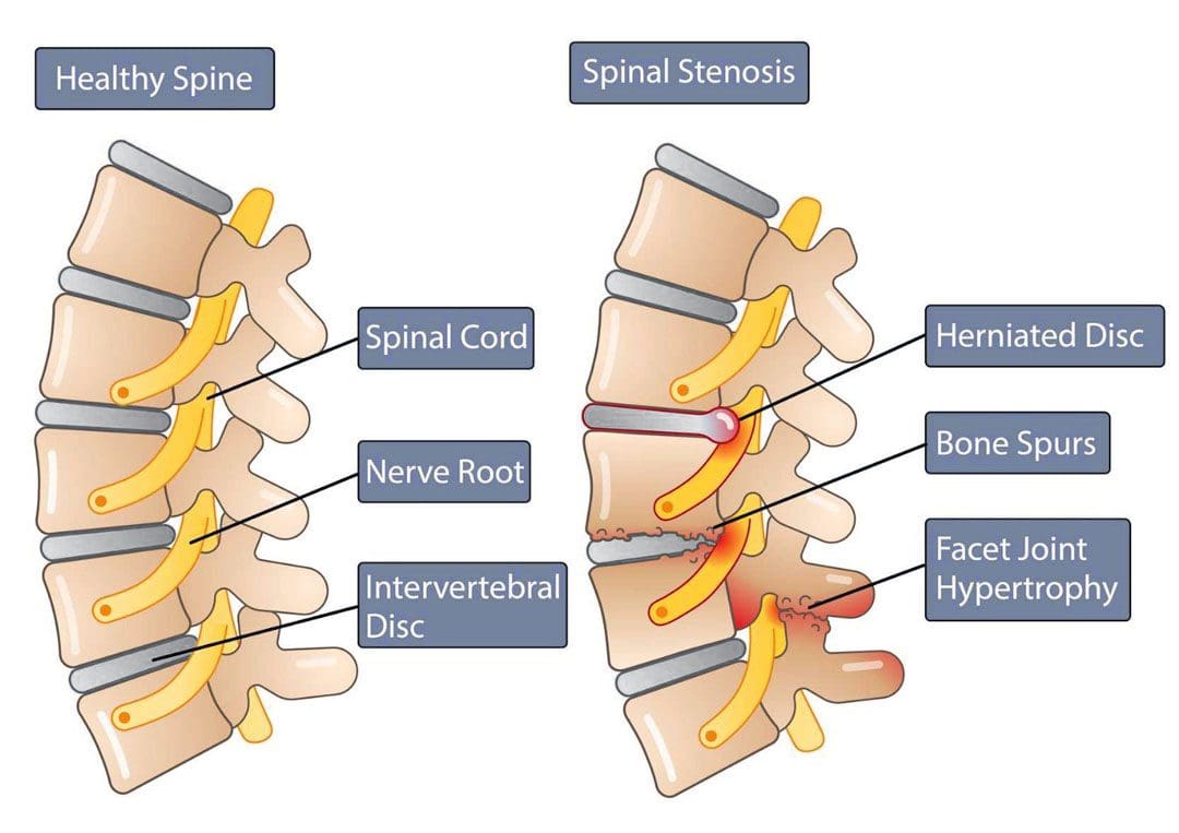
Facet hypertrophy is an incurable, chronic disease that affects the facet joints in the spine. Can recognizing symptoms, help in diagnosis, and treatment?
Facet Hypertrophy
Facet hypertrophy causes the facet joints in the spine to enlarge. They are found where the vertebrae come into contact on the back of the vertebrae that form the backbone. These joints stabilize the spine when twisting and bending. Hypertrophy results when damage wears down the cartilage that cushions the bones that meet in the joint. This can include:
Aging
Wear and tear
Arthritis
Other joint diseases can damage facet joints.
Swelling, new bone growth, and bone spurs can occur as the joint tries to repair the damaged cartilage. The swelling and new bone growth can narrow the spinal canal and compress surrounding nerves, causing pain and other sensation symptoms. This ailment does not have a cure and worsens over time. The objective of treatment is to manage the pain symptoms and slow down the disease’s progress.
Types
Facet hypertrophy can be described as unilateral or bilateral.
Symptoms can have a wide range of intensity, from a dull ache to chronic, disabling pain. The location of symptoms depends on the affected joint and the nerves involved, Pain manifests when the enlarged joints and new bone growth compresses the nearby nerves. The result leads to nerve damage and the following symptoms: (Weill Cornell Medicine Brain & Spine Center. 2023) (Cedars Sinai. 2022)
Stiffness, especially when standing up or getting out of a chair.
Inability to stand straight when walking.
Inability to look up to the left or right without turning the whole body.
Reduced range of motion and mobility.
Numbness or a tingling sensation of pins and needles.
Radiating pain from the affected joint into the buttocks, hips, and upper thigh when the affected joint/s are in the lower back.
Radiating pain from the affected joint into the shoulder, neck, and back of the head when the affected joint/s are in the upper back.
Headaches when the affected joint is in the neck.
Causes
A common cause is the age-related degeneration of the joints, called spondylosis. Research indicates that more than 80% of individuals who are 40 or older have radiologic evidence of spondylosis, even though they may not have symptoms. (The University of Toledo Medical Center. N.D.) The following conditions can also increase the risk of facet hypertrophy (Weill Cornell Medicine Brain & Spine Center. 2023)
Unhealthy posture
Being overweight or obese
Sedentary lifestyle
Injury or trauma to the spine
Inflammatory conditions like rheumatoid arthritis or ankylosing spondylitis
Osteoarthritis
Genetic predisposition to the condition
Diagnosis
Diagnosis can be challenging when neck or back pain is the main complaint, as symptoms can mimic conditions such as sciatica from a herniated disc or hip arthritis. (Weill Cornell Medicine Brain & Spine Center. 2023)
A healthcare provider will conduct a complete physical examination, medical history, and discussion of symptoms.
CT scans with or without myelogram – the use of contrast dye in the space around the spinal cord.
MRI
X-rays with or without a myelogram
A diagnosis is confirmed by injecting a diagnostic block that involves administering an anesthetic injection, sometimes with an anti-inflammatory like cortisone, into the joint or nerves near the affected joint. Two injections are given at different times to confirm the effect. (Romain Perolat et al., 2018)
If immediate relief improves after each injection, the facet joint is confirmed as the source of the pain symptoms.
If the block does not decrease the pain, the facet joint is probably not the source of the pain symptoms. (Brigham and Women’s Hospital. 2023)
Treatment
There is no cure for facet hypertrophy.
The goal of treatment is to make the pain more manageable.
Conservative treatment is usually successful in making a difference.
Nonsteroidal anti-inflammatory – aspirin, ibuprofen, and naproxen.
Muscle relaxers – cyclobenzaprine or metaxalone.
Steroid injection into the facet joints.
Injection of platelet-rich plasma/PRP into the joints.
Medial Branch or Facet Block
A medial branch block injects local anesthetic near the medial nerves that connect to an inflamed joint.
Medial nerves are small nerves outside the joint space near the nerve that transmit signals and other impulses to the brain.
A facet block injects the medication outside the joint space near the nerve that supplies the joint called the medial branch.
Neurolysis
Neurolysis, also known as rhizotomy or neurotomy, is a procedure that destroys affected nerve fibers to relieve pain, reduce disability, and reduce the need for analgesics. This treatment can relieve pain for six to 12 months until the nerve regenerates, where further treatments may be necessary. (Matthew Smuck et al., 2012) Neurolysis can be performed using one of the following techniques (Romain Perolat et al., 2018)
Radiofrequency ablation RFA – the application of heat through radiofrequency.
Cryoneurolysis – the application of cold temperatures to the targeted nerve.
Chemical neurolysis – applying chemical agents, like a combination of phenol and alcohol.
Severing the nerves with surgical instrumentation.
Surgery
When one or more facet joints are severely damaged, they can become nonfunctional and painful. Surgery may be recommended when symptoms are not relieved by other therapies. (Ali Fahir Ozer, et al., 2015)
Prognosis
Facet hypertrophy is a chronic condition that progresses with age and does not affect life expectancy. (Weill Cornell Medicine Brain & Spine Center. 2023) The disorder is incurable, but symptoms can be managed with conservative therapies
A healthcare provider can help develop a treatment plan based on the extent and location of the joint affected.
Early diagnosis and treatment can help individuals achieve the best results.
Maintaining an active lifestyle and healthy weight can help prevent further joint stress. Individuals may be recommended to do regular stretching and strengthening exercises to lower inflammation, reduce stress, and improve overall health.
Facet Syndrome Treatment
References
Perolat, R., Kastler, A., Nicot, B., Pellat, J. M., Tahon, F., Attye, A., Heck, O., Boubagra, K., Grand, S., & Krainik, A. (2018). Facet joint syndrome: from diagnosis to interventional management. Insights into imaging, 9(5), 773–789. https://doi.org/10.1007/s13244-018-0638-x
Smuck, M., Crisostomo, R. A., Trivedi, K., & Agrawal, D. (2012). Success of initial and repeated medial branch neurotomy for zygapophysial joint pain: a systematic review. PM & R : the journal of injury, function, and rehabilitation, 4(9), 686–692. https://doi.org/10.1016/j.pmrj.2012.06.007
Ozer, A. F., Suzer, T., Sasani, M., Oktenoglu, T., Cezayirli, P., Marandi, H. J., & Erbulut, D. U. (2015). Simple facet joint repair with dynamic pedicular system: Technical note and case series. Journal of craniovertebral junction & spine, 6(2), 65–68. https://doi.org/10.4103/0974-8237.156049
IFM's Find A Practitioner tool is the largest referral network in Functional Medicine, created to help patients locate Functional Medicine practitioners anywhere in the world. IFM Certified Practitioners are listed first in the search results, given their extensive education in Functional Medicine